FLIPR Calcium Assay Evaluation Kit
FLIPR Calcium Assay Evaluation Kit
FLIPR Calcium Assay Evaluation Kit
Create successful ePaper yourself
Turn your PDF publications into a flip-book with our unique Google optimized e-Paper software.
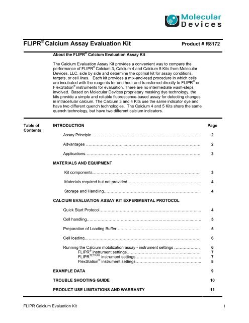
<strong>FLIPR</strong> ® <strong>Calcium</strong> <strong>Assay</strong> <strong>Evaluation</strong> <strong>Kit</strong>Product # R8172About the <strong>FLIPR</strong> ® <strong>Calcium</strong> <strong>Evaluation</strong> <strong>Assay</strong> <strong>Kit</strong>The <strong>Calcium</strong> <strong>Evaluation</strong> <strong>Assay</strong> <strong>Kit</strong> provides a convenient way to compare theperformance of <strong>FLIPR</strong> ® <strong>Calcium</strong> 3, <strong>Calcium</strong> 4 and <strong>Calcium</strong> 5 <strong>Kit</strong>s from MolecularDevices, LLC. side by side and determine the optimal kit for assay conditions,targets, or cell lines. Each kit provides a mix-and-read procedure in which cellsare incubated with the reagents for one hour and transferred directly to <strong>FLIPR</strong> ® orFlexStation ® instruments for evaluation. There are no intermediate wash-stepsinvolved. Based on Molecular Devices proprietary masking dye technology, thekits provide a simple and reliable fluorescence-based assay for detecting changesin intracellular calcium. The <strong>Calcium</strong> 3 and 4 <strong>Kit</strong>s use the same indicator dye andhave two different quench technologies. The <strong>Calcium</strong> 4 and 5 <strong>Kit</strong>s share the samequench technology, but have two different calcium indicators.Table ofContentsINTRODUCTION<strong>Assay</strong> Principle………………………………………………………………………Advantages ………………………...……………………………………………….Applications………………………………………………………………………….MATERIALS AND EQUIPMENT<strong>Kit</strong> components……………………………………………………………………..Materials required but not provided……………………………………….……...Storage and Handling……………………………………………………………...CALCIUM EVALUATION ASSAY KIT EXPERIMENTAL PROTOCOLQuick Start Protocol…………………………………..………………….………....Cell handling……………………………….…………………………………..…….Preparation of Loading Buffer…………..…………..……………………………..Cell loading………………………………………..…………………………………Running the <strong>Calcium</strong> mobilization assay - instrument settings ……………….<strong>FLIPR</strong> ® instrument settings………………………………………...…….<strong>FLIPR</strong> TETRA® instrument settings……….………………………….……..FlexStation ® instrument settings…….………………………….………..EXAMPLE DATATROUBLE SHOOTING GUIDEPRODUCT USE LIMITATIONS AND WARRANTYPage2233444556677891011<strong>FLIPR</strong> <strong>Calcium</strong> <strong>Evaluation</strong> <strong>Kit</strong> 1
About the <strong>FLIPR</strong> ® <strong>Calcium</strong> <strong>Evaluation</strong> <strong>Assay</strong> <strong>Kit</strong>Introduction<strong>Calcium</strong> assays from Molecular Devices employ sensitive calcium indicators andmasking dyes. The evaluation kit provides the opportunity to determine whichcombination of calcium indicator and quench technology will provide optimal responsedepending on assay conditions. The <strong>Calcium</strong> 5 <strong>Assay</strong> contains a new calcium indicator;however, it has the same quench technology that’s used in the <strong>Calcium</strong> 4 <strong>Assay</strong>. Thiscombination further enhances the calcium flux assay with an increased signal window.<strong>Kit</strong> components are mixed with buffer and incubated for approximately one hour withcells. During incubation, the indicator passes through the cell membrane whereesterases in the cytoplasm cleave the AM portion of the molecule. Some cell lines havean anion-exchange protein that requires the use of an anion reuptake inhibitor such asprobenecid to retain the calcium indicator. After incubation, the cells are ready to beassayed. The masking dye does not enter the cell, but significantly reducesbackground originating from residual extracellular fluorescence of calcium indicator,media and other components. Once the target is activated, direct measurement ofintracellular fluorescence change due to increased calcium concentration is enabled.<strong>Assay</strong>PrincipleFigure 1: <strong>Calcium</strong> assay principleAdvantagesConventional calcium mobilization protocols are multi-step procedures which begin bypre-washing the cells, loading them with a calcium indicator (e.g.: Fluo-3 or Fluo-4),followed by extensive cell washing prior to running the assay. This type of wash protocolcan introduce the following problems:Cells removed from plates during the wash procedureReduced responsiveness (competence) of cells after washing due to perturbationSpontaneous calcium flux in the negative control cells upon buffer additionVariation in residual volume of wash buffer, leading to variation in the concentrationof test compoundIncomplete washing, resulting in a significant signal drop upon addition of testcompound<strong>FLIPR</strong> <strong>Calcium</strong> <strong>Evaluation</strong> <strong>Kit</strong> 2
AdvantagesMolecular Devices developed its line of <strong>FLIPR</strong> ® <strong>Calcium</strong> <strong>Assay</strong> <strong>Kit</strong>s to eliminate the causeof data variability and reduce the number of steps in the conventional wash protocol usingFluo-3 or Fluo-4. <strong>Calcium</strong> 3 <strong>Assay</strong> contains a calcium indicator and an earlier quenchtechnology. Building on this experience, the <strong>Calcium</strong> 4 <strong>Kit</strong> uses the same calciumindicator, with advanced quench technology. <strong>Calcium</strong> 5 kit uses the proven <strong>Calcium</strong> 4quench technology in combination with a novel calcium indicator. The homogenousapproach of all 3 kits introduces the following improvements over other kits as well as theconventional wash protocol:Enhanced signal dynamic rangeImproved data qualityReduced well-to-well variationEase of use with both adherent and non-adherent cellsRapid procedure with less hands-on timeFewer assay steps, resulting in higher sample throughputMinimal cell perturbation, reducing spontaneous calcium fluxesBroad range of applications for GPCR targets and calcium channelsAdaptable for use in 96-, 384-, or 1536-well formatsThe <strong>Calcium</strong> <strong>Evaluation</strong> <strong>Kit</strong> includes 3 vials each of <strong>Calcium</strong> 3, <strong>Calcium</strong> 4 and <strong>Calcium</strong> 5dye with corresponding quench technology. Identification of the optimal detection systemfor each assay will result in the following improvements for most cell systems:Increased assay signal,Reduced background,Increased z-factorMinimized addition artifact (“dip”)ApplicationsThe kit provides three different homogeneous assays for calcium flux. It is designed tohelp you evaluate which of the <strong>FLIPR</strong> ® <strong>Kit</strong>s will work best with your target, whether it beGPCR or calcium channel.Materials<strong>Kit</strong> ComponentsTable 1: <strong>FLIPR</strong> ® <strong>Calcium</strong> <strong>Evaluation</strong> <strong>Assay</strong> <strong>Kit</strong> (P/N R8172) contentsReagentR8172 (<strong>Evaluation</strong> <strong>Kit</strong>)Description 3 vials Component A of the <strong>Calcium</strong> 3 kit 3 vials Component A of the <strong>Calcium</strong> 4 kit 3 vials Component A of the <strong>Calcium</strong> 5 kit 1 bottle Component B1X Hank’s Balanced Salt solution (HBSS)plus 20 mM HEPES buffer, pH 7.4 Each vial is sufficient for assaying one 96-, 384-, or1536-well plate.<strong>FLIPR</strong> <strong>Calcium</strong> <strong>Evaluation</strong> <strong>Kit</strong> 3
Materials Required but Not ProvidedTable 2: Reagents and suppliesItemProbenecid: Inhibitor for the anionexchangeprotein may be required withsome cell lines. Prepare a stock solution of500 mM in 1N NaOH, then dilute to 250mM in HBSS plus 20mM HEPES buffer.Prepare loading buffer such that the finalin-well concentration of probenecid is 2.5mM after adding to cells.<strong>Assay</strong> plates: 96- or 384-well black-wall, clear bottomplates (assay plates) OR 1536-well low-base black-wall, clearbottom plates (assay plates) 1536-well lidsCompound plates: 96- or 384-well polypropylene plates 1536-well polystyrene platesSuggested Vendor Sigma (# P8761) or other chemicalsuppliersCostar, Nunc, BD or GreinerGreiner #783092 or E&K Scientific#EK16092Greiner #656191 or E&K Scientific#EK26191Costar, Nunc, BD or GreinerCostar, Nunc, BD or GreinerStorage and HandlingOn receipt of the <strong>FLIPR</strong> ® <strong>Calcium</strong> <strong>Evaluation</strong> <strong>Assay</strong> <strong>Kit</strong>, store contents at 20 o C. Underthese conditions the reagents are stable for six months in the original packaging.After formulation, the Loading Buffer is stable for up to eight hours at roomtemperature. Aliquots can be frozen and stored for up to 5 days without loss ofactivity.Quick Startprotocol Plate cells the previous night in microplates and incubate over night The following day prepare the Loading Buffer for each of the <strong>Calcium</strong> kits. Remove cell plates from the incubator and add an equal volume of the differentloading buffers to each well (i.e. 25µL of loading buffer to 25µL of cells and mediafor a 384 well plate) Return plates to the incubator and incubate 1h at 37°C Prepare compound platesRun experiment on <strong>FLIPR</strong> ® or FlexStation ® instrument<strong>FLIPR</strong> <strong>Calcium</strong> <strong>Evaluation</strong> <strong>Kit</strong> 4
ExperimentalProtocolA. Cell HandlingThe <strong>FLIPR</strong> ® <strong>Calcium</strong> <strong>Evaluation</strong> <strong>Assay</strong> <strong>Kit</strong> is designed to identify the optimal MolecularDevices <strong>Calcium</strong> kit for your assay of choice. Standard procedures vary across laboratoriesand we recognize that a variety of cell handling conditions might be adopted at the discretionof the user. In this section, we provide general guidelines for preparing cells for use with theassay kits. For optimal comparison of the kits, we recommend running them side-by-side onthe same plate.Adherent cells are the most frequently used cells with the kits. They are typically plated theday prior to an experiment and then incubated in a 5% CO 2 , 37C incubator overnight. SeeTable 3 for suggested plating volumes and seeding densities to create an 80-90% confluentcell monolayer before placing the plates in the <strong>FLIPR</strong> ® or FlexStation ® instruments.Table 3: Suggested plating volumes and seeding densitiesCell Type(cells/well)96-well plate(100 L growthmedium)384-well plate(25 L growthmedium)1536-well plate(4 L growthmedium)Adherent cells 20,000 – 80,000 5,000 – 30,000 1,500 – 5,000Non-adherent cells 40,000 – 200,000 10,000 – 60,000 3,000 – 10,000For non-adherent cells, we recommend centrifuging cells from culture medium and resuspendingthe pellet in culture medium on the day of the experiment. It is recommendedafter the cells are plated to centrifuge the plates at 100 x g for up to 4 minutes (with brakeoff). Alternatively, non-adherent cells can be treated like adherent cells, plating the daybefore the assay using the same plating volumes and seeding densities, as long as the cellsare seeded onto coated plates (e.g.: poly-D-lysine or collagen) to ensure good attachmentto the plate bottom.B. Preparation of Loading BufferThe following procedure is designed for preparation of the Loading Buffer per 1 vial each ofthe <strong>Evaluation</strong> <strong>Kit</strong> (R8172).1. Remove one vial of Component A each (<strong>Calcium</strong> 3, <strong>Calcium</strong> 4, <strong>Calcium</strong> 5) from the<strong>FLIPR</strong> ® <strong>Calcium</strong> <strong>Evaluation</strong> kit and equilibrate to room temperature.2. Dissolve contents of Component A vials by adding the appropriate amount ofComponent B (1X HBSS Buffer plus 20 mM HEPES, pH 7.4) to each as outlined inTable 4. Mix by vortexing (~1-2 min) until contents of vials are dissolved. It isimportant that contents are completely dissolved to ensure reproducibility betweenexperiments.Table 4: Quantities of 1X HBSS necessary to dissolve Component A contents.Plate Format96- or 384-well 10 mL1536-well6.5 mL<strong>Evaluation</strong> <strong>Kit</strong> (R8172)Explorer kit sized vials<strong>FLIPR</strong> <strong>Calcium</strong> <strong>Evaluation</strong> <strong>Kit</strong> 5
Note: If your cells require probenecid, then a 500 mM stock solution should be prepared byadding 1N NaOH, vortexing, and diluting to 250 mM with 1X HBSS buffer plus 20mMHEPES. Prepare the Loading Buffer so that the final in-well working concentration is 2.5mM. Adjust Loading Buffer pH to 7.4 after addition of probenecid. Refer to the procedurefor making probenecid on page 4. Do not store frozen aliquots of Loading Buffer withprobenecid and always prepare fresh probenecid on the day of the experiment. Watersoluble probenecid may also be used following supplier instructions.Warning: The components supplied are sufficient for proper cell loading. For optimumresults it is important NOT to add any additional reagents or change volumes andconcentrations.C. Cell Loading using Loading Buffer1. Remove cell plates from the incubator or centrifuge. Do not remove the supernatant.Add an equal volume of Loading Buffer to each well (100 µL per well for 96-wellplates, 25 µL for 384-well plate. Note: Add 2 L per well for 1536-well plate by usinga cell dispensing device.Note: Although Molecular Devices does not recommend washing cells before dyeloading, growth medium and serum may interfere with certain assays. In this case,the supernatant can be aspirated and replaced with an equal volume of serum-freeHBSS plus 20 mM HEPES buffer before adding the Loading Buffer. Alternatively,cells can be grown in low serum or serum-free conditions.2. Incubate cell plates for 1 hour at 37C and then keep the plates at room temperatureuntil used (loading time should be optimized for your cell line).Note: Some assays perform optimally when the plates are incubated at roomtemperature.Warning: Do NOT wash the cells after dye loading.D. Running the <strong>Calcium</strong> Mobilization <strong>Assay</strong><strong>FLIPR</strong> ® Instrument1. After incubation, transfer the plates directly to <strong>FLIPR</strong> ® read position and begin thecalcium assay as described in the system manual.2. When performing a signal test prior to an experiment, typical average baseline countsrange from 7,000–12,000 RFU (<strong>FLIPR</strong> 1, <strong>FLIPR</strong> 384 or <strong>FLIPR</strong> 3 ) or 700–1,200 RFU onthe <strong>FLIPR</strong> TETRA® system with EMCCD camera, or 5,000-7,000 RFU on the<strong>FLIPR</strong> TETRA® system with ICCD camera.3. Suggested experimental setup parameters for each <strong>FLIPR</strong> ® system are as follows:Faster addition speeds close to the cell monolayer are recommended to ensure bettermixing of compounds and lower signal variance across the plate. However, furtherassay development, adjustment of the volume, height and speed of dispense, isrecommended to optimize your cell response.<strong>FLIPR</strong> <strong>Calcium</strong> <strong>Evaluation</strong> <strong>Kit</strong> 6
Table 5. Experimental setup parameters for <strong>FLIPR</strong> 1, <strong>FLIPR</strong>384 & <strong>FLIPR</strong>3Parameters96-well plate<strong>FLIPR</strong> 1, <strong>FLIPR</strong> 384384-well plate<strong>FLIPR</strong> 384384-well plate<strong>FLIPR</strong> 3Exposure (sec) 0.4 0.4 0.4Camera Gain N/A N/A 50-80Addition Volume (L) 50 12.5 12.5Addition Height (L) 210-230 35-45 35-45Compound Concentration 5X 5X 5X(Fold)Addition Speed (L/sec) 50-100 10-20 25-40Adherent CellsAddition Speed (L/sec)Non adherent Cells10-20 5-10 10-25Table 6. Experimental setup parameters for <strong>FLIPR</strong> TETRA® system with EMCCD CameraParameters 96-well plate 384-well plate 1536-well plateExposure (sec) 0.4 0.4 0.4Camera Gain 50-130 50-130 50-130Addition Volume (L) 50 12.5 1Compound Concentration 5X 5X 7X(Fold)Excitation LED (nm) 470-495 470-495 470-495Emission Filter (nm) 515-575 515-575 515-575Intensity (%) 80 80 80Addition Height (L) 210-230 35-45 2Tip Up Speed (mm/sec) 10 10 5Addition Speed50-100 30-40 4-7Adherent Cells (L/sec)Addition SpeedNon Adherent Cells(L /sec)10-20 10-20 1-5Table 7. Experimental setup parameters for <strong>FLIPR</strong> TETRA® system with ICCD cameraParameters 96-well plate 384-well plate 1536-well plateExposure (sec) 0.53 0.53 0.53Camera Gain Fixed at 2,000 Fixed at 2,000 Fixed at 2,000Camera Gate 6% 6% 6%Addition Volume (L) 50 12.5 1Compound Concentration 5X 5X 7X(Fold)Excitation LED (nm) 470-495 470-495 470-495Emission Filter (nm) 515-575 515-575 515-575LED Intensity (%) 50 50 50Addition Height (L) 210-230 35-45 2Tip Up Speed (mm/sec) 10 10 5Addition Speed50-100 30-40 4-7Adherent Cells (L/sec)Addition SpeedNon Adherent Cells(L /sec)10-20 10-20 1-5<strong>FLIPR</strong> <strong>Calcium</strong> <strong>Evaluation</strong> <strong>Kit</strong> 7
FlexStation ® Instrument1. Recommended experimental setup parameters for the FlexStation ® instrument are asfollows. Set up your FlexStation ®® instrument using SoftMax ® Pro software before youread the plate.Table 8. Experimental setup parameters for 96- and 384-well plates on FlexStation ®instrumentFluorescence Parameters 96-well 384-wellExcitation Wavelength (nm) 485 485Emission Wavelength (nm) 525 525Auto Emission Cut-Off (nm) 515 515Parameters 96-well 384-wellPMT Sensitivity 6 6Pipette Height (L) 230 50Transfer Volume (L) 50 12.5Compound Concentration (Fold) 5X 5XAddition Speed (Rate)2 2-3Adherent CellsAddition Speed (Rate)Non Adherent Cells1 12. After incubation (see notes in <strong>FLIPR</strong> ® instrument section), transfer the assay platedirectly to the FlexStation ® instrument assay plate carriage and run the assay.3. The calcium flux signal peak should be complete within 1 to 3 min after addition. Foran entire plate however, the plate will not be complete until all chosen columns arefinished. We recommend collecting data for a minimum of 6 min for a single columnduring assay development to determine appropriate assay time prior to running theentire plate. Adjust the time accordingly4. Analyze the data using SoftMax ® Pro Software.<strong>FLIPR</strong> <strong>Calcium</strong> <strong>Evaluation</strong> <strong>Kit</strong> 8
Data Analysis<strong>FLIPR</strong> ® <strong>Calcium</strong> <strong>Assay</strong> ExamplesF/F (max-min)3.53.02.52.01.51.00.5Acetylcholine Agonism ofMuscarinic M1 Receptor<strong>Calcium</strong> 5 <strong>Kit</strong>EC 50 = 3.1 nM<strong>Calcium</strong> 4 <strong>Kit</strong>EC 50 = 3.0 nMC alcium 3 <strong>Kit</strong>EC 50 = 2.7 nM0.010 -3 10 -2 10 -1 10 0 10 1 10 2 10 3[Acetylcholine] nMFigure 2. Acetylcholine CRC in CHO M1 cells. Cells were seeded overnight at 25 L per well in a 384-well black clear bottom-plate. Cells were incubated with 25 L of each of the three <strong>Calcium</strong> <strong>Assay</strong> dyesincluding probenecid for 45 minutes at 37 o C 5% and CO 2 followed by 15 minutes at roomtemperature. During simultaneous detection on a <strong>FLIPR</strong> TETRA® system with ICCD camera, a 5Xconcentration of acetylcholine was added (12.5 L /well) to achieve the final indicated concentration.<strong>FLIPR</strong> ® <strong>Calcium</strong> 5 <strong>Assay</strong>:Antagonism of <strong>Calcium</strong> Flux inResponse to Acetylcholine inCHO M1 CellsF/F (max-min)3.53.02.52.01.51.00.5AtropineIC 50 = 2.5 nMScopolamineIC 50 = 1.1 nMEC 80 ACh = 10 nM0.010 -2 10 -1 10 0 10 1 10 2 10 3[Antagonist] nMFigure 3. CHO M1 cells were seeded overnight at 25 L per well in a 384-well black well clear bottomplate. Cells were incubated with 25 L of <strong>Calcium</strong> 5 <strong>Assay</strong> <strong>Kit</strong> including probenecid for 45 minutes at37 o C 5% CO 2 followed by 15 minutes at room temperature. 12.5 uL 5X antagonist was added followedby a fifteen minute incubation time at room temperature. A final concentration of 10 nM acetylcholinewas added as challenge agonist during detection on the <strong>FLIPR</strong> TETRA® instrument with EMCCD camera.<strong>FLIPR</strong> <strong>Calcium</strong> <strong>Evaluation</strong> <strong>Kit</strong> 9
TroubleshootingGuideFluorescence drop upon compound addition (“dip”)The <strong>Calcium</strong> 3 <strong>Assay</strong> has been observed to have a more pronounced fluorescence dropupon compound addition. This may be the result of compound or target interference with thequencher. As a result, <strong>Calcium</strong> 4 and 5 <strong>Assay</strong>s contain an advanced quench technology.There are, however, some target responses best detected by <strong>Calcium</strong> 3 assay. If the signaldrops due to cell blow-off during pipetting, lowering the addition/dispense speed or adjustingaddition height or both should solve the problem. This can be diagnosed by saving imagesduring detection and looking for areas with significantly less illumination at the center of thewells during playback.Another potential reason might be the dilution of the non-fluorescent compound into a platewith media containing fluorescent components (like DMEM media). Adding volumes greaterthan those recommended may increase the initial fluorescence drop. In these cases it may benecessary to adjust the volumes of the components. The recommended volume of theLoading Buffer is 100 L for 96-well plates, 25 L for 384-well plates and 2 L for 1536-wellplates.Warning: Decreasing the final in-well concentration of the Loading Buffer may decrease theresponse of the assay. Therefore, if only one addition is required, adding a higherconcentration of compound in low volume could help reduce any fluorescence drop uponaddition.Serum-sensitive cells or targetsSome cells are serum-sensitive resulting in oscillations of intracellular calcium that couldinterfere with results. Also, some target receptors or test compounds may interact with serumfactors. In these cases, serum-containing growth medium should be removed prior to additionof loading buffer. The volume of growth medium removed should be replaced with an equalvolume of 1X HBSS plus 20mM HEPES buffer before loading. Alternatively cells could beincubated overnight in lower concentrations of FBS and not washed prior to the addition ofDye Loading Buffer.Cells tested with buffer plus DMSO show a signal response.Buffer used for the negative control wells should contain the same final concentration ofDMSO as is present in the wells containing the test compounds. However, this concentrationof DMSO could cause a calcium flux. In these cases, add DMSO to the Loading Buffer suchthat the final concentration of DMSO in the wells does not change after buffer addition.Precipitation in the Reagent Buffer.The <strong>FLIPR</strong> ® <strong>Calcium</strong> <strong>Evaluation</strong> <strong>Assay</strong> <strong>Kit</strong> is compatible with numerous buffers. Use buffersshown to work in previously established assays, if available.Response is smaller than expected.Agonists and antagonists may stick to the tips and trays. Use 0.1% BSA in all compoundbuffer diluents and presoak tips in compound buffer containing 0.1% BSA. (Note: Do not usethe same compound plate for presoaking and compound addition when using a 384 Pipettorhead in the <strong>FLIPR</strong> ® System. Instead, use an open reservoir in SBS plate format ‘Boat’ for thepresoak.)Apparent well-to-well variation is observed.A liquid dispenser compatible with cell handling is recommended for use with all additions offthe <strong>FLIPR</strong> ® or FlexStation ® instruments if apparent well-to-well variation is observed. In somecases allowing the plates to stand at room temp prior to use in the assay may decrease wellto-wellvariation.<strong>FLIPR</strong> <strong>Calcium</strong> <strong>Evaluation</strong> <strong>Kit</strong> 10
Product Use Limitations and WarrantyAll Molecular Devices, LLC. reagent products are sold for research use only and are not intended for use indiagnostic procedures. Reagents may contain chemicals that are harmful. Due care should be exercised toprevent direct human contact with the reagent.Each product is shipped with documentation stating specifications and other technical information. MolecularDevices, LLC. products are warranted to meet or exceed the stated specifications. The sole obligation ofMolecular Devices, LLC. and the customer’s sole remedy are limited to replacement of the products free ofcharge in the event that the product fails to perform as warranted.Molecular Devices, LLC. makes no other warranties, either expressed or implied, including without limitation theimplied warranties of merchantability and fitness for a particular purpose or use.This product or portions thereof is manufactured under several license rights including the Bayer license.Molecular Devices, LLC. is the licensee of a patented assay technology from Bayer A.G. (U.S. Patents6,420,183; 7,063,952; and 7,138,280, European Patent 0,906,572 and family members throughout the world).The purchase of this assay kit from Molecular Devices, LLC. includes a non-exclusive right to practice BayerA.G., U.S. Patents 6,420,183; 7,063,952; and 7,138,280, European Patent 0,906,572, and foreign counterpartsthereof, in conjunction with and only in conjunction with the use of this assay kit.Sales Offices_________________________________________________________________________________________________________________USA & Canada 800-635-5577 • UK+44-118-944-8000 • Germany +49-89-960588-0 • Japan +81-3-5282-5261 • Australia +61-3-9896-4700Check our web site for a current listing of our worldwide distributors. www.moleculardevices.comFOR RESEARCH USE ONLY. NOT FOR USE IN DIOAGNOSTIC PROCEDURES.The trademarks mentioned herein are the property of Molecular Devices, LLC. or their respective owners.©2011 Molecular Devices, LLC. Printed in U.S.A R3564 Rev. F 10/28/2011<strong>FLIPR</strong> <strong>Calcium</strong> <strong>Evaluation</strong> <strong>Kit</strong> 11


