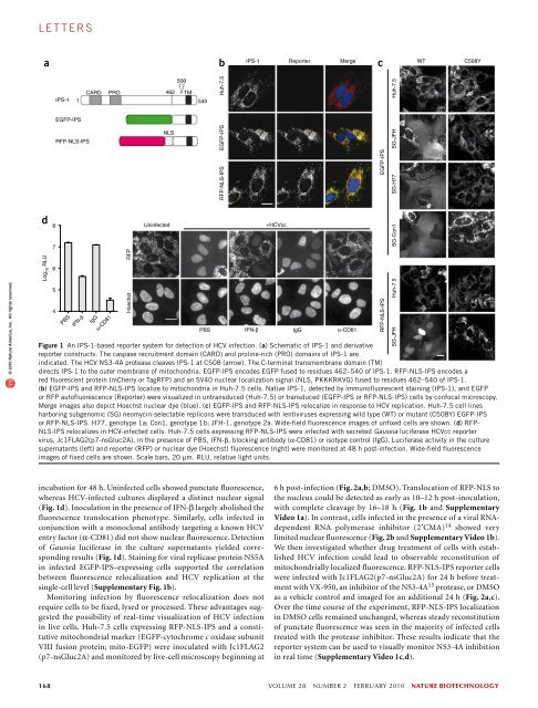
l e t t e r sReal-time imaging of hepatitis C virus infection using afluorescent cell-based reporter systemChristopher T Jones 1 , Maria Teresa Catanese 1 , Lok Man J Law 1 , Salman R Khetani 2,5 , Andrew J Syder 1,5 ,Alexander Ploss 1 , Thomas S Oh 1 , John W Schoggins 1 , Margaret R MacDonald 1 , Sangeeta N Bhatia 2–4 &Charles M Rice 1© 2010 Nature America, Inc. All rights reserved.Hepatitis C virus (HCV), which infects 2–3% of the worldpopulation, is a causative agent of chronic hepatitis and theleading indication for liver transplantation 1 . The ability topropagate HCV in cell culture (HCVcc) is a relatively recentbreakthrough and a key tool in the quest for specific antiviraltherapeutics. Monitoring HCV infection in culture generallyinvolves bulk population assays, use of genetically modifiedviruses and/or terminal processing of potentially precioussamples. Here we develop a cell-based fluorescent reportersystem that allows sensitive distinction of individual HCVinfectedcells in live or fixed samples. We demonstrate use ofthis technology for several previously intractable applications,including live-cell imaging of viral propagation and hostresponse, as well as visualizing infection of primary hepatocytecultures. Integration of this reporter with modern image-basedanalysis methods could open new doors for HCV research.For over two decades, advances in HCV assay systems have been hardwon.Methodologies have ranged from adapted selectable genomesand detection methods that require fixation or cell lysis, such asimmunostaining and quantitative RT PCR, to the use of infectiousreporter viruses 2,3 . Broadening the scope of HCV research, however,will require versatile new assays that allow sensitive single-cell analysisof infection events using unmodified viral genomes.To construct a cellular marker of HCV infection, we adapted aknown substrate of the HCV NS3-4A protease 4–6 , the mitochondriallytethered interferon (IFN)-β promoter stimulator protein 1 (IPS-1;ref. 7), also termed MAVS 8 , VISA 9 or Cardif 4 . The C-terminal region ofIPS-1, encompassing the NS3-4A recognition site and a mitochondrialtargeting sequence, was fused to green fluorescent protein (EGFP-IPS,Fig. 1a) or to the red fluorescent proteins (RFPs) mCherry or TagRFP.We also introduced an SV40 nuclear localization sequence (NLS)between the RFP variant and IPS-1 segment (RFP-NLS-IPS, Fig. 1a).Human hepatoma (Huh-7.5) cells stably transduced with lentivirusesencoding EGFP-IPS or RFP-NLS-IPS exhibited punctate fluorescenceconsistent with mitochondrial localization of the reporter, which wasconfirmed by colocalization with native IPS-1 (Fig. 1b).We determined the reporter phenotype of the EGFP-IPS or RFP-NLS-IPS constructs in the presence of NS3-4A by transduction intoHuh-7.5 cells stably expressing an autonomously replicating HCVsubgenome 10 (JFH-1 strain, SG-JFH). Replicon-harboring cellsexpressing EGFP-IPS showed diffuse fluorescence, whereas an NS3-4Acleavage–resistant form 11 of the reporter (EGFP-IPS(C508Y); Fig. 1c)exhibited a punctate pattern. Similarly, replicon-containing Huh-7.5 cells expressing RFP-NLS-IPS, but not RFP-NLS-IPS(C508Y),showed nuclear translocation of fluorescence (Fig. 1c). Both reportersdisplayed a punctate pattern in the absence of the HCV replicon(Fig. 1c). These results indicate that cleavage of EGFP-IPS and RFP-NLS-IPS are dependent on an intact NS3-4A recognition site and thatHCV-dependent fluorescence relocalization (HDFR) can be used asa marker of viral replication.HCV exists as multiple genotypes, which exhibit extensive sequencedivergence as well as differences in pathogenesis and treatment susceptibility12 . Evasion of the innate immune response by cleavage ofnative IPS-1, however, is likely to be a conserved feature of HCV infection.In addition to JFH-1 (genotype 2a), Huh-7.5 cells harboring H77(genotype 1a) or Con1 (genotype 1b) subgenomes 10 were transducedwith EGFP-IPS or EGFP-IPS(C508Y). Regardless of the HCV strain,EGFP-IPS transduction resulted in diffuse fluorescence, and EGFP-IPS(C508Y) expression led to punctate EGFP (Fig. 1c). Whereas thelack of replicon systems for other genotypes precludes comprehensiveanalysis, these results indicate that cleavage of EGFP-IPS can be usedas a marker of several diverse HCV strains. In contrast, replicationof other positive-strand RNA viruses, such as yellow fever virus orVenezuelan equine encephalitis virus, did not lead to fluorescencerelocalization (Supplementary Fig. 1a). These results suggest thatthe HDFR reporter system achieves a high level of HCV specificitycombined with genotype independence.Although replicon-containing cells constitutively express theviral proteins, monitoring authentic virus infection is important foranalyses of HCV biology and therapeutic inhibition. To determinethe ability of HDFR to detect infection, we inoculated Huh-7.5 cellsexpressing RFP-NLS-IPS with an HCVcc reporter virus expressingsecreted Gaussia luciferase, Jc1FLAG2(p7-nsGluc2A) 13 , followed by1 Center for the Study of Hepatitis C, Laboratory of Virology and Infectious Disease, The Rockefeller University, New York, New York, USA. 2 Division of Health Sciencesand Technology, Department of Electrical Engineering and Computer Science, Massachusetts Institute of Technology, Cambridge, Massachusetts, USA. 3 HowardHughes Medical Institute, 4 Division of Medicine, Brigham & Women’s Hospital, Boston, Massachusetts, USA. 5 Present addresses: Hepregen Corporation, Medford,Massachusetts, USA (S.R.K.) and iTherX Pharmaceuticals, San Diego, California, USA (A.J.S.). Correspondence should be addressed to C.M.R. (ricec@rockefeller.edu).Received 17 August 2009; accepted 4 January 2010; published online 31 January 2010; doi:10.1038/nbt.1604nature biotechnology VOLUME 28 NUMBER 2 FEBRUARY 2010 167
l e t t e r sabIPS-1 Reporter Merge WT C508YcIPS-11CARDPRO508462 TM540Huh-7.5Huh-7.5EGFP-IPSRFP-NLS-IPSNLSEGFP-IPSSG-JFH© 2010 Nature America, Inc. All rights reserved.RFP-NLS-IPSEGFP-IPSSG-H77dLog 10 RLU87654PBSIFN-βIgGα-CD81RFPHoechstUninfected+HCVccPBS IFN-β IgG α-CD81Figure 1 An IPS-1-based reporter system for detection of HCV infection. (a) Schematic of IPS-1 and derivativereporter constructs. The caspase recruitment domain (CARD) and proline-rich (PRO) domains of IPS-1 areindicated. The HCV NS3-4A protease cleaves IPS-1 at C508 (arrow). The C-terminal transmembrane domain (TM)directs IPS-1 to the outer membrane of mitochondria. EGFP-IPS encodes EGFP fused to residues 462–540 of IPS-1. RFP-NLS-IPS encodes ared fluorescent protein (mCherry or TagRFP) and an SV40 nuclear localization signal (NLS, PKKKRKVG) fused to residues 462–540 of IPS-1.(b) EGFP-IPS and RFP-NLS-IPS localize to mitochondria in Huh-7.5 cells. Native IPS-1, detected by immunofluorescent staining (IPS-1), and EGFPor RFP autofluorescence (Reporter) were visualized in untransduced (Huh-7.5) or transduced (EGFP-IPS or RFP-NLS-IPS) cells by confocal microscopy.Merge images also depict Hoechst nuclear dye (blue). (c) EGFP-IPS and RFP-NLS-IPS relocalize in response to HCV replication. Huh-7.5 cell linesharboring subgenomic (SG) neomycin-selectable replicons were transduced with lentiviruses expressing wild type (WT) or mutant (C508Y) EGFP-IPSor RFP-NLS-IPS. H77, genotype 1a; Con1, genotype 1b; JFH-1, genotype 2a. Wide-field fluorescence images of unfixed cells are shown. (d) RFP-NLS-IPS relocalizes in HCV-infected cells. Huh-7.5 cells expressing RFP-NLS-IPS were infected with secreted Gaussia luciferase HCVcc reportervirus, Jc1FLAG2(p7-nsGluc2A), in the presence of PBS, IFN-β, blocking antibody (α-CD81) or isotype control (IgG). Luciferase activity in the culturesupernatants (left) and reporter (RFP) or nuclear dye (Hoechst) fluorescence (right) were monitored at 48 h post-infection. Wide-field fluorescenceimages of fixed cells are shown. Scale bars, 20 µm. RLU, relative light units.RFP-NLS-IPSSG-Con1Huh-7.5SG-JFHincubation for 48 h. Uninfected cells showed punctate fluorescence,whereas HCV-infected cultures displayed a distinct nuclear signal(Fig. 1d). Inoculation in the presence of IFN-β largely abolished thefluorescence translocation phenotype. Similarly, cells infected inconjunction with a monoclonal antibody targeting a known HCVentry factor (α-CD81) did not show nuclear fluorescence. Detectionof Gaussia luciferase in the culture supernatants yielded correspondingresults (Fig. 1d). Staining for viral replicase protein NS5Ain infected EGFP-IPS–expressing cells supported the correlationbetween fluorescence relocalization and HCV replication at thesingle-cell level (Supplementary Fig. 1b).Monitoring infection by fluorescence relocalization does notrequire cells to be fixed, lysed or processed. These advantages suggestedthe possibility of real-time visualization of HCV infectionin live cells. Huh-7.5 cells expressing RFP-NLS-IPS and a constitutivemitochondrial marker (EGFP-cytochrome c oxidase subunitVIII fusion protein; mito-EGFP) were inoculated with Jc1FLAG2(p7-nsGluc2A) and monitored by live-cell microscopy beginning at6 h post-infection (Fig. 2a,b; DMSO). Translocation of RFP-NLS tothe nucleus could be detected as early as 10–12 h post-inoculation,with complete cleavage by 16–18 h (Fig. 1b and SupplementaryVideo 1a). In contrast, cells infected in the presence of a viral RNAdependentRNA polymerase inhibitor (2′CMA) 14 showed verylimited nuclear fluorescence (Fig. 2b and Supplementary Video 1b).We then investigated whether drug treatment of cells with establishedHCV infection could lead to observable reconstitution ofmitochondrially localized fluorescence. RFP-NLS-IPS reporter cellswere infected with Jc1FLAG2(p7-nsGluc2A) for 24 h before treatmentwith VX-950, an inhibitor of the NS3-4A 15 protease, or DMSOas a vehicle control and imaged for an additional 24 h (Fig. 2a,c).Over the time course of the experiment, RFP-NLS-IPS localizationin DMSO cells remained unchanged, whereas steady reconstitutionof punctate fluorescence was seen in the majority of infected cellstreated with the protease inhibitor. These results indicate that thereporter system can be used to visually monitor NS3-4A inhibitionin real time (Supplementary Video 1c,d).168 VOLUME 28 NUMBER 2 FEBRUARY 2010 nature biotechnology
- Page 3 and 4:
volume 28 number 2 february 2010COM
- Page 5 and 6:
in this issue© 2010 Nature America
- Page 7 and 8:
© 2010 Nature America, Inc. All ri
- Page 10 and 11:
NEWS© 2010 Nature America, Inc. Al
- Page 12 and 13:
NEWS© 2010 Nature America, Inc. Al
- Page 14 and 15:
NEWS© 2010 Nature America, Inc. Al
- Page 16 and 17:
© 2010 Nature America, Inc. All ri
- Page 18 and 19:
© 2010 Nature America, Inc. All ri
- Page 20 and 21:
© 2010 Nature America, Inc. All ri
- Page 22 and 23:
NEWS feature© 2010 Nature America,
- Page 24 and 25: uilding a businessComing to termsDa
- Page 26 and 27: uilding a business© 2010 Nature Am
- Page 28 and 29: correspondence© 2010 Nature Americ
- Page 30 and 31: correspondence© 2010 Nature Americ
- Page 32 and 33: correspondence© 2010 Nature Americ
- Page 34 and 35: correspondence© 2010 Nature Americ
- Page 36 and 37: case studyNever againcommentaryChri
- Page 38 and 39: COMMENTARY© 2010 Nature America, I
- Page 40 and 41: COMMENTARY© 2010 Nature America, I
- Page 42 and 43: patents© 2010 Nature America, Inc.
- Page 44 and 45: patents© 2010 Nature America, Inc.
- Page 46 and 47: news and viewsChIPs and regulatory
- Page 48 and 49: news and viewsFrom genomics to crop
- Page 50 and 51: news and views© 2010 Nature Americ
- Page 52 and 53: news and views© 2010 Nature Americ
- Page 54 and 55: e s o u r c eRational association o
- Page 56 and 57: e s o u r c e© 2010 Nature America
- Page 58 and 59: e s o u r c e© 2010 Nature America
- Page 60 and 61: e s o u r c e© 2010 Nature America
- Page 62 and 63: © 2010 Nature America, Inc. All ri
- Page 64 and 65: B r i e f c o m m u n i c at i o n
- Page 66 and 67: i e f c o m m u n i c at i o n sAUT
- Page 68 and 69: lettersa1.5 kb hVPrIntron 112.5 kbA
- Page 70 and 71: letters© 2010 Nature America, Inc.
- Page 72 and 73: letters© 2010 Nature America, Inc.
- Page 76 and 77: l e t t e r sFigure 2 Time-lapse li
- Page 78 and 79: l e t t e r s© 2010 Nature America
- Page 80 and 81: l e t t e r sRational design of cat
- Page 82 and 83: l e t t e r s© 2010 Nature America
- Page 84 and 85: l e t t e r s© 2010 Nature America
- Page 86 and 87: sample fluorescence was measured as
- Page 88 and 89: careers and recruitmentFourth quart