

2003; baxter - Supplements - Haematologica
2003; baxter - Supplements - Haematologica
2003; baxter - Supplements - Haematologica
- No tags were found...

You also want an ePaper? Increase the reach of your titles
YUMPU automatically turns print PDFs into web optimized ePapers that Google loves.
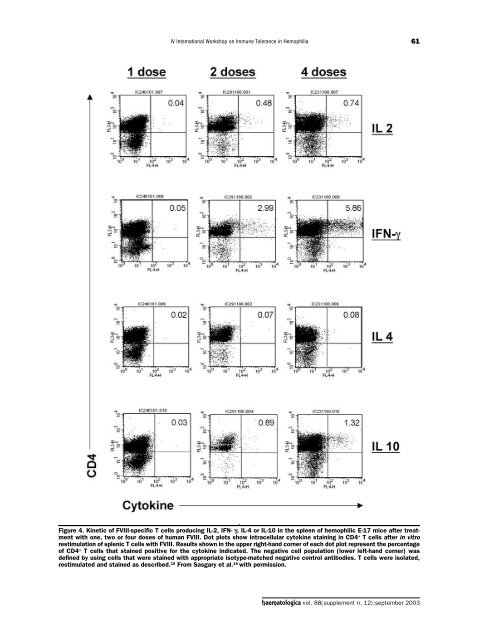
IV International Workshop on Immune Tolerance in Hemophilia61Figure 4. Kinetic of FVIII-specific T cells producing IL-2, IFN- γ, IL-4 or IL-10 in the spleen of hemophilic E-17 mice after treatmentwith one, two or four doses of human FVIII. Dot plots show intracellular cytokine staining in CD4 + T cells after in vitrorestimulation of splenic T cells with FVIII. Results shown in the upper right-hand corner of each dot plot represent the percentageof CD4 + T cells that stained positive for the cytokine indicated. The negative cell population (lower left-hand corner) wasdefined by using cells that were stained with appropriate isotype-matched negative control antibodies. T cells were isolated,restimulated and stained as described. 16 From Sasgary et al. 16 with permission.haematologica vol. 88(supplement n. 12):september <strong>2003</strong>


