Microbe Hunter Microbe Hunter - MicrobeHunter.com
Microbe Hunter Microbe Hunter - MicrobeHunter.com
Microbe Hunter Microbe Hunter - MicrobeHunter.com

Create successful ePaper yourself
Turn your PDF publications into a flip-book with our unique Google optimized e-Paper software.
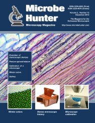
<strong>Microbe</strong><br />
<strong>Hunter</strong><br />
Microscopy Magazine<br />
ISSN 2220-4962 (Print)<br />
ISSN 2220-4970 (Online)<br />
Volume 2, Number 1<br />
January 2012<br />
The Magazine for the<br />
Enthusiast Microscopist<br />
http://www.microbehunter.<strong>com</strong><br />
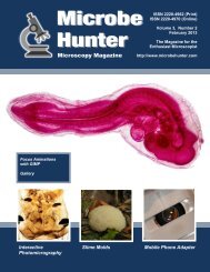
Victorian “Live Box”<br />
Microscope<br />
High Dynamic<br />
Range (HDR)<br />
Imaging<br />
Essay: Amateur<br />
Microscopy - Good<br />
for the Soul<br />
Desmid Taxonomy<br />
Stereoscopic<br />
Micrographs<br />
Mosquito Wing as<br />
a Lens Test Subject<br />
Desmid Taxonomy<br />
Testing with a<br />
Mosquito Wing<br />
Victorian “Live Box”<br />
Microscope<br />
<strong>Microbe</strong><strong>Hunter</strong> Microscopy Magazine - January 2012 - 1
ABOUT<br />
<strong>Microbe</strong>hunter Microscopy Magazine<br />
The magazine for the enthusiast microscopist<br />
<strong>Microbe</strong><strong>Hunter</strong> Magazine is a non-<strong>com</strong>mercial project.<br />
Volume 2, Number 1, January 2012<br />
ISSN 2220-4962 (Print)<br />
ISSN 2220-4970 (Online)<br />
Download: <strong>Microbe</strong>hunter Microscopy Magazine can be downloaded<br />
at: http://www.microbehunter.<strong>com</strong><br />
Print version: The printed version can be ordered at:<br />
http://microbehunter.magcloud.<strong>com</strong><br />
Publisher and editor:<br />
Oliver Kim, Ziegeleistr. 10-3, A-4490 St.Florian, Austria<br />
Email: editor@microbehunter.<strong>com</strong><br />
Web: http://www.microbehunter.<strong>com</strong><br />
Tel.: +43 680 2115051<br />
Images and Articles by:<br />
Bianchi, Gino I.<br />
Borg, David B.<br />
Clinedienst, Jeff<br />
Crosby, James D.<br />
Guwak, Mike<br />
Kim, Oliver<br />
Kreindler, R. Jordan<br />
Monzo, Luca<br />
Thomas, Anthony<br />
Copyright: By submitting articles and pictures, the authors<br />
have confirmed that they are the full copyright owners of the material.<br />
Creative <strong>com</strong>mons and public domain images are indicated<br />
with a small text next to the image or in the caption. The<br />
copyright of all other images is with the author of the article. You<br />
are not allowed to distribute this magazine by email, file sharing<br />
sites, web sites or by any other means. If you want to have a<br />
copy of this magazine, either order one from Magcloud (see link<br />
above) or vistit www.microbehunter.<strong>com</strong>.<br />
Editorial: Article and image submissions are wel<strong>com</strong>e and<br />
should be sent to: editor@microbehunter.<strong>com</strong>.<br />
For submission guidelines, consult the website at:<br />
http://www.microbehunter.<strong>com</strong>/submission<br />
Disclaimer: Articles that are published in <strong>Microbe</strong>hunter Microscopy<br />
Magazine and the blog do not necessarily reflect the position<br />
or opinion of the publisher. The publication of these articles<br />
does not constitute an endorsement of views they may express.<br />
Advice provided in <strong>Microbe</strong>hunter Microscopy Magazine is provided<br />
as a service and neither the authors nor the publisher can<br />
be held liable and responsible for any errors, omissions or inaccuracies,<br />
or for any consequences (health, hardware, etc.) arising<br />
from the use of information of this magazine and the blog (or<br />
anything else). Conduct all lab work and (microscopy) hardware<br />
modifications at your own risk and always follow the instructions<br />
of the manufacturers.<br />
Front Cover:<br />
Large image: Oliver Kim (synthetic fibers)<br />
Left image: Mike Guwak<br />
Middle image: Anthony Thomas<br />
Right image: R. Jordan Kreindler<br />
ANNOUNCEMENT<br />
Visit the Forum!<br />
It is now possible to discuss the individual articles<br />
of the magazine. Every issue has a separate subforum<br />
for discussion.<br />
www.microbehunter.<strong>com</strong>/forum<br />
Facebook<br />
Do you have any microscopy links to share?<br />
Do it here on facebook:<br />
www.facebook.<strong>com</strong>/microbehunter<br />
CONTRIBUTE!<br />
Write for <strong>Microbe</strong>hunter!<br />
Please contribute both articles and pictures. Share your experiences,<br />
problems and microscopic adventures. If you are a<br />
researcher using microscopes, tell the readers what your research<br />
is about. Please contribute, even if you consider yourself<br />
inexperienced. If you are a struggling beginner, tell us<br />
something about the problems that you encountered. If you<br />
are an active enthusiast microscopist then share your projects,<br />
experiences and observations. Are you a teacher or lecturer?<br />
Share your microscopic experiences from school or<br />
university. This magazine is made by an enthusiast microscopist<br />
for other enthusiasts. Let‘s work together to make this<br />
project a successful one.<br />
Please send all contributions to:<br />
editor@microbehunter.<strong>com</strong><br />
You must own the copyright of the contributions and you retain<br />
the copyright of all submitted articles and pictures. While<br />
we are not able to pay you for your efforts, we will, of course,<br />
give you full credit for your contributions.<br />
Guest Bloggers! Yes, guest blogging is also a possibility.<br />
Write microscopy-related blog posts, send them to me and I<br />
will publish them on the web site. Naturally, I’ll put a link to<br />
your blog. Condition: it must be original content and you must<br />
be the copyright holder of the text (obviously). When submitting<br />
articles, please indicate if you want to have them published<br />
on the blog or in the magazine (or both).<br />
Before submitting anything, please read the submissions<br />
page on the website: www.microbehunter.<strong>com</strong>/submissions.<br />
2 - <strong>Microbe</strong><strong>Hunter</strong> Microscopy Magazine - January 2012
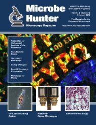
CONTENTS<br />
4 Victorian “Live Box” Microscope Capability<br />
in 40mm<br />
These small microscopes are not only attractive<br />
collectibles, but also practical excursion <strong>com</strong>panions.<br />
R. Jordan Kreindler<br />
4<br />
10 High Dynamic Range (HDR) Imaging of<br />
Micrographs<br />
Sometimes it is impossible to show both bright and<br />
dark areas of a micrograph correctly exposed. HDR<br />
imaging <strong>com</strong>bines micrographs of different exposures<br />
to obtain a single, correctly exposed picture.<br />
Oliver Kim<br />
15 Closterium costatum CORDA ex RALF<br />
Starting this issue, Mike Guwak presents a different<br />
Desmid every month.<br />
Mike Guwak<br />
15<br />
17 Making Stereoscopic Micrographs<br />
The free program Picolay can be used to <strong>com</strong>pute<br />
stereoscopic images from a focus stack.<br />
Oliver Kim<br />
17<br />
20 Gallery of Micrographs<br />
Images by Jeff Clinedienst, Gino I. Bianchi, James D.<br />
Crosby, Anthony Thomas and Luca Monzo.<br />
28 Amateur Microscopy - Good for the Soul<br />
Thinking about eye-diving into a microscope? Or are<br />
you already a devotee? A life-long fan ruminates on<br />
our fondness for looking at little things.<br />
David B. Borg<br />
32 Mosquito wing as a lens test subject<br />
How sharp is your objective?<br />
Anthony Thomas<br />
32<br />
10<br />
Answer to the puzzle (back cover):Cross section conifer needle. Pseudo DIC<br />
<strong>Microbe</strong><strong>Hunter</strong> Microscopy Magazine - January 2012 - 3
HISTORICAL<br />
MICROSCOPY<br />
“Live Box” Microscope<br />
These small microscopes are not only attractive collectibles, but also practical<br />
excursion <strong>com</strong>panions.<br />
R. Jordan Kreindler<br />
Small cylindrical microscopes<br />
were popular at the end of the<br />
19th century and the beginning of<br />
the 20th. Figure 1 presents some varieties<br />
that are still <strong>com</strong>monly available at<br />
antique malls, flea markets, and through<br />
on-line auctions. These are sometimes<br />
referred to as "Pocket Microscopes" or<br />
"Pocket Magnifiers". They are descendants<br />
of the microscope first invented<br />
by Nicolas Hartsocker (Dutch) c. 1689,<br />
and popularized by James Wilson (England)<br />
c. 1702. The cylindrical instruments<br />
shown in Figure 1 <strong>com</strong>e from a<br />
variety of makers usually with only minor<br />
variations and are generally quite<br />
similar in size. Many include a removable<br />
magnifying glass (Fig. 2).<br />
The examples in Figure 1 include<br />
two somewhat atypical microscopes at<br />
the right. The rightmost microscope displays<br />
a somewhat more conical than<br />
cylindrical shape, although of relatively<br />
similar size. See below and Figure 4 for<br />
a brief discussion of the second microscope<br />
from the right. At the far left a<br />
modern Episcope, mounted on a stand,<br />
is included for <strong>com</strong>parison.<br />
Earlier examples of cylindrical microscopes<br />
can be found in the Royal<br />
Microscopical Society collection<br />
(RMS) i which also has a related example<br />
ii . Other related examples are in the<br />
Science Museum, London iii . The Billings<br />
collection provides additional varieties<br />
iv .<br />
Some of these microscopes <strong>com</strong>e<br />
with a spring-loaded stage which is<br />
moved downward by two short lateral<br />
metal dowels to allow small slides to be<br />
viewed. (See three instruments to the<br />
rear of Fig 1.) Like the centuries earlier<br />
Wilson screw-barrel microscope, these<br />
instruments' slides are inserted in the<br />
sides of the body tube. They were often<br />
provided with a Stanhope lens.<br />
Spring-loaded stage pocket microscopes<br />
were frequently sold in cardboard<br />
or wood boxes with 3 to 6 small<br />
slides, often with one or more plain<br />
slides, a well slide, and some prepared<br />
slides with slots to store the slides.<br />
Some kits included a stand containing<br />
an adjustable mirror. The microscope<br />
could be mounted in the stand and secured<br />
with a built-in knurled screw.<br />
These microscopes were sold under<br />
a variety of names including various<br />
<strong>com</strong>binations of the words universal,<br />
pocket, and microscope. These included,<br />
e.g., "The Microscope", "Universal<br />
Pocket Microscope", "Pocket Microscope",<br />
"Universal Microscope". They<br />
were also sold as, "The Thavos Microscope",<br />
"Achromatisches Universal<br />
Figure 1. Examples of small<br />
cylindrical microscopes<br />
4 - <strong>Microbe</strong><strong>Hunter</strong> Microscopy Magazine - January 2012
“Live Box” Microscope<br />
HISTORICAL<br />
MICROSCOPY<br />
Figure 2.: A basic cylindrical<br />
microscope disassembled to show<br />
removable magnifying glass.<br />
Figure 3 (left): Storage box for Thavos microscope<br />
Figure 4 (right): Stage focusing pocket microscope<br />
<strong>Microbe</strong><strong>Hunter</strong> Microscopy Magazine - January 2012 - 5
HISTORICAL<br />
MICROSCOPY<br />
“Live Box” Microscope<br />
Figure 5: "Insectoscope", of a design<br />
sometimes attributed to Raspail.<br />
Taschenmikroskop", "The Midgard",<br />
etc. Two stands with microscopes<br />
mounted are shown at the center rear of<br />
Figure 1, A storage box with microscope,<br />
stand, and slides is shown in Fig.<br />
3. Many of these microscopes were<br />
made in Europe, particularly in Germany<br />
and France. A spring-loaded pocket<br />
microscope was discussed in an interesting<br />
paper by Martin Mach, in a Micscape<br />
article v some years ago.<br />
While many of the cylindrical microscopes<br />
shown in Figure 1 focus by<br />
movement of pressure fitting <strong>com</strong>ponents,<br />
there are cylindrical microscopes<br />
that focus via rotating threads. The microscope<br />
shown in Figure 4 has a base<br />
similar to that of a typical drum microscope.<br />
It has a threaded cylinder which<br />
can be screwed up and down the main<br />
body tube to achieve focus by effectively<br />
moving the stage. Other examples<br />
were also sold with integral mounted<br />
mirrors.<br />
One additional cylindrical microscope<br />
worthy of mention is the "Insectoscope",<br />
Fig. 5. Its design is sometimes<br />
attributed to the French political reformer<br />
and scientist François-Vincent<br />
Raspail (1794-1878). In use, an insect<br />
is placed inside the glass cylinder and<br />
the instrument focused by screwing the<br />
magnifier, located in the top section of<br />
the instrument, up or down. This "Insectoscope"<br />
is somewhat similar in purpose<br />
to the Live Box microscope.<br />
However, with its approximately 1-1/2"<br />
diameter and 4-1/2" height when open,<br />
it is almost twice the size and less portable<br />
than the approximately 2-1/3rd" tall<br />
(in working position) “Live Box"<br />
microscope. Weiner vi presents an interesting<br />
account of Raspail. While that<br />
biography does not present an "Insecto-<br />
Figure 6. Betrand "Furnace" and<br />
Pocket 'Live Box' Microscope open<br />
and closed.<br />
6 - <strong>Microbe</strong><strong>Hunter</strong> Microscopy Magazine - January 2012
“Live Box” Microscope<br />
HISTORICAL<br />
MICROSCOPY<br />
scope", it does include a picture of one<br />
of Raspail's earliest box-based instruments.<br />
It might be easy for professional<br />
microscopists to miss the importance of<br />
these small and relatively inexpensive<br />
microscopes which at the turn of the<br />
century cost about 1s vii . However, for<br />
many microscope users these were often<br />
the first, and perhaps only, microscopes<br />
they had. Their low price made<br />
these microscopes quite popular as<br />
demonstrated by the variety of manufacturers<br />
and the number available in<br />
the marketplace, even today.<br />
The microscope discussed here in its<br />
fully open state is similar in height to<br />
the Bertrand "Furnace" pocket microscope,<br />
c 1840 viii (Fig. 6), but somewhat<br />
wider. Closed it is considerably smaller<br />
than the Bertrand "Furnace" or any of<br />
the brass microscopes shown in Fig 1.<br />
Like those microscopes, it can only accept<br />
smaller slides, but unlike those<br />
instruments it is unique in having its<br />
own convenient built-in live box.<br />
The 'Live Box' microscope can be<br />
disassembled into three parts: (1) the<br />
lower section is the live box bottom,<br />
i.e., a brass cylinder terminating in a<br />
clear glass plate, (2) the middle section<br />
is the body tube with lateral openings<br />
for small slides, and also terminating in<br />
a glass plate, and (3) the upper section<br />
contains the eyepiece with pressure fitting<br />
collar (Fig. 7).<br />
The microscope focuses with a sliding<br />
tube inside a friction collar (Fig. 8),<br />
allowing it to change focus easily from<br />
slide to live box. The eyepiece is <strong>com</strong>posed<br />
of two air separated glass elements.<br />
The bottom of the lower element<br />
of the eyepiece extends below its housing<br />
and is convex. The eyepiece lens is<br />
approximately 19 mm in diameter, and<br />
the live box about 25.4 mm.<br />
Unlike most similar small microscopes<br />
the top lens of the eyepiece is not<br />
flush with the top of the instrument, but<br />
recessed approximately 7 mm.<br />
The microscope was sold in a single<br />
hinged wood box, with single front<br />
latch approximately 38.5 mm high x 50<br />
mm x 67 mm at its longest dimension.<br />
Although protective of the instrument<br />
when stored, it was not an ideal <strong>com</strong>panion<br />
for the pocket. See Fig. 12 for a<br />
more convenient carrying case. The microscope's<br />
size and design, in particular<br />
its integral live box, should make it a<br />
nice <strong>com</strong>panion for field trips. So, this<br />
raises the question of its optical performance.<br />
On page 8 are pictures taken though<br />
the microscope of two small, approximately<br />
55mm x 17mm, paper covered<br />
Victorian slides of a 'flea' and 'hair'; live<br />
box subject resolution is similar. These<br />
were adjusted in Photoshop to approach<br />
the visual view through the microscope.<br />
In use, actual views are somewhat crisper<br />
than the photographs suggest. Fig. 9<br />
shows a flea as seen in its entirety<br />
through the microscope's eyepiece. Fig.<br />
11 is a photograph through an Olympus<br />
microscope of the hair shown in Fig. 10,<br />
providing a <strong>com</strong>parison with a modern<br />
benchtop instrument. Performance is<br />
surprisingly good for an instrument only<br />
40mm tall when closed.<br />
Figure 7. 'Live Box' Microscope<br />
dissembled.<br />
Figure 8: Top view<br />
<strong>Microbe</strong><strong>Hunter</strong> Microscopy Magazine - January 2012 - 7
HISTORICAL<br />
MICROSCOPY<br />
“Live Box” Microscope<br />
Figure 9: Flea seen through 'Live<br />
Box' microscope.<br />
Figure 10 (bottom left): Hair seen<br />
through 'Live Box' microscope.<br />
Figure 11 (bottom right): Hair seen<br />
through an Olympus benchtop at<br />
100x for <strong>com</strong>parison.<br />
The full provenance of the instrument<br />
is unknown. Before I obtained the<br />
microscope, it was owned by several<br />
generations of a Western US family. It<br />
had been used extensively as demonstrated<br />
by the occasional slippage of the<br />
friction focusing tube, which was easily<br />
repaired.<br />
Although, perhaps, not quite as optically<br />
capable as e.g., a later Seibert<br />
Wetzlar Emoscope, its ability to accept<br />
both slides and its integral live box, in<br />
which specimens can be quickly enclosed,<br />
makes it an almost ideal, easy<br />
to carry <strong>com</strong>panion for field trips. I<br />
now pocket this microscope for excursions<br />
stowed in a nicely fitting case,<br />
originally made for an Edmund N.J.<br />
optical measurement viewer (Fig. 12).<br />
Small cylindrical brass microscopes<br />
are still widely available and are usually<br />
quite inexpensive, in today's, 2011,<br />
market from $10 to $50 depending on<br />
condition, <strong>com</strong>pleteness, and of course<br />
the buyers present at the time of sale. A<br />
similar example to the one presented<br />
here sold with wood case on eBay in<br />
Apr 2011 for $50 ix .<br />
The 'Live Box' style of pocket microscope<br />
discussed here occasionally<br />
<strong>com</strong>es to market, and although not up to<br />
professional quality, these instruments<br />
are still quite capable microscopes that<br />
make excellent, <strong>com</strong>fortable to carry,<br />
and convenient <strong>com</strong>panions for casual<br />
outings.<br />
The author wel<strong>com</strong>es any suggestions<br />
for corrections or improvement.<br />
He is always pleased to learn about any<br />
unique small brass cylindrical microscopes.<br />
He can be contacted at,<br />
R. Jordan Kreindler:<br />
leona111@bellsouth.net<br />
©2011 Text and photographs by the<br />
author.<br />
■<br />
8 - <strong>Microbe</strong><strong>Hunter</strong> Microscopy Magazine - January 2012
“Live Box” Microscope<br />
HISTORICAL<br />
MICROSCOPY<br />
Figure 12. Carrying case for 'Live<br />
Box' Microscope, adapted from an<br />
Edmund, N.J. measurement tool<br />
References<br />
i Turner, G.L'E. The Great Age of the Microscope. The Collection of the Royal Microscopical Society through 150 Years.<br />
Adam Hilger: Bristol, England and New York, 1989, Figs. 262, 265, 266, 267, and 268<br />
ii Ibid, Fig. 310.<br />
iii Bracegirdle, Brain. A Catalog of the Microscopy Collection of The Science Museum, London. 2005, Item 1/32 - "Pocket<br />
Microscope", 1/33 "Pocket Microscopes", 4/31 "Pocket Microscope", etc.<br />
iv Purtle, Helen R. (ed.). The Billings Microscope Collection. Second Edition. Armed Forces Institute of Pathology:<br />
Washington, D.C., 1974, Figs 294, 296, 314, 393<br />
v Mach, Martin. 30 g of microscope please! Micscape Magazine, August 2000<br />
vi Weiner, Dora B. RASPAIL SCIENTIST and REFORMER. New York: Columbia University Press, 1968<br />
vii Ibid iii, 4/27 "Pocket Microscope"<br />
viii Moe, Harald. The Story of the Microscope. Rhodos: Denmark, 2004, p 189<br />
ix US eBay, April 14, 2011, Item ID 290553686109, 4 bidders, 9 bids, USD<br />
<strong>Microbe</strong><strong>Hunter</strong> Microscopy Magazine - January 2012 - 9
IMAGE PROCESSING<br />
Exposure control<br />
Sometimes it is impossible to show both bright and dark areas of a micrograph correctly<br />
exposed. HDR imaging <strong>com</strong>bines micrographs of different exposures to obtain a single,<br />
correctly exposed picture. This article shows three ways on how to do this.<br />
Oliver Kim<br />
Some microscopic specimens can<br />
have a large difference in bright<br />
and dark areas. Insects and other<br />
arthropods, for example, have a dark<br />
chitin exoskeleton which does not allow<br />
much light to go through. In order to see<br />
structural details of the insect's body, it<br />
is necessary to turn the microscope's<br />
lamp up and it is also necessary to open<br />
the condenser aperture diaphragm. This,<br />
however, may cause other parts of the<br />
specimens, such as the thinner legs, to<br />
be<strong>com</strong>e overexposed. In extreme cases,<br />
the finer structures are not visible at all,<br />
as they are <strong>com</strong>pletely flooded by light.<br />
Often contrast adjustment using digital<br />
image processing is not a solution.<br />
One may consider to use the “levels”<br />
tool of image editing software to make<br />
the dark areas of the image brighter and<br />
the bright areas darker. This only works<br />
to a point, however. Image information<br />
which is not there in the first place can<br />
not be recovered this way (Figure 3).<br />
Many microscopic cameras are not<br />
able to capture the full range of brightness.<br />
Some pixels of the digital camera<br />
may go into saturation (in the bright<br />
parts), and are thus not capable of capturing<br />
the image information in these<br />
areas. Other parts of the camera’s sensor<br />
receive so little light that even increasing<br />
the brightness in these areas<br />
will not reveal much image detail (Figure<br />
3 illustrates this).<br />
This issue can be over<strong>com</strong>e to a<br />
certain extent by using digital SLR cameras<br />
that have the capability of recording<br />
image in the RAW format. This<br />
format is capable of storing a far greater<br />
color depth than JPG images, and it may<br />
1 2<br />
D<br />
A<br />
B<br />
C<br />
3<br />
E<br />
Figures 1 and 2: The original images used show the mandibles of a cockroach.<br />
Figure 1 correctly shows small hair and irregularities in the chitin exoskeleton (A).<br />
At the same time the dark structures (B) reveal very little detail and appear to<br />
conglomerate into one large mass. Opening the condenser will reveal more<br />
details (C) but also results in highly over exposed areas (D). The hair disappear<br />
<strong>com</strong>pletely.<br />
Figure 3: This image shows what happens if one attempts to increase the<br />
brightness of figure 1. The image starts to look posterized, but not much more<br />
image detail is revealed in the dark areas (E).<br />
10 - <strong>Microbe</strong><strong>Hunter</strong> Microscopy Magazine - January 2012
Exposure control<br />
IMAGE PROCESSING<br />
4: PhotoShop: simple blending 5: PhotoShop: smart blending<br />
6: Enfuse blending 7: PhotoShop: 50% opacity of layers<br />
<strong>Microbe</strong><strong>Hunter</strong> Microscopy Magazine - January 2012 - 11
IMAGE PROCESSING<br />
Exposure control<br />
Figures 8 and 9: The original images to be blended (a tick). This was a difficult specimen. The condenser had to be opened<br />
widely to allow light to pass through the specimen. This resulted in very over exposed parts, of especially the legs.<br />
Figures 4-7 (previous page) and 10-13: Comparison of different blending tools. PhotoShop’s “simple blending” as well as the<br />
blending done by Enfuse resulted in the best images. Smart blending did produce considerable artefacts, especially around<br />
the edges of the specimen. Adjusting the opacity of the layer (image 13) also produced acceptable results.<br />
therefore be possible to recover more<br />
details from the darker areas of the picture.<br />
I have not yet tried this, however.<br />
Combining several images of<br />
different exposures<br />
There is, however, also a different<br />
approach of solving the HDR problem.<br />
If the dynamic range of the specimen is<br />
too large, then it is also possible to<br />
<strong>com</strong>bine several images of different<br />
brightnesses into one correctly exposed<br />
final image.<br />
The specimen is first photographed<br />
several times, with different condenser<br />
and light intensity settings. The different<br />
images will then have different parts<br />
of the specimen correctly exposed, with<br />
other parts either being too dark or too<br />
bright.<br />
Figures 1 and 2 show the mandibles<br />
of a cockroach. Some parts of figure 1<br />
are underexposed (the black circular<br />
structures at the center-bottom). In figure<br />
2 these structures are correctly exposed,<br />
but the area between the<br />
mandibles is now white and lacks detail.<br />
The hair on the cockroach are also not<br />
visible anymore. Both, the dark and the<br />
bright part, has experienced a loss of<br />
image information. These two images<br />
can now be <strong>com</strong>bined into an image<br />
with an overall correct exposure (Figures<br />
4-7). Figures 8 and 9 show an even<br />
more extreme example (a tick), with<br />
highly underexposed and overexposed<br />
parts.<br />
Enfuse<br />
I have experimented with wo different<br />
programs. The program Enfuse can<br />
be freely downloaded from<br />
http://enblend.sourceforge.net/. It does<br />
not require installation. Enfuse is used<br />
over the <strong>com</strong>mand line in Windows. I<br />
have found out that the standard settings<br />
of the program produces very satisfactory<br />
results. Blending a large number of<br />
images together is easy, as one only has<br />
to specify the file names in a separate<br />
text file.<br />
Photomerge<br />
The second program that I used was<br />
the <strong>com</strong>mercial program PhotoShop Elements<br />
11.0. The Photomerge Exposure<br />
tool offers several modes (simple blending<br />
and smart blending), including a<br />
manual mode. The simple blending<br />
mode is a single-click option, in which<br />
the program uses a pre-defined set of<br />
parameters. The smart-blending mode<br />
allows the user to control color contrast<br />
and saturation over sliders.<br />
This tool can be accessed by over<br />
the menu (File - New - Photomerge<br />
Exposure). PhotoShop blending is done<br />
<strong>com</strong>pletely graphically and requires only<br />
a few mouse clicks. This may be an<br />
advantage for those people who are not<br />
familiar with the <strong>com</strong>mand line in the<br />
DOS box of Windows. You can download<br />
a 30 day trial version of this program<br />
from the Adobe website, to check<br />
if you are satisfied with the functionality<br />
of this tool.<br />
Layer Opacity Control<br />
Last, I tried to increase the dynamic<br />
range by blending two layers. This can<br />
be done also with PhotoShop or with the<br />
free program Gimp (or any other program<br />
supporting layers). First, the two<br />
original images are copied each into one<br />
separate layer. The opacity control is<br />
then used to make the top layer transparent.<br />
Choose a value of 50% to weigh<br />
the two images equally. PhotoShop’s<br />
Simple Blending tool and the 50%<br />
blending using layers produced very<br />
similar images. I suppose that the algorithm<br />
used to <strong>com</strong>pute these images is<br />
the same.<br />
On page 14, I have included a brief<br />
explanation on how to blend images<br />
using Enfuse and layers.<br />
■<br />
12 - <strong>Microbe</strong><strong>Hunter</strong> Microscopy Magazine - January 2012
Exposure control<br />
IMAGE PROCESSING<br />
10: PhotoShop:<br />
simple blending<br />
11: PhotoShop:<br />
smart blending<br />
12: Enfuse blending<br />
13: PhotoShop:<br />
50% opacity of layers<br />
<strong>Microbe</strong><strong>Hunter</strong> Microscopy Magazine - January 2012 - 13
IMAGE PROCESSING<br />
Exposure Control<br />
Using Enfuse<br />
Using Layers<br />
Here you need a graphics program<br />
which is capable of supporting layers.<br />
The screenshots show Photo-<br />
Shop.<br />
Step 1: Download enfuse from<br />
enblend.sourceforge.net and copy<br />
the file enfuse.exe into the directory<br />
containing your original images.<br />
(no installation required).<br />
Step 2: Use a text editor to make a<br />
list (called list.txt) of original files<br />
that are to be processed. Press Enter<br />
at the end of each line. The last<br />
line must be empty.<br />
Step 1: Open the first original image<br />
and click on the Create a new layer<br />
button<br />
Step 2: A new empty layer is created<br />
on top of the image.<br />
Step 3: Open the second image,<br />
press CTRL-A to select the whole image<br />
and CTRL-C to copy the image to<br />
the clipboard.<br />
Step 3: Open the <strong>com</strong>mand line window<br />
by running the program cmd<br />
from the start menu. Change into<br />
the directory containing the program<br />
enfuse and the original images.<br />
Type:<br />
enfuse @list.txt<br />
to process the files.<br />
Step 4: The program has now generated<br />
a file called a.tif. This is the<br />
output file containing the HDR image.<br />
Step 4: Switch back to the first image<br />
and press CTRL-V to paste the second<br />
image into the empty layer 1, on<br />
top of the first image.<br />
Step 5: Use the horizontal slider<br />
(green circle) to adjust the opacity of<br />
the top layer. The two images will be<br />
averaged this way.<br />
Step 6: Merge all layers or flatten the<br />
image (menu Layers). Then save the<br />
new image.<br />
14 - <strong>Microbe</strong><strong>Hunter</strong> Microscopy Magazine - January 2012
Reference Plate<br />
DESMIDS<br />
Closterium costatum CORDA ex RALF<br />
Mike Guwak<br />
1<br />
Name: Closterium costatum<br />
CORDA ex RALFS var. costatum<br />
Original publication: Ralfs, J.<br />
(1848) p. 170 and p. 312.<br />
Synonym: Closterium striolatum<br />
EHR. var. costatum (CORDA)<br />
KLEBS<br />
Origin of name: The name costatum<br />
refers to the characteristic costae, the<br />
striations in the cell wall (Fig. 2).<br />
Size: 200-400mm by 30-45mm<br />
Shape: The cells are 6-10 times longer<br />
than wide and are arched. The<br />
cell wall is striated. In contrast to<br />
other Closterium species, the striae<br />
are conspicuous, far apart and riblike<br />
(costate). Slight dots are visible<br />
between the striae. This costate appearance<br />
is a distinguishing characteristic<br />
for this species and allows for a differentiation<br />
from the similar looking C.<br />
striolatum, whose striae are much<br />
denser. The apices (Figs. 3, 4) are<br />
widely rounded and possess a thickened<br />
cell wall.<br />
Occurrence: This is a very adaptable<br />
and widely distributed species. It has<br />
been found in acidic moorland pools as<br />
well as in neutral and even weakly<br />
alkaline lake shore zones.<br />
Note: The images show dead, empty<br />
cells. Live cells are green due to their<br />
chloroplasts. The striae can be seen<br />
better in dead cells.<br />
References<br />
"Closterium costatum Corda ex Ralfs" Algaebase:<br />
Listing the World's Algae. N.p., n.d.<br />
Web. 15 Jan. 2012.<br />
.<br />
Lenzenweger, Rupert (1996). Desmidiaceenflora<br />
von Österreich: Teil 1. Berlin, Stuttgart:<br />
J. Cramer<br />
Ralfs, J. (1848). The British Desmidieae. London:<br />
Reeve, Benham & Reeve.<br />
Image Credit<br />
The images are copyrighted by<br />
Mike Guwak (2012).<br />
http://www.mikroskopie-main-taunus.de<br />
mike.guwak@mikroskopie-main-taunus.de<br />
<strong>Microbe</strong><strong>Hunter</strong> Microscopy Magazine - January 2012 - 15
DESMIDS<br />
Reference Plate<br />
2<br />
3 4<br />
16 - <strong>Microbe</strong><strong>Hunter</strong> Microscopy Magazine - January 2012
Radiolarians in 3D<br />
STACKING<br />
Yes, the pictures on these pages are real micrographs and not drawings! The free program<br />
Picolay can be used to <strong>com</strong>pute stereoscopic images from a focus stack.<br />
Oliver Kim<br />
On these pages, I want to show<br />
you some stereoscopic imaged<br />
of radiolarians. The images on<br />
page 16 and 17 can be viewed by crosseyed<br />
viewing. The two images on page<br />
18 are anaglyphs, which require the use<br />
of red-cyan 3D glasses.<br />
Cross-eyed viewing does not need<br />
glasses. Look at the images and cross<br />
your eyes until the two images start to<br />
appear as four (out of focus) images.<br />
Then slowly uncross your vision so that<br />
the middle two of the four images overlap.<br />
You will then see the radiolarian in<br />
3D.<br />
The images on page 18 require<br />
anaglyph glasses. The right eye looks<br />
through a blue/cyan foul, while the left<br />
eye looks through a red foil. These<br />
glasses can also be made at home, by<br />
using an ink-jet printer and printing the<br />
two colors on an overhead transparency<br />
foil.<br />
How the images were made<br />
The free program Picolay was used<br />
to make these images. The program is a<br />
stacking software, which is able to <strong>com</strong>bine<br />
different images that have different<br />
parts in focus into one single image, one<br />
which is in focus throughout. The program<br />
is also able to <strong>com</strong>pute stereoscopic<br />
images from the stack. The<br />
condenser diaphragm should be opened<br />
to lower the depth of field. This way<br />
many pictures have to be taken and it is<br />
the task of the program to assemble the<br />
many pictures into a final image. The<br />
intensity of the stereoscopic effect can<br />
be specified as well.<br />
The images were originally in color,<br />
but the anaglyphs can be can be better<br />
viewed in grayscale. The program allows<br />
for a conversion to grayscale.<br />
The procedure was relatively simple. A<br />
series of about 20 images were taken,<br />
each picture at a different focus. The<br />
program assumes that the first image is<br />
the top most image and that all further<br />
images are deeper into the sample. The<br />
images were loaded into Picolay and<br />
stacking was started by pressing the F1<br />
key. After <strong>com</strong>pletion, a new menu (3D<br />
edit) starts to appear. It is then possible<br />
to convert the stacked image into either<br />
a red-cyan anaglyph or into images for<br />
cross-eyed viewing. The newly generated<br />
images are all automatically saved.<br />
The program is relatively elaborate<br />
and allows for much fine-tuning. The<br />
program can be downloaded from:<br />
www.picolay.de. It is advisable to study<br />
the documentation to fully understand<br />
all of the features. I have found out that<br />
the standard settings provide very good<br />
results for radiolarians, however.<br />
Help for cross-eyed viewing:<br />
http://www.starosta.<strong>com</strong>/3dshowcase/ihelp.html<br />
<strong>Microbe</strong><strong>Hunter</strong> Microscopy Magazine - January 2012 - 17
STACKING<br />
Radiolarians in 3D<br />
18 - <strong>Microbe</strong><strong>Hunter</strong> Microscopy Magazine - January 2012
Radiolarians in 3D<br />
STACKING<br />
These images erquire red-cyan 3D<br />
glasses (red over the left eye, cyan<br />
over the right).<br />
<strong>Microbe</strong><strong>Hunter</strong> Microscopy Magazine - January 2012 - 19
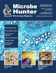
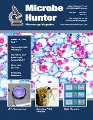
GALLERY<br />
Crystals<br />
A <strong>com</strong>mon pain relief medication with acetaminophen, aspirin, and caffeine as the main constituents. Stack of 5 images using<br />
CombineZP. Partially polarized and oblique lighting. Amscope T490, 10x objective, Canon 7D. By Jeff Clinedinst (www.jclinedinst.<strong>com</strong>)<br />
Crystalized B/W film developer containing phenidone, hydroquinone, sodium sulfite and sodium bisulfite. Polarized lighting using Amscope<br />
T490, 4x objective, Canon 7D. By Jeff Clinedinst<br />
20 - <strong>Microbe</strong><strong>Hunter</strong> Microscopy Magazine - December January 2012 2011 - Send images to editor@microbehunter.<strong>com</strong>
Larva and Histology<br />
GALLERY<br />
Mosquito larva in darkfield.<br />
Amscope T490, 10x objective,<br />
Canon 7D. By Jeff Clinedinst<br />
Top: A 7 micron histologic slide of human breast with lactational change , colored with hematoxilin and eosin. Camera used: Coolpix 5000<br />
with adapter. Labophot microscope with Nikon e-plan objective. By Gino I. Bianchi<br />
Send images to editor@microbehunter.<strong>com</strong> - <strong>Microbe</strong><strong>Hunter</strong> Microscopy Magazine - December - January 2011 2012 - 21
GALLERY<br />
Lichens<br />
Top and right: Lichen from King’s<br />
Canyon, CA<br />
Page 23, top: Easter moss<br />
Page 24, bottom: “The lonely<br />
sporophyte” (moss).<br />
All images on page 23 and 24 by<br />
James D. Crosby<br />
22 - <strong>Microbe</strong><strong>Hunter</strong> Microscopy Magazine - December January 2012 2011 - Send images to editor@microbehunter.<strong>com</strong>
Moss<br />
GALLERY<br />
Send images to editor@microbehunter.<strong>com</strong> - <strong>Microbe</strong><strong>Hunter</strong> Microscopy Magazine - December - January 2012 2011 - 23
GALLERY<br />
Butterfly Eggs<br />
The three images show the eggs of Mourning Cloak butterfly (Nymphalis antiopa)<br />
Right: yellow freshly laid egg mass.<br />
Bottom: Images of single eggs darker than when freshly laid due to developing<br />
caterpillar; top view and side view.<br />
By Anthony Thomas<br />
24 - <strong>Microbe</strong><strong>Hunter</strong> Microscopy Magazine - December January 2012 2011 - Send images to editor@microbehunter.<strong>com</strong>
Aquarium Life<br />
GALLERY<br />
The top two images were obtained with<br />
a Bresser trinocular microscope (Trino<br />
Researcher II model). They show an<br />
amoeba with cyanobacteria inside. The<br />
sample was taken from the algal<br />
material of my aquarium. I used a patch<br />
of black paper to obtain an oblique<br />
illumination. The photos were processed<br />
with Acdsee software, increasing the<br />
contrast and detail with the tool<br />
“unsharp mask”.<br />
The image on the left was obtained with<br />
the same technique of the previous<br />
images. For all the pictures I used a<br />
SLR Canon Eos 350D at 1600 ISO and<br />
automatic exposure with the software<br />
"DsrlRemote" to control remotely the<br />
camera from my laptop PC.<br />
By Luca Monzo<br />
Send images to editor@microbehunter.<strong>com</strong> - <strong>Microbe</strong><strong>Hunter</strong> Microscopy Magazine - December - January 2011 2012 - 25
GALLERY<br />
Water Life<br />
The images on this double page show water fleas of my little<br />
water tank. The original source is a fountain located in the public<br />
garden of Udine, Italy (see photo on the left). I used a piece of<br />
black paper to obtain oblique illumination on some images. I<br />
processed them with Helicon Focus or ACDSee software.<br />
By Luca Monzo<br />
26 - <strong>Microbe</strong><strong>Hunter</strong> Microscopy Magazine - December January 2012 2011 - Send images to editor@microbehunter.<strong>com</strong>
Water Life<br />
GALLERY<br />
Send images to editor@microbehunter.<strong>com</strong> - <strong>Microbe</strong><strong>Hunter</strong> Microscopy Magazine - December - January 2011 2012 - 27
ESSAY<br />
Thinking about eye-diving into a microscope? Or are you already a devotee?<br />
A life-long fan ruminates on our fondness for looking at little things.<br />
David B. Borg<br />
Oliver Kim, the exemplary<br />
founder of <strong>Microbe</strong> <strong>Hunter</strong>,<br />
states the website and magazine's<br />
main mission is to encourage and<br />
motivate. He wants this project to induce<br />
enthusiasts to broaden their interests<br />
in the miniature world and share the<br />
results. He also hopes to inspire new<strong>com</strong>ers<br />
to enter into the magical realm<br />
of microscopes for the first time. In<br />
support of these laudable aspirations I<br />
offer a few thoughts – some 'philosophizing'<br />
– for everyone's consideration,<br />
but especially for novices, on what<br />
makes our “great hobby and pastime”<br />
so satisfying and worthwhile.<br />
On microscopy itself<br />
“The development of microscopy<br />
revolutionized biology and remains an<br />
essential technique in the life and biological<br />
sciences.” (From an article in<br />
Wikipedia)<br />
The <strong>com</strong>pelling word in that sentence<br />
is: “remains”. Consider that light<br />
microscopy, invented centuries ago, has<br />
not yet been made obsolete and replaced<br />
by engineering advancements based on<br />
totally different chemistry and physics.<br />
How many other vital technologies are<br />
there in our lives today about which the<br />
same may be said? Very few.<br />
Breakthroughs such as electricity,<br />
internal <strong>com</strong>bustion engines, electromagnetic<br />
wave transmission, electronics<br />
and digital technologies have<br />
consigned scores of antiquated technologies<br />
to the dustbin over the last century<br />
or so. Nothing strange about that – in<br />
fact it is expected. That's why it is<br />
notable (even thrilling, if you are the<br />
contemplative sort) to find a venerable<br />
way of doing something important –<br />
very important – still in the forefront.<br />
Optical magnifying lenses have not<br />
only survived, they continue to triumph.<br />
Thus far, no one has <strong>com</strong>e up with a<br />
better, more economical way of resolving<br />
small objects, particularly tiny living<br />
things. And note that people have<br />
been crafting glass for this purpose for<br />
four hundred years. (The invention of<br />
the <strong>com</strong>pound microscope is often credited<br />
to Dutch spectacle-makers Hans<br />
Jansen and his son Zacharias in 1590.)<br />
Engineering continues to improve<br />
lens-making and microscope design,<br />
but the basic physics has not been superseded<br />
in any practical way. That lets<br />
you, the amateur microscopist, stand<br />
side by side with the ghosts of Antonie<br />
van Leeuwenhoek and Robert Hooke.<br />
You can feel the same astonishment and<br />
exhilaration they did in seeing cells and<br />
“animalcules” for the first time in your<br />
life, in exactly the same way, which is<br />
still the best way.<br />
Empathy bridging centuries. Microscopy<br />
is an venture whose living<br />
history is continuous and progressive,<br />
not a resuscitated tradition whose time<br />
of relevance and innovation came and<br />
went. There are no “re-enactors” in our<br />
endeavor.<br />
On the noble tradition<br />
of the amateur<br />
“Amateur” is a word whose reputation<br />
has been degraded by changing<br />
times. Currently, the adjective shares in<br />
most people's minds a dual definition.<br />
One is neutral (engaging in a pursuit<br />
without payment; nonprofessional).<br />
But the other is disparaging (contemptibly<br />
inept or unskillful). How much<br />
worse could one's sincere efforts be<br />
denigrated than by being described as<br />
“amateurish”?<br />
It is a shame to have <strong>com</strong>e this.<br />
There was a time when amateur scientists<br />
were so highly regarded that prestigious<br />
institutions were <strong>com</strong>missioned<br />
by courts and governments to encourage<br />
their efforts and allow the benefits<br />
of their discoveries to accrue to the<br />
greater good of all citizens and subjects.<br />
(The Royal Society of London for Improving<br />
Natural Knowledge, was<br />
founded in November 1660 by King<br />
Charles II's grant of Royal Charter. It is<br />
possibly the oldest such society in existence.)<br />
Back then, an Amateur was someone<br />
with time on his hands, which<br />
meant he (or alas, rarely, she) was educated<br />
and had money – someone of<br />
aristocratic birth, inherited wealth or<br />
deserving of patronage by way of an<br />
evident and prodigious brilliance.<br />
These scientists were often free of<br />
the stifling constraints of <strong>com</strong>mercial<br />
considerations; they pursued their interests<br />
with abandon and without deadlines,<br />
and most shared the results of<br />
their efforts with the world without<br />
avarice. In our avocation, we 21st century<br />
amateur microscopists can be just<br />
as free.<br />
We can rejuvenate the older definition's<br />
sense of altruistic virtue with our<br />
own attitudes and behavior. And what<br />
better attitude to have than that which is<br />
at the etymological heart of the word<br />
itself:<br />
ORIGIN late 18th cent.:<br />
from French amateur, 'lover of',<br />
from Italian amatore,<br />
from Latin amator ‘lover,’<br />
from amare ‘to love.’<br />
28 - <strong>Microbe</strong><strong>Hunter</strong> Microscopy Magazine - January 2012
Amateur Microscopy – Good for the Soul<br />
ESSAY<br />
(Image: Oliver Kim)<br />
Citric acid in polarized light.<br />
And reflect upon whose footsteps<br />
we can humbly follow, in earnestness if<br />
not necessarily with similarly towering<br />
intellects: Charles Darwin and Gregor<br />
Mendel were “amateurs”.<br />
On left brain/right brain<br />
Thinking and feeling. Analyzing<br />
and creating. Science and Art. Do<br />
these divergent human faculties ever<br />
intersect? Constantly – often in moments<br />
of supreme insight.<br />
Take the music of Mozart for example.<br />
Are there mathematics in its beauty<br />
or beauty in its mathematics? Or Einstein's<br />
formulations. Did he think the<br />
sublime or did the sublime condescend<br />
to “think” him?<br />
As a musician (formerly professional<br />
and now happily “amateur” again) I<br />
sometimes feel a tangible expression of<br />
music, already existing in perfection far<br />
above me, show me the way by allowing<br />
a tiny shadow of itself to alight for a<br />
moment in the lower world of sound<br />
waves in air. It turns me into its temporary<br />
vessel, a self-less screen on which<br />
to project the slightest hint of its grace.<br />
Afterwards I ponder, with all the<br />
self-love ego can muster, “Wow, how<br />
did I do that?” Of course, “I” was not<br />
there at the time and had nothing to do<br />
with it. But my rapture in such experiences<br />
is transcendent. Hardly my private<br />
domain – anybody doing anything<br />
can find themselves in the “Zone” if<br />
they work at it. Many do.<br />
What work? My job is to strive with<br />
my best effort and be fully present with<br />
no expectations. My job is to study and<br />
practice never-endingly. My job, as the<br />
vocabulary of technical proficiency and<br />
mental understanding grows, is to open<br />
the door to emotion, let the vocabulary<br />
flow in to find expression and quietly<br />
invite beauty to join me.<br />
The job description is exactly the<br />
same with microscopy. Follow your<br />
bliss, as Joseph Campbell said, and joy<br />
will be yours if you apply yourself.<br />
There is an infinity of science to learn<br />
with an organized mind, and no less to<br />
work with creatively. One side of your<br />
brain stimulates the other.<br />
The microscope magnifies and resolves.<br />
Your mind and heart perceive<br />
and synthesize. The more you understand<br />
about what you are looking at, the<br />
more you see, and the more beauty is<br />
disclosed and admired. This all leads to<br />
critical insight and new understanding.<br />
Art and science merge and advance<br />
together.<br />
I must admit, biology might never<br />
have been my favorite subject, nor<br />
would microscopes have held so much<br />
interest for me had it not been for the<br />
pictures! Images feed my soul, as does<br />
music. And, just as with other fine art,<br />
the interpretation and meaning associated<br />
with elegant microscopic images expands<br />
appreciation far beyond the mere<br />
perception of abstract designs.<br />
<strong>Microbe</strong><strong>Hunter</strong> Microscopy Magazine - January 2012 - 29
ESSAY<br />
Amateur Microscopy – Good for the Soul<br />
(Image: Oliver Kim)<br />
Today, with the digital technology<br />
of photo-microscopy so available and<br />
diverse, an arena of boundless creativity<br />
is open to us that was mostly closed to<br />
the frugal amateur just a decade or two<br />
ago. And there is a booming <strong>com</strong>munity<br />
out there (of which <strong>Microbe</strong> <strong>Hunter</strong><br />
is a very friendly subset) with which to<br />
share splendid images. I cite, as the<br />
lofty tip of the iceberg, the yearly festivals<br />
put on by Nikon (Small World) and<br />
Olympus (BioScapes). Google, gawk,<br />
emulate and enter. Join the party; post<br />
a picture.<br />
On serendipity<br />
You never know what, exactly, you<br />
are going to see, and what you do see<br />
through a microscope is always unique<br />
to you. Even if your subject is <strong>com</strong>monplace,<br />
your awareness, if sharp<br />
enough, and your mind, if open enough,<br />
will discover something new, possibly<br />
even “important”, but certainly fascinating<br />
to you, if no one else. Developing<br />
these qualities in yourself –<br />
awareness and openness – rewards you<br />
with the sustained elation of continuous<br />
adventure!<br />
Microscopy is also a perfect domain<br />
in which to test the truth of the old<br />
maxim “Luck is where opportunity<br />
meets preparation”. There is opportunity<br />
every time you examine a specimen.<br />
Diligence and persistence define preparation.<br />
When you reflexively snap the shutter<br />
on that bewitching image that suddenly<br />
– unexpectedly – appears in your<br />
field of view, or when that technical<br />
“accident” produces results ten times<br />
better than anticipated, some may think<br />
it is good fortune. But we know better.<br />
You get to enjoy the private pleasure of<br />
appearing to be smiled upon by “luck”<br />
while knowing that you yourself were a<br />
willing and essential participant, and<br />
that your next piece of luck is right<br />
around the corner.<br />
On the directness of perception<br />
Imagine accessing information –<br />
truth – that has not been debased by spin<br />
doctors.<br />
Imagine this truth emanating from<br />
the moment to moment reality of an<br />
unfolding universe of being.<br />
Imagine yourself perceiving this<br />
truth directly from its source in the form<br />
of visual images produced by your eyes<br />
and brain.<br />
Stem of a sunflower, inverted colors.<br />
Imagine your perception being assisted<br />
only by the applied understanding<br />
of natural laws, developed over several<br />
millennia by synergistic scientific discovery,<br />
in the form of instruments and<br />
techniques that make the formerly unseeable,<br />
seeable.<br />
Intrigued? This is the great gift of<br />
microscopy. It is pure. It is unlike so<br />
many readily available “entertainments”<br />
and distractions, digital and otherwise.<br />
There is no interposed opinion,<br />
political agenda, religious belief, cultural<br />
bias, market research or profit motive<br />
shaping your perception or influencing<br />
your interpretation of it.<br />
You are sitting there in your chair.<br />
Something real is sitting there on a<br />
slide. A tiny slice of the electromagnetic<br />
spectrum of radiant energy is mediating<br />
the space between you and that<br />
something real through a handful of<br />
chunks of carefully crafted clear glass.<br />
And suddenly you are in direct <strong>com</strong>munication<br />
with a formerly invisible, unimaginable<br />
world of incalculable<br />
vastness and gracefully organized <strong>com</strong>plexity.<br />
You are touching the infinite with<br />
your own eyes, unfiltered – undefiled –<br />
by narrow human interests. Realize<br />
that, be receptive enough, and awe will<br />
sweep through you. This kind of awe is<br />
good for what ails all of us. It is what<br />
keeps me <strong>com</strong>ing back to the microscope<br />
for another peek.<br />
On humility through the<br />
experience of scale<br />
Would you like to generate within yourself<br />
the healthiest of humblenesses?<br />
Here's a recipe:<br />
1. Calculate the linear measurements<br />
of various specimen features under<br />
magnification every time you focus<br />
your microscope. In other words, always<br />
know the actual sizes of the objects<br />
you are looking at.<br />
2. Consciously relate them again<br />
and again to your own body's measurements<br />
and those of planets, molecules,<br />
galaxies, subatomic particles and the<br />
30 - <strong>Microbe</strong><strong>Hunter</strong> Microscopy Magazine - January 2012
Amateur Microscopy – Good for the Soul<br />
ESSAY<br />
expanding, curving vastness of<br />
space/time.<br />
Make a personal study of scale as a<br />
concept. Consider nanometers and light<br />
years and everything in between and<br />
beyond in both directions; feel them in<br />
your bones as units and multiples of<br />
arbitrary increments on one impossibly<br />
long tape measure, the ends of which<br />
never end.<br />
3. Reflect on the limitless elegance<br />
of this continuum of scale, inside of<br />
which you are apparently sandwiched<br />
exactly at the theoretical center-point.<br />
Yes, think about that.<br />
Look at your hand. Know deeply<br />
that there is a bacterium, 3 micrometers<br />
long, one million five hundred thousand<br />
times smaller than yourself, with the<br />
same survival imperative as yourself,<br />
alive on the surface of that hand even<br />
though you cannot see it.<br />
Look at the horizon. Know deeply<br />
that as big as the earth seems, the sun is<br />
large enough to contain over one million<br />
earths. And it is so far away, it<br />
takes over eight minutes for its light,<br />
traveling at 671 million miles per hour<br />
(1.08 x 10 9 kilometers per hour), to<br />
reach us.<br />
Perform these exercises regularly<br />
and each time your self-importance will<br />
suffer a fresh diminishment. And what<br />
a relief that will continue to be for you!<br />
Awareness of one's insignificance gently<br />
banishes anxieties of all kinds, and<br />
vigorously tempers the ego by removing<br />
its burden of false pride.<br />
On the cultivation of gratitude<br />
and trust<br />
Microscopy, as a vehicle of profound<br />
pleasure and insight, invites you<br />
to be grateful for your life and the gifts<br />
of sight and reason. You can accept the<br />
invitation and live better, longer.<br />
Someone said, “You can't be angry if<br />
you are grateful.”<br />
The perfection of what you see also<br />
engenders a sense of deep trust, a stressdissolving<br />
antidote to the media's relentless<br />
panic-pandering. How can you<br />
remain fearful while contemplating the<br />
consummate form and function in such<br />
stunningly beguiling, ancient creatures<br />
as diatoms and protozoa? Thus far, they<br />
have survived evolution's toughest tests,<br />
and when it is time, they will depart the<br />
stage, as will all species, to be replaced<br />
by new ones. Life trusts itself. It<br />
doesn't watch the news.<br />
On cheap thrills<br />
And you can have all this for a song.<br />
Aspiring to afford an adequate microscope<br />
is not limited to the wealthiest<br />
among us. To get started, costs for<br />
equipment and supplies can be quite<br />
modest. (As a financial investment, the<br />
risk-to-reward ratio is extremely favorable!)<br />
And <strong>com</strong>pared with so many other<br />
flamboyant and extravagant leisure pursuits,<br />
amateur microscopy leaves a tiny<br />
environmental footprint. Microscopy<br />
does not, in its inherent organizing principles,<br />
abet our subconscious existential<br />
desire to consume ever more resources<br />
just to feel alive for a moment. Neither<br />
does it promote <strong>com</strong>pulsive acquisition.<br />
(Some collectors of antique instruments<br />
may disagree!)<br />
Microscopy could be the prototypical<br />
avocation for the adage “Less Is<br />
More”. All tools are limited in one way<br />
or another, so even the most elaborate<br />
set-up can't give you everything you<br />
could hope for. The trick is making the<br />
most of what you have in front of you<br />
now. No matter what that may be, you<br />
can imagine innovative and enthralling<br />
ways of using it.<br />
And there are many micro-technique<br />
“do-it-yourself” articles and video<br />
tutorials available enabling you to<br />
expand your capabilities for little or no<br />
money. Microscopists are a generous<br />
clan when it <strong>com</strong>es to offering information.<br />
Satisfaction through striving to have<br />
what you want is fleeting, because, once<br />
you have it, you will soon want more,<br />
and that is not a happy or tranquil state.<br />
Conversely, happiness through wanting<br />
what you do have is permanent and<br />
calm, even as what you have changes<br />
toward more or toward less.<br />
On solitude<br />
Microscopy creates superb space in<br />
your life to be silent and alone. We all<br />
need time away from the inundating<br />
cacophony of the 'infotainment' stream<br />
and the demands of all our social networks.<br />
It's good to let go of the i-devices<br />
for a while.<br />
In the stillness of your solitary retreat,<br />
the quiet white light streams up<br />
through the column of crystals, freeing<br />
the micro-genies to reveal their mysteries<br />
– for your eyes only.<br />
Let it be a meditative practice. You<br />
can watch extraneous thoughts <strong>com</strong>e<br />
and go while you purposefully bring<br />
your attention back to observing all that<br />
is going on inside the microscope and<br />
yourself. (Nine out of ten enlightened<br />
beings re<strong>com</strong>mend it.....)<br />
In conclusion<br />
Clara Schumann, the wife of nineteenth<br />
century romantic <strong>com</strong>poser Robert<br />
Schumann, herself a noted concert<br />
pianist and <strong>com</strong>poser, left us a poignant<br />
quotation musing on the quality of her<br />
inner life while she <strong>com</strong>posed music. It<br />
applies succinctly to all creative activities,<br />
and I will paraphrase it, with a few<br />
italicized words, into my own personal<br />
experience:<br />
Microscopy gives me great pleasure…<br />
there is nothing that surpasses<br />
the joy of creative observation, if only<br />
because through it one wins hours of<br />
self-forgetfulness, when one lives in<br />
an infinitesimal world of visual splendor.<br />
This essay is dedicated with deep affection<br />
and respect to Richard A. Cloney,<br />
Ph.D., Professor Emeritus of Zoology,<br />
University of Washington in Seattle, my<br />
microscopy mentor and friend, whose<br />
famously rigorous undergraduate<br />
course in <strong>com</strong>parative vertebrate histology<br />
I had the greatest delight and honor<br />
of attending both as a student and subsequently<br />
as a teaching assistant and<br />
laboratory instructor.<br />
For those readers who wish to respond<br />
to this essay: please post <strong>com</strong>ments to<br />
the forum About <strong>Microbe</strong><strong>Hunter</strong><br />
Magazine on microbehunter.<strong>com</strong>. ■<br />
<strong>Microbe</strong><strong>Hunter</strong> Microscopy Magazine - January 2012 - 31
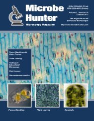
TESTING<br />
Sharpness of Objectives<br />
How sharp is your objective?<br />
Anthony Thomas<br />
Compound microscopes<br />
vary enormously in price.<br />
The newest infinity-type<br />
are likely not within reach of anyone<br />
not associated with a research<br />
laboratory. The older used<br />
finite-type microscopes, especially<br />
the Olympus BH2 series<br />
and the Nikon Labophot, are popular<br />
with keen amateur microscopists<br />
but <strong>com</strong>mand a high price,<br />
anywhere between $1,000.00 to<br />
$3,000.00. In contrast, some new<br />
models from certain manufactures<br />
can be obtained for around<br />
$500.00.<br />
I have a trinocular Olympus<br />
BH2 with a set of S Plan Apo<br />
objectives. I thought it may be<br />
worthwhile to <strong>com</strong>pare image<br />
quality from my setup with images<br />
obtained from a new lowerpriced<br />
microscope. Hopefully<br />
these new lower-priced models<br />
will give images as good as the<br />
older used (expensive) scopes.<br />
However, I have only this one<br />
scope and will leave it to others<br />
to post images for <strong>com</strong>parison.<br />
For <strong>com</strong>parison purpose subject<br />
matter is critical. I am using<br />
the wing of a northern mosquito,<br />
possibly an Aedes or Culex species,<br />
mosquitoes of these genera<br />
are to be found in many parts of<br />
the world. The wing was detached<br />
from the fly, dehydrated<br />
in Isopropyl Alcohol, rinsed in<br />
Toluene and mounted in a pine<br />
resin.<br />
An Olympus 2.5x NFK relay<br />
lens fits in the photo tube of the<br />
BH2 and projects an image,<br />
4,288 x 2,848 pixels, onto the<br />
23.6 x15.8 mm sensor of a Nikon<br />
D90 camera. Obviously one cannot<br />
display the images at full size<br />
so I have reduced them to 1,000<br />
pixels wide (but they may be further<br />
reduced to fit on the page)<br />
but have also included images of<br />
actual pixels. i.e., images of<br />
1,000 x 1,000 pixels as recorded<br />
by the camera's sensor. Because<br />
of the very limited depth of field<br />
with the higher power objectives<br />
the following images are stacks<br />
of several frames using Zerene<br />
Stacker software.<br />
The first image is an overall<br />
image of the detached wing taken<br />
with a 2x Olympus S Plan FL2<br />
and shows the area along the<br />
trailing edge that is used in the<br />
other images centering on the<br />
isolated wing scale which is<br />
about 13 microns (0.013 mm)<br />
wide.<br />
The second image shows the<br />
entire image of 4,288 x 2,848<br />
pixels recorded by the camera<br />
with the 10x objective but reduced<br />
to 1,000 pixels wide to fit<br />
on the page. Below this image,<br />
image 3 shows an area of 1,000 x<br />
1,000 actual pixels as recorded<br />
by the camera with the 10x objective.<br />
The fourth image is with the<br />
20x objective, and image #5 is<br />
the 1,000 x 1,000 pixels crop.<br />
The 6th image is with the 40x<br />
objective, and image #7 is the<br />
1,000 x 1,000 pixel crop.<br />
You can contact the author at:<br />
mothman@nbnet.nb.ca ■<br />
Figure 1: Mosquito wing<br />
32 - <strong>Microbe</strong><strong>Hunter</strong> Microscopy Magazine - January 2012
Sharpness of Objectives<br />
TESTING<br />
Figure 2: Portion of wing, 10x objective +<br />
2.5x relay lens<br />
Figure 3: 1,000 x 1,000 pixels crop of 10x<br />
image<br />
<strong>Microbe</strong><strong>Hunter</strong> Microscopy Magazine - January 2012 - 33
TESTING<br />
Sharpness of Objectives<br />
Figure 4: Portion of wing, 20x objective +<br />
2.5x relay lens<br />
Figure 5: 1,000 x 1,000 pixels crop of 20x<br />
image<br />
34 - <strong>Microbe</strong><strong>Hunter</strong> Microscopy Magazine - January 2012
Sharpness of Objectives<br />
TESTING<br />
Figure 6: Portion of wing, 40x objective<br />
+ 2.5x relay lens<br />
Figure 7: 1,000 x 1,000 pixels crop of 40x<br />
image<br />
<strong>Microbe</strong><strong>Hunter</strong> Microscopy Magazine - January 2012 - 35
What’s this? Answer on page 3.<br />
36 - <strong>Microbe</strong><strong>Hunter</strong> Microscopy Magazine - January 2012