
Create successful ePaper yourself
Turn your PDF publications into a flip-book with our unique Google optimized e-Paper software.
152 Ill. PROLES ARACHNES<br />
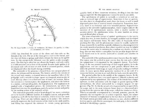
FIG. 52. Leg of spider. (i) Coxa; (ii) trochanter; (iii) femur; (iv) patella; (v) tibia;<br />
(vi) metatarsus; (vii) tarsus.<br />
( 1932) has described the action of the claws and claw-tufts on the<br />
threads of the web. The set of the claws gives a twist to the thread, and<br />
the claw-tuft forms a springy pad which releases the claws as the spider<br />
runs. In this unexpectedly elaborate way the spider avoids entanglement.<br />
The first leg is often but not always the longest, and only rarely<br />
is it held stretched out in front of the animal. In some male spiders it is<br />
decorated with tufts of setae or with black or coloured patches, which<br />
are displayed before the female during courtship.<br />
The underside of the prosoma is formed from two unequal plates of<br />
chitin, the labium and the sternum. The former, which is the sternite of<br />
the second body somite, is situated between the maxillary lobes of the<br />
pedipalpi, and is variable in shape: square or elongated, semicircular or<br />
oval. Directly above it is a flattened cone of tissue, the rostrum. Below<br />
the rostrum is a chitinous plate, the epipharynx, and above the labium<br />
is a similar, corresponding plate, the hypopharynx. The epipharynx is<br />
marked with a longitudinal groove, the stomodaeum, up which the<br />
liquid food rises into the oesophagus, partly by surface tension and partly<br />
by the sucking action of the stomach within.<br />
The ste~num is generally oval or heart-shaped, but many variations<br />
are to be found in different families. It is slightly convex, and generally<br />
marked on each side by four bays or acetabula, which receive the coxae<br />
of the legs. There are also lyriform organs on its surface. The sternum<br />
probably represents the fused sternites of the third to the sixth somites,<br />
and Giltay has reported the existence in certain young specimens,<br />
18. THE ORDER ARANEAE 153<br />
suitably fixed, of three transverse striations, dividing it into the four<br />
regions expected. But this appearance cannot be seen in any adult.<br />
The opisthosoma of spiders is normally a cylindrical or oval sac,<br />
with no outward sign of segmentation. Sometimes it has no pattern,<br />
but often there is a longitudinal mark above the heart, as well as small<br />
depressed points due to internal muscle attachments. Sometimes, however,<br />
there is an elaborate and even beautiful pattern. Segmentation<br />
persists in the sub-order Liphistiomorphae, where tcrgites indicate the<br />
existence of 12 opisthosomatic somites, while a smaller number of<br />
sternites protect the opisthosoma below. In some families an unsegmented<br />
dorsal plate is found.<br />
The most remarkable feature of spiders' opisthosomas is the way in<br />
which they may, in some families, be strangely modified and developed<br />
into bizarre and fantastic forms. No other order of <strong>Arachnida</strong> shows<br />
anything like this, and the phenomenon cannot be readily explained.<br />
It may reasonably be said that a prickly abdomen is a discouragement to<br />
the tender mouths of predators, that a short rounded one may be helpful<br />
because the owner may be mistaken for a mollusc or a beetle, or that a<br />
long, thin one may mimic a caterpillar; but the peculiar shapes that are<br />
to be found seem to be merely awkward to control and valueless in<br />
themselves.<br />
The underside of the opisthosoma shows more features than the upper.<br />
The region near the pedicel is more convex than the rest and is called<br />
the epigastrium: it is separated by the epigastric furrow. Two booklungs,<br />
or the two anterior book-lungs of four-lunged spiders, lie in the<br />
epigastrium, and are conspicuous as pale patches. Behind them are<br />
either the posterior book-lungs or a pair of spiracles leading to tracheal<br />
tubes. In many species these spiracular tracheae lie at the ends of a<br />
transverse furrow, not easy to see and often far back, near the spinnerets.<br />
The genital orifice lies in the middle of the epigastric furrow. In the<br />
male the vas deferens has a very small aperture, difficult to discern and<br />
unprotected by any epiandrium. The oviduct of the female has a larger<br />
aperture, in close association with the single or paired opening of the<br />
spermathecae, the whole surrounded by a complex epigyne (or epigynum)<br />
(Fig. 53). In this the actual vulva is protected by an operculum,<br />
the scape, and in the most elaborate forms there is a downward projection<br />
from the anterior side of the scape, called the crochet or clavus.<br />
This is occasionally accompanied by a second, posterior, process, the<br />
parmula: the two together act as a short ovipositor. Again, the unique<br />
character of Araneae is emphasized by the fact that in no other order is<br />
the female system provided with so elaborate an exit, which is different<br />
in every species, and, like its counterpart, the male palp, is the invariable<br />
method of identifying a species.