Radiation in Cath lab.pdf - TransradialWorld.com
Radiation in Cath lab.pdf - TransradialWorld.com
Radiation in Cath lab.pdf - TransradialWorld.com

Create successful ePaper yourself
Turn your PDF publications into a flip-book with our unique Google optimized e-Paper software.
<strong>Cath</strong> conference<br />
K<strong>in</strong>tur Sanghvi MD<br />
December 10, 2007
Fundamentals of X-ray imag<strong>in</strong>g<br />
Cardiology department,<br />
SVCMC
Generator and X-ray tube<br />
<br />
X-ray tube is a vacuum tube<br />
Electrons:generator to the cathode,<br />
white-hot (about 3,000 000°F) F). Electrons<br />
virtually boil off the cathode at this<br />
temperature (thermionic emission)<br />
jump from the cathode to the anode by<br />
the large voltage potential across the X-<br />
ray tube.<br />
The maximal (peak) voltage potential<br />
across the X-ray tube is referred to as<br />
the kVp of the system.<br />
cup sets up a local voltage potential -<br />
focus and control the number of e<br />
<br />
The number of electrons sent from the<br />
cathode to the anode is referred to as the<br />
mA of the system and directly correlates<br />
with the eventual number of X-rays<br />
produced.<br />
Cardiology department,<br />
SVCMC
Generator and X-ray tube<br />
The anode rotates rapidly (3,500 –<br />
10,000 rpm) to help dissipate the<br />
tremendous heat generated.<br />
X-ray tube surrounded by circulat<strong>in</strong>g oil<br />
The whole system is remarkably<br />
<strong>in</strong>efficient<br />
i Anode is tungsten - converts the<br />
electrons <strong>in</strong>to X-rays.- steeply angulated<br />
(8 –15°) - X-rays <strong>com</strong>e from as much a<br />
po<strong>in</strong>t source<br />
S<strong>in</strong>ce low-frequency X-rays contribute<br />
to radiation exposure but not image<br />
quality- alum<strong>in</strong>um or copper filters<br />
The beam is also shaped by the use of<br />
collimators at this po<strong>in</strong>t to help focus<br />
the eventual direction the X-rays travel.<br />
Cardiology department,<br />
SVCMC
X-ray generation<br />
<br />
<br />
Electrons -close to the nucleus<br />
and slow down - the loss of<br />
energy when the electron slows<br />
results <strong>in</strong> Bremsstrahlung<br />
(brak<strong>in</strong>g) X-rays be<strong>in</strong>g released.<br />
Electrons - knock out an electron<br />
from one of the shells of the<br />
tungsten atom – outer shell<br />
electron moves to fill the vacant<br />
lower shell, energy is lost <strong>in</strong> the<br />
form of an X-ray- Characteristic<br />
X-ray<br />
Cardiology department,<br />
SVCMC
Effect of KVp on X-ray generation<br />
Cardiology department,<br />
SVCMC
Effect of KVp on Iod<strong>in</strong>e’s contrast effect<br />
<br />
<br />
<br />
The energy of the X-rays<br />
<strong>in</strong>crease, there is less absorption<br />
by iod<strong>in</strong>e until suddenly this is a<br />
sudden <strong>in</strong>crease <strong>in</strong> the<br />
absorption.<br />
This sudden <strong>in</strong>crease <strong>in</strong> the<br />
absorption occurs at the energy<br />
of the K-shell electrons of iod<strong>in</strong>e;<br />
Energies below this edge do not<br />
contribute to the iod<strong>in</strong>e image<br />
Cardiology department,<br />
SVCMC
Effect of KVp on Iod<strong>in</strong>e’s contrast effect<br />
<br />
<br />
<br />
When the kVp is too high, there<br />
are many high-energy photons<br />
wasted that are not absorbed by<br />
the iod<strong>in</strong>e.<br />
These high-energy photons<br />
overpenetrate the iod<strong>in</strong>e column,<br />
creat<strong>in</strong>g a gray or washedout<br />
image.<br />
Better image contrast occurs if<br />
the kVp is kept at a reasonable<br />
low level (usually around 70–80<br />
kVp).<br />
Cardiology department,<br />
SVCMC
Effect of mA on Iod<strong>in</strong>e’s contrast effect<br />
<br />
<br />
<br />
The result of <strong>in</strong>creas<strong>in</strong>g the mA<br />
(and subsequently the number of<br />
X-rays) is demonstrated.<br />
When the mA is <strong>in</strong>creased, but<br />
the kVp kept optimal for image<br />
contrast, these extra X-rays<br />
contribute to overall image<br />
quality by provid<strong>in</strong>g for improved<br />
exposure.<br />
Also adds to scatter radiation<br />
Cardiology department,<br />
SVCMC
Once the X-rays leave the X-ray tube, they diverge and travel<br />
through hthe table and dthe patient ttoward dthe image <strong>in</strong>tensifier.<br />
ifi<br />
Most never make it to the image <strong>in</strong>tensifier as they are absorbed,<br />
attenuated, or scattered <strong>in</strong> the table or <strong>in</strong> the patient.<br />
Much of the occupational X-ray scatter important to the <strong>in</strong>vasive<br />
cardiologist occurs on the entrance side of the patient.<br />
When the X-ray tube is near the operator(cranial LAO) six times<br />
a (caudal RAO view)<br />
Cardiology department,<br />
SVCMC
Image Intensifier<br />
<br />
<br />
<br />
<br />
<br />
Grid that helps screen out scattered<br />
X-rays<br />
The <strong>in</strong>put face- covered with a<br />
phosphor of cesium iodide- converts<br />
x-ray to photon (sc<strong>in</strong>tillation)<br />
light photons strike a photocathode<br />
that converts the light energy back<br />
to electrons<br />
These electrons are then accelerated<br />
through the image <strong>in</strong>tensifier and<br />
strike a smaller output phosphor on<br />
the other end of the II.<br />
The output image is both m<strong>in</strong>ified<br />
(made smaller) and about 1,000<br />
times brighter than the <strong>in</strong>put image.<br />
Cardiology department,<br />
SVCMC
Image Distributor<br />
<br />
This visual output image is then sent<br />
through a series of lenses <strong>in</strong> the<br />
image distributor to a video camera<br />
or a charged-coupled device (CCD).<br />
Cardiology department,<br />
SVCMC
Control of the X-ray output<br />
<br />
<br />
<br />
<br />
<br />
<br />
With<strong>in</strong> the image distributor, a partially<br />
silvered mirror or prism - to a<br />
photodetector.<br />
Information from photodetector-to the<br />
generator to control the output<br />
Exposure is a function of the number of<br />
electrons made <strong>in</strong>to X-rays (the mA)<br />
times the maximum voltage across the<br />
X-ray tube (kVp) times the pulse width<br />
(the length of time the X-rays are sent<br />
per frame).<br />
Too high kVp -washed-out image<br />
Too high mA-excessive scatter radiation<br />
Too high pulse width-blurr<strong>in</strong>g of image.<br />
Cardiology department,<br />
SVCMC
Control of the X-ray output<br />
<br />
<br />
<br />
<br />
<br />
<br />
X-rays emitted - pulsed by<br />
electronics- allows fram<strong>in</strong>g rates of<br />
7.5–60<br />
Dur<strong>in</strong>g c<strong>in</strong>e - frame to frame<br />
Puls<strong>in</strong>g dur<strong>in</strong>g fluoroscopy reduce<br />
total exposure<br />
Most studies are now acquired at<br />
15–30 frames/sec <strong>in</strong> adults.<br />
Higher frame rates young<br />
children(HR)<br />
Dense structure – needs more<br />
exposure (bones) – angulated views<br />
<strong>in</strong> obese people – more scatter<br />
radiation<br />
Cardiology department,<br />
SVCMC
Effect of magnification on X-ray output<br />
<br />
<br />
<br />
<br />
Magnification <strong>in</strong>creases X-ray<br />
exposure<br />
Electronic magnification at the<br />
image <strong>in</strong>tensifier – by us<strong>in</strong>g less and<br />
less of the face of the II - results <strong>in</strong><br />
the image appear<strong>in</strong>g larger on the<br />
output phosphor<br />
To use less of the <strong>in</strong>put phosphor<br />
means a greater dose of X-rays<br />
An image acquired at 5” mode,-<br />
may require over three times the X-<br />
ray <strong>in</strong> <strong>com</strong>pare to 9” mode<br />
Cardiology department,<br />
SVCMC
Effect of II distance on X-ray output<br />
<br />
Source-to-image distance (SID)<br />
Chang<strong>in</strong>g the SID affects both X-<br />
ray exposure and image<br />
magnification<br />
<br />
As the SID is <strong>in</strong>creased, there is<br />
greater loss of X-rays, and the<br />
image appears more magnified<br />
(means more X-rays needed -<br />
exposure equation)<br />
Cardiology department,<br />
SVCMC
Older camera<br />
<br />
<br />
To c<strong>in</strong>e and yet still see an image - a<br />
partially silvered mirror (up to 85%<br />
of the beam is diverted to the c<strong>in</strong>e<br />
camera)<br />
The human bra<strong>in</strong> is unable to<br />
dist<strong>in</strong>guish flicker at fram<strong>in</strong>g rates<br />
of greater than 50 frames/sec<br />
So even after acquir<strong>in</strong>g at 30<br />
frames/sec- by the projector each<br />
image is shown twice thus fool<strong>in</strong>g<br />
the bra<strong>in</strong> by display<strong>in</strong>g the images<br />
at a rate of 60 images/sec.<br />
Cardiology department,<br />
SVCMC
Digital Imag<strong>in</strong>g<br />
<br />
<br />
<br />
<br />
<br />
<br />
<br />
<br />
Conversion of the video<br />
signal to digital <strong>in</strong>formation<br />
flicker-free view<strong>in</strong>g<br />
freeze-frame displays<br />
high-resolution image data<br />
immediately<br />
digital format<br />
network<br />
short- or long-term archival<br />
Digital i Communication i <strong>in</strong><br />
Medic<strong>in</strong>e (DICOM)<br />
Cardiology department,<br />
SVCMC
Factors effect<strong>in</strong>g Angiographic Image<br />
contrast<br />
Subject Contrast<br />
Vessel size<br />
X-ray y KV (lower contrast at > 75 KV<br />
Scatter:<br />
Patient e t thickness along beam<br />
X-ray beam area (FOV & Collimation)<br />
Image <strong>in</strong>tensifier veil<strong>in</strong>g glare<br />
Digital image process<strong>in</strong>g
Relationship between image quality and dose
Scatter and Veil<strong>in</strong>g glare depend on FOV
Effect of X-ray beam area on scatter
Raw image <strong>in</strong>tensifier versus process flat<br />
panel Detector
Effect of Digital image on the X-ray dose
Activity of Radioactive Material<br />
Curie: equivalent to the activity of 1 gram of radium<br />
1 becquerel = amount of material which will produce<br />
1 nuclear decay per second<br />
1 curie = 3.7 x 10¹⁰ becquerels<br />
1 curie = amount of material that<br />
will produce 3.7 x 10¹⁰ nuclear<br />
decays per second<br />
Measurement of Exposure<br />
• The emitted x- or gamma ray <strong>in</strong>teracts with tissue or<br />
air that it passes through, caus<strong>in</strong>g ionization events<br />
• Ionization i <strong>in</strong> air is measured <strong>in</strong> units of Coulombs/kg<br />
or (frequently <strong>in</strong> the US) <strong>in</strong> milliroentgens, or mr/hr<br />
• Roentgen: Ionization liberat<strong>in</strong>g a charge equal to<br />
2.58 X 10 -4 coulombs/kg of air<br />
• Coulombs: 1 coulomb/kg = 3876 R<br />
Cardiology department,<br />
SVCMC
Measurement of Dose (Absorbed)<br />
RAD<br />
• Measure of energy deposition <strong>in</strong> tissue from<br />
radiation<br />
• Units: rads, ergs, or Grays<br />
• RAD – is the deposition of one hundred ergs of<br />
energy <strong>in</strong> one gram of any material (NRC<br />
Regulations use per gram of body tissue) due to<br />
the ionization from any type of radiation<br />
• 100 rads = 1 Gray (gy)<br />
Cardiology department,<br />
SVCMC
Measurement of Dose (Equivalent)<br />
Measurement of Dose (Equivalent)<br />
REM<br />
• REM estimates biological damage caused<br />
by ionization <strong>in</strong> human body tissue (term for<br />
dose equivalence)<br />
• 1 REM = biological damage that would be<br />
caused by one RAD of dose<br />
• REM = RAD X Quality Factor<br />
Cardiology department,<br />
SVCMC
QUALITY FACTOR: amount<br />
of biological damage caused<br />
by the different types of<br />
radiation<br />
– Q = 1 for gamma rays, Xrays,<br />
and beta particles<br />
– Q = 20 for alpha particles<br />
Cardiology department,<br />
SVCMC
Effective dose<br />
<br />
<br />
<br />
Same probability of <strong>in</strong>duc<strong>in</strong>g a<br />
cancer or genetic disease<br />
whether the irradiation is<br />
uniformly delivered to the whole<br />
body or nonuniformly to part of<br />
the body or to specific organs<br />
The weight<strong>in</strong>g factors: different<br />
radiosensitivity of different<br />
tissues<br />
Operator risk not easily related<br />
s<strong>in</strong>gle collar film badge: the<br />
radiation field <strong>in</strong> the <strong>lab</strong>oratory is<br />
highly hl variable; superficial<br />
i tissues attenuate X-rays; lead<br />
aprons and thyroid collars<br />
significantly reduce the dose<br />
delivered to shielded organs.<br />
Cardiology department,<br />
SVCMC
Cardiology department,<br />
SVCMC
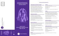
Basics of <strong>Radiation</strong> <strong>in</strong> <strong>Cath</strong><strong>lab</strong><br />
<strong>Radiation</strong> to Patient
Basics of <strong>Radiation</strong> <strong>in</strong> the cath <strong>lab</strong><br />
Cardiology department,<br />
SVCMC
Basics of <strong>Radiation</strong> <strong>in</strong> the cath <strong>lab</strong><br />
<br />
<br />
<br />
The total amount of scatter is<br />
proportional to the <strong>in</strong>tensity of the<br />
primary beam and the area of the<br />
entrance port<br />
the effect of beam size on the<br />
amount of stray radiation<br />
collimator is closed - meter only<br />
detects leakage from the tube<br />
A partially opened collimator -<br />
Leakage plus scatter from the<br />
small field<br />
<br />
A fully open collimator yields the<br />
maximum beam size Leakage<br />
plus the greater degree of scatter<br />
Cardiology department,<br />
SVCMC
Basics of <strong>Radiation</strong> <strong>in</strong> the cath <strong>lab</strong><br />
The distribution of scatter- highly dependent on the angiographic<br />
projection<br />
The distribution of radiation described <strong>in</strong> the form of isokerma<br />
curves (Air kerma is the unit of dose delivered to air. Higher<br />
values- more <strong>in</strong>tense radiation)<br />
Outside a particular isokerma curve -less than the the curve<br />
More stray radiation on the X-ray tube side of the patient than II<br />
Entrance surface of the patient is the ma<strong>in</strong> source of scatter -<br />
patient’s tissues provide shield<strong>in</strong>g on the image <strong>in</strong>tensifier side<br />
less stray radiation at waist and eye level - X-ray tube is close to<br />
the floor - Distance- advantage of large size <strong>lab</strong>s
90 LAO 150 cm above floor (at eye level)
90 LAO 100 cm above floor (at west level)
60 LAO 150 cm above floor (at eye level)
60 LAO 100 cm above floor (at west level)
90 RAO 150 cm above floor (at eye level)
90 RAO 100 cm above floor (at west level)
Typical exposure <strong>in</strong> Cardiac Angiography
Biological Effect<br />
Dose Modify<strong>in</strong>g Factors<br />
• Amount of exposure<br />
• Duration of exposure<br />
– Given rads over m<strong>in</strong>utes > months or years<br />
• Type of radiation<br />
– Alpha particles and fission fragments > ionis<strong>in</strong>g radiation beta<br />
particles or gamma rays<br />
• Biological variability<br />
– Age (Children > adults)<br />
– Health status (Sick > healthy)<br />
• The part of body exposed<br />
– Total body > partial body exposure<br />
– Major organs > limbs<br />
• Gonads, lens, blood form<strong>in</strong>g organs more radiosensitive<br />
iti<br />
Shield<strong>in</strong>g<br />
- Inversely proportaional to half value layer- atomic number of shield<strong>in</strong>g<br />
material, density, size<br />
Cardiology department,<br />
SVCMC
Biologic Effect<br />
<br />
<br />
Stochastic effect<br />
Determ<strong>in</strong>istic effect<br />
Cardiology department,<br />
SVCMC
Biologic effects of radiation<br />
Cardiology department,<br />
SVCMC
<strong>Radiation</strong> Dose limit<br />
Maximum permissible dose, annual limits:<br />
<br />
Occupational workers:<br />
Whole body = 5,000 mrem/yr<br />
Eyes = 15,000 mrem/yr<br />
Sk<strong>in</strong> = 50,000 mrem/yr<br />
If pregnant = 500 mrem/gestational period, not to exceed<br />
50 mrem/month<br />
General public = 500 mrem, whole body, <strong>in</strong>frequentexposure<br />
<br />
Patient: No described limit by NRC. Risk vs benefit evaluation by<br />
physician<br />
Cardiology department,<br />
SVCMC
M<strong>in</strong>imiz<strong>in</strong>g the risk of <strong>Radiation</strong>
Determ<strong>in</strong>istic <strong>Radiation</strong> effect <strong>in</strong> Cardiac<br />
Angiography
Practical Tips
Practical Tips
Collimate to reduce Scatter, Glare, Exposure
Practical Tips
Practical Tips
How to m<strong>in</strong>imize radiation exposure<br />
Technical Method<br />
X-ray system<br />
Lower dose per frame<br />
Increase filtration<br />
Use pulsed flouroscopy<br />
High image <strong>in</strong>tensifier<br />
reception<br />
Digital system<br />
Instant replay<br />
Edge enhansement<br />
Lower flouro frame rate<br />
Imag<strong>in</strong>g Technique<br />
Time<br />
Shorter “picks”<br />
Fewer “picks”<br />
Don’t record flouro<br />
<br />
Distance<br />
Stand back<br />
Increase SSD<br />
M<strong>in</strong>imize air gap<br />
Shield<strong>in</strong>g<br />
Collimate<br />
M<strong>in</strong>imize air gap<br />
Use portable shields<br />
Wear 2- piece aprons
Graphical Presentation of Cancer Risk and<br />
<strong>Radiation</strong> Dose<br />
Picano, E. BMJ 2004;328:578-580<br />
Cardiology department,<br />
SVCMC
<strong>Radiation</strong> Dose From Selected Nuclear<br />
Cardiology Procedures<br />
RC Thomas and J Cullom. J Nucl Cardiol 2006;13:19-23<br />
Cardiology department,<br />
SVCMC
<strong>Radiation</strong> Dose From Selected<br />
Cardiac CT Procedures<br />
RC Thomas and J Cullom. J Nucl Cardiol 2006;13:19-23<br />
Cardiology department,<br />
SVCMC