A simple method for measuring glomerular filtration rate in dogs
A simple method for measuring glomerular filtration rate in dogs
A simple method for measuring glomerular filtration rate in dogs

You also want an ePaper? Increase the reach of your titles
YUMPU automatically turns print PDFs into web optimized ePapers that Google loves.
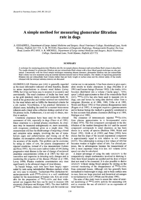
Research <strong>in</strong> Veter<strong>in</strong>ary Science 1995, 59, 118-123<br />
A <strong>simple</strong> <strong>method</strong> <strong>for</strong> <strong>measur<strong>in</strong>g</strong> <strong>glomerular</strong> <strong>filtration</strong><br />
<strong>rate</strong> <strong>in</strong> <strong>dogs</strong><br />
A. GLEADHILL, Department of Large Animal Medic<strong>in</strong>e and Surgery, Royal Veter<strong>in</strong>ary College, Hawkshead Lane, North<br />
Mymms, Hatfield AL9 7TA, A. M. PETERS, Department of Diagnostic Radiology, Hammersmith Hospital, Du Cane<br />
Road, London W12 ONN, A. R. MICHELL, Department of Large Animal Medic<strong>in</strong>e and Surgery, Royal Veter<strong>in</strong>ary<br />
College, Hawkshead Lane, North Mymms, Hatfield AL9 7TA<br />
SUMMARY<br />
A technique <strong>for</strong> <strong>measur<strong>in</strong>g</strong> <strong>glomerular</strong> <strong>filtration</strong> <strong>rate</strong> (by six-sample plasma clearance) and extracellular fluid volume is described.<br />
The results obta<strong>in</strong>ed <strong>for</strong> <strong>glomerular</strong> <strong>filtration</strong> <strong>rate</strong> per extracellular fluid volume with a three blood sample technique are also presented.<br />
Concurrently with the blood sample techniques <strong>measur<strong>in</strong>g</strong> plasma clearance, <strong>glomerular</strong> <strong>filtration</strong> <strong>rate</strong> per extracellular<br />
fluid volume was also measured us<strong>in</strong>g an external radiation detector and no blood samples. The validity of express<strong>in</strong>g <strong>glomerular</strong><br />
<strong>filtration</strong> <strong>rate</strong> per extracellular fluid volume (rather than per body weight or surface area) and the cl<strong>in</strong>ical utility of the results<br />
obta<strong>in</strong>ed with the external radiation detector are discussed.<br />
GLOMERULAR <strong>filtration</strong> <strong>rate</strong> (OFR) is generally regarded<br />
as the most <strong>in</strong><strong>for</strong>mative <strong>in</strong>dicator of renal function, despite<br />
its m<strong>in</strong>or imperfections <strong>in</strong> chronic renal failure (Levey<br />
1990), but it has not been easy to measure it accu<strong>rate</strong>ly and<br />
conveniently. The renal clearance of <strong>in</strong>ul<strong>in</strong> has been used<br />
as the gold standard; <strong>in</strong>ul<strong>in</strong> is a small molecule freely filtered<br />
at the glomerulus and neither secreted nor reabsorbed<br />
by the renal tubule and so fulfils the theoretical criteria <strong>for</strong><br />
a ~vP, marker. Nevertheless, it has practical limitations <strong>in</strong><br />
cl<strong>in</strong>ical cases, <strong>in</strong>clud<strong>in</strong>g the need <strong>for</strong> a constant <strong>in</strong>travenous<br />
<strong>in</strong>fusion and a timed ur<strong>in</strong>e collection dur<strong>in</strong>g a period of stable<br />
plasma <strong>in</strong>ul<strong>in</strong>. Furthermore, it is not easy to obta<strong>in</strong>, sterilise<br />
or analyse.<br />
Creat<strong>in</strong><strong>in</strong>e clearances have been used <strong>for</strong> the cl<strong>in</strong>ical<br />
assessment of GFR, especially <strong>in</strong> <strong>dogs</strong> (F<strong>in</strong>co et al 1981).<br />
S<strong>in</strong>ce creat<strong>in</strong><strong>in</strong>e is a normal plasma constituent, its clearance<br />
may be measured endogenously or, more accu<strong>rate</strong>ly,<br />
with exogenous creat<strong>in</strong><strong>in</strong>e given as a s<strong>in</strong>gle dose. This<br />
measurement still has the disadvantage of requir<strong>in</strong>g a timed<br />
and complete ur<strong>in</strong>e sample, <strong>in</strong>volv<strong>in</strong>g the use of metabolism<br />
cages and catheterisation and r<strong>in</strong>s<strong>in</strong>g out of the<br />
bladder. An <strong>in</strong>complete collection of ur<strong>in</strong>e seriously affects<br />
the accuracy of the results. This problem can be overcome<br />
by us<strong>in</strong>g an <strong>in</strong>dex of GFR, the plasma disappearance of<br />
<strong>in</strong>jected creat<strong>in</strong><strong>in</strong>e, <strong>in</strong>stead of a true measurement (Labato<br />
and Ross 1991). An even more <strong>in</strong>direct <strong>in</strong>dex of GFR dur<strong>in</strong>g<br />
progressive renal disease is the <strong>rate</strong> of change of reciprocal<br />
creat<strong>in</strong><strong>in</strong>e concentrations (Allen et al 1987) but they are<br />
<strong>in</strong>sensitive and imprecise <strong>in</strong> detect<strong>in</strong>g early changes and <strong>in</strong><br />
monitor<strong>in</strong>g the progress of the disease (Walser et al 1989,<br />
Levey 1990). Plasma creat<strong>in</strong><strong>in</strong>e concentration only rises<br />
significantly <strong>in</strong> the advanced stages of renal disease.<br />
In human be<strong>in</strong>gs, such tests have been largely superseded<br />
by tests us<strong>in</strong>g radiolabelled <strong>filtration</strong> markers which<br />
are very easy to measure. Technetium-99m diethylene-triam<strong>in</strong>e-pentacetic<br />
acid (99mTC-DTPA) is a well established<br />
marker <strong>for</strong> the measurement of human GFR. Kits are available<br />
<strong>for</strong> the preparation of 99mTc-DTPA and their widespread<br />
use suggests that they can be used to measure GFR<br />
accu<strong>rate</strong>ly (Sampson and Keegan 1985). The short half-life<br />
of 99mTc (six hours), makes it relatively safe <strong>for</strong> rout<strong>in</strong>e use<br />
rout<strong>in</strong>e use <strong>in</strong> out-patients. It has been shown to give equivalent<br />
results to <strong>in</strong>ul<strong>in</strong> clearances <strong>in</strong> <strong>dogs</strong> (McAfee et al<br />
1981) and human be<strong>in</strong>gs (Perrone 1992). Like <strong>in</strong>ul<strong>in</strong>, DTPA<br />
becomes distributed <strong>in</strong>to a volume ('<strong>in</strong>ul<strong>in</strong> space', 'DTPA<br />
space') which approximates to that of the extracellular fluid<br />
(ECF). 99mTC-DTPA has also been used to measure GFR <strong>in</strong><br />
<strong>dogs</strong>, cats and horses by us<strong>in</strong>g data derived either from a<br />
renogram (Krawiec et al 1986, 1988, Uribe et al 1992,<br />
Walsh and Royal 1992) or from plasma disappearance <strong>rate</strong>s<br />
(Rogers et al 1991). A renogram requires a gamma-camera<br />
and <strong>in</strong> human be<strong>in</strong>gs the <strong>method</strong> is generally considered to<br />
be less accu<strong>rate</strong> than plasma clearances (Rehl<strong>in</strong>g et al 1984,<br />
Mulligan et al 1990).<br />
The clearance of <strong>in</strong>travenously <strong>in</strong>jected 99mTC-DTPA<br />
from plasma approximates to a bi-exponential curve<br />
(Cohen 1974). The first component corresponds to the distribution<br />
of the marker throughout the ECF and the second<br />
component corresponds to renal clearance, as has been<br />
shown <strong>in</strong> a variety of species (Robb<strong>in</strong>s 1984, Walsh and<br />
Royal 1992). Groth et al (1983) and Peter (1992) have<br />
shown that the slope of the second exponential component<br />
is approximately equal to GFR relative to ECF volume<br />
(ECFV). This relationship is strong at low values of GFR but<br />
becomes weaker at higher values of GFR. For cl<strong>in</strong>ical purposes,<br />
this is advantageous because the measurement of<br />
GFR is most important when it is low. ECF volume (DTPA<br />
space) can also be calculated.<br />
A major advantage of us<strong>in</strong>g the ratio GFR/ECFV is the<br />
comparatively <strong>simple</strong> mathematics <strong>in</strong>volved <strong>in</strong> deriv<strong>in</strong>g it.<br />
Only three blood samples are needed and the dose of tracer<br />
does not need to be measured. It is also possible to derive<br />
GFR/ECFV without tak<strong>in</strong>g blood samples by rely<strong>in</strong>g on the<br />
external monitor<strong>in</strong>g of the disappearance of 99mTC-DTPA<br />
from extrarenal tissues. Although GFR has generally been<br />
normalised to bodyweight <strong>in</strong> <strong>dogs</strong>, and to body surface area<br />
<strong>in</strong> man, there are large practical and theoretical advantages<br />
to normalisation to ECFV (White and Strydom 1991).<br />
The aims of this study were to assess the value of external<br />
monitor<strong>in</strong>g of 99mTC-DTPA activity <strong>for</strong> <strong>measur<strong>in</strong>g</strong> GFR<br />
and to exam<strong>in</strong>e the use of GFR/ECFV as an expression of<br />
renal function.<br />
118
A <strong>simple</strong> <strong>method</strong> <strong>for</strong> <strong>measur<strong>in</strong>g</strong> <strong>glomerular</strong><strong>filtration</strong> <strong>rate</strong> <strong>in</strong> <strong>dogs</strong> 119<br />
I<br />
.Z:"<br />
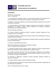
FIG 1 : The three positions of the external detector: counter-clockwise from top right, head 1, head 2 and heart<br />
MATERIALS AND METHODS<br />
GFR was measured <strong>in</strong> 120 <strong>dogs</strong> by giv<strong>in</strong>g an <strong>in</strong>travenous<br />
<strong>in</strong>jection of 50 to 200 MBq of 99mTC-DTPA. Dogs with<br />
known or suspected renal disease were exam<strong>in</strong>ed <strong>in</strong> an<br />
ef<strong>for</strong>t either to confirm a diagnosis of renal <strong>in</strong>sufficiency or<br />
to monitor the progression of renal disease. The <strong>dogs</strong> were<br />
all fasted overnight (to avoid the post prandial rise <strong>in</strong> GFR)<br />
but allowed free access to water dur<strong>in</strong>g the procedure.<br />
Hepar<strong>in</strong>ised venous blood samples were taken at 10, 20 and<br />
30 m<strong>in</strong>utes and two, three and four hours after the <strong>in</strong>jection.<br />
The dose was measured and a standard prepared with a<br />
measured aliquot of the 99rnTC-DTPA which was diluted and<br />
counted at the same time as the blood samples. The blood<br />
samples were centrifuged and between 0.1 and 0.25 ml of<br />
plasma was counted <strong>for</strong> one m<strong>in</strong>ute <strong>in</strong> a gamma-counter<br />
(Canberra Packard). The activity was expressed <strong>in</strong> counts<br />
ml q .<br />
Between one and four hours after the <strong>in</strong>jection of 99mTc-<br />
DTPA, an external radiation detector (Sc<strong>in</strong>tillation Detector,<br />
Oakfields Instruments) was used to measure the disappearance<br />
of radioactivity from the dog (result<strong>in</strong>g from both<br />
renal clearance and the radioactive decay) by external<br />
count<strong>in</strong>g. The detector was placed over the follow<strong>in</strong>g three<br />
regions at 30-m<strong>in</strong>ute <strong>in</strong>tervals between one-and-a-half and<br />
four hours after the adm<strong>in</strong>istration of the 99mTC-DTPA (Fig<br />
1). Over the head, the detector was first centred over the<br />
middle of the dog's <strong>for</strong>ehead, mid-way between the eyes<br />
and ears; it was directed downwards and the dog's neck<br />
was extended so that only the head was be<strong>in</strong>g counted. It<br />
was then centred over the back of the dog's head and<br />
directed caudocranially. Over the heart, the detector was<br />
centred over the apex beat and directed laterally. These<br />
areas were chosen fh'st because the counts were reproducible,<br />
and secondly to avoid counts from the kidneys and<br />
bladder, thus avoid<strong>in</strong>g the radioactivity which accumulates<br />
<strong>in</strong> the ur<strong>in</strong>e dur<strong>in</strong>g the procedure.<br />
The <strong>dogs</strong> were taken <strong>for</strong> a walk <strong>in</strong> a designated area to<br />
encourage them to ur<strong>in</strong>ate dur<strong>in</strong>g the procedure and be<strong>for</strong>e<br />
they were discharged, and they were sent home about one<br />
half-life after the adm<strong>in</strong>istration of the isotope. The proce-<br />
dure was not per<strong>for</strong>med on <strong>dogs</strong> which were not house<br />
tra<strong>in</strong>ed and the owners were <strong>in</strong>structed to take their <strong>dogs</strong><br />
outside to ur<strong>in</strong>ate regularly dur<strong>in</strong>g the even<strong>in</strong>g.<br />
The curve of the disappearance of the 99mTC-DTPA from<br />
the plasma was fitted to a biexponential equation:<br />
P(t) = A e -cdt + B e -c~2t<br />
where P = plasma concentration of 99mTC-DTPA at time t,<br />
A is the <strong>in</strong>tercept and al the <strong>rate</strong> constant <strong>for</strong> the first exponential<br />
component (ma<strong>in</strong>ly a reflection of the distribution of<br />
the isotope through the ECFV), and B is the <strong>in</strong>tercept and a2<br />
the <strong>rate</strong> constant of the second exponential component<br />
(ma<strong>in</strong>ly a reflection of the renal clearance of the isotope).<br />
By us<strong>in</strong>g the follow<strong>in</strong>g equations, the absolute GFR, the<br />
ratio GFR/ECFV and the ECFV were calculated. Follow<strong>in</strong>g the<br />
measurement of absolute GFR, the GFR bodyweight -1 and<br />
body surface area -1 were derived.<br />
Dose (1)<br />
GFR -- (A/a 1 + B/a2 )<br />
t = The transit time of a <strong>filtration</strong> marker (2)<br />
hence<br />
Volume of distribution<br />
<strong>rate</strong> of clearance<br />
ECFV<br />
GFR<br />
GFR 1 (3)<br />
ECFV t<br />
t = A/(a 1)2 + B/(a2)2 (4)<br />
A/a 1 + B/a 2<br />
GFR _ A/a 1 + B/a 2<br />
ECFV a/(al) 2 + B~(a2) 2 (5)<br />
If the concentration of the marker <strong>in</strong> plasma is the same as<br />
<strong>in</strong> the extracellular fluid, then the transit time is <strong>in</strong>versely<br />
proportional to the <strong>rate</strong> constant of the second exponential:<br />
t<br />
1<br />
There<strong>for</strong>e,<br />
a 2<br />
GFR<br />
ECFV -- a2 6)
120 A. Gleadhill, A. M. Peters, A. R. Michell<br />
"7,<br />
"6<br />
E<br />
2000<br />
1500<br />
©<br />
O<br />
1000 ~)<br />
500<br />
relationships between GFR/ECFV and the various measurements<br />
of ct 2 were there<strong>for</strong>e fitted to second order polynomials<br />
(Brochner-Mortensen 1972, Peters 1992). The ratio of<br />
GFR/ECFV to Ot 2 as a function of GFR/ECFV was plotted to<br />
assess the divergence of the latter from ot 2 at higher levels<br />
of renal function which results from an <strong>in</strong>creas<strong>in</strong>g difference<br />
between the concentrations of the tracer <strong>in</strong> plasma and<br />
<strong>in</strong>terstitial fluid. This relationship was fitted with an exponential<br />
function (Fig 6).<br />
In all the analyses, correction factors <strong>for</strong> the radioactive<br />
decay of 99mTc were <strong>in</strong>cluded because significant physical<br />
decay occurs dur<strong>in</strong>g the procedure.<br />
Plasma creat<strong>in</strong><strong>in</strong>e concentrations were measure on a<br />
Gil<strong>for</strong>d Selective Batch Analyser (SBA 300) by the Jaffe<br />
<strong>method</strong>.<br />
0<br />
0 10 20 30<br />
o<br />
20<br />
Head 1 /<br />
GFR/ECFV (ml m<strong>in</strong> ~ litre ~)<br />
(Six samples)<br />
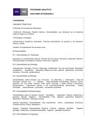
FIG 2: Relationship between plasma creat<strong>in</strong><strong>in</strong>e concentration and GFR/ECFV<br />
L<strong>in</strong>ear regression analysis was applied to the tracer concentrations<br />
<strong>in</strong> the last three blood samples (two, three and<br />
four hours) to obta<strong>in</strong> the <strong>rate</strong> constant (Gt2) of the second<br />
exponential, which represents the ratio GFR/ECFV. L<strong>in</strong>ear<br />
regression was also applied to the read<strong>in</strong>gs of the external<br />
detector to obta<strong>in</strong> an <strong>in</strong>dependent estimate of the <strong>rate</strong> constant<br />
of the second exponential<br />
The relationship between 6FR/ECFV and a 2 (from the<br />
blood samples or external count<strong>in</strong>g) was non-l<strong>in</strong>ear; the<br />
'7<br />
._c<br />
E<br />
E<br />
r~<br />
15<br />
10<br />
20<br />
1 01<br />
I~r"2 i i i<br />
0 5 10 15 20 25<br />
Head 2 o /<br />
20<br />
e~<br />
v E 15<br />
.-= g<br />
w<br />
0<br />
,~ o<br />
°o° oo<br />
5<br />
0<br />
0<br />
I ! I I I<br />
5 10 15 20 25<br />
GFR/ECV (ml m<strong>in</strong> ~ litre -~)<br />
(Six samples)<br />
FIG 3: Second order polynomial relationship between the value of GFPJECFV<br />
derived from ~2 calculated from the last three blood samples and the value of<br />
GFR/ECFV derived from measurements of all six blood samples<br />
m<br />
"7<br />
w<br />
E<br />
15<br />
10<br />
I I I I I<br />
0 5 10 15 20 25<br />
_.-'~- 20 15 Heart o ~ o / f<br />
E 10 . Z ~ o q~<br />
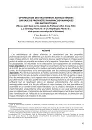
TABLE 1: Polynomial equations describ<strong>in</strong>g the relationships between<br />
the values of c~ 2 determ<strong>in</strong>ed from the last three blood samples and from<br />
the external detecor and GFR/ECFV (ml m<strong>in</strong> -1 litre -1) determ<strong>in</strong>ed from six<br />
blood samples<br />
~2 derived from Equation r<br />
Three blood samples = -0.326 + 1.146 x + 0.0020 x 2 0-984<br />
External detector<br />
Head 1 = -0.335 + 1-090 x -0.0191 x 2 0.943<br />
Head2 = -0.14 + 1.068x-0.0172x 2 0-938<br />
Heart = +0.142 + 0-899 x-0-0010 x 2 0.896<br />
X = GFPJECFV<br />
I ! I I I I<br />
0 5 10 15 20 25<br />
GFR/ECV (ml ra<strong>in</strong> 1 litre -1)<br />
(Six samples)<br />
FIG 4: Non-l<strong>in</strong>ear relationships between the values of c~ 2 derived from the<br />
measurements with the external detector held <strong>in</strong> the three positions and the<br />
value of GFR/ECFV derived from measurements of all six blood samples
A <strong>simple</strong> <strong>method</strong> <strong>for</strong> <strong>measur<strong>in</strong>g</strong> <strong>glomerular</strong><strong>filtration</strong> <strong>rate</strong> <strong>in</strong> <strong>dogs</strong> 121<br />
"7<br />
t-<br />
20<br />
10<br />
0<br />
20<br />
Head 1 1.6<br />
O.<br />
o o E 1.4<br />
o g~<br />
o8o~,.,q~-8~;;" o b ~ 1.2<br />
o<br />
la_ e~<br />
~E<br />
o~o"<br />
o<br />
o<br />
~ 1.o<br />
I !<br />
0 10 20<br />
Head 2<br />
X<br />
~2<br />
0.8<br />
o Oo/<br />
o °/o<br />
O A~ OOfo<br />
0 G~ 0"~ 0 0 ~F 0<br />
o o oo2¢ oo8<br />
~o°~<br />
I I I I I<br />
0 5 10 15 20 25<br />
GFR/ECFV: (ml m<strong>in</strong> 4 litre 4)<br />
(Six samples)<br />
FIG 6: Relationship between the value of GFR/ECFV derived from measurements<br />
of all six blood samples and the ratio of GFR/ECFV to (z 2 derived from<br />
measurements of the last three blood samples<br />
.--<br />
'7<br />
10<br />
S°<br />
J °<br />
! !<br />
0 10 20<br />
20 Heart<br />
,; °o oo<br />
~ A~o
122 A. Gleadhill, A. M. Peters, A. R. MichelI<br />
TABLE 4: Polynomial equations describ<strong>in</strong>g the relationship between<br />
GFR/ECFV and the value of ~ derived from the external detector from<br />
which GFR/ECFV can be derived (ml m<strong>in</strong> -1 litre -1)<br />
~2 derived from Equation r<br />
Last three<br />
blood samples<br />
GFR/ECFV = 0.217 + 0.882ct 2 + 0.0237c~22 0.975<br />
Head 1 GFR/ECFV = -0.454 + 1.40(x 2 -0.014(x22 0.934<br />
Head 2 GFR/ECFV = -0.132 + 1.32ct 2-0.0127c~22 0.931<br />
Heart GFR/ECFV= -0.566 + 1.665ct 2-0.0356ot22 0.902<br />
be obta<strong>in</strong>ed from the values of a2 derived either from the<br />
three last blood samples or from the external detector at any<br />
level of renal function. The correlation between the value<br />
of ~2 derived from three blood samples and GFR/ECFV was<br />
0.975 (Table 4).<br />
DISCUSSION<br />
Rout<strong>in</strong>e tests <strong>for</strong> the detection and evaluation of the<br />
severity of can<strong>in</strong>e renal disease <strong>in</strong>clude the measurement of<br />
blood levels of urea and creat<strong>in</strong><strong>in</strong>e. However, the nonl<strong>in</strong>ear<br />
relationship between plasma creat<strong>in</strong><strong>in</strong>e and GFR<br />
means that <strong>in</strong>creases <strong>in</strong> creat<strong>in</strong><strong>in</strong>e above the normal<br />
range do not occur until much renal function has been<br />
lost (Fig 2). In the early stages of renal failure, large<br />
changes <strong>in</strong> function are accompanied by only small changes<br />
<strong>in</strong> plasma creat<strong>in</strong><strong>in</strong>e, and as a result renal failure cannot be<br />
detected early. Normal plasma creat<strong>in</strong><strong>in</strong>e concentration is<br />
also affected by an animal's muscle mass which varies<br />
between <strong>in</strong>dividuals; moreover, <strong>in</strong> advanced renal disease,<br />
there are changes <strong>in</strong> the metabolism and renal handl<strong>in</strong>g<br />
of creat<strong>in</strong><strong>in</strong>e, although the extent of these changes <strong>in</strong> <strong>dogs</strong><br />
rema<strong>in</strong>s uncerta<strong>in</strong> (Rob<strong>in</strong>son et al 1974, Walser et al 1989,<br />
Levey 1990).<br />
The relationship between plasma creat<strong>in</strong><strong>in</strong>e and<br />
GFR/ECFV (Fig 2) confirmed how little change <strong>in</strong> plasma<br />
creat<strong>in</strong><strong>in</strong>e occurs until after there has been a significant fall<br />
<strong>in</strong> GFR (Biewenga and Van Den Brom 1981, Levey et al<br />
1988). Although this is well known among nephrologists,<br />
its importance is often <strong>in</strong>sufficiently appreciated by practitioners.<br />
A creat<strong>in</strong><strong>in</strong>e <strong>in</strong> the high-normal range, ie 140 gM,<br />
may be normal <strong>in</strong> some <strong>dogs</strong> but may be associated with a<br />
marked reduction <strong>in</strong> renal function <strong>in</strong> others. Blood urea<br />
concentration is even less satisfactory as an <strong>in</strong>dicator of<br />
renal function because its concentration is affected by factors<br />
such as pyrexia, liver function, hydration and dietary<br />
prote<strong>in</strong> content (Epste<strong>in</strong> et al 1984).<br />
Although isotopic plasma clearance techniques have<br />
been described previously <strong>in</strong> <strong>dogs</strong> (Van Den Brom and<br />
Biewenga 1981), they have used eight or more blood sampies.<br />
The present results demonst<strong>rate</strong> the adequacy of a<br />
<strong>method</strong> based on only three blood samples and the potential<br />
value of a <strong>method</strong> rely<strong>in</strong>g solely on external monitor<strong>in</strong>g.<br />
GFR has been measured by us<strong>in</strong>g an external detector <strong>in</strong> a<br />
small group of cats (Rogers et al 1991), but a blood sample<br />
was also required.<br />
The choice of the position of the external detector is particularly<br />
important <strong>for</strong> the accuracy of the measurement of<br />
GFR. The correlation coefficients between the values of ~2<br />
derived from the three blood samples and the external<br />
detector were best when the detector was placed on the<br />
head, and these results were much more consistent than<br />
those obta<strong>in</strong>ed when the detector was directed towards the<br />
heart. This is probably because the positions on the head<br />
were more reproducible; it is likely that the probe was<br />
angled slightly differently towards the heart <strong>for</strong> each measurement.<br />
Furthermore, <strong>in</strong> very small <strong>dogs</strong>, it is difficult<br />
when direct<strong>in</strong>g the detector towards the heart to avoid the<br />
kidneys and bladder, which would gene<strong>rate</strong> erroneously<br />
high counts by <strong>in</strong>clud<strong>in</strong>g counts from ur<strong>in</strong>e as well as<br />
plasma.<br />
This <strong>method</strong> is non-<strong>in</strong>vasive, <strong>simple</strong> and accu<strong>rate</strong>.<br />
Because it uses radio-isotopes, it is unlikely to be widely<br />
used <strong>in</strong> general practice <strong>in</strong> the <strong>for</strong>eseeable future, but it<br />
would be possible <strong>in</strong> a referral centre. The measurement<br />
would ideally be used as a quantitative assessment of renal<br />
function <strong>in</strong> <strong>dogs</strong> <strong>in</strong> which renal disease has been suspected<br />
from the result of some other suitable screen<strong>in</strong>g test.<br />
The technique presupposes that the ratio GFR/ECFV is a<br />
valid way of normalis<strong>in</strong>g GFR <strong>for</strong> body size. This is particularly<br />
important <strong>in</strong> <strong>dogs</strong> because of the wide range of body<br />
sizes <strong>in</strong> different breeds, rang<strong>in</strong>g from the chihuahua to the<br />
St Bernard. In <strong>dogs</strong> the GFR has by convention been<br />
expressed per kg, but <strong>in</strong> human be<strong>in</strong>gs GFR values are usually<br />
normalised with reference to the body surface area. In<br />
the present experiments, normalis<strong>in</strong>g the GFR to the ECFV<br />
has the major advantage that the GFR can then be measured<br />
directly with the external detector. It also simplifies the<br />
mathematics <strong>in</strong> calculat<strong>in</strong>g GFR/ECFV compared with that <strong>in</strong><br />
calculat<strong>in</strong>g GFR kg -1. Many <strong>method</strong>s have been described<br />
<strong>for</strong> calculat<strong>in</strong>g the absolute GFR <strong>in</strong> human be<strong>in</strong>gs by us<strong>in</strong>g<br />
one to three blood samples taken two to four hours after the<br />
<strong>in</strong>jection of TC-DTPA; it is not considered cl<strong>in</strong>ically practical<br />
to take more blood samples (Mulligan et al 1990). It has<br />
been shown that the error <strong>in</strong> us<strong>in</strong>g ~2 to represent GFR/ECFV<br />
is less than the error <strong>in</strong> calculat<strong>in</strong>g GFR absolutely from two<br />
to three blood samples taken between two and four hours<br />
after an <strong>in</strong>jection of DTI'A (Peters 1992).<br />
In addition to these considerations, there is a strong<br />
theoretical reason why the GFR normalised with respect to<br />
ECFV may be a more suitable way of express<strong>in</strong>g GFR<br />
(McCance and Widdowson 1952, White and Strydom<br />
1991), <strong>for</strong> it is the role of the kidney to filter the extracellular<br />
fluid and regulate its composition and volume<br />
(Brochner-Mortensen 1980). Barratt (1985) concluded that<br />
the 'normalisation' of renal function to body surface area<br />
has little theoretical foundation and that the ECFV may be<br />
more logical. In human be<strong>in</strong>gs the GFR per body surface<br />
area <strong>in</strong>dicates that GFR is higher <strong>in</strong> men than women but<br />
expressed per ECFV there is no gender difference<br />
(Brochner-Mortensen 1982). In human be<strong>in</strong>gs with<br />
acromegaly, the absolute GFR is <strong>in</strong>creased but so is the<br />
ECFV and as a result the GFR/ECFV rema<strong>in</strong>s constant (Peters<br />
1992, Brochner-Mortensen and Ditzel 1982). In addition,<br />
the ECFV is more closely correlated with lean body mass<br />
than with weight (Boer 1984). Thus normalisation with<br />
respect to ECFV may be more physiologically correct, <strong>in</strong><br />
addition to simplify<strong>in</strong>g the procedure. The changes <strong>in</strong> ECrV<br />
which are with<strong>in</strong> the cl<strong>in</strong>ically probable range are unlikely<br />
to affect the GFR/ECFV measurement grossly unless there is<br />
oedema or ascites.<br />
This technique allows renal dysfunction to be diagnosed<br />
at an early stage, which may be particularly helpful with<br />
familial nephropathies, and it should facilitate the assessment<br />
of the progression of renal disease. Such an assessment<br />
is essential to the evaluation of the effects of therapeutic<br />
regimens or dietary measures on the disease, and<br />
such evaluations will be of <strong>in</strong>terest <strong>for</strong> comparative medic<strong>in</strong>e<br />
as well as improv<strong>in</strong>g the management of renal disease<br />
<strong>in</strong> companion animals.
A <strong>simple</strong> <strong>method</strong> <strong>for</strong> <strong>measur<strong>in</strong>g</strong> <strong>glomerular</strong><strong>filtration</strong> <strong>rate</strong> <strong>in</strong> <strong>dogs</strong> 123<br />
ACKNOWLEDGEMENTS<br />
The authors would like to acknowledge the help which<br />
they have received from Dr P. M. Webbon, Miss N.<br />
Manfield, Miss R. Leahy, Mr A. J. Wagstaff, Mrs R.<br />
Forster and the cl<strong>in</strong>icians and owners who sought further<br />
<strong>in</strong>vestigation of their cl<strong>in</strong>ical cases; they are particularly<br />
g<strong>rate</strong>ful to Hills Pet Products <strong>for</strong> support<strong>in</strong>g this work and<br />
to Miss H. Brooks <strong>for</strong> the art work.<br />
REFERENCES<br />
ALLEN, T. A., JAENKE, R. S. & FETTMAN, M. J. (1987) A technique <strong>for</strong> estimat<strong>in</strong>g<br />
progression of chronic renal failure <strong>in</strong> the dog. Journal of the American<br />
Veter<strong>in</strong>ary Medical Association 190, 866-867<br />
BARRATT, T. M. (1985) The kidney <strong>in</strong> childhood. In Postgraduate Nephrology. Ed<br />
F. Marsh. London, W. He<strong>in</strong>emann Medical Books. pp 459-498<br />
BIEWENGA, W. J. & VAN DEN BROM, W. E. (1981) Assessment of glomemlar<br />
<strong>filtration</strong> <strong>rate</strong> <strong>in</strong> <strong>dogs</strong> with renal <strong>in</strong>sufficiency: analysis of the 51 Cr-EDTA clearance<br />
and its relation to the plasma concentrations of urea and creat<strong>in</strong><strong>in</strong>e. Research<br />
<strong>in</strong> Veter<strong>in</strong>ary Science 30, 158-160<br />
BOER, P. (1984) Estimated lean body mass as an <strong>in</strong>dex <strong>for</strong> normalization of body<br />
fluid volumes <strong>in</strong> man. American Journal of Physiology 247, F632-F635<br />
BROCHNER-MORTENSEN, J. (1972) A <strong>simple</strong> <strong>method</strong> <strong>for</strong> the determ<strong>in</strong>ation of<br />
<strong>glomerular</strong> <strong>filtration</strong> <strong>rate</strong>. Scand<strong>in</strong>avian Journal of Cl<strong>in</strong>ical Laboratory<br />
Investigation 30, 271-274<br />
BROCHNER-MORTENSEN, J. (1980) A <strong>simple</strong> s<strong>in</strong>gle <strong>in</strong>jection <strong>method</strong> <strong>for</strong> determ<strong>in</strong>ation<br />
of the extracellular fluid volume. Scand<strong>in</strong>avian Journal of Cl<strong>in</strong>ical<br />
Laboratory Investigation 40, 567-573<br />
BROCHNER-MORTENSEN, J. (1982) The extracellular fluid volume of normal<br />
man determ<strong>in</strong>ed as the distribution volume of 51 -Cr EDTA. Scand<strong>in</strong>avian Journal<br />
of Cl<strong>in</strong>ical Laboratory Investigation 42, 261-264<br />
BROCHNER-MORTENSEN, J. & DITZEL, J. (1982) Glomemlar <strong>filtration</strong> <strong>rate</strong> and<br />
extracellular fluid volume <strong>in</strong> <strong>in</strong>sul<strong>in</strong>-dependent patients with diabetes mellims.<br />
Kidney International 21, 696-698<br />
COHEN, M. L. (1974) Radionuclide clearance techniques. Sem<strong>in</strong>ars <strong>in</strong> Nuclear<br />
Medic<strong>in</strong>e 4, 23-37<br />
EPSTEIN, M. E., BARSANTI, J. A., FINCO, D. R. & COWGILL, L. M. (1984)<br />
Post-prandial changes <strong>in</strong> plasma urea nitrogen and plasma creat<strong>in</strong>e <strong>in</strong> <strong>dogs</strong> fed<br />
commercial diets. Journal of the American Animal Hospitals Association 20,<br />
779-782<br />
FINCO, D. R., COULTER, D. B. & BARSANTI, J. A. (1981) Simple, accu<strong>rate</strong><br />
<strong>method</strong> <strong>for</strong> cl<strong>in</strong>ical estimation of <strong>glomerular</strong> <strong>filtration</strong> <strong>rate</strong> <strong>in</strong> the dog. American<br />
Journal of Veter<strong>in</strong>ary Research 42, 1874-1877<br />
GROTHS, S., CHRISTENSEN, A. B. & NIELSEN, H. (1983) CdTe-detector registration<br />
of 99mTc-DTPA clearance. European Journal of Nuclear Medic<strong>in</strong>e 8, 242-<br />
244<br />
KRAWIEC, D. R., BADERTSCHER, R. R., TWARDOCK, A. R., RUBIN, S. I. &<br />
GELBERG, H. B. (1986) Evaluation of 99mTc-diethylenetriam<strong>in</strong>e-pentacetic acid<br />
nuclear imag<strong>in</strong>g <strong>for</strong> quantitative determ<strong>in</strong>ation of the <strong>glomerular</strong> <strong>filtration</strong> <strong>rate</strong> of<br />
<strong>dogs</strong>. American Journal of Veter<strong>in</strong>ary Research 47, 2175-2179<br />
KRAWIEC, D. R., TWARDOCK, A. R,, BADERTSCHER, R. R., DANIEL, G. B.<br />
& DUGAN, S. J. (1988) Use of 99mTe-diethylenetriam<strong>in</strong>e-pentacetic acid <strong>for</strong><br />
assessment of renal function <strong>in</strong> <strong>dogs</strong> with suspected renal disease. Journal of the<br />
American Veter<strong>in</strong>ary Medical Association 192, 1077-1080<br />
LABATO, M. A. & ROSS, L. A. (1991) Plasma disappearance of creat<strong>in</strong><strong>in</strong>e as a<br />
renal function test <strong>in</strong> the dog. Research <strong>in</strong> Veter<strong>in</strong>ary Science 50, 253-258<br />
LEVEY, A. S. (1990) Measurement of renal function <strong>in</strong> chronic renal disease.<br />
Kidney lnternationa138, 167-184<br />
LEVEY, A. S., PERRONE, R. D. & MADIAS, N. E. (1988) Serum creat<strong>in</strong><strong>in</strong>e and<br />
renal function. Annual Review of Medic<strong>in</strong>e 39, 465-490<br />
McAFEE, J. G., GROSSMAN, Z. D., GANGE, G., ZENS, A. L., SUBRAMANIAN,<br />
G., THOMAS, F. D., FERNANDEZ, P. & ROSKOPF, M. L. (1981) Comparison<br />
of renal extraction efficieneies <strong>for</strong> radioactive agents <strong>in</strong> the normal dog. Journal of<br />
Nuclear Medic<strong>in</strong>e 22, 333-338<br />
McCANCE, R. A. & WIDDOWSON, E. M. (1952) The correct basis on which to<br />
compare <strong>in</strong>fant and adult renal function. Lancet 2, 860-862<br />
MULLIGAN, J. S., BLUE, P.W. & HARSBARGEN, J. A. (1990) Methods <strong>for</strong> <strong>measur<strong>in</strong>g</strong><br />
GFR with TC99-DTPA. An analysis of several common <strong>method</strong>s. Journal of<br />
Nuclear Medic<strong>in</strong>e 31, 1211-1219<br />
PERRONE, R. D. (1992) Means of evaluation of renal disease progression. Kidney<br />
International 41, $26-$32<br />
PETERS, A. M. (1992) Expression glomemlar <strong>filtration</strong> <strong>rate</strong> <strong>in</strong> terms of extracellular<br />
fluid volume. Nephrology, Dialysis and Transplantation 7, 205-210<br />
PETERS, A. M., GORDON, I. & SIXT, R. (1994) Normalisation of <strong>glomerular</strong><br />
<strong>filtration</strong> <strong>rate</strong> <strong>in</strong> children; body surface area, body weight or extracellular fluid<br />
vohnne? Journal of Nuclear Medic<strong>in</strong>e 35, 438-444<br />
REHLING, M., MOLLER, M. L., THOMDROP, B., LUND, J. V. & TRAP-JENSEN,<br />
J. (1984) Simultaneous measurement of renal clearance and plasma clearance of 99<br />
Tc-DTPA and 51 Cr-EDTA and <strong>in</strong>ul<strong>in</strong> <strong>in</strong> man. Cl<strong>in</strong>ical Science 66, 613-619<br />
ROBBINS, M. E. C. (1984) S<strong>in</strong>gle <strong>in</strong>jection techniques <strong>in</strong> determ<strong>in</strong><strong>in</strong>g age-related<br />
changes <strong>in</strong> porc<strong>in</strong>e renal function. International Journal of Applied Radiation and<br />
Isotopes 35, 85-91<br />
ROBINSON, T., HARBINSON, M. & BOVEE, K. C. (1974) Influence of reduced<br />
renal mass on tubular secretion of ereat<strong>in</strong><strong>in</strong>e <strong>in</strong> the dog. American Journal of<br />
Veter<strong>in</strong>ary Research 5, 487-489<br />
ROGERS, K. S., KOMKOV, A., BROWN, S. A., LEES, G. E., HIGHTOWER, D. &<br />
RUSSO, E. A. (I991) Comparison of four <strong>method</strong>s of estimat<strong>in</strong>g glomerutar illtration<br />
<strong>rate</strong> <strong>in</strong> cats. American Journal of Veter<strong>in</strong>ary Research 52, 961-964<br />
SAMPSON, C. B. & KEEGAN, J. (1985) Stability of 99Tc-DTPA <strong>in</strong>jection: effect of<br />
delay after preparation, dilution, generator oxidant, air and oxygen. Journal of<br />
Nuclear Medic<strong>in</strong>e Communications 6, 313-318<br />
UPdBE, D., KRAWIEC, D. R., TWARDOCK, A. R. & GELBERG, H. B. (1992)<br />
Quantitative renal sc<strong>in</strong>tigraphic determ<strong>in</strong>ation of the <strong>glomerular</strong> <strong>filtration</strong> <strong>rate</strong> <strong>in</strong><br />
cats with normal and abnormal kidney function us<strong>in</strong>g 99mTc-diethytenetriam<strong>in</strong>epeutacetie<br />
acid. American Journal of Veter<strong>in</strong>ary Research 53, 1101 - 1107<br />
VAN DEN BROM, W. E. & BIEWENGA, W. L (1981) Assessment of glomemlar<br />
<strong>filtration</strong> <strong>rate</strong> <strong>in</strong> normal <strong>dogs</strong>: analysis of the 51Cr-EDTA clearance and its relation<br />
to several endogenous parameters of <strong>glomerular</strong> <strong>filtration</strong>. Research <strong>in</strong> Veter<strong>in</strong>ary<br />
Science 30, 152-157<br />
WALSER, M., DREW, H. H. & LaFRANCE, N. D. (1989) Reciprocal creat<strong>in</strong><strong>in</strong>e<br />
slopes often give erroneous estimates of progression of chronic renal failure.<br />
Kidney International 36, $81 -$85<br />
WALSH, D. M. & ROYAL, H. D. (1992) Evaluation of a s<strong>in</strong>gle <strong>in</strong>jection of 99Tc-<br />
DTPA <strong>for</strong> <strong>measur<strong>in</strong>g</strong> glomemlar <strong>filtration</strong> <strong>rate</strong> <strong>in</strong> horses. American Journal of<br />
Veter<strong>in</strong>ary Research 53, 776-780<br />
WHITE, A. 3. & STRYDOM, W. J. (1991) Normalization of <strong>glomerular</strong> <strong>filtration</strong><br />
<strong>rate</strong> measurements. European Journal of Nuclear Medic<strong>in</strong>e 18, 385-390<br />
Received January 18, 1994<br />
Accepted December 8, 1994