Guidelines on the Management of Atopic Dermatitis ... - Dermatology
Guidelines on the Management of Atopic Dermatitis ... - Dermatology
Guidelines on the Management of Atopic Dermatitis ... - Dermatology
Create successful ePaper yourself
Turn your PDF publications into a flip-book with our unique Google optimized e-Paper software.
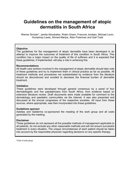
<str<strong>on</strong>g>Guidelines</str<strong>on</strong>g> <strong>on</strong> <strong>the</strong> management <strong>of</strong> atopic<br />
dermatitis in South Africa<br />
Werner Sinclair*, Jamila Aboobaker, Robin Green, Francois Jordaan, Michael Levin,<br />
Humphrey Lewis, Ahmed Manjra, Allan Puterman and Gail Todd<br />
Objective<br />
The guidelines for <strong>the</strong> management <strong>of</strong> atopic dermatitis have been developed in an<br />
attempt to improve <strong>the</strong> outcomes <strong>of</strong> treatment <strong>of</strong> this c<strong>on</strong>diti<strong>on</strong> in South Africa. This<br />
c<strong>on</strong>diti<strong>on</strong> has a major impact <strong>on</strong> <strong>the</strong> quality <strong>of</strong> life <strong>of</strong> sufferers and it is expected that<br />
<strong>the</strong>se guidelines, if implemented, will play a role in achieving this.<br />
Recommendati<strong>on</strong>s<br />
All health care workers involved in <strong>the</strong> management <strong>of</strong> atopic dermatitis should take note<br />
<strong>of</strong> <strong>the</strong>se guidelines and try to implement <strong>the</strong>m in clinical practice as far as possible. All<br />
treatment methods and procedures not substantiated by evidence from <strong>the</strong> literature<br />
should be disc<strong>on</strong>tinued and avoided to decrease <strong>the</strong> financial burden <strong>of</strong> dermatitis<br />
treatment.<br />
Validati<strong>on</strong><br />
These guidelines were developed through general c<strong>on</strong>sensus by a panel <strong>of</strong> four<br />
dermatologists and five paediatricians from South Africa, from evidence based <strong>on</strong><br />
extensive literature review. Draft documents were made available for comment to <strong>the</strong><br />
dermatology and paediatric communities via <strong>the</strong> internet. It was also presented and<br />
discussed at <strong>the</strong> annual c<strong>on</strong>gresses <strong>of</strong> <strong>the</strong> respective societies. All input from <strong>the</strong>se<br />
sources, where appropriate, was <strong>the</strong>n incorporated into <strong>the</strong>se guidelines.<br />
<str<strong>on</strong>g>Guidelines</str<strong>on</strong>g> sp<strong>on</strong>sor<br />
Astellas and Galderma co-sp<strong>on</strong>sored <strong>the</strong> meeting <strong>of</strong> <strong>the</strong> work group and all costs<br />
generated by <strong>the</strong> meeting.<br />
Disclaimer<br />
These guidelines do not represent all <strong>the</strong> possible methods <strong>of</strong> management applicable to<br />
all patients, do not exclude any o<strong>the</strong>r reas<strong>on</strong>able methods and will not ensure successful<br />
treatment in every situati<strong>on</strong>. The unique circumstances <strong>of</strong> each patient should be taken<br />
into account by <strong>the</strong> resp<strong>on</strong>sible physician regarding decisi<strong>on</strong>s <strong>on</strong> any specific <strong>the</strong>rapy.<br />
*Chair <strong>of</strong> work group
Introducti<strong>on</strong> and methods<br />
<strong>Atopic</strong> dermatitis (AD) is a very comm<strong>on</strong>, chr<strong>on</strong>ic, inflammatory eczematous skin disease,<br />
affecting up to 20% <strong>of</strong> children in Western Europe and Australia. The prevalence <strong>of</strong> AD in<br />
adults is less well defined, but it is believed that about 40% <strong>of</strong> childhood cases will c<strong>on</strong>tinue<br />
into adulthood. The morbidity and impact <strong>on</strong> quality <strong>of</strong> life <strong>of</strong> <strong>the</strong>se patients can be very<br />
severe 1 and <strong>the</strong> psychological distress suffered correlates well with <strong>the</strong> severity <strong>of</strong> <strong>the</strong><br />
dermatitis. 2<br />
There is c<strong>on</strong>siderable c<strong>on</strong>fusi<strong>on</strong> about uniform criteria for <strong>the</strong> diagnosis <strong>of</strong> AD and<br />
management is <strong>of</strong>ten arbitrary and empirical, <strong>of</strong>ten with poor outcomes. There is a need for<br />
standardised guidelines <strong>on</strong> diagnosing and managing this c<strong>on</strong>diti<strong>on</strong>.<br />
A panel <strong>of</strong> dermatologists and paediatricians was c<strong>on</strong>vened and tasked with writing and<br />
publishing <strong>the</strong>se guidelines. The compilati<strong>on</strong> <strong>of</strong> <strong>the</strong> panel was endorsed by <strong>the</strong><br />
Dermatological, Paediatric (SAPA) and Allergy (ALLSA) Societies <strong>of</strong> South Africa.<br />
The panel used an evidence-based module <strong>of</strong> evidence obtained by means <strong>of</strong> a thorough<br />
literature search published <strong>on</strong> <strong>the</strong> topic over <strong>the</strong> last 15 years. Evidence was graded<br />
according to <strong>the</strong> SIGN 3 grading system and recommendati<strong>on</strong>s and statements in <strong>the</strong> text are<br />
marked according to <strong>the</strong>se levels <strong>of</strong> evidence to denote <strong>the</strong> strength <strong>of</strong> evidence and<br />
<strong>the</strong>refore <strong>the</strong> validity and weight <strong>of</strong> recommendati<strong>on</strong>:<br />
Levels <strong>of</strong> evidence<br />
1++ High quality meta-analyses, systematic reviews <strong>of</strong> randomised c<strong>on</strong>trolled trials<br />
(RCTs), or RCTs with very low risk <strong>of</strong> bias<br />
1+ Well-c<strong>on</strong>ducted meta-analyses, systematic reviews <strong>of</strong> RCTs, or RCTs with low risk <strong>of</strong><br />
bias<br />
1- Meta-analyses, systematic reviews <strong>of</strong> RCTs, or RCTs with a high risk <strong>of</strong> bias<br />
2++ High quality systematic reviews <strong>of</strong> case-c<strong>on</strong>trol or cohort studies; high quality casec<strong>on</strong>trol<br />
or cohort studies with a very low risk <strong>of</strong> c<strong>on</strong>founding, bias or chance and a<br />
high probability that <strong>the</strong> relati<strong>on</strong>ship is causal<br />
2+ Well-c<strong>on</strong>ducted case-c<strong>on</strong>trol or cohort studies with a low risk <strong>of</strong> c<strong>on</strong>founding, bias or<br />
chance and a moderate probability that <strong>the</strong> relati<strong>on</strong>ship is causal<br />
2- Case-c<strong>on</strong>trol or cohort studies with a high risk <strong>of</strong> c<strong>on</strong>founding, bias or chance and a<br />
significant risk that <strong>the</strong> relati<strong>on</strong>ship is not causal.<br />
3 N<strong>on</strong>-analytical studies, e.g. case reports, case series<br />
4 Expert opini<strong>on</strong><br />
Grades <strong>of</strong> recommendati<strong>on</strong> for interventi<strong>on</strong>s<br />
A<br />
Highly recommended as method <strong>of</strong> choice, supported by at least <strong>on</strong>e metaanalysis,<br />
systematic review or RCTs rated as1++ and directly applicable to <strong>the</strong> target<br />
populati<strong>on</strong>; or
A systematic review <strong>of</strong> RCTs or a body <strong>of</strong> evidence c<strong>on</strong>sisting principally <strong>of</strong> studies<br />
rated as 1+, directly applicable to <strong>the</strong> target populati<strong>on</strong> and dem<strong>on</strong>strating overall<br />
c<strong>on</strong>sistency <strong>of</strong> results.<br />
B<br />
C<br />
D<br />
Str<strong>on</strong>gly recommended, supported by a body <strong>of</strong> evidence including studies rated as<br />
2++, directly applicable to <strong>the</strong> target populati<strong>on</strong> and dem<strong>on</strong>strating overall<br />
c<strong>on</strong>sistency <strong>of</strong> results; or<br />
Extrapolated evidence from studies rated as 1++ or 1+.<br />
Recommended as possible alternative opti<strong>on</strong>, supported by a body <strong>of</strong> evidence<br />
including studies rated as 2+, directly applicable to <strong>the</strong> target populati<strong>on</strong> and<br />
dem<strong>on</strong>strating overall c<strong>on</strong>sistency <strong>of</strong> results; or<br />
Extrapolated evidence from studies rated as 2++.<br />
Weak recommendati<strong>on</strong>, supported by evidence level 3 or 4; or<br />
Extrapolated evidence from studies rated as 2+.<br />
0 Not recommendable due to no supporting evidence or available evidence proves<br />
lack <strong>of</strong> effectiveness.<br />
Scope<br />
These guidelines were developed to address <strong>the</strong> diagnosis and management <strong>of</strong> patients<br />
who suffer from atopic dermatitis.<br />
References<br />
1. Evers AW, Duller P, Van der Valk PG, Kraaimaat FW, Van de Kerkh<strong>of</strong> PC. Comm<strong>on</strong> burden<br />
<strong>of</strong> chr<strong>on</strong>ic diseases? C<strong>on</strong>tributors to psychological distress in adults with psoriasis and atopic<br />
dermatitis. Br J Dermatol 2005;152(6):1275-81<br />
2. Taieb C, Ngyuen C, My<strong>on</strong> E. Adults with atopic dermatitis: Quality <strong>of</strong> life impact. J Am Acad<br />
Dermatol 2006;54(3):AB80<br />
3. SIGN 50. A Guideline Developers Handbook. Nati<strong>on</strong>al Institute for Clinical Excellence:<br />
Guideline Development Methods. 2004;7.5-7.8<br />
Definiti<strong>on</strong>s and terminology<br />
The word “eczema” comes from <strong>the</strong> Greek for “to boil over”. In patients with eczema, <strong>the</strong><br />
c<strong>on</strong>diti<strong>on</strong> periodically flares up (or “boils over”). During a flare, eczema in remissi<strong>on</strong> or stable<br />
sub-acute or chr<strong>on</strong>ic eczema transforms into acute eczema. Flares may happen<br />
sp<strong>on</strong>taneously or be precipitated by a number <strong>of</strong> factors.<br />
The term “dermatitis” refers to inflammati<strong>on</strong> <strong>of</strong> <strong>the</strong> skin, analogous to “appendicitis”<br />
(inflammati<strong>on</strong> <strong>of</strong> <strong>the</strong> appendix), “hepatitis” (inflammati<strong>on</strong> <strong>of</strong> <strong>the</strong> liver), etc. Nowadays <strong>the</strong><br />
terms “dermatitis” and “eczema” are generally regarded as syn<strong>on</strong>yms. Some authors still use<br />
<strong>the</strong> term “dermatitis” to include all types <strong>of</strong> cutaneous inflammati<strong>on</strong>, so that all eczema is<br />
dermatitis, but not all dermatitis is eczema. The term “dermatitis”, however, should be used<br />
with care, as some patients regard it as implying an occupati<strong>on</strong>al cause. Unfortunately, <strong>the</strong>re<br />
is still no internati<strong>on</strong>al agreement <strong>on</strong> <strong>the</strong> use <strong>of</strong> <strong>the</strong>se terms. Ackerman 1,2 has argued that, as<br />
<strong>the</strong> term eczema cannot be defined in a way that meets with universal approval, it should be<br />
dropped from <strong>the</strong> dermatological lexic<strong>on</strong>. There seems to be c<strong>on</strong>sensus that <strong>the</strong> term still
serves a useful purpose for <strong>the</strong> clinician. Much energy has been needlessly wasted in<br />
debating which term should be used. Geographically <strong>the</strong>re is unexplained preference for <strong>on</strong>e<br />
or <strong>the</strong> o<strong>the</strong>r. The word “eczema” tends to be used more as a layman‟s term and “dermatitis”<br />
more in <strong>the</strong> scientific c<strong>on</strong>text.<br />
Eczema/dermatitis is not a diagnosis. The most comm<strong>on</strong> forms <strong>of</strong> eczema/dermatitis are<br />
atopic, seborrhoeic, primary irritant, allergic c<strong>on</strong>tact, photoallergic, phototoxic, nummular,<br />
asteatotic, stasis and dyshidrotic. Eczema/dermatitis associated with infecti<strong>on</strong> (e.g.<br />
dermatophyte) or infestati<strong>on</strong> (e.g. scabies) – <strong>the</strong> so-called “ide” reacti<strong>on</strong>s – are additi<strong>on</strong>al<br />
variants.<br />
“Atopy” comes from <strong>the</strong> Greek “atopos” meaning strange or unusual. In 1892, Besnier was<br />
<strong>the</strong> first to describe <strong>the</strong> associati<strong>on</strong> <strong>of</strong> atopic dermatitis with allergic rhinitis and asthma. The<br />
term “atopy” was first coined in medicine in 1923 by two allergists, Coca and Cook. They<br />
defined atopy clinically as a proclivity to develop <strong>the</strong> triad <strong>of</strong> atopic eczema, allergic rhinitis<br />
and asthma. Patients whom <strong>the</strong>y c<strong>on</strong>sidered to be atopic possessed a distinctive antibody,<br />
which <strong>the</strong>y called “regain” or “skin-sensitising antibody”, because intradermal skin tests to a<br />
variety <strong>of</strong> inhalant allergens, e.g. trees, weeds, grasses, dust, moulds and danders, elicited<br />
wheals at sites <strong>of</strong> some injecti<strong>on</strong>s. When Hill and Sulzberger in <strong>the</strong> early 1930s encountered<br />
atopic patients with skin lesi<strong>on</strong>s that favoured antecubital and popliteal fossae, <strong>the</strong>y initially<br />
called <strong>the</strong> disorder “neurodermatitis <strong>of</strong> atopic type”, <strong>the</strong>n “atopic eczema” and finally “atopic<br />
dermatitis”.<br />
In <strong>the</strong> 1980s, Hanifin and Rajka proposed a list <strong>of</strong> criteria, and unity in <strong>the</strong> clinical c<strong>on</strong>cept <strong>of</strong><br />
atopic dermatitis was established. In 1994 <strong>the</strong> UK Working Party refined <strong>the</strong>se criteria into a<br />
c<strong>on</strong>cise and validated set <strong>of</strong> survey-based diagnostic criteria useful for <strong>the</strong> purposes <strong>of</strong><br />
epidemiological studies (vide infra). The word “atopy” can be defined as “a clinical<br />
hypersensitivity state that is subject to hereditary influences; included are hay fever, asthma<br />
and eczema”. These c<strong>on</strong>diti<strong>on</strong>s develop against a complex genetic background: <strong>the</strong> socalled<br />
atopic dia<strong>the</strong>sis.<br />
The word “atopic” in <strong>the</strong> term “atopic eczema” is simply an indicator <strong>of</strong> <strong>the</strong> frequent<br />
associati<strong>on</strong> with atopy and <strong>the</strong> need to separate this clinical phenotype from <strong>the</strong> o<strong>the</strong>r forms<br />
<strong>of</strong> eczema which have o<strong>the</strong>r causes and distinct patterns. The terms “atopic eczema” and<br />
“atopic dermatitis” are syn<strong>on</strong>ymous.<br />
According to <strong>the</strong> positi<strong>on</strong> paper from <strong>the</strong> Nomenclature Review Committee <strong>of</strong> <strong>the</strong> World<br />
Allergy Associati<strong>on</strong> 3 <strong>the</strong> term “atopic eczema/dermatitis syndrome” (AEDS) should be used<br />
as <strong>the</strong> umbrella term to cover <strong>the</strong> different subtypes <strong>of</strong> AD. The new nomenclature (AEDS)<br />
underlines <strong>the</strong> fact that AD is not <strong>on</strong>e single disease, but ra<strong>the</strong>r an aggregati<strong>on</strong> <strong>of</strong> several<br />
diseases with certain clinical characteristics in comm<strong>on</strong>.<br />
Intrinsic AD (n<strong>on</strong>-allergic AEDS = NAAEDS, a.k.a. atopiform dermatitis) fulfils <strong>the</strong> most<br />
comm<strong>on</strong>ly used diagnostic criteria for AD. These patients have no associated respiratory<br />
diseases, such as br<strong>on</strong>chial asthma or allergic rhinitis, show normal total serum IgE levels,<br />
no specific IgE, and negative skin-prick tests to aeroallergens or foods. In <strong>on</strong>e study, intrinsic<br />
AD was more comm<strong>on</strong> in females and disease <strong>on</strong>set was later. Palmar hyperlinearity,<br />
pityriasis alba, recurrent c<strong>on</strong>junctivitis, and hand and/or foot eczema were uncomm<strong>on</strong>. The<br />
Dennie-Morgan fold associated positively with this type <strong>of</strong> eczema. This group comprises at<br />
least 20% (up to 60%) <strong>of</strong> cases 4 .<br />
Extrinsic AD (allergic AEDS = AAEDS) is comm<strong>on</strong>ly associated with respiratory allergies<br />
such as rhinitis and asthma, a high level <strong>of</strong> serum IgE, specific IgE and positive skin-prick<br />
tests to aeroallergens or foods. Immunological differences between NAAEDS and AAEDS<br />
can be found in <strong>the</strong> cell and cytokine pattern in peripheral blood and in <strong>the</strong> affected skin, and
also by phenotyping characterisati<strong>on</strong> <strong>of</strong> epidermal dendritic cells. The current explanati<strong>on</strong> <strong>of</strong><br />
this distincti<strong>on</strong> is based <strong>on</strong> differences in genetics and/or envir<strong>on</strong>mental c<strong>on</strong>diti<strong>on</strong>s. This<br />
group comprises 40 - 80% <strong>of</strong> patients 4 .<br />
The classificati<strong>on</strong> into AAEDS and NAAEDS at each stage <strong>of</strong> life, i.e. infancy, childhood,<br />
teenage and adult, is essential for <strong>the</strong> allergological management <strong>of</strong> patients in respect <strong>of</strong><br />
allergen avoidance, sec<strong>on</strong>dary allergy preventi<strong>on</strong>, and immuno<strong>the</strong>rapy. The risk <strong>of</strong> an “atopy<br />
march” is significantly lower in children with NAAEDS. 5-13<br />
This subdivisi<strong>on</strong> is currently c<strong>on</strong>troversial. Cases may transform from <strong>on</strong>e type to <strong>the</strong> o<strong>the</strong>r.<br />
This divisi<strong>on</strong> may not be applicable to adults. It is quite possible that <strong>the</strong>re are distinct<br />
subsets <strong>of</strong> atopic eczema, e.g. those cases associated with atopy and those who have<br />
severe disease with recurrent infecti<strong>on</strong>s. Until <strong>the</strong> exact genetic and causative agents are<br />
known, it is wiser to c<strong>on</strong>sider <strong>the</strong> clinical disease as <strong>on</strong>e c<strong>on</strong>diti<strong>on</strong>. Perhaps sensitivity<br />
analyses should be d<strong>on</strong>e within clinical trials, or am<strong>on</strong>g those who are thought to represent<br />
distinct subsets, e.g. those who are definitely atopic with raised circulating IgE to allergens,<br />
and those with severe disease and associated asthma. 14,15<br />
Acute eczema/dermatitis is characterised by oedema, ery<strong>the</strong>ma, vesiculati<strong>on</strong>, exudati<strong>on</strong> and<br />
crusting. Microscopy shows collecti<strong>on</strong>s <strong>of</strong> serum in <strong>the</strong> stratum corneum, moderate to<br />
marked sp<strong>on</strong>giosis, intraepidermal vesiculati<strong>on</strong>, moderate to marked sub-epidermal oedema<br />
in <strong>the</strong> papillary dermis and lymphocytes in <strong>the</strong> upper dermis. Chr<strong>on</strong>ic eczema/dermatitis is<br />
characterised by lichenificati<strong>on</strong>. Lichenificati<strong>on</strong> refers to thickening <strong>of</strong> <strong>the</strong> skin with<br />
exaggerati<strong>on</strong> <strong>of</strong> <strong>the</strong> normal markings. Flat-topped, shiny, quadrilateral coalescing papules<br />
are enclosed. Microscopy shows compact orthokeratosis, acanthosis, mild sp<strong>on</strong>giosis,<br />
collagen in vertical streaks in <strong>the</strong> dermal papillae and lymphocytes in <strong>the</strong> upper dermis. Subacute<br />
eczema/dermatitis shows features overlapping with acute and chr<strong>on</strong>ic<br />
eczema/dermatitis. Lesi<strong>on</strong>s are comm<strong>on</strong>ly slightly elevated, are red, brownish or purplish in<br />
colour, and with variable scaling. Generally, physicians most comm<strong>on</strong>ly encounter <strong>the</strong> subacute<br />
presentati<strong>on</strong> <strong>of</strong> eczema/dermatitis.<br />
The morphology, distributi<strong>on</strong> and evoluti<strong>on</strong> <strong>of</strong> eczema/dermatitis in atopic eczema/dermatitis<br />
are highly characteristic. During <strong>the</strong> infant phase (birth to 2 years) red scaly lesi<strong>on</strong>s develop<br />
typically <strong>on</strong> <strong>the</strong> cheeks, usually sparing <strong>the</strong> perioral and perinasal areas. The chin is typically<br />
involved and cheilitis is comm<strong>on</strong>. A small but significant number <strong>of</strong> infants develop a<br />
generalised erupti<strong>on</strong>. Involvement <strong>of</strong> <strong>the</strong> scalp is not uncomm<strong>on</strong>. The diaper area is <strong>of</strong>ten<br />
spared. Sometimes <strong>the</strong> cubital/popliteal fossae or o<strong>the</strong>r parts <strong>of</strong> <strong>the</strong> limbs are involved.<br />
During <strong>the</strong> childhood phase (2-12 years) eczema/dermatitis involves <strong>the</strong> flexural areas (i.e.<br />
<strong>the</strong> antecubital fossae and popliteal fossae) but also <strong>the</strong> neck, wrist and ankles.<br />
During <strong>the</strong> adult phase (12 years to adult) lesi<strong>on</strong>s involve similar areas to those affected<br />
during <strong>the</strong> childhood phase. Additi<strong>on</strong>ally, hand eczema/dermatitis, periocular<br />
eczema/dermatitis and anogenital eczema/dermatitis are comm<strong>on</strong>. In some cases lesi<strong>on</strong>s<br />
occur mostly <strong>on</strong> extensor surfaces and follicular accentuati<strong>on</strong> may be prominent.<br />
Morphologically, lesi<strong>on</strong>s may be acute, sub-acute or chr<strong>on</strong>ic. Importantly for <strong>the</strong> clinician, <strong>the</strong><br />
diagnosis <strong>of</strong> atopic eczema/dermatitis is based <strong>on</strong> <strong>the</strong> aforementi<strong>on</strong>ed criteria, namely age<br />
<strong>of</strong> <strong>the</strong> patient, distributi<strong>on</strong> <strong>of</strong> <strong>the</strong> rash and morphology <strong>of</strong> <strong>the</strong> rash. <strong>Atopic</strong> eczema is a difficult<br />
disease to define, as <strong>the</strong> clinical features are highly variable in morphology, body site and<br />
time.<br />
There is no specific diagnostic test that encompasses all people with typical eczema that can<br />
serve as a reference standard. Diagnosis is, <strong>the</strong>refore, essentially a clinical <strong>on</strong>e. 16,17,18
References<br />
1. Ackerman AB. „Eczema‟ revisited. A status report based up<strong>on</strong> current textbooks <strong>of</strong>dermatology.<br />
Am J Dermatopathol 1994; 16: 517-22; discussi<strong>on</strong> 523-531<br />
2. Ackerman AB. A plea to expunge <strong>the</strong> word „eczema‟ from <strong>the</strong> lexic<strong>on</strong> <strong>of</strong> dermatology and<br />
dermatopathology. Arch Dermatol Res 1982;272:407-20<br />
3. Johanss<strong>on</strong> SGO, Bieber T, Dahl R, et al. Revised nomenclature for allergy for global use:<br />
report <strong>of</strong> <strong>the</strong> Nomenclature Review Committee <strong>of</strong> <strong>the</strong> World Allergy Organizati<strong>on</strong>, October<br />
2003. J Allergy Clin Immunol 2004; 113:832-6<br />
4. Flohr C, Johanss<strong>on</strong> SGO, Wahlgren C-F and Williams H. How atopic is atopic dermatitis? J<br />
Allergy Clin Immunol 2004;114:150-8<br />
5. Folster-Holst R, Pape M, Buss YL, Christophers E, Weichenthal M. Low prevalence <strong>of</strong> <strong>the</strong><br />
intrinsic form <strong>of</strong> atopic dermatitis am<strong>on</strong>g adult patients. Allergy 2006; 61: 629-632.<br />
6. Akdis CA, Akdis M. Immunological differences between intrinsic and extrinsic types <strong>of</strong> atopic<br />
dermatitis. Clin Exp Allergy 2003; 33:1618-21<br />
7. Novak N, Bieber T. Allergic and n<strong>on</strong>-allergic forms <strong>of</strong> atopic diseases. J Allergy Clin Immunol<br />
2003; 112:252-62<br />
8. Wuthrich B, Schmid-Grendelmeier P. The atopic eczema/dermatitis syndrome. Epidemiology,<br />
natural course, and immunology <strong>of</strong> <strong>the</strong> IgE-associated („extrinsic‟) and <strong>the</strong> n<strong>on</strong>-allergic<br />
(„intrinsic‟) AEDS. J Invest Allergol Clin Immunol 2003;13:1-5<br />
9. Tokura Y. Extrinsic and intrinsic types <strong>of</strong> atopic dermatitis. J Dermatol Sci 2010;58:1-7<br />
10. Kerschenlohr K, Decard S, Przybilla B, Wollenberg A. Atopy patch test reacti<strong>on</strong>s show a rapid<br />
influx <strong>of</strong> inflammatory dendritic epidermal cells in patients with extrinsic atopic dermatitis and<br />
patients with intrinsic atopic dermatitis. J Allergy Clin Immunol 2003;111:869-74<br />
11. Ingordo V, D‟Andria G, D‟Andria C, Tortora A. Results <strong>of</strong> atopy patch tests with house dust<br />
mites in adults with „intrinsic‟ and „extrinsic‟ atopic dermatitis. J Eur Acad Dermatol Venereol<br />
2002;16:450-4<br />
12. Schmid-Grendelmeier P, Sim<strong>on</strong> D, Sim<strong>on</strong> HU, Akdis CA, Wuthrich B. Epidemiology, clinical<br />
features, and immunology <strong>of</strong> <strong>the</strong> „intrinsic‟ (n<strong>on</strong>-IgE-mediated) type <strong>of</strong> atopic dermatitis<br />
(c<strong>on</strong>stituti<strong>on</strong>al dermatitis). Allergy 2001;56:841-9<br />
13. Benkmeijer EE, Spuls PI, Legierse CM, et al. Clinical differences between atopic and atopiform<br />
dermatitis J Am Acad Dermatol 2008;58(3):407-14<br />
14. Williams HC, Johanss<strong>on</strong> SGO. Two types <strong>of</strong> eczema – or are <strong>the</strong>re? J Allergy Clin Immunol<br />
2005;116:1064-1066<br />
15. Bieber T. Putative mechanisms underlying chr<strong>on</strong>icity in atopic eczema. Acta Derm Venereol<br />
2005; Suppl 215:7-10<br />
16. Friedmann PS, Holden CA. <strong>Atopic</strong> dermatitis. In: Burns DA, Breathnach SM, Cox NH,Griffiths<br />
CEM, eds. Rook‟s Textbook <strong>of</strong> <strong>Dermatology</strong>, Vol 1, 7th ed. Oxford: Blackwell Publishing,<br />
2004:18.1-18.31<br />
17. Habif TB. <strong>Atopic</strong> dermatitis: In: Clinical <strong>Dermatology</strong>, 4th ed. St Louis: Mosby, 2004: 105-125<br />
18. Simps<strong>on</strong> EL, Hanifin JM. <strong>Atopic</strong> dermatitis. Med Clin N Am 2006;90:149-67<br />
Aetiopathogenesis <strong>of</strong> atopic dermatitis<br />
This is probably multifactorial. Current thinking favours a skin barrier defect as <strong>the</strong> most<br />
significant predisposing factor where mutati<strong>on</strong>s in <strong>the</strong> filaggrin gene feature str<strong>on</strong>gly. 1-8 Most<br />
studies investigating <strong>the</strong> causes <strong>of</strong> atopic dermatitis deal with children. There is little to<br />
suggest that adult atopic dermatitis should have a different aetiopathogenesis apart from<br />
some clinical features that differ, such as <strong>the</strong> predominant involvement <strong>of</strong> <strong>the</strong> hands and <strong>the</strong><br />
head and neck. 9<br />
Genetics<br />
Populati<strong>on</strong>-based family studies in Europe suggest that in atopic families, up to 50% <strong>of</strong><br />
<strong>of</strong>fspring will have atopic dermatitis. 10 Twin studies showing a c<strong>on</strong>cordance rate for atopic
dermatitis <strong>of</strong> 0.75 for m<strong>on</strong>ozygotic twins compared to 0.20 for dizygotic twins support a<br />
genetic basis for atopic dermatitis. 10,11,12 Fur<strong>the</strong>r evidence for a genetic predispositi<strong>on</strong> to<br />
atopic dermatitis is <strong>the</strong> finding <strong>of</strong> candidate genes. 1,10,12<br />
Allergic sensitisati<strong>on</strong><br />
The predispositi<strong>on</strong> for IgE hyper-resp<strong>on</strong>siveness to allergens defines <strong>the</strong> term atopy. 13 A<br />
systematic review <strong>of</strong> <strong>the</strong> published evidence for allergic sensitisati<strong>on</strong> and dermatitis in 12<br />
populati<strong>on</strong> studies from around <strong>the</strong> world has shown that IgE hyper-resp<strong>on</strong>siveness does not<br />
necessarily equate to atopic dermatitis, even though it may be associated with <strong>the</strong> disease<br />
phenotype, especially those with severe disease. 14,15 Am<strong>on</strong>gst those with atopic dermatitis,<br />
up to 20 - 60% were not atopic per definiti<strong>on</strong>. Geographic locati<strong>on</strong> was associated with <strong>the</strong><br />
risk <strong>of</strong> being atopic, am<strong>on</strong>gst those with atopic dermatitis as compared to normal healthy<br />
c<strong>on</strong>trols.<br />
In five studies that included adolescents and adults, <strong>the</strong> findings were essentially similar. In a<br />
cross-secti<strong>on</strong>al household survey from Ethiopia, which included adults and children, 15% <strong>of</strong><br />
those with atopic dermatitis and 8% <strong>of</strong> those without atopic dermatitis were atopic by skinprick<br />
testing. 16 This lack <strong>of</strong> associati<strong>on</strong> between atopic dermatitis and allergen sensitisati<strong>on</strong><br />
was c<strong>on</strong>firmed in a cross-secti<strong>on</strong>al survey and nested case c<strong>on</strong>trolled study <strong>of</strong> children. 17<br />
Envir<strong>on</strong>ment<br />
A documented increasing prevalence <strong>of</strong> atopic dermatitis over <strong>the</strong> last 50 years is not<br />
c<strong>on</strong>sistent with genetic drift al<strong>on</strong>e, 18,19,20 but supports a str<strong>on</strong>g envir<strong>on</strong>mental influence as<br />
evidenced by populati<strong>on</strong> migrati<strong>on</strong> studies. 21,22 These envir<strong>on</strong>mental influences, which affect<br />
initial disease expressi<strong>on</strong> or aggravati<strong>on</strong> <strong>of</strong> established disease, are summarised in Table 1.<br />
Populati<strong>on</strong> studies from Africa seldom c<strong>on</strong>firm a role for <strong>the</strong>se envir<strong>on</strong>mental factors. 16,17,23<br />
Interestingly, many are surrogate markers <strong>of</strong> urbanisati<strong>on</strong> 21,22 and increased socio-ec<strong>on</strong>omic<br />
status, 24 which appear to be <strong>the</strong> <strong>on</strong>ly fairly c<strong>on</strong>sistent associati<strong>on</strong> across all populati<strong>on</strong><br />
groups*. More detailed informati<strong>on</strong> can be obtained from published reviews <strong>on</strong> geneenvir<strong>on</strong>ment<br />
interacti<strong>on</strong>s. 1,10,12,21,25-40<br />
The aetiopathogenesis <strong>of</strong> atopic dermatitis is best explained by <strong>the</strong> c<strong>on</strong>cept <strong>of</strong> a damaged<br />
barrier functi<strong>on</strong>, whe<strong>the</strong>r intrinsically normal or dysfuncti<strong>on</strong>al, that induces a state <strong>of</strong><br />
epidermal repair, coupled with aberrant resp<strong>on</strong>ses to epidermal insults in <strong>the</strong> affected<br />
skin. 2,3,4,8,10,12,24,27,41,42,43 In Africa this hypo<strong>the</strong>sis has still not been validated. A novel filaggrin<br />
gene defect has been documented in a single Ethiopian case <strong>of</strong> atopic dermatitis 44 . What<br />
evoluti<strong>on</strong>ary advantage <strong>the</strong> skin barrier defect c<strong>on</strong>veyed to <strong>the</strong> populati<strong>on</strong>s now exposed to<br />
envir<strong>on</strong>mental influences precipitating atopic disease is unknown.<br />
Table 1: Envir<strong>on</strong>mental influences <strong>on</strong> atopic dermatitis<br />
Envir<strong>on</strong>mental factor Effect <strong>on</strong> atopic dermatitis References<br />
Rural compared to urban<br />
living (hygiene hypo<strong>the</strong>sis)<br />
Increased risk with urbanisati<strong>on</strong> 21, 22, 24,<br />
25, 38<br />
*Socioec<strong>on</strong>omic status Increased risk with higher socio-ec<strong>on</strong>omic status 16, 17, 24,<br />
28, 45<br />
*Educati<strong>on</strong> level <strong>of</strong> parents Increased risk with higher level <strong>of</strong> educati<strong>on</strong> 29<br />
*Family size (hygiene<br />
hypo<strong>the</strong>sis)<br />
Increased risk with decreasing family size 25, 29
*Day-care attendance<br />
(hygiene hypo<strong>the</strong>sis)<br />
Animal exposure in early life<br />
(hygiene hypo<strong>the</strong>sis)<br />
*Endotoxin exposure in early<br />
infancy (hygiene hypo<strong>the</strong>sis)<br />
*Basic hygiene (hygiene<br />
hypo<strong>the</strong>sis)<br />
*Early life infecti<strong>on</strong>s (hygiene<br />
hypo<strong>the</strong>sis)<br />
*Parasite exposure in early<br />
life (hygiene hypo<strong>the</strong>sis)<br />
Decreased risk with day-care attendance in 25<br />
infancy<br />
May be protective with specific pet exposure in 25, 36, 37,<br />
early life<br />
41, 46<br />
Decreased risk <strong>of</strong> dermatitis development with 25<br />
early life exposure<br />
Increased risk with improved pers<strong>on</strong>al hygiene 25<br />
No clear evidence for protecti<strong>on</strong> from specific<br />
organism exposure<br />
Protecti<strong>on</strong> may be related to microbial burden in<br />
early life<br />
Little evidence for a protective effect <strong>on</strong> atopic<br />
dermatitis development<br />
Exposure reduces IgE sensitisati<strong>on</strong><br />
Protective effect suggested but associati<strong>on</strong> with<br />
vaccinati<strong>on</strong> type unclear.<br />
Increased risk with increasing antibiotic<br />
25<br />
25, 41, 45,<br />
47, 48<br />
Vaccinati<strong>on</strong> (hygiene<br />
hypo<strong>the</strong>sis)<br />
Early life antibiotic use<br />
(hygiene hypo<strong>the</strong>sis) prescribing in infants<br />
*Mode <strong>of</strong> delivery Increased risk with caesarean secti<strong>on</strong> 37, 51<br />
*Maternal age Increased risk with increased age 30, 33<br />
Maternal diet:<br />
- Probiotics<br />
- Food avoidance<br />
- Specific foods<br />
Envir<strong>on</strong>mental tobacco<br />
smoke<br />
Maternal risk (parent-<strong>of</strong>origin<br />
effect)<br />
Diet:<br />
- Formulae<br />
- Solid food introducti<strong>on</strong><br />
- Inclusi<strong>on</strong> – specific food<br />
- Exclusi<strong>on</strong> – specific food<br />
- Diet restricti<strong>on</strong><br />
- Elemental diet<br />
- Organic food<br />
Diet supplementati<strong>on</strong>:<br />
- Anti-oxidants<br />
- Essential fatty acids<br />
- Early fish as solid food<br />
- Probiotics<br />
Possible decreased risk with probiotic use<br />
Little evidence for decreased risk with food<br />
avoidance if applied in pregnancy or while breast<br />
feeding<br />
Evidence for decreased risk if fish c<strong>on</strong>sumed<br />
during pregnancy and breast feeding<br />
Major risk factor for development <strong>of</strong> atopic<br />
dermatitis<br />
25, 30, 49,<br />
50<br />
25, 36<br />
30, 34, 35,<br />
36<br />
30, 34, 35,<br />
36, 40<br />
38, 40, 52<br />
70, 71<br />
Increased risk if mo<strong>the</strong>r had atopic dermatitis 12, 30, 38<br />
Fully hydrolysed formula – decreased risk<br />
Partially hydrolysed formula – no clear evidence<br />
for role in c<strong>on</strong>trol or preventi<strong>on</strong><br />
No clear evidence for role in c<strong>on</strong>trol or preventi<strong>on</strong><br />
Evidence for delayed <strong>on</strong>set with fish in early life<br />
Decreased risk with maternal probiotic use<br />
Disease c<strong>on</strong>trol in children
Breastfeeding<br />
Allergen exposure:<br />
- House dust mite<br />
- Super-antigen<br />
- Type IV allergens<br />
Irritant exposure:<br />
- Climate<br />
- Hard water<br />
- Clothing<br />
- Occlusive wraps<br />
- Clo<strong>the</strong>s s<strong>of</strong>teners<br />
Bath additives:<br />
- Emollients<br />
- Antiseptics<br />
- Salt<br />
Polluti<strong>on</strong>:<br />
- Urbanisati<strong>on</strong><br />
- Smoking<br />
- Solvents<br />
Co-morbidities:<br />
- HIV<br />
- BMI<br />
- Smoking<br />
- Psychosocial stress<br />
- Malaria<br />
Some evidence for decreased risk in breastfed<br />
infants with a family history <strong>of</strong> atopic dermatitis<br />
Decreased exposure/load – no effect preventi<strong>on</strong><br />
but may have an effect <strong>on</strong> c<strong>on</strong>trol in severe cases<br />
Staphylococcal c<strong>on</strong>trol – no effect c<strong>on</strong>trol<br />
C<strong>on</strong>tact dermatitis – increased associati<strong>on</strong><br />
Increased risk in cooler climates<br />
Unclear risk in climate extremes<br />
Hard water – no effect if calcium carb<strong>on</strong>ate<br />
removed<br />
Disease aggravati<strong>on</strong> by fibre roughness<br />
Disease aggravati<strong>on</strong> by heavy textiles<br />
Silk clothing – no clear evidence for role in c<strong>on</strong>trol<br />
Wet or dry – no clear evidence for role in c<strong>on</strong>trol<br />
No clear evidence for role in c<strong>on</strong>trol<br />
No clear evidence for role in c<strong>on</strong>trol<br />
No clear evidence for role in c<strong>on</strong>trol<br />
No clear evidence for role in c<strong>on</strong>trol<br />
Increased risk with urbanisati<strong>on</strong><br />
Increased risk in adults linked to lifetime exposure<br />
Increased risk in established dermatitis<br />
No increased risk<br />
Increased risk in preventi<strong>on</strong> and dermatitis c<strong>on</strong>trol<br />
Increased risk in adults linked to lifetime exposure<br />
Increased risk in preventi<strong>on</strong> (episodes in early life)<br />
and dermatitis c<strong>on</strong>trol<br />
Increased risk in Africa<br />
* Possible surrogate markers <strong>of</strong> urbanisati<strong>on</strong> and increased socio-ec<strong>on</strong>omic status.<br />
30, 39, 40,<br />
58, 59<br />
32, 35, 36,<br />
38<br />
35, 36, 38,<br />
60<br />
61, 62<br />
21<br />
21, 36<br />
28, 63<br />
35<br />
35<br />
38, 64<br />
38, 65<br />
35<br />
38, 66<br />
35, 36, 38,<br />
60<br />
35<br />
21, 31<br />
67<br />
31<br />
68<br />
24<br />
67<br />
36, 37, 69<br />
17<br />
References<br />
1. Cork MJ, Robins<strong>on</strong> DA, Vasilopoulos Y et al. New perspectives <strong>on</strong> epidermal barrier<br />
dysfuncti<strong>on</strong> in atopic dermatitis: Gene-envir<strong>on</strong>ment interacti<strong>on</strong>s. J Allergy Clin Immunol<br />
2006;118:3-21<br />
2. Wolf R, Wolf D. Abnormal epidermal barrier in <strong>the</strong> pathogenesis <strong>of</strong> atopic dermatitis. Clinics in<br />
Dermatol 2012;30:329-34<br />
3. Irvine AD, Irwin McLean WH, Leung DYM. Filaggrin mutati<strong>on</strong>s associated with skin and allergic<br />
disease. N Engl J Med 2011;365:1315-27<br />
4. Cork MJ, Danby SG, Vasilopoulos Y et al. Epidermal barrier dysfuncti<strong>on</strong> in atopic dermatitis. J<br />
Invest Dermatol 2009;129:1892-908<br />
5. Rodriquez E, Baurecht H, Herberich E et al. Meta-analysis <strong>of</strong> filaggrin polymorphisms in<br />
dermatitis and asthma: robust risk factors in atopic disease. J Allergy Clin Immunol<br />
2009;123:1361-70<br />
6. Van den Oord RA, Sheikh A. Filaggrin gene defects and risk <strong>of</strong> developing allergic sensitisati<strong>on</strong><br />
and allergic disorders: a systematic review and meta-analysis. Br Med J 2009;339:b2433<br />
7. Baurecht H, Irvine AD, Novak N et al. Towards a major risk factor for atopic dermatitis: metaanalyses<br />
<strong>of</strong> filiggrin polymorphism data. J allergy Clin Immunol 2007;120:1406-12<br />
8. Huds<strong>on</strong>. Skin barrier functi<strong>on</strong> and allergic risk. Nature Genetics 2006;38:399-400
9. Sandstrom Falk MH and Faergemann J. <strong>Atopic</strong> dermatitis in adults: Does it disappear with<br />
age? Acta Derm Venereol 2006;86:135-9<br />
10. Schultz Larsen F. Genetic epidemiology <strong>of</strong> atopic dermatitis. In <strong>Atopic</strong> dermatitis. The<br />
epidemiology, causes and preventi<strong>on</strong> <strong>of</strong> atopic dermatitis. Ed HC Williams. Cambridge<br />
University Press 2000;113-124<br />
11. Schultz Larsen F, <strong>Atopic</strong> dermatitis: a genetic-epidemiologic study in a populati<strong>on</strong>-based twin<br />
sample. J Am Acad Dermatol 1993;28:719-23<br />
12. Morar N, Willis-Owen SAG, M<strong>of</strong>fatt MF and Cooks<strong>on</strong> WOCM. The genetics <strong>of</strong> atopic dermatitis.<br />
J Allergy Clin Immunol 2006;118:24-34<br />
13. Johanss<strong>on</strong> SGO, Bieber T, Dahl R et al Revised nomenclature review committee <strong>of</strong> <strong>the</strong> world<br />
allergy organisati<strong>on</strong>, October 2003. Allergy Clin Immunol Int - J World Allergy Org 2005;17:4-8<br />
14. Flohr C, Johanss<strong>on</strong> SGO, Wahlgren C-F and Williams H. How atopic is atopic dermatitis? J<br />
Allergy Clin Immunol 2004;114:150-8<br />
15. Williams H, Flohr C. How epidemiology has challenged 3 prevailing c<strong>on</strong>cepts about atopic<br />
dermatitis. J Allergy Clin Immunol 2006;118:209-13<br />
16. Yemaneberhan H, Flohr C, Bekele Z et al. Prevalence and associated risk factors <strong>of</strong> atopic<br />
dermatitis symptoms in rural and urban Ethiopia. Clin Exp Allergy 2004;34:779-85<br />
17. Haileamlak A, Dagoye D, Williams H et al. Early risk factors for atopic dermatitis in Ethiopian<br />
children. J Allergy Clin Immunol 2005;115:370-6<br />
18. Williams HC. <strong>Atopic</strong> dermatitis – why we should look to <strong>the</strong> envir<strong>on</strong>ment. Br Med J<br />
1995;311:1241-2<br />
19. Williams HC. Is <strong>the</strong> prevalence <strong>of</strong> atopic dermatitis increasing? Clin Exp Dermatol 1992;17:385-<br />
91<br />
20. Diepgen T. Is <strong>the</strong> prevalence <strong>of</strong> atopic dermatitis increasing? In <strong>Atopic</strong> dermatitis. The<br />
epidemiology, causes and preventi<strong>on</strong> <strong>of</strong> atopic dermatitis. Ed HC Williams. Cambridge<br />
University Press 2000;96-112<br />
21. Burrell-Morris C, Williams HC. <strong>Atopic</strong> dermatitis in migrant populati<strong>on</strong>. In <strong>Atopic</strong> dermatitis. The<br />
epidemiology, causes and preventi<strong>on</strong> <strong>of</strong> atopic dermatitis. Ed HC Williams. Cambridge<br />
University Press 2000;169-82<br />
22. Schram ME, Tedja Am Spijker R et al. Is <strong>the</strong>re a rural/urban gradient in <strong>the</strong> prevalence <strong>of</strong><br />
dermatitis? A systematic review. Br J Dermatol 2010;162:964-73<br />
23. Nnoruka EN. Current epidemiology <strong>of</strong> atopic dermatitis in south-eastern Nigeria. Int J Dermatol<br />
2004;43:739-44<br />
24. Obeng BB, Hartgers F, Boakye D, Yazdanbakhsh M. Out <strong>of</strong> Africa: what can be learned from<br />
<strong>the</strong> studies <strong>of</strong> allergic disorders in Africa and Africans? Curr Opini<strong>on</strong> Allergy Clin Immunol<br />
2008;8:391-7<br />
25. Flohr C, Pascoe D and Williams HC. <strong>Atopic</strong> dermatitis and <strong>the</strong> „hygiene hypo<strong>the</strong>sis‟: too clean<br />
to be true? Br J Dermatol 2005;152:202-16<br />
26. Akdis CA, Akdai M, Bieber T et. al. for <strong>the</strong> European Academy <strong>of</strong> Allergology and Clinical<br />
Immunology/American Academy <strong>of</strong> Allergy, Asthma and Immunology/PRACTALL C<strong>on</strong>sensus<br />
Group. Diagnosis and treatment <strong>of</strong> atopic dermatitis in children and adults: European Academy<br />
<strong>of</strong> Allergology and Clinical Immunology /American Academy <strong>of</strong> Allergy, Asthma and<br />
Immunology/PRACTALL C<strong>on</strong>sensus Report. J Allergy Clin Immunol 2006;118:152-89<br />
27. Taieb A, Hanifin J, Cooper K et al. Proceedings <strong>of</strong> <strong>the</strong> 4th Georg Rajka Internati<strong>on</strong>al<br />
Symposium <strong>on</strong> atopic dermatitis, Aacach<strong>on</strong>, France, September 15-17,2005. J Allergy Clin<br />
Immunol 2006;117:378-90<br />
28. McNally N, Phillips D. Geographical studies <strong>of</strong> atopic dermatitis. In <strong>Atopic</strong> dermatitis. The<br />
epidemiology, causes and preventi<strong>on</strong> <strong>of</strong> atopic dermatitis. Ed HC Williams. Cambridge<br />
University Press 2000;71-84<br />
29. McNally N, Phillips D. Social factors and atopic dermatitis. In <strong>Atopic</strong> dermatitis. The<br />
epidemiology, causes and preventi<strong>on</strong> <strong>of</strong> atopic dermatitis. Ed HC Williams. Cambridge<br />
University Press 2000;139-54<br />
30. Godfrey K. Fetal and perinatal origins <strong>of</strong> atopic dermatitis. In <strong>Atopic</strong> dermatitis. The<br />
epidemiology, causes and preventi<strong>on</strong> <strong>of</strong> atopic dermatitis. Ed HC Williams. Cambridge<br />
University Press 2000;125-38<br />
31. Schafer T, Ring J. The possible role <strong>of</strong> envir<strong>on</strong>mental polluti<strong>on</strong> in <strong>the</strong> development <strong>of</strong> atopic<br />
dermatitis. In <strong>Atopic</strong> dermatitis. The epidemiology, causes and preventi<strong>on</strong> <strong>of</strong> atopic dermatitis.<br />
Ed HC Williams. Cambridge University Press 2000;155-68
32. Kolmer H, Platts-Mills TAE. The role <strong>of</strong> inhalant allergens in atopic dermatitis. In <strong>Atopic</strong><br />
dermatitis. The epidemiology, causes and preventi<strong>on</strong> <strong>of</strong> atopic dermatitis. Ed HC Williams.<br />
Cambridge University Press 2000;1183-201<br />
33. Braae Olesen A, Thestrup-Petersen K. The „old mo<strong>the</strong>r‟ hypo<strong>the</strong>sis. In <strong>Atopic</strong> dermatitis. The<br />
epidemiology, causes and preventi<strong>on</strong> <strong>of</strong> atopic dermatitis. Ed HC Williams. Cambridge<br />
University Press 2000;148-54<br />
34. Williams HC, Wuthrich B. The natural history <strong>of</strong> atopic dermatitis. In <strong>Atopic</strong> dermatitis. The<br />
epidemiology, causes and preventi<strong>on</strong> <strong>of</strong> atopic dermatitis. Ed HC Williams. Cambridge<br />
University Press2000;41-59<br />
35. Hoare C, Li Wan Po A, Williams H. Systematic review <strong>of</strong> treatments for atopic dermatitis. Health<br />
Technology Assessment 2000;4(37):1-191<br />
36. Nati<strong>on</strong>al Institute for Health and Clinical Excellence. <strong>Atopic</strong> dermatitis in children. <strong>Management</strong><br />
<strong>of</strong> atopic dermatitis in children from birth up to <strong>the</strong> age <strong>of</strong> 12 years. NICE clinical guideline 57.<br />
L<strong>on</strong>d<strong>on</strong>: NICE, 2007<br />
37. Williams HC, Grindlay DJC. What‟s new in atopic dermatitis? An analysis <strong>of</strong> systematic reviews<br />
published in 2007 and 2008. Part 1. Definiti<strong>on</strong>s, causes and c<strong>on</strong>sequences <strong>of</strong> dermatitis. Clin<br />
Exp Dermatol 2009;35:12-5<br />
38. Shams K, Grindlay DJC, Williams HC. What‟s new in atopic dermatitis? An analysis <strong>of</strong><br />
systematic reviews published in 2009-2010. Clin Exp Dermatol 2011;36:573-8<br />
39. Batchelor JM, Grindlay DJC, Williams HC. What‟s new in atopic dermatitis? An analysis <strong>of</strong><br />
systematic reviews published in 2008 and 2009. Clin Exp Dermatol 2010;35:823-8<br />
40. Muche-Borowski C, Koop M, Reese I et al. Allergy preventi<strong>on</strong>. Dtsch Arztebl Int 2009;106:625-<br />
31<br />
41. Roduit C, Wohlgensinger J, Frei R et al. Prenatal animal c<strong>on</strong>tact and gene expressi<strong>on</strong> <strong>of</strong> innate<br />
immunity receptors at birth are associated with atopic dermatitis. J Allergy Clin Immunol<br />
2011;127:179-85<br />
42. Van den Biggelaar AH, Hua TD, Rodrigues LC et al. Genetic variati<strong>on</strong> in IL-10 is associated<br />
with atopic reactivity in Gab<strong>on</strong>ese schoolchildren. J Allergy Clin Immunol 2007;120:973-5<br />
43. De Benedetto A, Agnihothri R, Mc Girt LY, et al. <strong>Atopic</strong> dermatitis: a disease caused by innate<br />
immune defects. J Invest Dermatol 2009;129(1):14-30.<br />
44. Winge MCG, Bilcha KD, Lieden A et al. Novel filaggrin mutati<strong>on</strong> but no loss-<strong>of</strong>-functi<strong>on</strong> variants<br />
found in Ethiopian patients with atopic dermatitis. Br J Dermatol 2011;165:1074-80<br />
45. Flohr C, Tuyen LN, Lewis S et.al. Poor sanitati<strong>on</strong> and helminth infecti<strong>on</strong> protect against skin<br />
sensitisati<strong>on</strong> in Vietnamese children: A cross-secti<strong>on</strong>al study. J Allergy Clin Immunol<br />
2006;118:1305-11<br />
46. Langan SM, Flohr C, Williams HC. The role <strong>of</strong> furry pets in dermatitis: a systematic review. Arch<br />
Dermatol 2007;143:1570-7<br />
47. Feary J, Britt<strong>on</strong> J, Le<strong>on</strong>ardi-Bee J. Atopy and current intestinal parasite infecti<strong>on</strong>: a systematic<br />
review and meta-analysis. Allergy 2011;66:569-78<br />
48. Mpairwe H, Webb EL, Muhangi L et al. An<strong>the</strong>lminthic treatment during pregnancy is associated<br />
with increased risk <strong>of</strong> infantile dermatitis: randomized-c<strong>on</strong>trolled trial results. Pediatr Allergy<br />
Immunol 2011;22:305-12<br />
49. Adler UC. The influence <strong>of</strong> childhood infecti<strong>on</strong>s and vaccinati<strong>on</strong> <strong>on</strong> <strong>the</strong> development <strong>of</strong> atopy: A<br />
systematic review <strong>of</strong> direct epidemiological evidence. Homeopathy 2005;94:182-95<br />
50. Gruber C, Warner J, Hill D et al. Early atopic disease and early childhood immunizati<strong>on</strong> – is<br />
<strong>the</strong>re a link? Allergy 2008;63:1464-72<br />
51. Bager P, Wohlfahrt J, Westergaard T. Caesarean delivery and risk <strong>of</strong> atopy and allergic<br />
disease: meta-analyses. Clin Exp Allergy 2008;38:634-42<br />
52. Kremmyda L-S, Vlachava M, Noakes PS et al. Atopy risk in infants and children in relati<strong>on</strong> to<br />
early exposure to fish, oily fish, or l<strong>on</strong>g-chain omega-3 fatty acids: a systematic review. Clin<br />
Rev Allergy Immunol 2011;41:36-66<br />
53. Bath-Hextall F, Delamere FM, Williams HC. Dietary exclusi<strong>on</strong>s for improving established atopic<br />
dermatitis in adults and children: systematic review. Allergy 2009;64:258-64<br />
54. Osborn DA, Sinn J. Formulas c<strong>on</strong>taining hydrolysed protein for preventi<strong>on</strong> <strong>of</strong> allergy and food<br />
intolerance in infants. Cochrane Database System Rev 2006;4:CD003664<br />
55. Lowe AJ, Hosking CS, Bennett CM et al. Effect <strong>of</strong> a partially hydrolyzed whey infant formula at<br />
weaning <strong>on</strong> risk <strong>of</strong> allergic disease in high-risk children: A randomized c<strong>on</strong>trolled trial. J Allergy<br />
Clin Immunol 2011;128:360-5<br />
56. Dangour AD, Lock K Hayter A et al. Nutriti<strong>on</strong>-related health effects <strong>of</strong> organic foods: a<br />
systematic review. Am J Clin Nutr 2010;92:203-10
57. Booyle RJ, Bath-hextall FM, Le<strong>on</strong>ardi-Bee J et al. Probiotics for <strong>the</strong> treatment <strong>of</strong> dermatitis: a<br />
systematic review. Clin Exp Allergy 2009;39:1117-27<br />
58. Kramer MS. Breastfeeding and allergy: The evidence. Ann Nutr Metab 2011; 59(suppl 1):20-6<br />
59. Yang YW, Tsai CL, Lu CY. Exclusive breastfeeding and incident atopic dermatitis in childhood:<br />
a systematic review and meta-analysis <strong>of</strong> prospective cohort studies. Br J Dermatol<br />
2009;161:737-83<br />
60. Bath-Hextall FJ, Birnie AJ, Ravenscr<strong>of</strong>t JC, Williams HC. Interventi<strong>on</strong>s to reduce<br />
Staphylococcus aureus in <strong>the</strong> management <strong>of</strong> atopic dermatitis: an updated Cochrane review.<br />
Br J Dermatol 2010;163:12-26<br />
61. B<strong>on</strong>itas NG, Tatsi<strong>on</strong>i A, Bassioukas K, Ioannidis PA. Allergens resp<strong>on</strong>sible for allergic c<strong>on</strong>tact<br />
dermatitis am<strong>on</strong>gst children: a systematic review and meta-analysis. C<strong>on</strong>tact dermatitis<br />
2011;64:245-57<br />
62. Czarnobilska E, Obtulowicz K, Dyga W, Spiewak R. A half <strong>of</strong> schoolchildren with „ISAAC<br />
dermatitis‟ are ill with allergic c<strong>on</strong>tact dermatitis. J Eur Acad Dermatol Venereol 2011;25:1104-7<br />
63. Thomas KS, Dean T, O‟Leary C et al. A randomized c<strong>on</strong>trolled trial <strong>of</strong> i<strong>on</strong>-exchange water<br />
s<strong>of</strong>teners for <strong>the</strong> treatment <strong>of</strong> dermatitis in children. PLoS Med 2011; 8(2):<br />
e1000395.doi:10.1371/journal.pmed.1000395<br />
64. Vlachou C, Thomas KS, Williams HC. A case report and critical appraisal <strong>of</strong> <strong>the</strong> literature <strong>on</strong> <strong>the</strong><br />
use <strong>of</strong> DermaSilk in children with atopic dermatitis. Clin Exp Dermatol 2009; 34:e901-3<br />
65. Braham SJ, Pugashetti R, Koo J Maibach HI. Occlusive <strong>the</strong>rapy in atopic dermatitis: overview.<br />
J Dermatol Treat 2010;21:62-72<br />
66. Tarr A, Iheanacho I. Should we use bath emollients for atopic dermatitis? Br Med J<br />
2009;339:b4274<br />
67. Lee CH, Chuang HY, H<strong>on</strong>g CH et al. Lifetime exposure to cigarette smoking and <strong>the</strong><br />
development <strong>of</strong> adult-<strong>on</strong>set atopic dermatitis. Br J Dermatol 2011;164:483-9<br />
68. Masekela R, Moodley T, Mahlaba N et al. Atopy in HIV-infected children in Pretoria. S Afr Med<br />
J 2009;99:822-5<br />
69. Chida Y, Hamer M, Steptoe A. A bidirecti<strong>on</strong>al relati<strong>on</strong>ship between psychosocial factors and<br />
atopic disorders: a systematic review and meta-analysis. Psychosom Med 2008;70:102-16<br />
70. Yi O, Kw<strong>on</strong> HJ, Kim H et al. Effect <strong>of</strong> envir<strong>on</strong>mental tobacco smoke <strong>on</strong> atopic dermatitis am<strong>on</strong>g<br />
children in Korea. Envir<strong>on</strong> Res 2012;113:40-5<br />
71. Krämer U, Lemmen CH, Behrendt H,et al. The effect <strong>of</strong> envir<strong>on</strong>mental tobacco smoke <strong>on</strong><br />
eczema and allergic sensitizati<strong>on</strong> in children. Br J Dermatol 2004;150(1):111-8<br />
Epidemiology <strong>of</strong> atopic dermatitis<br />
How comm<strong>on</strong> is atopic dermatitis and who gets it?<br />
Much <strong>of</strong> <strong>the</strong> published work <strong>on</strong> <strong>the</strong> epidemiology <strong>of</strong> atopic dermatitis has been d<strong>on</strong>e <strong>on</strong><br />
children 1,2,3,4,5 and a variety <strong>of</strong> prevalence measures have been used, including lifetime<br />
prevalence, point prevalence and <strong>on</strong>e-year prevalence rates. The Internati<strong>on</strong>al Study <strong>of</strong><br />
Asthma and Allergies in Childhood ISAAC Phases I and III 6,7 have documented that <strong>the</strong> <strong>on</strong>eyear<br />
prevalence rate for atopic dermatitis symptoms varies worldwide dependent <strong>on</strong> <strong>the</strong><br />
populati<strong>on</strong> and geographic area studied (globally, nati<strong>on</strong>ally or locally). A comparis<strong>on</strong> <strong>of</strong> <strong>the</strong><br />
two studies documents a general decline or plateau <strong>on</strong>e-year prevalence rate in <strong>the</strong><br />
developed world, but an increasing prevalence in <strong>the</strong> developing world. 8<br />
There are few studies addressing <strong>the</strong> prevalence <strong>of</strong> atopic dermatitis in South African<br />
populati<strong>on</strong>s. The Phase I ISAAC study 6 <strong>of</strong> 13- to 14-year-old school children in Cape Town<br />
showed a 8.3% <strong>on</strong>e-year prevalence rate <strong>of</strong> atopic dermatitis symptoms, with 2,3% having<br />
severe disease (sleep disturbance for >1 night per week). The Phase III follow-up study 7
documented an increased <strong>on</strong>e-year prevalence <strong>of</strong> 13.3% am<strong>on</strong>gst children <strong>of</strong> <strong>the</strong> same age.<br />
No children 6 to 7 years <strong>of</strong> age were included for South Africa in ei<strong>the</strong>r study. In 3- to 11-<br />
year-old normal children, <strong>the</strong> <strong>on</strong>e-year prevalence rate was 1-2.5% in amaXhosa children,<br />
depending <strong>on</strong> <strong>the</strong> methodology used to define atopic dermatitis and whe<strong>the</strong>r <strong>the</strong> children<br />
came from urban or rural envir<strong>on</strong>ments. 9<br />
While it is accepted that atopic dermatitis is a particular problem in children, <strong>the</strong> burden <strong>of</strong><br />
disease is significant in adults. A study in adults in Scotland document a 0,2% <strong>on</strong>e-year point<br />
prevalence for atopic dermatitis in those over 40years <strong>of</strong> age. Adults accounted for 38% <strong>of</strong><br />
<strong>the</strong> atopic dermatitis populati<strong>on</strong>. 10 African studies from Nigeria and Ethiopia have<br />
documented that 40% to 60% <strong>of</strong> patients with atopic dermatitis were older than 19 years <strong>of</strong><br />
age. 11,12<br />
Few incidence studies have been d<strong>on</strong>e and <strong>the</strong>se are in cohorts <strong>of</strong> children in Europe. 5,13<br />
What is <strong>the</strong> natural history and severity <strong>of</strong> atopic dermatitis?<br />
Studies <strong>on</strong> <strong>the</strong> natural history <strong>of</strong> atopic dermatitis document up to 60% sp<strong>on</strong>taneous clearing<br />
by puberty. 3,5,10,13,14,15 <strong>Atopic</strong> dermatitis may recur in adults and <strong>the</strong> risk is associated with a<br />
family history, early <strong>on</strong>set, severity and persistence <strong>of</strong> childhood atopic dermatitis and <strong>the</strong><br />
presence <strong>of</strong> mucosal atopy. 10 In adults <strong>the</strong> clinical picture may be altered: patients<br />
presenting with hand dermatitis caused by exposure to additi<strong>on</strong>al insults such as irritants like<br />
wet work, detergents, chemicals and solvents or head and neck involvement. 16<br />
The historical c<strong>on</strong>cept <strong>of</strong> <strong>the</strong> “atopic march”, where children with atopic dermatitis evolve into<br />
mucosal forms <strong>of</strong> atopic disease, 17,18 has been challenged by cohort studies 19,20 . Early<br />
wheeze and specific sensitisati<strong>on</strong> pattern (wheat, cat, mite, soy and birch) were predictors <strong>of</strong><br />
wheezing at school age, irrespective <strong>of</strong> <strong>the</strong> presence <strong>of</strong> atopic dermatitis in a German-birth<br />
cohort study. 15 The development <strong>of</strong> rhinoc<strong>on</strong>junctivitis is more str<strong>on</strong>gly associated with<br />
atopic dermatitis than is asthma. 21 It is probable that <strong>the</strong>re are many subsets <strong>of</strong> <strong>the</strong> atopic<br />
dermatitis phenotype.<br />
Studies assessing <strong>the</strong> severity <strong>of</strong> atopic dermatitis in Europe revealed that in children, 84%<br />
have mild, 14% moderate and 2% severe disease. 22,23 In adult cohorts, those that had<br />
severe disease accounted for 12%, using <strong>the</strong> SCORAD scoring system. 16 In a Japanese<br />
populati<strong>on</strong> survey, 70 to 90% <strong>of</strong> cases were mild dependent <strong>on</strong> age group. Moderate to<br />
severe atopic dermatitis was found predominantly in early adolescence and adulthood. 24<br />
References<br />
1. Diepgen T. Is <strong>the</strong> prevalence <strong>of</strong> atopic dermatitis increasing? In: <strong>Atopic</strong> dermatitis. The<br />
epidemiology, causes and preventi<strong>on</strong> <strong>of</strong> atopic dermatitis. Williams HC ed. Cambridge,<br />
Cambridge University Press 2000; p96-112<br />
2. Charman CR, Williams HC. Epidemiology. In: Bieber T, Leung DYM, eds. <strong>Atopic</strong> <strong>Dermatitis</strong>.<br />
New York: Dekker; 2002; p21–42.<br />
3. Williams HC, Wuthrich B. The natural history <strong>of</strong> atopic dermatitis. In: <strong>Atopic</strong> dermatitis. The<br />
epidemiology, causes and preventi<strong>on</strong> <strong>of</strong> atopic dermatitis. Williams HC ed. Cambridge,<br />
Cambridge University Press 2000; p41-59<br />
4. Williams HC. Epidemiology <strong>of</strong> atopic dermatitis. Clin Expt Dermatol 2000;25:522-9<br />
5. Nati<strong>on</strong>al Institute for Health and Clinical Excellence. <strong>Atopic</strong> dermatitis in children. <strong>Management</strong><br />
<strong>of</strong> atopic dermatitis in children from birth up to <strong>the</strong> age <strong>of</strong> 12 years. NICE clinical guideline 57.<br />
L<strong>on</strong>d<strong>on</strong>: NICE, 2007
6. Williams H, Roberts<strong>on</strong> C, Stewart A et al. Worldwide variati<strong>on</strong>s in <strong>the</strong> prevalence <strong>of</strong> symptoms<br />
<strong>of</strong> atopic dermatitis in <strong>the</strong> Internati<strong>on</strong>al Study <strong>of</strong> Asthma and Allergies in Childhood. J Allergy<br />
Clin Immunol 1999;103:125-38<br />
7. Asher MI, M<strong>on</strong>tefort S, Bjorkstein B et al. Worldwide time trends in <strong>the</strong> prevalence <strong>of</strong> symptoms<br />
<strong>of</strong> asthma, allergic rhinoc<strong>on</strong>junctivitis, and dermatitis in childhood: ISAAC Phase One and<br />
Three repeat multicountry cross-secti<strong>on</strong>al surveys. Lancet 2006;368:733-43<br />
8. Williams H, Stewart A, V<strong>on</strong> Mutius E et al. Is dermatitis really <strong>on</strong> <strong>the</strong> increase worldwide? J<br />
Allergy Clin Immunol 2008;121:947-54<br />
9. Chalmers DA, Todd G, Saxe N et al. Validati<strong>on</strong> <strong>of</strong> <strong>the</strong> U.K. Working Party criteria for atopic<br />
dermatitis in a Xhosa-speaking African populati<strong>on</strong>. Br J Dermatol 2007;156:111-6<br />
10. Herd RM, Tidman MJ, Prescott RJ, Hunter JAA. Prevalence <strong>of</strong> atopic dermatitis in <strong>the</strong><br />
community: <strong>the</strong> Lothian atopic dermatitis study. Br J Dermatol 1996;135:18-9<br />
11. Nnoruka EN. Current epidemiology <strong>of</strong> atopic dermatitis in south-eastern Nigeria. Int J Dermatol<br />
2004;43:739-44<br />
12. Yemaneberhan H, Flohr C, Lewis SA et al. Prevalence and associated factors <strong>of</strong> atopic<br />
dermatitis symptoms in rural and urban Ethiopia. Clin Exp Allergy 2004;34:779-85<br />
13. Halkjaer LB Loland l, Buchvald FF et al. Development <strong>of</strong> atopic dermatitis during <strong>the</strong> first 3<br />
years <strong>of</strong> life. The Copenhagen prospective study <strong>on</strong> asthma in childhood cohort study in high<br />
risk children. Arch Dermatol 2006;142:561-6<br />
14. Williams HC. <strong>Atopic</strong> dermatitis. N Engl J Med 2005;352:2314-24<br />
15. Illi S, v<strong>on</strong> Mutius, Lau S et al. The natural course <strong>of</strong> atopic dermatitis from birth to age 7 years<br />
and <strong>the</strong> associati<strong>on</strong> with asthma. J Allergy Clin Immunol 2004;113:925-31<br />
16. Sandstrom Falk MH, Faergemann J. <strong>Atopic</strong> dermatitis in adults: Does it disappear with age?<br />
Acta Derm Venereol 2006;86:135-9<br />
17. Zheng T, Yu J, Oh MH, Zhu Z. The atopic march: Progressi<strong>on</strong> from atopic dermatitis to allergic<br />
rhinitis and asthma. Allergy Asthma Immunol Res 2011;3:67-73<br />
18. Santer M, Lewis-J<strong>on</strong>es S. Could good dermatitis care prevent development <strong>of</strong> o<strong>the</strong>r atopic<br />
c<strong>on</strong>diti<strong>on</strong>s? Br J Gen Pract 2011;61:246-7<br />
19. Williams H, Flohr C. How epidemiology has challenged 3 prevailing c<strong>on</strong>cepts about atopic<br />
dermatitis. J Allergy Clin Immunol 2006;118:209-13<br />
20. Flohr C, Johanss<strong>on</strong> SGO, Wahlgren C-F and Williams H. How atopic is atopic dermatitis? J<br />
Allergy Clin Immunol 2004;114:150-8<br />
21. Ait-Khaled N, Odhiambo J, Pearce N et al. Prevalence <strong>of</strong> symptoms <strong>of</strong> asthma, rhinitis and<br />
dermatitis in 13-to-14-year-old children in Africa: <strong>the</strong> Internati<strong>on</strong>al Study <strong>of</strong> Asthma and<br />
Allergies in Childhood Phase III. Allergy 2007; 62:247-58<br />
22. Emers<strong>on</strong> RM, Williams HC, Allen BR. Severity distributi<strong>on</strong> <strong>of</strong> atopic dermatitis in <strong>the</strong> community<br />
and its relati<strong>on</strong>ship to sec<strong>on</strong>dary referral. Br J Dermatol 1998;139:73-6<br />
23. Dotterud LK, Kvammen B, Lund E, Falk ES. Prevalence and some clinical aspects <strong>of</strong> atopic<br />
dermatitis in <strong>the</strong> community <strong>of</strong> Sor-Varanger. Acta Dermatol Venereol 1995;75:50-3<br />
24. Katayama I, Kohno Y, Akiyama K et al. Japanese guideline for atopic dermatitis. Allergol Int<br />
2011;60:205-20<br />
<strong>Atopic</strong> dermatitis and food allergy<br />
The inter-relati<strong>on</strong>ship between atopic dermatitis and food allergy is complex. Many patients<br />
and/or <strong>the</strong>ir carers believe that atopic dermatitis is caused by something in <strong>the</strong>ir diet;<br />
however, it is rarely diet al<strong>on</strong>e that triggers atopic dermatitis. In some patients with food<br />
allergy and atopic dermatitis, dietary modificati<strong>on</strong> may help atopic dermatitis, but all patients<br />
with eczema will need a good skin-care routine irrespective <strong>of</strong> whe<strong>the</strong>r <strong>the</strong>y have food<br />
allergies or not. 1,2 Investigati<strong>on</strong>s for food allergy should not be routine in all cases <strong>of</strong> atopic<br />
dermatitis. C<strong>on</strong>comitant or causative food allergy should be c<strong>on</strong>sidered in those patients with<br />
a c<strong>on</strong>vincing history <strong>of</strong> food allergy and those with moderate to severe eczema that does not<br />
resp<strong>on</strong>d to appropriate and adequate topical treatment.
Food allergy in atopic dermatitis:<br />
Sensitisati<strong>on</strong> to foods (presence <strong>of</strong> raised ImmunoCAP or positive skin-prick tests (SPT)) is<br />
comm<strong>on</strong> in atopic dermatitis, but is not syn<strong>on</strong>ymous with clinically relevant food allergy.<br />
About 60% <strong>of</strong> patients with moderate to severe atopic dermatitis are sensitised to food<br />
allergens, 3,4,5,6,7 which is much greater than <strong>the</strong> overall prevalence <strong>of</strong> food sensitisati<strong>on</strong> in <strong>the</strong><br />
general populati<strong>on</strong> <strong>of</strong> around 16%. 8,9 In 2009, South African infants with atopic dermatitis<br />
were shown to have frequent sensitisati<strong>on</strong> to foods, most comm<strong>on</strong>ly egg white (47.1%),<br />
cow‟s milk (28.4%) and peanuts (26.8%). 10 In 2011, infants attending a tertiary dermatology<br />
clinic for atopic dermatitis were shown to have even higher sensitisati<strong>on</strong> rates (66% to at<br />
least <strong>on</strong>e food), most comm<strong>on</strong>ly to egg (52%), peanuts (39%) and cow‟s milk (25%). 11<br />
Approximately 30-40% <strong>of</strong> children with moderate to severe atopic dermatitis seen at<br />
specialised units have co-existing food allergy, 3,4,5,6,12,13,14,15,16,17 but this is less comm<strong>on</strong> in<br />
unselected populati<strong>on</strong>s and in adults. In 50% <strong>of</strong> <strong>the</strong>se patients <strong>the</strong> reacti<strong>on</strong> is an immediate<br />
hypersensitivity reacti<strong>on</strong> coexisting with atopic dermatitis. These immediate c<strong>on</strong>comitant<br />
food allergy reacti<strong>on</strong>s usually have n<strong>on</strong>-eczematous cutaneous features 4 with or without<br />
gastrointestinal reacti<strong>on</strong>s, respiratory symptoms or anaphylaxis and occur within two hours<br />
<strong>of</strong> food ingesti<strong>on</strong>. South African data in children attending a tertiary dermatology clinic for<br />
atopic dermatitis (selected and severe cases) show 41% <strong>of</strong> patients have a c<strong>on</strong>comitant<br />
immediate type food allergy. 11 There is no data <strong>on</strong> delayed reacti<strong>on</strong>s in <strong>the</strong> South African<br />
setting. Internati<strong>on</strong>al literature <strong>on</strong> food reacti<strong>on</strong>s in children with moderate to severe disease<br />
shows isolated eczematous reacti<strong>on</strong>s occur in 10% <strong>of</strong> reacti<strong>on</strong>s (3-4% <strong>of</strong> such children) and<br />
are usually delayed >6 hours after food ingesti<strong>on</strong>. 1,13,14,17 A combinati<strong>on</strong> <strong>of</strong> n<strong>on</strong>-eczematous<br />
and eczematous reacti<strong>on</strong>s occurs in 40% <strong>of</strong> cases (12-16% <strong>of</strong> such children). 1,13,14<br />
The same food allergens that cause reacti<strong>on</strong>s in <strong>the</strong> general populati<strong>on</strong> are resp<strong>on</strong>sible for<br />
<strong>the</strong> majority <strong>of</strong> reacti<strong>on</strong>s in children with atopic dermatitis. Egg, milk, peanut, wheat and soy<br />
cause 90% <strong>of</strong> food reacti<strong>on</strong>s in children with AD. 18,19<br />
There is no specific diet for <strong>the</strong> treatment <strong>of</strong> unselected patients with atopic dermatitis so<br />
patients should not routinely be placed <strong>on</strong> exclusi<strong>on</strong> diets. 21 Eliminati<strong>on</strong> diets are potentially<br />
harmful. 24 Food allergy should <strong>on</strong>ly be c<strong>on</strong>sidered in specific cases, and eliminati<strong>on</strong> diets<br />
reserved for those children who have been proven to be allergic and tailored to <strong>the</strong> individual<br />
after appropriate investigati<strong>on</strong>s, including challenge tests where necessary, have been<br />
performed to assess possible food triggers. They must be d<strong>on</strong>e under <strong>the</strong> supervisi<strong>on</strong> <strong>of</strong> a<br />
dietician and should always be combined with atopic skincare. Food allergy is more comm<strong>on</strong><br />
in those with a very early <strong>on</strong>set <strong>of</strong> atopic dermatitis, 22 where atopic dermatitis is more<br />
severe 12 and where GIT symptoms are prominent.
Testing for food allergy<br />
Routine testing for food allergy is not recommended. Only patients who fulfil <strong>the</strong> following<br />
criteria for evaluati<strong>on</strong> for food allergy should be tested.<br />
A history <strong>of</strong> an immediate n<strong>on</strong>-eczematous reacti<strong>on</strong> to food<br />
A c<strong>on</strong>vincing history <strong>of</strong> food-induced flares <strong>of</strong> atopic dermatitis<br />
Cases <strong>of</strong> moderate to severe AD in an infant or child which is resistant to adequate<br />
and appropriate atopic skin care<br />
Cases <strong>of</strong> severe AD in teenagers or adults which is resistant to adequate and<br />
appropriate atopic skin care.<br />
An accurate diary <strong>of</strong> what <strong>the</strong> patient eats and <strong>of</strong> <strong>the</strong> c<strong>on</strong>diti<strong>on</strong> <strong>of</strong> <strong>the</strong> skin, both atopic<br />
dermatitis and acute reacti<strong>on</strong>s, can be useful to guide food related investigati<strong>on</strong>s.<br />
Tests for IgE sensitisati<strong>on</strong> (SPT and specific IgE ImmunoCAP tests) are useful in <strong>the</strong><br />
investigati<strong>on</strong> <strong>of</strong> food allergy but <strong>the</strong>re is no 100% reliable test for identifying which foods<br />
trigger atopic dermatitis. The aims <strong>of</strong> food allergy testing in a patient with atopic dermatitis<br />
may be to prove that a food allergy results in a n<strong>on</strong>-eczematous immediate hypersensitivity<br />
reacti<strong>on</strong>. Such food allergy testing is d<strong>on</strong>e in a similar way in patients with or without atopic<br />
dermatitis. An additi<strong>on</strong>al aim <strong>of</strong> testing may be to prove that a food allergy results in direct<br />
exacerbati<strong>on</strong> <strong>of</strong> atopic dermatitis. This is much more challenging. Tests with no or poor<br />
evidence to support <strong>the</strong>ir use include IgG testing, ELISA/ACT, applied kinesiology, ALCAT<br />
testing, analysis <strong>of</strong> hair samples, Vega testing, cytotoxic testing and o<strong>the</strong>rs.
Immediate (IgE mediated) hypersensitivity food reacti<strong>on</strong>s<br />
The diagnosis <strong>of</strong> immediate type (IgE mediated) food allergy is made by taking a thorough<br />
history, performing tests, looking for specific IgE sensitisati<strong>on</strong> (SPT and ImmunoCAP), and<br />
performing oral food challenges if indicated. Negative skin and ImmunoCAP tests are good<br />
for excluding an immediate type reacti<strong>on</strong>, but cannot exclude a delayed type reacti<strong>on</strong>. The<br />
presence <strong>of</strong> “positive” tests indicating sensitisati<strong>on</strong> is not syn<strong>on</strong>ymous with food allergy. The<br />
predictive values for a history <strong>of</strong> a food reacti<strong>on</strong>, positive SPT and positive food specific IgE<br />
in isolati<strong>on</strong> are all poor for diagnosing food allergy in AD. The level <strong>of</strong> sensitisati<strong>on</strong> must be<br />
interpreted in c<strong>on</strong>juncti<strong>on</strong> with <strong>the</strong> history, and in many cases where uncertainty remains, a<br />
food challenge test will be <strong>the</strong> best means to definitively prove food allergy or food tolerance.<br />
Skin-prick tests have high negative predictive values and are a good predictor that subjects<br />
will not have an immediate type reacti<strong>on</strong> <strong>on</strong> exposure but cannot exclude a delayed type<br />
reacti<strong>on</strong>. However, positive predictive values are low, 23,24 hence a “positive” result does not<br />
equal clinical reactivity. Published “cut-<strong>of</strong>f levels” for clinical relevance have been studied for<br />
selected allergens (child >2 years: milk ≥8mm, egg ≥7mm, peanuts ≥ 8mm; child
Delayed eczematous food allergy reacti<strong>on</strong>s<br />
The diagnosis <strong>of</strong> delayed eczematous reacti<strong>on</strong>s is more difficult than <strong>the</strong> diagnosis <strong>of</strong><br />
immediate reacti<strong>on</strong>s. In such cases specific IgE and skin prick tests may not correlate with<br />
<strong>the</strong> presence or absence <strong>of</strong> a delayed food reacti<strong>on</strong>. In <strong>the</strong>se cases an eliminati<strong>on</strong>reintroducti<strong>on</strong><br />
diet is <strong>the</strong> <strong>on</strong>ly reliable way <strong>of</strong> determining whe<strong>the</strong>r or not a food is a trigger.<br />
Such diets must be d<strong>on</strong>e under supervisi<strong>on</strong> <strong>of</strong> a dietician. If patients resp<strong>on</strong>d to any dietary<br />
interventi<strong>on</strong>, it is highly recommended that <strong>the</strong> food should be reintroduced to c<strong>on</strong>firm <strong>the</strong><br />
diagnosis. This may be a formal food challenge in hospital in <strong>the</strong> presence <strong>of</strong> any<br />
sensitisati<strong>on</strong> or history <strong>of</strong> immediate reacti<strong>on</strong>s to <strong>the</strong> food(s), or a home<br />
challenge/reintroducti<strong>on</strong> in <strong>the</strong> absence <strong>of</strong> sensitisati<strong>on</strong> or a history <strong>of</strong> <strong>on</strong>ly delayed<br />
symptoms. If a formal food challenge is performed for atopic dermatitis, <strong>the</strong> schedule may<br />
need to be prol<strong>on</strong>ged to observe <strong>the</strong> patient for up to six hours after <strong>the</strong> maximum dose for<br />
immediate and intermediate reacti<strong>on</strong>s. It is important to review <strong>the</strong> patient at 24 hours for<br />
scoring to formally document delayed-type worsening <strong>of</strong> atopic dermatitis. In cases <strong>of</strong><br />
prol<strong>on</strong>ged avoidance <strong>of</strong> a food, it is recommended to perform SPT or ImmunoCAP tests in<br />
patients prior to reintroducti<strong>on</strong> <strong>of</strong> <strong>the</strong> food – even in cases where <strong>the</strong>re has not been a<br />
history <strong>of</strong> any immediate reacti<strong>on</strong>s and such a challenge should be performed under<br />
c<strong>on</strong>trolled circumstances.<br />
The process <strong>of</strong> eliminati<strong>on</strong>-rechallenge testing involves: 20<br />
<br />
<br />
<br />
<br />
<br />
Removing all sources <strong>of</strong> <strong>the</strong> suspected food or foods for four to six weeks to bring<br />
about an improvement in <strong>the</strong> atopic dermatitis. If <strong>the</strong> atopic dermatitis does not<br />
improve within four weeks, it is unlikely that food allergy is a relevant trigger and oral<br />
food challenges are not necessary. In this case a normal diet should resume<br />
immediately.<br />
Even if <strong>the</strong> atopic dermatitis has resolved, foods should be reintroduced sequentially to<br />
assess for a return (or worsening) <strong>of</strong> <strong>the</strong> atopic dermatitis, prior to ascribing <strong>the</strong><br />
improvement to <strong>the</strong> exclusi<strong>on</strong> diet. This is because <strong>the</strong> improvement may be<br />
coincidental or reflect a placebo effect. C<strong>on</strong>comitant <strong>the</strong>rapies and o<strong>the</strong>r envir<strong>on</strong>mental<br />
factors should not be changed during <strong>the</strong> period <strong>of</strong> assessment for food allergies. In<br />
additi<strong>on</strong>, if multiple foods have been excluded it is imperative to see which <strong>of</strong> <strong>the</strong>se<br />
foods is truly resp<strong>on</strong>sible and exclude <strong>on</strong>ly those foods, while allowing <strong>the</strong> return <strong>of</strong><br />
n<strong>on</strong>-c<strong>on</strong>tributory foods into <strong>the</strong> diet.<br />
Food reintroducti<strong>on</strong> may be performed as a standard food challenge with a single food<br />
in incremental doses. If <strong>the</strong>re is no immediate reacti<strong>on</strong>, <strong>the</strong>n give <strong>the</strong> food for three to<br />
four days successively and m<strong>on</strong>itor atopic dermatitis scores daily. In selected cases a<br />
home challenge may be performed.<br />
Should <strong>the</strong> skin not react to <strong>the</strong> introducti<strong>on</strong> <strong>of</strong> this food, challenge with a new food<br />
every three to four days.<br />
However, should <strong>the</strong> food exacerbate <strong>the</strong> atopic dermatitis, it should now be<br />
c<strong>on</strong>sidered a causal food allergen and be removed from <strong>the</strong> diet to bring about <strong>the</strong><br />
improvement in <strong>the</strong> symptoms for <strong>the</strong> sec<strong>on</strong>d time.<br />
Removing foods from <strong>on</strong>e‟s diet requires support and educati<strong>on</strong>, especially in cases where<br />
<strong>the</strong> food is comm<strong>on</strong> and present in many hidden sources. A dietician must be c<strong>on</strong>sulted to<br />
ensure <strong>the</strong> allergen is completely eliminated from <strong>the</strong> diet, as well as to provide alternatives<br />
to ensure nutriti<strong>on</strong>al adequacy <strong>of</strong> <strong>the</strong> residual diet.<br />
The natural history <strong>of</strong> food allergy resoluti<strong>on</strong> is variable and may differ in those with and<br />
without atopic dermatitis. It varies between allergens, with milk, egg, soy and wheat<br />
resolving earlier, and more comm<strong>on</strong>ly than allergies to peanuts or tree nuts. 31 Allergy to fish<br />
and shellfish, which more comm<strong>on</strong>ly develops later, may be life-l<strong>on</strong>g. In atopic dermatitis,
approximately 25% <strong>of</strong> patients will outgrow <strong>the</strong>ir food allergy after <strong>on</strong>e year. 32 Patients with<br />
severe c<strong>on</strong>comitant IgE mediated food allergy/anaphylaxis should be followed up very<br />
frequently, but all patients should be reassessed after 12 m<strong>on</strong>ths. Repeat testing should be<br />
followed by food reintroducti<strong>on</strong> in <strong>the</strong> form <strong>of</strong> a formal food challenge to reduce <strong>the</strong> risk <strong>of</strong><br />
immediate reacti<strong>on</strong>s that may be present or may have developed, in order to restore a<br />
normal diet wherever possible.<br />
References<br />
1. Suh K-Y. Food allergy and atopic dermatitis: separating fact from ficti<strong>on</strong>. Semin Cutan Med<br />
Surg 2010;29:72-8<br />
2. Guillet G, Guillet MH: Natural history <strong>of</strong> sensitizati<strong>on</strong>s atopic dermatitis. A 3-year follow-up in<br />
250 children: Food allergy and high risk <strong>of</strong> respiratory symptoms. Arch Dermatol 1992;128:187-<br />
92<br />
3. Burks AW, James JM, Hiegel A et al. <strong>Atopic</strong> dermatitis and food hypersensitivity reacti<strong>on</strong>s. J<br />
Pediatrics 1998;132(1):132-6<br />
4. Eigenmann PA, Sicherer SH, Borkowski TA et al. Prevalence <strong>of</strong> IgE-Mediated Food Allergy<br />
Am<strong>on</strong>gChildren with <strong>Atopic</strong> <strong>Dermatitis</strong>. Pediatrics 1998;101(3):e8<br />
5. Eigenmann PA, Calza A-M. Diagnosis <strong>of</strong> IgE-mediated food allergy am<strong>on</strong>g Swiss children with<br />
atopic dermatitis. Pediatr Allergy Immunol 2000;11:95-100<br />
6. Garcia C, El-Qutob D, Martorell A et al. Sensitisati<strong>on</strong> in early age to food allergens in children<br />
with atopic dermatitis. Allergol Immunopath (Madr) 2007;35(1):15-20<br />
7. Eigenmann PA, Sicherer SH, Borkowski TA, et al. Prevalence <strong>of</strong> IgE-mediated food allergy<br />
am<strong>on</strong>g children with atopic dermatitis. Pediatrics in Review 1998;101(3):E8.<br />
8. Venter C, Arshad SH. Epidemiology <strong>of</strong> Food Allergy. Pediatr Clin N Am 2011;58:327-49<br />
9. Burney P, Summers C, Chinn S et al. Prevalence and distributi<strong>on</strong> <strong>of</strong> sensitizati<strong>on</strong> to foods in <strong>the</strong><br />
European Community Respiratory Health Survey: a EuroPrevall analysis. Allergy<br />
2010;65:1182-8<br />
10. De Benedictis FM, Franceschini F, Hill D, Naspitz C et al. The allergic sensitizati<strong>on</strong> in infants<br />
with atopic eczema from different countries. Allergy 2009;64:295-303<br />
11. Gray C. A prospective descriptive study to determine <strong>the</strong> prevalence <strong>of</strong> IgE-mediated food<br />
allergy in South African children with atopic dermatitis attending a tertiary medical centre:<br />
Review <strong>of</strong> <strong>the</strong> first 80 patients. Abstract. S Afr J Child Health 2011;5(3):99<br />
12. Hill DJ, Hosking CS. Food allergy and atopic dermatitis in infancy: an epidemiologic study.<br />
Pediatr Allergy Immunol 2004;15:421-7<br />
13. Niggemann B, Sielaff B, Beyer K, Binder C, Wahn U. Outcome <strong>of</strong> double-blind, placeboc<strong>on</strong>trolled<br />
food challenge tests in 107 children with atopic dermatitis. Clin Exp Allergy<br />
1999;29:91-6<br />
14. Niggemann B, Reibel S, Roehr C et al. Predictors <strong>of</strong> positive food challenge outcome in n<strong>on</strong>-<br />
IgE-mediated reacti<strong>on</strong>s to food in children with atopic dermatitis. J Allergy Clin Immunol<br />
2001;108(6):1053-8<br />
15. Samps<strong>on</strong> H. Role <strong>of</strong> immediate food hypersensitivity in <strong>the</strong> pathogenesis <strong>of</strong> atopic dermatitis. J<br />
Allergy Clin Immunol 1983;71(5):473–80<br />
16. Samps<strong>on</strong> H, McCaskill C. Food hypersensitivity and atopic dermatitis: evaluati<strong>on</strong> <strong>of</strong> 113<br />
patients. J Pediatrics 1985;107(5):669–75<br />
17. Breuer K, Heratizadeh A, Wulf A et al. Late eczematous reacti<strong>on</strong>s to food in children with atopic<br />
dermatitis. Clin Exp Allergy 2004;34:817-24<br />
18. Sicherer S, Samps<strong>on</strong> H. Food Hypersensitivity and <strong>Atopic</strong> <strong>Dermatitis</strong>: Pathophysiology,<br />
Epidemiology, Diagnosis and <strong>Management</strong>. J Allergy Clin Immunol 1999:104:S114-S122<br />
19. Ellmann LK, Chatchatee P, Sicherer SH, Samps<strong>on</strong> HA. Food hypersensitivity in two groups <strong>of</strong><br />
children and young adults with atopic dermatitis evaluated a decade apart. Pediatr Allergy<br />
Immunol 2002;13:295-8<br />
20. Werfel T, Ballmer-Weber B, Eigenmann P et al. Eczematous reacti<strong>on</strong>s to food in atopic<br />
dermatitis: positi<strong>on</strong> paper <strong>of</strong> <strong>the</strong> EAACI and GA2LEN. Allergy 2007;62:723-8<br />
21. Bath-Hextall F, Delamere FM, Williams HC. Dietary exclusi<strong>on</strong>s for improving established atopic<br />
eczema in adults and children: systemic review. Allergy 2009;64:258-64
22. Hill DJ, Hosking CS, De Benedictis FM et al. C<strong>on</strong>firmati<strong>on</strong> <strong>of</strong> <strong>the</strong> associati<strong>on</strong> between high<br />
levels <strong>of</strong> immunoglobulin E food sensitizati<strong>on</strong> and eczema in infancy: An internati<strong>on</strong>al study.<br />
Clin Exp Allergy 2008;38:161-8<br />
23. Samps<strong>on</strong> HA, Albergo R. Comparis<strong>on</strong> <strong>of</strong> results <strong>of</strong> skin tests, RAST, and double-blind, placeboc<strong>on</strong>trolled<br />
food challenges in children with atopic dermatitis. J Allergy Clin Immunol 1984;74:26-<br />
33<br />
24. <strong>Atopic</strong> Eczema in Children. <strong>Management</strong> <strong>of</strong> <strong>Atopic</strong> Eczema in Children from Birth up to <strong>the</strong><br />
Age <strong>of</strong> 12 Years. NICE Clinical <str<strong>on</strong>g>Guidelines</str<strong>on</strong>g>, No. 57. Nati<strong>on</strong>al Collaborating Centre for Women's<br />
and Children's Health (UK). L<strong>on</strong>d<strong>on</strong>: RCOG Press; 2007<br />
25. Sporik R, Hill DJ, Hosking CS. Specificity <strong>of</strong> allergen skin testing in predicting positive open<br />
food challenges to milk, egg and peanut in children. Clin Exp Allergy 2000;30:1540-6<br />
26. Werfel T, Breuer K. Role <strong>of</strong> food allergy in atopic dermatitis. Curr Opini<strong>on</strong> Allergy Clin Immunol<br />
2004;4(5):379-85<br />
27. Samps<strong>on</strong> HA. Utility <strong>of</strong> food-specific IgE c<strong>on</strong>centrati<strong>on</strong>s in predicting symptomatic food allergy.<br />
J Allergy Clin Immuno. 2001;107(5):891-6<br />
28. Lipozencic J, Wolf R. The diagnostic value <strong>of</strong> atopy patch testing and prick testing in atopic<br />
dermatitis: facts and c<strong>on</strong>troversies. Clin Dermatol 2010;28(1):38-44<br />
29. Turjanmaa K. The role <strong>of</strong> atopy patch tests in <strong>the</strong> diagnosis <strong>of</strong> allergy in atopic dermatitis. Curr<br />
Opin Allergy Clin Immunol 2005;5:425-8<br />
30. Osborne NJ, Koplin JJ, Martin PE, Gurrin LC, Thiele L, Tang ML, P<strong>on</strong>s<strong>on</strong>by AL, Dharmage SC,<br />
Allen KJ; HealthNuts Study Investigators. The HealthNuts populati<strong>on</strong>-based study <strong>of</strong> paediatric<br />
food allergy: validity, safety and acceptability. Clin Exp Allergy 2010;40(10):1516-22<br />
31. R<strong>on</strong>a RJ, Keil T, Summers C, Gislas<strong>on</strong> D, Zuidmeer L, Sodergren E, Sigurdardottir ST, Lindner<br />
T, Goldhahn K, Dahlstrom J, McBride D, Madsen C. The prevalence <strong>of</strong> food allergy: a metaanalysis.<br />
J Allergy Clin Immunol 2007;120(3):638-46.<br />
32. Samps<strong>on</strong> HA, Scanl<strong>on</strong> SM. Natural history <strong>of</strong> food hypersensitivity in children with atopic<br />
dermatitis. J Pediatr 1989;115:23-7<br />
Diagnosis <strong>of</strong> atopic dermatitis<br />
The diagnosis <strong>of</strong> this c<strong>on</strong>diti<strong>on</strong> is <strong>of</strong>ten not straightforward, especially in adults. Several o<strong>the</strong>r<br />
c<strong>on</strong>diti<strong>on</strong>s have to be c<strong>on</strong>sidered in <strong>the</strong> differential diagnosis (vide infra). Where <strong>the</strong> disease<br />
represents mere c<strong>on</strong>tinuati<strong>on</strong> <strong>of</strong> atopic dermatitis from childhood, <strong>the</strong> diagnosis is usually<br />
easy and <strong>the</strong> clinical picture also typical. Difficulty arises where <strong>on</strong>set occurs after <strong>the</strong> age <strong>of</strong><br />
18 years (adult-<strong>on</strong>set atopic dermatitis) and in <strong>the</strong>se cases <strong>the</strong> disease pattern is <strong>of</strong>ten not<br />
obvious, although it may still present with <strong>the</strong> usual flexural dermatitis seen in children. N<strong>on</strong>typical<br />
morphology and localisati<strong>on</strong> are comm<strong>on</strong> with nummular, prurigo-like, follicular and<br />
seborrhoeic patterns <strong>of</strong>ten seen 1 . Erythroderma is a rare manifestati<strong>on</strong> <strong>of</strong> atopic dermatitis in<br />
adults 2 . The physical and envir<strong>on</strong>mental factors at play in adults differ from that in children<br />
and this is resp<strong>on</strong>sible for <strong>the</strong> different patterns <strong>of</strong> involvement.<br />
The traditi<strong>on</strong>al criteria set out by Hanifin and Rajka 3 are not used anymore. Williams et al<br />
developed a revised set <strong>of</strong> criteria 4 and this was validated in <strong>the</strong> hospital setting 5 and in <strong>the</strong><br />
community 6 . It is <strong>the</strong> opini<strong>on</strong> <strong>of</strong> this work group that <strong>the</strong>se criteria should be adopted for <strong>the</strong><br />
diagnosis <strong>of</strong> atopic dermatitis, even though a recent study 7 d<strong>on</strong>e <strong>on</strong> children has shown that<br />
<strong>the</strong>se criteria are not reliable when applied in <strong>the</strong> low-prevalence rural areas <strong>of</strong> <strong>the</strong> Eastern<br />
Cape.
These criteria are set out as follows in Table 2:<br />
The diagnosis <strong>of</strong> atopic dermatitis is primarily clinical; special investigati<strong>on</strong>s <strong>on</strong>ly<br />
c<strong>on</strong>tribute in identifying external aggravating factors.<br />
Revised criteria for <strong>the</strong> diagnosis <strong>of</strong> atopic dermatitis 4 :<br />
a. Must have:<br />
Pruritus / Itching<br />
b. Plus three or more <strong>of</strong> <strong>the</strong> following:<br />
History <strong>of</strong> flexural dermatitis (fr<strong>on</strong>t <strong>of</strong> elbows, back <strong>of</strong> knees, fr<strong>on</strong>t <strong>of</strong> ankles,<br />
neck, around <strong>the</strong> eyes) or involvement <strong>of</strong> cheeks and/or extensor surfaces in<br />
children aged up to 18 m<strong>on</strong>ths<br />
Visible flexural dermatitis involving <strong>the</strong> skin creases (or involvement <strong>of</strong> cheeks<br />
and/or extensor surfaces in children aged up to 18 m<strong>on</strong>ths)<br />
History <strong>of</strong> a generally dry skin in <strong>the</strong> past year<br />
Pers<strong>on</strong>al history <strong>of</strong> asthma or hay fever (or history <strong>of</strong> atopic disease in a first<br />
degree relative in children aged under four years)<br />
Onset under <strong>the</strong> age <strong>of</strong> two years (used <strong>on</strong>ly for children aged four years or<br />
older at time <strong>of</strong> diagnosis)<br />
The histological findings <strong>on</strong> skin biopsy can be suggestive <strong>of</strong> <strong>the</strong> diagnosis, but <strong>on</strong> <strong>the</strong> whole<br />
it is not helpful and cannot be relied up<strong>on</strong> to make <strong>the</strong> diagnosis. 4<br />
Total IgE-levels are significantly raised in about 80% <strong>of</strong> cases, being normal in <strong>the</strong> rest,<br />
<strong>the</strong>refore reducing <strong>the</strong> value <strong>the</strong>re<strong>of</strong> in <strong>the</strong> diagnosis. The level <strong>of</strong> IgE does not correlate with<br />
severity <strong>of</strong> <strong>the</strong> dermatitis and 15% <strong>of</strong> n<strong>on</strong>-atopic individuals have raised IgE levels.<br />
Several c<strong>on</strong>diti<strong>on</strong>s have to be c<strong>on</strong>sidered in <strong>the</strong> differential diagnosis <strong>of</strong> AD, as listed below.<br />
These have to be excluded <strong>on</strong> clinical grounds and by employing appropriate investigati<strong>on</strong>s.<br />
Table 3: Differential diagnosis <strong>of</strong> atopic dermatitis in adults:<br />
<br />
<br />
<br />
<br />
<br />
<br />
<br />
<br />
<br />
<br />
<br />
<br />
Seborrhoeic dermatitis<br />
Discoid (nummular) dermatitis<br />
Irritant c<strong>on</strong>tact dermatitis (especially <strong>of</strong> <strong>the</strong> hands)<br />
Allergic c<strong>on</strong>tact dermatitis and airborne c<strong>on</strong>tact dermatitis<br />
Photo-allergic and photo-irritant dermatitis<br />
“HIV-dermatitis”<br />
Drug-induced dermatitis<br />
Cutaneous T-cell lymphoma<br />
Psoriasis, especially <strong>the</strong> erythrodermic type<br />
Scabies<br />
Insect bites<br />
Filariasis
Measuring <strong>the</strong> severity <strong>of</strong> atopic dermatitis<br />
The severity <strong>of</strong> dermatitis in individual cases can be measured and m<strong>on</strong>itored in several<br />
ways. The SCORAD index (SCORing <strong>Atopic</strong> <strong>Dermatitis</strong>), 8 <strong>the</strong> Objective Severity<br />
Assessment <strong>of</strong> <strong>Atopic</strong> <strong>Dermatitis</strong> (OSAAD) 9,10,11,12 and <strong>the</strong> Three Item Severity Score 13 all<br />
have merit in research c<strong>on</strong>text, but are not practical for daily clinical use.<br />
It is <strong>the</strong> opini<strong>on</strong> <strong>of</strong> this work group that <strong>the</strong> severity assessment should be simplified to make<br />
it easy to use in practice. The aim is to stratify treatment accordingly in individual patients.<br />
We propose:<br />
1. A measurement <strong>of</strong> <strong>the</strong> area involved in percentage <strong>of</strong> body surface, where 1% body<br />
surface is equal to <strong>the</strong> size <strong>of</strong> <strong>on</strong>e hand (including <strong>the</strong> fingers) <strong>of</strong> <strong>the</strong> patient.<br />
2. Acute, sub-acute or chr<strong>on</strong>ic changes, where acute changes would imply more severe<br />
dermatitis. These changes have been explained under <strong>the</strong> heading <strong>of</strong> definiti<strong>on</strong>s.<br />
3. Impact <strong>on</strong> <strong>the</strong> quality <strong>of</strong> life <strong>of</strong> <strong>the</strong> patient as measured by sleep disturbance,<br />
absenteeism, visible scratching, social withdrawal, etc., according to <strong>the</strong> judgment <strong>of</strong><br />
<strong>the</strong> clinician managing <strong>the</strong> case.<br />
<strong>Dermatitis</strong> can <strong>the</strong>n be classified as mild, moderate or severe as outlined below.<br />
Stratificati<strong>on</strong> <strong>of</strong> treatment according to this scheme will also be explained.<br />
A flare <strong>of</strong> dermatitis can be defined as any episode <strong>of</strong> “upgrading” <strong>of</strong> <strong>the</strong> dermatitis from <strong>on</strong>e<br />
group to <strong>the</strong> next, e.g. mild to moderate.<br />
Table 4: Assessing <strong>the</strong> severity <strong>of</strong> atopic dermatitis:<br />
<br />
<br />
<br />
Mild: Less than 5% body surface involved<br />
• No acute changes<br />
• No significant impact <strong>on</strong> quality <strong>of</strong> life<br />
Moderate: 5-30% body surface involved<br />
• Mild dermatitis with acute changes<br />
• Mild dermatitis with significant impact <strong>on</strong> quality <strong>of</strong> life<br />
Severe: More than 30% body surface involved<br />
• Moderate dermatitis with acute changes<br />
• Moderate dermatitis with significant impact <strong>on</strong> quality <strong>of</strong> life<br />
References<br />
1. Ozkaya E. Adult-<strong>on</strong>set atopic dermatitis. J Am Acad Dermatol 2005;52:579-82<br />
2. Rym BM, Mourad M, Bechir Z, Dalenda E, Faika C, Iadh AM, Amel BO. Erythroderma in adults:<br />
a report <strong>of</strong> 80 cases. Int J Dermatol 2005;44(9):731-5<br />
3. Hanifin JM, Rajka G. Diagnostic features <strong>of</strong> atopic dermatitis. Acta Derm Venereol (Stockh)<br />
1980;(Suppl 92):44-7<br />
4. Williams HC, Burney PG, Hay RJ et al. The U.K. Working Party‟s diagnostic criteria for atopic<br />
dermatitis. I. Derivati<strong>on</strong> <strong>of</strong> a minimum set <strong>of</strong> discriminators for atopic dermatitis. Br J Dermatol<br />
1994;131:383-96<br />
5. Williams HC, Burney PG, Pembroke AC, Hay RJ. The U.K. Working Party‟s diagnostic criteria<br />
for atopic dermatitis. III. Independent hospital validati<strong>on</strong>. Br J Dermatol 1994;131:406-16<br />
6. Williams HC, Burney PG, Strachan D, Hay RJ. The U.K. Working Party‟s diagnostic criteria for<br />
atopic dermatitis. II. Observer variati<strong>on</strong> <strong>of</strong> clinical diagnosis and signs <strong>of</strong> atopic dermatitis. Br J<br />
Dermatol 1994;131:397-405
7. Chalmers DA, Todd G, Saxe N, et al. Validati<strong>on</strong> <strong>of</strong> <strong>the</strong> U.K. Working Party diagnostic criteria for<br />
atopic eczema in a Xhosa-speaking African populati<strong>on</strong>. Br J Dermatol 2007;156(1):111-6<br />
8. Severity scoring <strong>of</strong> atopic dermatitis. The SCORAD index: C<strong>on</strong>sensus report <strong>of</strong> <strong>the</strong> European<br />
Task Force <strong>on</strong> <strong>Atopic</strong> <strong>Dermatitis</strong>. <strong>Dermatology</strong> 1993;186:23-31<br />
9. Sugarman J, Fluhr J, Fowler A, et al. The Objective Severity Assessment <strong>Atopic</strong> <strong>Dermatitis</strong><br />
score. Arch Dermatol 2003;139:1417-22<br />
10. Angelova-Fischer I, Bauer A, Hipler UC, et al. The Objective Severity Assessment <strong>of</strong> <strong>Atopic</strong><br />
<strong>Dermatitis</strong> (OSAAD) score: validity, reliability and sensitivity in adult patients with atopic<br />
dermatitis. Br J Dermatol 2005;153:767-73<br />
11. Frezzolini A, Paradisi M, Faffiro A, et al. Circulating interleukin-16 (Il-16) in children with atopic<br />
eczema/dermatitis syndrome (AEDS): a novel marker <strong>of</strong> disease activity. Acta Derm Venereol<br />
2002;57:815-20<br />
12. Masuda K, Katoh N, Okuda F, Kishimoto S. Increased levels <strong>of</strong> serum interleukin-16 in adult<br />
type atopic dermatitis. Acta Derm Venereol 2003;83:49-53<br />
13. Charman CR, Venn AJ, Williams HC. Measuring atopic eczema severity visually: which<br />
variables are most important to patients? Arch Dermatol 2005;141(9):1146-51<br />
Special investigati<strong>on</strong>s in atopic dermatitis<br />
To make a diagnosis <strong>of</strong> atopic dermatitis, special investigati<strong>on</strong>s are rarely necessary. The<br />
investigati<strong>on</strong>s are d<strong>on</strong>e mainly to identify trigger factors that flare up or aggravate dermatitis<br />
so that patients may be advised to avoid <strong>the</strong>se. However, <strong>the</strong>se investigati<strong>on</strong>s are useful in<br />
<strong>the</strong> management <strong>of</strong> atopic dermatitis, mainly in children. 1,2<br />
A skin biopsy is d<strong>on</strong>e occasi<strong>on</strong>ally and may be useful if an adult presents with generalised<br />
exfoliative erythroderma to differentiate it from o<strong>the</strong>r causes <strong>of</strong> erythroderma. 1<br />
Estimati<strong>on</strong> <strong>of</strong> total serum IgE, ImmunoCAP assays for specific IgE and skin-prick tests<br />
(SPTs) are usually d<strong>on</strong>e to c<strong>on</strong>firm <strong>the</strong> atopic nature <strong>of</strong> <strong>the</strong> individual patient‟s c<strong>on</strong>diti<strong>on</strong>. It<br />
may be <strong>of</strong> value occasi<strong>on</strong>ally to c<strong>on</strong>firm <strong>the</strong> atopic nature <strong>of</strong> <strong>the</strong> rash in adult-<strong>on</strong>set<br />
dermatitis. However it must be remembered that 20 - 60% <strong>of</strong> patients with atopic dermatitis<br />
have normal total serum IgE levels and negative ImmunoCAP assay, whereas 15% <strong>of</strong><br />
apparently healthy people have raised IgE 3 .<br />
The skin-prick test is <strong>the</strong> most comm<strong>on</strong> procedure used to c<strong>on</strong>firm food allergy and inhaled<br />
aeroallergens. Ei<strong>the</strong>r commercially available extracts or fresh foods may be used, most<br />
comm<strong>on</strong>ly cow‟s milk, hen's egg, wheat, soy, fish and peanuts. Aeroallergens (mites,<br />
pollens, moulds, and animal dander) can be tested in <strong>the</strong> same way. Commercial extracts<br />
are placed directly <strong>on</strong> <strong>the</strong> skin and <strong>the</strong> skin is pricked through <strong>the</strong> liquid. Aqueous fresh<br />
foods (or solid native foods crushed with saline) may be tested in <strong>the</strong> same way. Solid<br />
native foods such as fruit may also be tested by pricking <strong>the</strong> food with <strong>the</strong> lancet and <strong>the</strong>n<br />
pricking <strong>the</strong> skin, <strong>the</strong> “prick-prick test.” A new lancet should be used for each skin-prick. The<br />
test site is observed for 15-20 minutes and <strong>the</strong> mean wheal reacti<strong>on</strong> (largest diameter + 90<br />
degrees to this divided by 2) is measured and recorded in millimetres. A positive c<strong>on</strong>trol with<br />
histamine should be 3mm or more; a negative c<strong>on</strong>trol with saline is also d<strong>on</strong>e. 4,5,6,7<br />
Variants <strong>of</strong> SPT include <strong>the</strong> scratch patch test where <strong>the</strong> skin is scratched and <strong>the</strong> food<br />
applied under an occlusive patch, and <strong>the</strong> skin applicati<strong>on</strong> test where <strong>the</strong> food is applied to<br />
<strong>the</strong> skin without scratching and examined at 10 minute intervals for a reacti<strong>on</strong>. These tests<br />
are not comm<strong>on</strong>ly used as no additi<strong>on</strong>al value is added to <strong>the</strong> routine SPT. 6,7<br />
The indicati<strong>on</strong>s for and interpretati<strong>on</strong> <strong>of</strong> <strong>the</strong>se investigati<strong>on</strong>s are discussed fully in <strong>the</strong><br />
secti<strong>on</strong> c<strong>on</strong>cerning diet in atopic dermatitis.
In adults with head and neck dermatitis a positive ImmunoCAP assay to Malassezia species<br />
may be useful as such patients may benefit from treatment with oral anti-yeast treatment. 1,2<br />
Patch tests involve formal patch testing to diagnose superimposed allergic c<strong>on</strong>tact<br />
dermatitis, e.g. in adult patients with chr<strong>on</strong>ic hand dermatitis. The aetiology <strong>of</strong> <strong>the</strong> c<strong>on</strong>tinuing<br />
dermatitis is elucidated. 1 Formal patch tests are also useful for deteriorating dermatitis that is<br />
being intensively treated. The patient may have an allergic c<strong>on</strong>tact dermatitis to <strong>on</strong>e <strong>of</strong> <strong>the</strong><br />
comp<strong>on</strong>ents <strong>of</strong> <strong>the</strong> current topical treatment. 1<br />
The Atopy Patch Test (APT) is used to diagnose type IV hypersensitivity reacti<strong>on</strong>s and is<br />
used to determine food and aeroallergen triggers for dermatitis. It is a specialised, timec<strong>on</strong>suming<br />
procedure that is highly specific. but has lower levels <strong>of</strong> sensitivity. 6 It is not <strong>of</strong>ten<br />
used in adults with atopic dermatitis. 1,2,5,6<br />
If immunodeficiency is suspected with atopic dermatitis, tests like HTLV-1, HIV,<br />
immunoglobulin subtypes, T and B cell numbers and functi<strong>on</strong>s will need to be d<strong>on</strong>e. If<br />
sec<strong>on</strong>dary bacterial infecti<strong>on</strong>s are suspected, pus swabs will be necessary. For suspected<br />
herpes infecti<strong>on</strong> a Tzanck smear, an immun<strong>of</strong>luorescence slide test or electr<strong>on</strong><br />
microscopy will give a quick c<strong>on</strong>firmati<strong>on</strong>. 1<br />
References<br />
1. Friedmann PS, Arden-J<strong>on</strong>es MR, Holden CA. <strong>Atopic</strong> <strong>Dermatitis</strong>. In: Burns T, Breathnach S,<br />
Cox N, Griffiths C, editors. Rook‟s Textbook <strong>of</strong> <strong>Dermatology</strong>. 8th ed. West Sussex: Wiley<br />
Blackwell Publishers; 2010.P.24.27-24.28<br />
2. Katsarou A, Armenaka MC. <strong>Atopic</strong> dermatitis in older patients: particular points. J Eur Acad<br />
Dermatol Venereol 2011;25(1):12-8<br />
3. M. Amarasekera. Immunoglobulin E in health and disease. Asia Pac Allergy 2011; 1(1):12-5<br />
4. Čelakovská J, Ettlerová K, Ettler K, Vaněčková J. Egg allergy in patients over 14 years old<br />
suffering from atopic eczema. Int J Dermatol 2011;50(7):811-8<br />
5. Suh KY. Food Allergy and <strong>Atopic</strong> <strong>Dermatitis</strong>: Separating Fact from Ficti<strong>on</strong>. Semin Cutan Med<br />
Surg 2010;29(2):72-8<br />
6. Čelakovská J, Ettlerová K, Ettler K, Vaněčková J. Atopy patch test in diagnosis <strong>of</strong> food allergy<br />
to egg in adult patients suffering from atopic eczema. Three case reports. Acta Medica<br />
(Hradec Kralove) 2011;54(1):29-35<br />
7. Bath-Hextall F, Delamere FM, Williams HC. Dietary exclusi<strong>on</strong> for improving established atopic<br />
eczema in adults and children: systematic review. Allergy 2009;64(2):258-64<br />
Preventi<strong>on</strong> <strong>of</strong> atopic dermatitis<br />
Preventi<strong>on</strong> <strong>of</strong> atopic dermatitis, next to a cure for <strong>the</strong> c<strong>on</strong>diti<strong>on</strong>, would be first prize for<br />
practiti<strong>on</strong>ers treating and researching this c<strong>on</strong>diti<strong>on</strong>. Many exciting strategies for preventi<strong>on</strong><br />
are being investigated, but need fur<strong>the</strong>r research. Our existing advice has not met with much<br />
progress in <strong>the</strong> last decade <strong>of</strong> investigati<strong>on</strong>.<br />
Primary preventi<strong>on</strong><br />
Attempts to provide allergen-free diets to pregnant mo<strong>the</strong>rs, so popular in <strong>the</strong> early 1990s,<br />
have failed to prevent <strong>the</strong> development <strong>of</strong> allergy or atopic dermatitis. Food allergen<br />
avoidance during pregnancy and early life is unhelpful; in fact, it may even promote<br />
atopy. 1 Allergen avoidance in breastfeeding mo<strong>the</strong>rs (avoiding potentially allergenic foods),<br />
even in <strong>the</strong> case <strong>of</strong> high-risk infants, is unhelpful. 2 [1+;A] Cigarette exposure is now
c<strong>on</strong>clusively linked to atopic aetiology and it is a major factor in cause <strong>of</strong> flares <strong>of</strong><br />
dermatitis. 3,4 [1-;B] Whilst all comp<strong>on</strong>ents <strong>of</strong> <strong>the</strong> “hygiene hypo<strong>the</strong>sis” may c<strong>on</strong>tribute to a<br />
rising prevalence <strong>of</strong> allergic disorders, it must be remembered that <strong>the</strong>y are<br />
epidemiologically linked to atopy, but would certainly not be important or useful in individual<br />
families. Such factors include birth by vaginal delivery, living <strong>on</strong> a farm, reduced<br />
c<strong>on</strong>sumpti<strong>on</strong> <strong>of</strong> antibiotics in infants, living in less hygienic circumstances, day-care<br />
attendance and living within <strong>the</strong> c<strong>on</strong>text <strong>of</strong> more siblings. 5<br />
The current role <strong>of</strong> probiotic supplementati<strong>on</strong> is unclear. [1++; 0] Unfortunately, <strong>the</strong>re are<br />
studies supporting benefit <strong>of</strong> probiotics in preventi<strong>on</strong>, 6,7,8 and studies that fail to support<br />
benefit. 9,10,11 The ultimate answer to <strong>the</strong> probiotic strategy may well depend <strong>on</strong> actual<br />
bacterial strains (not all organisms have equal benefit), dose, viability and timing <strong>of</strong><br />
interventi<strong>on</strong>. There is insufficient evidence at present to give a clear recommendati<strong>on</strong>.<br />
The role <strong>of</strong> breastfeeding is also unclear. Studies <strong>of</strong> exclusive breastfeeding are equivocal in<br />
<strong>the</strong>ir findings. 12,13,14 There is now mounting evidence that <strong>the</strong> most valuable allergy<br />
preventi<strong>on</strong> strategy is four m<strong>on</strong>ths <strong>of</strong> exclusive breastfeeding, followed by weaning between<br />
four and six m<strong>on</strong>ths <strong>of</strong> age, to a diet that incorporates gradual and c<strong>on</strong>sistent introducti<strong>on</strong> <strong>of</strong><br />
a wide range <strong>of</strong> foods – even those known to be allergenic (cow‟s milk, egg, peanuts, wheat<br />
and fish). 15,16 [1++; A] In high-risk infants who cannot be breastfed, <strong>the</strong>re is evidence that<br />
hypo-allergenic milk formulae may be beneficial. 17,18 [2+; B] These include partially<br />
hydrolysed whey formulae and extensively hydrolysed casein- and whey-based formulae.<br />
Where cost is an important c<strong>on</strong>siderati<strong>on</strong>, <strong>the</strong>re is <strong>on</strong>e study that has documented <strong>the</strong><br />
benefit <strong>of</strong> partially hydrolysed whey milk. 18 There is evidence that omega-3 polyunsaturated<br />
fatty acids (n-3 PUFA) supplementati<strong>on</strong> during pregnancy will reduce some food allergy<br />
sensitivities, 19 however, this effect was not seen for supplementati<strong>on</strong> <strong>of</strong> maternal diet during<br />
lactati<strong>on</strong>. 19 [2+; C]There is no clear evidence for preventi<strong>on</strong> <strong>of</strong> atopic dermatitis by<br />
supplementing <strong>the</strong> ne<strong>on</strong>atal or infant diet with n-3 PUFA. 20<br />
Primary Preventi<strong>on</strong> <strong>of</strong> Atopy:<br />
The <strong>on</strong>ly practical advice that can be given to <strong>the</strong> parents <strong>of</strong> so<strong>on</strong>-to-be born or newborn<br />
high-risk infants is to exclusively breastfeed for <strong>the</strong> first four m<strong>on</strong>ths <strong>of</strong> life and<br />
not to smoke during pregnancy or around young children.<br />
Sec<strong>on</strong>dary preventi<strong>on</strong> – preventi<strong>on</strong> <strong>of</strong> <strong>the</strong> atopic march<br />
The administrati<strong>on</strong> <strong>of</strong> newer anti-histamines to clearly atopic infants with dermatitis had<br />
initially shown some promise in <strong>the</strong> overall preventi<strong>on</strong> <strong>of</strong> asthma, but this is no l<strong>on</strong>ger <strong>the</strong><br />
case. The ETAC study found that a sub-set <strong>of</strong> atopic infants had a reduced prevalence <strong>of</strong><br />
wheeze after receiving cetirizine 21 . A study <strong>of</strong> levocetirizine for asthma preventi<strong>on</strong>, however,<br />
failed to provide benefit. 22 [2+; B]Specific immuno<strong>the</strong>rapy shows promise, but is clearly<br />
impractical unless orally available. 23 Newer immuno<strong>the</strong>rapy vaccines, and especially those<br />
that include bacterial products, are showing promise. 23 [3; C]The role <strong>of</strong> probiotics in<br />
sec<strong>on</strong>dary or tertiary preventi<strong>on</strong> is unclear. 24 The use <strong>of</strong> aqueous creams <strong>on</strong> <strong>the</strong> skin <strong>of</strong> newborn<br />
infants has been linked to an increased rate <strong>of</strong> dermatitis in children. 25 [3; B]<br />
Tertiary preventi<strong>on</strong>
The avoidance <strong>of</strong> allergens, irritants and triggers in <strong>the</strong> established dermatitis patient is an<br />
important adjunct to treatment and preventi<strong>on</strong> <strong>of</strong> acute flares. Flares <strong>of</strong> atopic dermatitis,<br />
toge<strong>the</strong>r with unc<strong>on</strong>trolled symptoms, are <strong>the</strong> main cost drivers in this disease and certainly<br />
impact significantly <strong>on</strong> <strong>the</strong> quality <strong>of</strong> life <strong>of</strong> <strong>the</strong> patient. 2 It should be stated that <strong>the</strong> successful<br />
treatment <strong>of</strong> dermatitis could improve <strong>the</strong> outcome <strong>of</strong> o<strong>the</strong>r atopic c<strong>on</strong>diti<strong>on</strong>s such as<br />
asthma. 26 [2+; B]<br />
Avoidance <strong>of</strong> <strong>the</strong> following trigger factors is important in atopic dermatitis patients: [1+; A]<br />
Exposure to pers<strong>on</strong>al and/or sec<strong>on</strong>d-hand tobacco smoke;<br />
Irritants and sensitisers in <strong>the</strong> home or in <strong>the</strong> workplace; and<br />
Proven allergens.<br />
Since varicella infecti<strong>on</strong> is more comm<strong>on</strong> and more severe in eczematous skin, varicella<br />
vaccine seems a prudent strategy in children with AD. 27 [2+; B]<br />
References<br />
1. Kramer MS, Kakuma R. Maternal dietary antigen avoidance during pregnancy and/or lactati<strong>on</strong><br />
for preventing or treating atopic disease in <strong>the</strong> child. Cochrane Database Syst Rev<br />
2006;(3):CD000133<br />
2. Fa¨lth-Magnuss<strong>on</strong> K, Kjellman NI. Development <strong>of</strong> atopic disease in babies whose mo<strong>the</strong>rs<br />
were receiving exclusi<strong>on</strong> diet during pregnancy: a randomized study. J Allergy Clin Immunol<br />
1987;80:868-75<br />
3. Yi O, Kw<strong>on</strong> HJ, Kim H et al. Effect <strong>of</strong> envir<strong>on</strong>mental tobacco smoke <strong>on</strong> atopic dermatitis am<strong>on</strong>g<br />
children in Korea. Envir<strong>on</strong> Res 2012;113:40-5<br />
4. Krämer U, Lemmen CH, Behrendt H,et al. The effect <strong>of</strong> envir<strong>on</strong>mental tobacco smoke <strong>on</strong><br />
eczema and allergic sensitizati<strong>on</strong> in children. Br J Dermatol 2004;150(1):111-8<br />
5. Wills-Karp M, Santeliz J, Karp CL. The germless <strong>the</strong>ory <strong>of</strong> allergic disease: revisiting <strong>the</strong><br />
hygiene hypo<strong>the</strong>sis. Nat Rev Immunol 2001;1:69-75<br />
6. Doege K, Grajecki D, Zyriax BC, Detinkina E, Zu Eulenburg C, Buhling KJ. Impact <strong>of</strong> maternal<br />
supplementati<strong>on</strong> with probiotics during pregnancy <strong>on</strong> atopic eczema in childhood – ametaanalysis.<br />
Br J Nutr 2012;107(1):1-6<br />
7. Dotterud CK, Storrø O, Johnsen R, Oien T. Probiotics in pregnant women to prevent allergic<br />
disease: a randomized, double-blind trial. Br J Dermatol 2010;163(3):616-23<br />
8. Lee J, Seto D, Bielory L. Meta-analysis <strong>of</strong> clinical trials <strong>of</strong> probiotics for preventi<strong>on</strong> and<br />
treatment <strong>of</strong> pediatric atopic dermatitis. J Allergy Clin Immunol 2008;121:116–21<br />
9. Vandenplas Y, Veereman-Wauters G, De Greef E, et al. Probiotics and prebiotics in preventi<strong>on</strong><br />
and treatment <strong>of</strong> diseases in infants and children. J Pediatr 2011;87:292-300<br />
10. Osborn DA, Sinn JK. Probiotics in infants for preventi<strong>on</strong> <strong>of</strong> allergic disease and food<br />
hypersensitivity. Cochrane Database Syst Rev 2007;(4):CD006475<br />
11. Boyle RJ, Ismail IH, Kivivuori S, et al. Lactobacillus GG treatment during pregnancy for <strong>the</strong><br />
preventi<strong>on</strong> <strong>of</strong> eczema: a randomized c<strong>on</strong>trolled trial. Allergy 2011;66:509–16<br />
12. Chuang CH, Hsieh WS, Chen YC, Chang PJ, Hurng BS, Lin SJ, Chen PC. Infant feeding<br />
practices and physician diagnosed atopic dermatitis: a prospective cohort study in Taiwan.<br />
Pediatr Allergy Immunol 2011;22(1 Pt 1):43-9<br />
13. Dattner AM. Breastfeeding and atopic dermatitis: protective or harmful? Facts and<br />
c<strong>on</strong>troversies. Clin Dermatol 2010;28(1):34-7<br />
14. Han Y, Chung SJ, Kim J, Ahn K, Lee SI. High sensitizati<strong>on</strong> rate to food allergens in breastfed<br />
infants with atopic dermatitis. Ann Allergy Asthma Immunol 2009;103(4):332-6<br />
15. Greer FR, Sicherer SH, Burks AW. Effects <strong>of</strong> early nutriti<strong>on</strong>al interventi<strong>on</strong>s <strong>on</strong> <strong>the</strong> development<br />
<strong>of</strong> atopic disease in infants and children: <strong>the</strong> role <strong>of</strong> maternal dietary restricti<strong>on</strong>, breastfeeding,<br />
timing <strong>of</strong> introducti<strong>on</strong> <strong>of</strong> complementary foods, and hydrolyzed formulas. Pediatrics<br />
2008;121:183-91<br />
16. Prescott SL, Smith P, Tang MLK, Palmer DJ, Sinn J, Huntley SJ, et al. The importance <strong>of</strong> early<br />
complementary feeding in <strong>the</strong> development <strong>of</strong> oral tolerance: c<strong>on</strong>cerns and c<strong>on</strong>troversies.<br />
Pediatr Allergy Immunol 2008; in press
17. Iskedjian M, Szajewska H, Spieldenner J, Farah B, Berbari J. Meta-analysis <strong>of</strong> a partially<br />
hydrolyzed 100%-whey infant formula vs. extensively hydrolyzed infant formulas in <strong>the</strong><br />
preventi<strong>on</strong> <strong>of</strong> atopic dermatitis. Current Med Res Opin 2010;26:2599-606<br />
18. Iskedjian M, Dup<strong>on</strong>t C, Spieldenner J, Kanny G, Raynaud F, Farah B, Hascke F. Ec<strong>on</strong>omic<br />
evaluati<strong>on</strong> <strong>of</strong> a 100% whey-based, partially hydrolyzed formula in <strong>the</strong> preventi<strong>on</strong> <strong>of</strong> atopic<br />
dermatitis am<strong>on</strong>g French children. Current Med Res Opin 2010;26:2607-26<br />
19. Klemens CM, Berman DR, Mozurkewich EL. The effect <strong>of</strong> perinatal omega-3 fatty acid<br />
supplementati<strong>on</strong> <strong>on</strong> inflammatory markers and allergic diseases: a systematic review. BJOG<br />
2011;118:916-25<br />
20. Kremmyda LS, Vlachava M, Noakes PS, Diaper ND, Miles EA, Calder PC. Atopy risk in infants<br />
and children in relati<strong>on</strong> to early exposure to fish, oily fish, or l<strong>on</strong>g-chain omega-3 fatty acids: a<br />
systematic review. Clin Rev Allergy Immunol 2011;41(1):36-66<br />
21. Diepgen TL; Early Treatment <strong>of</strong> <strong>the</strong> <strong>Atopic</strong> Child Study Group. L<strong>on</strong>g-term treatment with<br />
cetirizine <strong>of</strong> infants with atopic dermatitis: a multi-country, double-blind, randomised, placeboc<strong>on</strong>trolled<br />
trial (<strong>the</strong> ETAC trial) over 18 m<strong>on</strong>ths. Pediatr Allergy Immunol 2002;13(4):278-86<br />
22. Sim<strong>on</strong>s FE; Early Preventi<strong>on</strong> <strong>of</strong> Asthma in <strong>Atopic</strong> Children (EPAAC) Study Group. Safety <strong>of</strong><br />
levocetirizine treatment in young atopic children: An 18-m<strong>on</strong>th study. Pediatr Allergy Immunol<br />
2007;18(6):535-42<br />
23. Mastrandrea F. Immuno<strong>the</strong>rapy in atopic dermatitis. Expert Opin Investig Drugs 2001;10(1):49-<br />
63<br />
24. Michail S. The role <strong>of</strong> probiotics in allergic diseases. Allergy Asthma Clin Immunol 2009;5(1):5<br />
25. Blume-Peytavi U, Cork MJ, Faergemann J, Szczapa J, Vanaclocha F, Gelmetti C. Bathing and<br />
cleansing in newborns from day 1 to first year <strong>of</strong> life: recommendati<strong>on</strong>s from a European round<br />
table meeting. J Eur Acad Dermatol Venereol 2009;23(7):751-9<br />
26. Gord<strong>on</strong> BR. The allergic march: can we prevent allergies and asthma? Otolaryngol Clin North<br />
Am 2011;44(3):765-77<br />
27. Silverberg JI, Norowitz KB, Kleiman E, Silverberg NB, Durkin HG, Joks R, Smith-Norowitz TA.<br />
Associati<strong>on</strong> between varicella zoster virus infecti<strong>on</strong> and atopic dermatitis in early and late<br />
childhood: a case-c<strong>on</strong>trol study. J Allergy Clin Immunol 2010;126(2):300-5<br />
Patient educati<strong>on</strong> in atopic dermatitis<br />
Educati<strong>on</strong> <strong>of</strong> patients with atopic dermatitis is an essential and unavoidable comp<strong>on</strong>ent <strong>of</strong><br />
management. 1 Where <strong>the</strong> disease involves young children, educati<strong>on</strong> <strong>of</strong> parents is<br />
mandatory. Without adequate attenti<strong>on</strong> to educati<strong>on</strong>, all <strong>the</strong>rapies are futile and <strong>the</strong> patient is<br />
doomed to an impaired quality <strong>of</strong> life. Practiti<strong>on</strong>ers treating individuals with atopic dermatitis<br />
must be aware that <strong>the</strong> disease is <strong>on</strong>e <strong>of</strong> <strong>the</strong> most important medical c<strong>on</strong>diti<strong>on</strong>s to affect<br />
quality <strong>of</strong> life. 2 Quality <strong>of</strong> life impairment usually involves <strong>the</strong> entire family. 3 Whilst much <strong>of</strong><br />
<strong>the</strong> educati<strong>on</strong>al principles centre <strong>on</strong> an adequate explanati<strong>on</strong> <strong>of</strong> <strong>the</strong> <strong>the</strong>rapies and <strong>the</strong>ir<br />
appropriate applicati<strong>on</strong> and timing, <strong>the</strong>re are a number <strong>of</strong> messages that must be menti<strong>on</strong>ed<br />
independently. 4<br />
These include:<br />
An explanati<strong>on</strong> <strong>of</strong> <strong>the</strong> disease process, its aetiology and its pathology.<br />
Avoidance <strong>of</strong> generic (cigarette smoke, irritants) and individual specific (allergens)<br />
trigger factors.<br />
Attenti<strong>on</strong> to skin hygiene and care.<br />
Attenti<strong>on</strong> to itch preventi<strong>on</strong> (avoiding hot bedrooms, cutting <strong>of</strong> nails, avoidance <strong>of</strong><br />
woollen and o<strong>the</strong>r rough, scratchy clothing, avoidance <strong>of</strong> overdressing, avoidance <strong>of</strong><br />
soaps and adequate moisturisati<strong>on</strong>).<br />
An explanati<strong>on</strong> <strong>of</strong> <strong>the</strong> chr<strong>on</strong>ic and relapsing nature <strong>of</strong> <strong>the</strong> disease.<br />
An explanati<strong>on</strong> that all <strong>the</strong>rapies are able <strong>on</strong>ly to treat <strong>the</strong> c<strong>on</strong>diti<strong>on</strong> but not cure it.<br />
A discussi<strong>on</strong> about <strong>the</strong> scientific basis <strong>of</strong> alternative <strong>the</strong>rapies (homeopathy,<br />
reflexology, naturopathy, acupuncture and herbal <strong>the</strong>rapy). Such discussi<strong>on</strong> should
suggest that whilst such <strong>the</strong>rapies are not grounded by <strong>the</strong> same evidence required for<br />
allopathic medicine, many individuals feel compelled to try <strong>the</strong>m when desperate. Such<br />
desperati<strong>on</strong> can usually be overcome by careful attenti<strong>on</strong> to skin care and medicine<br />
use.<br />
H<strong>on</strong>est discussi<strong>on</strong> about medicati<strong>on</strong> side effects.<br />
Educati<strong>on</strong>al messages need to be frequently repeated. Patients and parents must be given<br />
adequate opportunity to raise c<strong>on</strong>cerns and ask questi<strong>on</strong>s. <strong>Atopic</strong> dermatitis c<strong>on</strong>sultati<strong>on</strong>s –<br />
even follow-up visits – usually require a l<strong>on</strong>g c<strong>on</strong>sultati<strong>on</strong>.<br />
Educati<strong>on</strong> is greatly aided by informati<strong>on</strong> leaflets, reputable website addresses and c<strong>on</strong>tact<br />
details for support groups 5 .<br />
References<br />
1. Kupfer J, Gieler U, Diepgen TL, et al. Structured educati<strong>on</strong> program improves <strong>the</strong> coping with<br />
atopic dermatitis in children and <strong>the</strong>ir parents – a multicenter, randomized c<strong>on</strong>trolled trial. J<br />
Psychosom Res 2010;68(4):353-8<br />
2. Holm EA, Wulf HC, Stegmann H, Jemec GB. Life quality assessment am<strong>on</strong>g patients with<br />
atopic eczema. Br J Dermatol 2006;154:719-25<br />
3. Moore EJ, Williams A, Manias E, Varigos G, D<strong>on</strong>ath S. Eczema workshops reduce severity <strong>of</strong><br />
childhood atopic eczema. Australas J Dermatol 2009;50(2):100-6<br />
4. Ogawa S, Uchi H, Fukagawa S. Development <strong>of</strong> atopic dermatitis – specific communicati<strong>on</strong><br />
tools: Interview form and questi<strong>on</strong> and answer brochure. J Dermatol 2007;34(3):164-71<br />
5. Chisolm SS, Taylor SL, Balkrishnan R, Feldman SR. Written acti<strong>on</strong> plans: potential for<br />
improving outcomes in children with atopic dermatitis. J Am Acad Dermatol 2008;59(4):677-83<br />
N<strong>on</strong>-pharmacological treatment modalities for atopic<br />
dermatitis<br />
These n<strong>on</strong>-pharmacological interventi<strong>on</strong>s are used toge<strong>the</strong>r with standard treatments to<br />
prevent or improve c<strong>on</strong>trol <strong>of</strong> atopic dermatitis. Most <strong>of</strong> <strong>the</strong> studies involve children; a few<br />
include adolescents and occasi<strong>on</strong>ally adults.<br />
Occlusive dressings [1-; B]<br />
A critical review <strong>of</strong> wet wraps as an interventi<strong>on</strong> for severe and refractory atopic dermatitis 1<br />
found that <strong>the</strong> wet wrap method varied widely between studies, making comparis<strong>on</strong>s difficult.<br />
Their findings suggest that wet wraps (cream or ointment applied to <strong>the</strong> skin <strong>the</strong>n covered by<br />
a double layer <strong>of</strong> cott<strong>on</strong> bandages, with a moist first layer and a dry sec<strong>on</strong>d layer and kept in<br />
place for 24 hours) were safe short-term interventi<strong>on</strong>s. They were more efficacious when<br />
used toge<strong>the</strong>r with topical steroids and reduced <strong>the</strong> absolute amount <strong>of</strong> topical steroid<br />
required – thus it could be recommended as a sec<strong>on</strong>d-line short-term (14 days) interventi<strong>on</strong><br />
to limit systemic absorpti<strong>on</strong>. The number <strong>of</strong> adolescents and adults included in <strong>the</strong> review<br />
was small and <strong>the</strong> authors raise <strong>the</strong> c<strong>on</strong>cern <strong>of</strong> possible increased risk <strong>of</strong> striae if wet wraps<br />
were to be used with topical steroids during puberty.<br />
Studies in mild to moderate dermatitis comparing <strong>the</strong> additi<strong>on</strong> <strong>of</strong> wet wraps to standard<br />
<strong>the</strong>rapy showed no additi<strong>on</strong>al benefit from wet wraps in children. 2
A more recent review <strong>on</strong> <strong>the</strong> use <strong>of</strong> occlusive dressing (wet or dry) found little evidence for<br />
<strong>the</strong>ir use, but noted that <strong>the</strong> studies were <strong>of</strong> poor quality. 3<br />
No such studies have been d<strong>on</strong>e in South Africa.
Bathing practices [1+; A]<br />
Although salt has been used for centuries for treating skin diseases – including dermatitis –<br />
according to a systematic review 5 <strong>the</strong>re is little evidence to support <strong>the</strong> use <strong>of</strong> salt baths for<br />
treating atopic dermatitis. C<strong>on</strong>trary to this practice, hard water has been thought to play a<br />
role in triggering atopic dermatitis. In a randomised c<strong>on</strong>trolled study d<strong>on</strong>e in <strong>the</strong> United<br />
Kingdom, <strong>the</strong>re were no benefits dem<strong>on</strong>strated when i<strong>on</strong> exchange water s<strong>of</strong>teners were<br />
used. 6 These s<strong>of</strong>teners removed predominantly calcium, but not o<strong>the</strong>r c<strong>on</strong>taminants.<br />
The use <strong>of</strong> emollients is a cornerst<strong>on</strong>e <strong>of</strong> atopic dermatitis management, but in a systematic<br />
review <strong>of</strong> randomised c<strong>on</strong>trolled studies, Tarr et al reported little evidence for <strong>the</strong> additi<strong>on</strong> <strong>of</strong><br />
emollients to <strong>the</strong> bath water. 7<br />
Infecti<strong>on</strong>s are accepted as triggers for atopic dermatitis and have been implicated as <strong>the</strong><br />
cause in those with poor c<strong>on</strong>trol. Previous reviews have shown no benefit 5 and an updated<br />
Cochrane systematic review c<strong>on</strong>firmed lack <strong>of</strong> benefit for <strong>the</strong> use <strong>of</strong> bath antiseptics for<br />
infected or n<strong>on</strong>-infected atopic dermatitis patients. 8<br />
Fur<strong>the</strong>r study is required to determine <strong>the</strong> efficacy <strong>of</strong> various cleansers used in bathing, <strong>the</strong><br />
role <strong>of</strong> bathing as a steroid-sparing modality, and <strong>the</strong> optimal durati<strong>on</strong> <strong>of</strong> bathing. Emollients<br />
applied during or after bathing provide a surface lipid film retarding evaporative water loss<br />
from <strong>the</strong> epidermis.<br />
Table 5: The current recommendati<strong>on</strong>s for bathing:<br />
<br />
<br />
<br />
<br />
<br />
<br />
Regular bathing to hydrate <strong>the</strong> skin and debride crust – useful for most patients for<br />
both cleansing and hydrating <strong>the</strong> skin;<br />
Ba<strong>the</strong> <strong>on</strong>ce daily for several minutes in warm (not hot) water;<br />
Use a moisturising cleanser;<br />
Avoid antibacterial cleansers (may lead to bacterial resistance);<br />
After bathing, pat dry; and<br />
Emollients should be applied immediately after bathing.<br />
Antiseptics and antimicrobials [1+; A]<br />
Although sec<strong>on</strong>dary infecti<strong>on</strong> and col<strong>on</strong>isati<strong>on</strong> <strong>of</strong> damaged skin by bacteria is<br />
understandable, <strong>the</strong> role <strong>of</strong> bacteria in <strong>the</strong> pathogenesis <strong>of</strong> atopic dermatitis is not clear. 9<br />
Evidence for <strong>the</strong> regular use <strong>of</strong> antimicrobial and antiseptic agents in <strong>the</strong> c<strong>on</strong>trol <strong>of</strong><br />
uninfected dermatitis has been reported in a systematic review 5 <strong>of</strong> trials that included<br />
children, adolescents and adults. There was little evidence to support <strong>the</strong> use <strong>of</strong> antiseptics<br />
in ei<strong>the</strong>r bath water or if applied directly to <strong>the</strong> skin. The intermittent use <strong>of</strong> short-term<br />
mupirocin was <strong>of</strong> benefit for relapsing atopic dermatitis that was not overtly infected, but<br />
<strong>the</strong>re is c<strong>on</strong>cern for emerging resistant organisms. There were no randomised c<strong>on</strong>trol trials<br />
supporting <strong>the</strong> use <strong>of</strong> oral antibiotics.<br />
No benefits for <strong>the</strong> regular use <strong>of</strong> topical antifungals in additi<strong>on</strong> to topical steroids were found<br />
for 60 adult patients with head and neck involvement, although yeast col<strong>on</strong>isati<strong>on</strong> fell<br />
significantly.<br />
In a more recent review <strong>of</strong> <strong>the</strong> subject <strong>of</strong> antimicrobial use in atopic dermatitis c<strong>on</strong>trol, no<br />
benefit when used as a topical agent or bath additive was found. 8
Laundry practices<br />
Patients are <strong>of</strong>ten advised to avoid certain products based <strong>on</strong> reported cases <strong>of</strong> skin irritati<strong>on</strong><br />
or allergy after exposure. Evidence for this advice is scarce. 10,11<br />
Enzyme-c<strong>on</strong>taining washing powders [1-; C]<br />
One randomised double-blind crossover study in adults failed to show any benefit from<br />
avoiding enzyme c<strong>on</strong>taining washing powders for laundry.<br />
Fabric s<strong>of</strong>teners [1-; C]<br />
For both <strong>the</strong> irritated and normal skin, <strong>the</strong> “s<strong>of</strong>tened” fabric was less irritating by all<br />
measured parameters, in a right/left comparis<strong>on</strong> study <strong>of</strong> atopic dermatitis volunteers with no<br />
active dermatitis.<br />
There is limited evidence suggesting that laundry practices have effect <strong>on</strong> atopic dermatitis<br />
c<strong>on</strong>trol. Logic would support adopting practices that are simple and reduce undue exposure<br />
to potential allergens and irritants in those with impaired skin barrier functi<strong>on</strong>. The benefit <strong>of</strong><br />
using fabric s<strong>of</strong>teners that reduce fabric roughness is indirectly supported by evidence<br />
relating to clothing choices (see below). The development <strong>of</strong> fabric s<strong>of</strong>teners free <strong>of</strong><br />
perfumes would be in keeping with <strong>the</strong> simple approach alluded to above.<br />
Clothing [1-; C]<br />
Wool intolerance is a minor criteri<strong>on</strong> for diagnosis in atopic dermatitis, according to Hannifin.<br />
Despite this <strong>the</strong>re are few studies <strong>on</strong> <strong>the</strong> benefits <strong>of</strong> specific clothing in atopic dermatitis. In a<br />
systemic review 5 two small, randomised c<strong>on</strong>trolled trials were reported. Itching and<br />
discomfort caused by garments made <strong>of</strong> various fibres, including cott<strong>on</strong>, and <strong>of</strong> different<br />
weave, weight and roughness were related to fibre weight and roughness <strong>on</strong>ly. Sweating<br />
increased discomfort for all tested fibres.<br />
Fabric roughness determines skin irritati<strong>on</strong>, and not <strong>the</strong> type <strong>of</strong> textile. This is indirectly<br />
supported by <strong>the</strong> finding that cloth washed with fabric “s<strong>of</strong>teners” causes less irritati<strong>on</strong> than<br />
c<strong>on</strong>trol cloth.<br />
There is little evidence to support <strong>the</strong> recent fashi<strong>on</strong> for silk clothing use in atopic dermatitis<br />
c<strong>on</strong>trol according to a systemic review based <strong>on</strong> poor quality studies. 4<br />
As <strong>the</strong>se studies were d<strong>on</strong>e in Europe, <strong>the</strong>y may have no bearing to <strong>the</strong> South African<br />
climate where <strong>the</strong> more occlusive syn<strong>the</strong>tic materials may provoke sweating and itch.<br />
Allergen avoidance<br />
Recent evidence has dem<strong>on</strong>strated that up to two-thirds <strong>of</strong> those with <strong>the</strong> atopic dermatitis<br />
phenotype do not show IgE hyper-resp<strong>on</strong>siveness and are thus not atopic by definiti<strong>on</strong>. 12,13<br />
Despite this, it remains fashi<strong>on</strong>able for patients with atopic dermatitis to be treated with<br />
allergen avoidance, with or without supporting evidence <strong>of</strong> allergy despite increasing support<br />
for a primary skin barrier defect underlying atopic dermatitis. 14-21<br />
Avoidance <strong>of</strong> allergen exposure can be associated with preventi<strong>on</strong> <strong>of</strong> atopic disease or<br />
aggravati<strong>on</strong> <strong>of</strong> established disease. 22,23 Once sensitised, desensitising <strong>the</strong>rapeutic
interventi<strong>on</strong>s can be attempted. Only <strong>the</strong> evidence for a role <strong>of</strong> allergen avoidance in<br />
established atopic dermatitis will be addressed here.<br />
A. Inhalant allergy<br />
House dust mite [1+; B]<br />
Circumstantial evidence suggests an important precipitating role for house dust mite<br />
allergens in patients with atopic dermatitis. Reducing exposure to <strong>the</strong> allergen should<br />
<strong>the</strong>oretically be beneficial for cases <strong>of</strong> established dermatitis.<br />
A review <strong>of</strong> <strong>the</strong> evidence for house dust mite reducti<strong>on</strong> revealed that <strong>the</strong>re is little evidence<br />
to support a clinical benefit from house dust mite allergen reducti<strong>on</strong> in preventing atopic<br />
dermatitis or c<strong>on</strong>trolling established disease, although it may help to reduce severity in<br />
severe atopic dermatitis. 5,10<br />
Pollens [1-; C]<br />
Seas<strong>on</strong>al variati<strong>on</strong>s in atopic dermatitis could be related to exposure to pollens. 9<br />
Animals [1+; B]<br />
There is currently c<strong>on</strong>flicting evidence for <strong>the</strong> role <strong>of</strong> pet allergens in atopic dermatitis. 9,24,25,26<br />
In a systematic review <strong>of</strong> cohort, case-c<strong>on</strong>trolled and cross-secti<strong>on</strong>al studies, Langan et al<br />
found no good evidence for a protective effect <strong>of</strong> furry pet avoidance. 33 In African studies <strong>of</strong><br />
atopic dermatitis <strong>the</strong>re is no associati<strong>on</strong> with animals in <strong>the</strong> home and atopic dermatitis. 27-30<br />
B. Ingestant allergy [1+; B]<br />
Diet manipulati<strong>on</strong> o<strong>the</strong>r than food avoidance:<br />
Diet supplementati<strong>on</strong> with a variety <strong>of</strong> products has been suggested both for preventi<strong>on</strong> <strong>of</strong><br />
atopic dermatitis (maternal manipulati<strong>on</strong>) and c<strong>on</strong>trol <strong>of</strong> existing disease. These aspects are<br />
dealt with fully under <strong>the</strong> secti<strong>on</strong> c<strong>on</strong>cerning food allergies and <strong>the</strong> secti<strong>on</strong> <strong>on</strong> preventi<strong>on</strong> <strong>of</strong><br />
atopic dermatitis.<br />
C. C<strong>on</strong>tact allergy [1-; C]<br />
Increasing evidence is being found for <strong>the</strong> role <strong>of</strong> c<strong>on</strong>tact allergens in atopic patients. It is<br />
well known that atopic dermatitis is a risk factor for developing c<strong>on</strong>tact allergies, but <strong>the</strong><br />
precise role <strong>of</strong> c<strong>on</strong>tact reacti<strong>on</strong>s (irritant and allergic) in atopic dermatitis pathogenesis is not<br />
clear. 9<br />
In a prospective subgroup analysis <strong>of</strong> 143 Polish children and adolescents classified as<br />
having atopic dermatitis by <strong>the</strong> ISAAC criteria, <strong>the</strong> authors found c<strong>on</strong>siderable overlap<br />
between atopic and allergic c<strong>on</strong>tact dermatitis (55.4% and 30% in children and 38.6% and<br />
51.7% in adolescents respectively). Of those diagnosed with atopic dermatitis based <strong>on</strong> a<br />
history <strong>of</strong> an itchy flexural rash, 19.5% (9/46) children and 52% (13/25) adolescent were<br />
found to be due to allergic c<strong>on</strong>tact dermatitis and lacked sufficient features for a diagnosis <strong>of</strong><br />
atopic dermatitis by Hanifin and Rajka‟s criteria. 31<br />
A systematic review and meta-analysis <strong>of</strong> allergens resp<strong>on</strong>sible for allergic c<strong>on</strong>tact<br />
dermatitis found that <strong>the</strong> top five allergens in children and adolescents included nickel,<br />
amm<strong>on</strong>ium persulfate, gold sodium thiosulfate, thimerosal and p-toluenediamine. 32 Most<br />
studies were from Europe and <strong>the</strong> United States and <strong>the</strong> proporti<strong>on</strong> <strong>of</strong> positive reacti<strong>on</strong>s<br />
increased from 1995.
Psychosocial factors [1+; B]<br />
Although stress and psychological factors appear to influence atopic dermatitis, evidence <strong>of</strong><br />
<strong>the</strong>ir impact is limited. 12<br />
Chida et al undertook a systemic review and meta-analysis <strong>of</strong> cohort studies <strong>of</strong> psychosocial<br />
factors and atopic disorders. They reported a bidirecti<strong>on</strong>al relati<strong>on</strong>ship between <strong>the</strong> two and<br />
noted that early childhood adverse psychosocial episodes were associated with an<br />
increased risk <strong>of</strong> developing atopic disease. Most studies dealt with asthma. 33<br />
References<br />
1. De Villiers ACA, Oranje AP. Efficacy and safety <strong>of</strong> „wet-wrap‟ dressings as an interventi<strong>on</strong><br />
treatment in children with severe and/or refractory atopic dermatitis: a critical review <strong>of</strong> <strong>the</strong><br />
literature. Br J Dermatol 2006;154:579-85<br />
2. Beattie PM, Lewis-J<strong>on</strong>es MS. A pilot study <strong>on</strong> <strong>the</strong> use <strong>of</strong> wet wraps in infants with moderate<br />
atopic eczema. Clin Exp Dermatol 2004;29:348-53<br />
3. Braham SJ, Pugashetti R, Koo J Maibach HI. Occlusive <strong>the</strong>rapy in atopic dermatitis: overview.<br />
J Dermatol Treat 2010;21:62-72<br />
4. Vlachou C, Thomas KS, Williams HC. A case report and critical appraisal <strong>of</strong> <strong>the</strong> literature <strong>on</strong> <strong>the</strong><br />
use <strong>of</strong> DermaSilk in children with atopic dermatitis. Clin Exp Dermatol 2009; 34:e901-3<br />
5. Hoare C, Li Wan Po A and Williams H. Systematic review <strong>of</strong> treatments for atopic eczema.<br />
Health Technology Assessment 2000;4(37):1-191<br />
6. Thomas KS, Dean T, O‟Leary C et al. A randomized c<strong>on</strong>trolled trial <strong>of</strong> i<strong>on</strong>-exchange water<br />
s<strong>of</strong>teners for <strong>the</strong> treatment <strong>of</strong> eczema in children. PLoS Med 2011; 8(2):<br />
e1000395.doi:10.1371/journal.pmed.1000395<br />
7. Tarr A, Iheanacho I. Should we use bath emollients for atopic eczema? Br Med J<br />
2009;339:b4274<br />
8. Bath-Hextall FJ, Birnie AJ, Ravenscr<strong>of</strong>t JC, Williams HC. Interventi<strong>on</strong>s to reduce<br />
Staphylococcus aureus in <strong>the</strong> management <strong>of</strong> atopic eczema: an updated Cochrane review. Br<br />
J Dermatol 2010;163:12-26<br />
9. Nati<strong>on</strong>al Institute for Health and Clinical Excellence. <strong>Atopic</strong> eczema in children <strong>Management</strong> <strong>of</strong><br />
atopic eczema in children from birth up to <strong>the</strong> age <strong>of</strong> 12 years. NICE clinical guideline 57.<br />
L<strong>on</strong>d<strong>on</strong>: NICE, 2007<br />
10. Williams H, Thomas K, Smethurst A et al. <strong>Atopic</strong> eczema In Evidenced-based dermatology. Ed<br />
Williams h BMJ Publishing Group, L<strong>on</strong>d<strong>on</strong>. 2003, chapter 17:144-218<br />
11. Williams HC. <strong>Atopic</strong> eczema. N Engl J Med 2005;352:2314-24<br />
12. Flohr C, Johanss<strong>on</strong> SGO, Wahlgren C-F and Williams H. How atopic is atopic dermatitis? J<br />
Allergy Clin Immunol 2004;114:150-8<br />
13. Williams H, Flohr C. How epidemiology has challenged 3 prevailing c<strong>on</strong>cepts about atopic<br />
dermatitis. J Allergy Clin Immunol 2006;118:209-13<br />
14. Cork MJ, Robins<strong>on</strong> DA, Vasilopoulos Y et al. New perspectives <strong>on</strong> epidermal barrier<br />
dysfuncti<strong>on</strong> in atopic dermatitis: Gene-envir<strong>on</strong>ment interacti<strong>on</strong>s. J Allergy Clin Immunol<br />
2006;118:3-21<br />
15. Wolf R, Wolf D. Abnormal epidermal barrier in <strong>the</strong> pathogenesis <strong>of</strong> atopic dermatitis. Clinics<br />
Dermatol 2012;30:329-34<br />
16. Irvine AD, Irwin McLean WH, Leung DYM. Filaggrin mutati<strong>on</strong>s associated with skin and allergic<br />
disease. N Engl J Med 2011;365:1315-27<br />
17. Cork MJ, Danby SG, Vasilopoulos Y et al. Epidermal barrier dysfuncti<strong>on</strong> in atopic dermatitis. J<br />
Invest Dermatol 2009;129:1892-908<br />
18. Rodriquez E, Baurecht H, Herberich E et al. Meta-analysis <strong>of</strong> filaggrin polymorphisms in<br />
eczema and asthma: robust risk factors in atopic disease. J Allergy Clin Immunol<br />
2009;123:1361-70<br />
19. Van den Oord RA, Sheikh A. Filaggrin gene defects and risk <strong>of</strong> developing allergic sensitisati<strong>on</strong><br />
and allergic disorders: a systematic review and meta-analysis. Br Med J 2009;339:b2433
20. Baurecht H, Irvine AD, Novak N et al. Towards a major risk factor for atopic eczema: metaanalyses<br />
<strong>of</strong> filaggrin polymorphism data. J Allergy Clin Immunol 2007;120:1406-12<br />
21. Huds<strong>on</strong>. Skin barrier functi<strong>on</strong> and allergic risk. Nature Genetics 2006;38:399-400<br />
22. Kolmer H, Platts-Mills TAE. The role <strong>of</strong> inhalant allergens in atopic dermatitis. In <strong>Atopic</strong><br />
dermatitis. The epidemiology, causes and preventi<strong>on</strong> <strong>of</strong> atopic eczema. Ed HC Williams.<br />
Cambridge University Press 2000;1183-201<br />
23. Muche-Borowski C, Koop M, Reese I et al. Allergy preventi<strong>on</strong>. Dtsch Arztebl Int 2009;106:625-<br />
31<br />
24. Flohr C, Pascoe D and Williams HC. <strong>Atopic</strong> dermatitis and <strong>the</strong> „hygiene hypo<strong>the</strong>sis‟: too clean<br />
to be true? Br J Dermatol 2005;152:202-16<br />
25. Roduit C, Wohlgensinger J, Frei R et al. Prenatal animal c<strong>on</strong>tact and gene expressi<strong>on</strong> <strong>of</strong> innate<br />
immunity receptors at birth are associated with atopic dermatitis. J Allergy Clin Immunol<br />
2011;127:179-85<br />
26. Langan SM, Flohr C, Williams HC. The role <strong>of</strong> furry pets in eczema: a systematic review. Arch<br />
Dermatol 2007;143:1570-7<br />
27. Yemaneberhan H, Flohr C, Bekele Z et al. Prevalence and associated risk factors <strong>of</strong> atopic<br />
dermatitis symptoms in rural and urban Ethiopia. Clin Exp Allergy 2004;34:779-85<br />
28. Haileamlak A, Dagoye D, Williams H et al. Early risk factors for atopic dermatitis in Ethiopian<br />
children. J Allergy Clin Immunol 2005;115:370-6<br />
29. Nnoruka EN. Current epidemiology <strong>of</strong> atopic dermatitis in south-eastern Nigeria. Int J Dermatol<br />
2004;43:739-44<br />
30. Obeng BB, Hartgers F, Boakye D, Yazdanbakhsh M. Out <strong>of</strong> Africa: what can be learned from<br />
<strong>the</strong> studies <strong>of</strong> allergic disorders in Africa and Africans? Curr Opini<strong>on</strong> Allergy Clin Immunol<br />
2008;8:391-7<br />
31. Czarnobilska E, Obtulowicz K, Dyga W, Spiewak R. A half <strong>of</strong> schoolchildren with „ISAAC<br />
eczema‟ are ill with allergic c<strong>on</strong>tact dermatitis. J Eur Acad Dermatol Venereol 2011;25:1104-7<br />
32. B<strong>on</strong>itas NG, Tatsi<strong>on</strong>i A, Bassioukas K, Ioannidis PA. Allergens resp<strong>on</strong>sible for allergic c<strong>on</strong>tact<br />
dermatitis am<strong>on</strong>gst children: a systematic review and meta-analysis. C<strong>on</strong>tact <strong>Dermatitis</strong><br />
2011;64:245-57<br />
33. Chida Y, Hamer M, Steptoe A. A bidirecti<strong>on</strong>al relati<strong>on</strong>ship between psychosocial factors and<br />
atopic disorders: a systematic review and meta-analysis. Psychosom Med 2008;70:102-16<br />
Emollients in atopic dermatitis<br />
Role <strong>of</strong> emollients<br />
Skin dryness is a very comm<strong>on</strong> feature <strong>of</strong> atopic dermatitis and is a diagnostic criteri<strong>on</strong> for<br />
<strong>the</strong> disease. The c<strong>on</strong>sequences <strong>of</strong> dry skin include:<br />
Inflammati<strong>on</strong>;<br />
Loss <strong>of</strong> suppleness leading to fissuring;<br />
Impaired barrier functi<strong>on</strong>; and<br />
Increased adherence <strong>of</strong> Staphylococcus aureus.<br />
Emollients (or moisturisers) act by occluding water loss from outer layers <strong>of</strong> <strong>the</strong> skin and by<br />
directly adding water to <strong>the</strong> dry outer layers <strong>of</strong> <strong>the</strong> skin, <strong>the</strong>reby providing a protective film<br />
over <strong>the</strong> skin to keep moisture in and irritants out. Emollients are classified according to <strong>the</strong>ir<br />
mechanism <strong>of</strong> acti<strong>on</strong> as occlusive (prevent water loss from <strong>the</strong> skin) or humectant (improve<br />
water binding in <strong>the</strong> skin). They are widely used to relieve symptoms in many skin diseases<br />
and may also reduce pruritus through an unknown mechanism 1 .They can be applied directly<br />
to <strong>the</strong> skin or used as a bath additive.<br />
Use <strong>of</strong> emollients [1-; B]
Emollients are universally recommended as first-line <strong>the</strong>rapy; however, despite this <strong>the</strong>re is<br />
a paucity <strong>of</strong> studies to justify <strong>the</strong>ir use – in fact, <strong>the</strong> quality and quantity <strong>of</strong> evidence is<br />
inversely proporti<strong>on</strong>al to <strong>the</strong> frequency <strong>of</strong> <strong>the</strong>ir use. 3 There are very few RCTs evaluating <strong>the</strong><br />
effectiveness and safety <strong>of</strong> emollients. 7,8,9 There was a lack <strong>of</strong> studies <strong>of</strong> any design that<br />
evaluated <strong>the</strong> effectiveness <strong>of</strong> emollients in children with atopic dermatitis. The available<br />
data c<strong>on</strong>sisted <strong>of</strong> isolated case series and case reports, with no c<strong>on</strong>trolled studies<br />
comparing emollients to placebo/no active interventi<strong>on</strong>. With no c<strong>on</strong>trol groups, it is not<br />
possible to quantify <strong>the</strong> benefits or harms <strong>of</strong> emollient <strong>the</strong>rapy. One RCT evaluated <strong>the</strong> use<br />
<strong>of</strong> emollients for <strong>the</strong> treatment <strong>of</strong> atopic dermatitis in children, in which emollients gave<br />
positive results, in particular <strong>on</strong> xerosis, pruritus and improvement <strong>of</strong> <strong>the</strong> quality <strong>of</strong> life, but no<br />
overall improvement in SCORAD counts 9 . Evidence from studies <strong>of</strong> various designs were<br />
identified for aqueous cream, emollients c<strong>on</strong>taining urea 6 or ceramide, 11,12,13,25 an<br />
antimicrobial emollient, 14 and bath emollient preparati<strong>on</strong>s. 15 These are discussed below.<br />
Emollients are available in a variety <strong>of</strong> formulati<strong>on</strong>s (ointments, creams, loti<strong>on</strong>s, gels and<br />
aerosol sprays). Ointments such as white s<strong>of</strong>t paraffin are greasy in nature, whereas creams<br />
and loti<strong>on</strong>s c<strong>on</strong>tain water and are more acceptable cosmetically. Creams, loti<strong>on</strong>s and gels<br />
c<strong>on</strong>tain preservatives to protect against microbial growth in <strong>the</strong> presence <strong>of</strong> water.<br />
Antiseptics added to emollients include triclosan, chlorhexidine hydrochloride and<br />
benzalk<strong>on</strong>ium chloride. 14,15<br />
Ointments and creams provide better barrier functi<strong>on</strong> than loti<strong>on</strong>s. In general, oilier<br />
preparati<strong>on</strong>s are better emollients but <strong>the</strong>se may be too messy for routine use. Different<br />
preparati<strong>on</strong>s may be needed for <strong>the</strong> face and body. Patients should be allowed to decide <strong>on</strong><br />
<strong>the</strong> most suitable emollient for <strong>the</strong>ir skin, i.e. an emollient that is effective and cosmetically<br />
acceptable. Emollients must be applied frequently. 3 The maximum durati<strong>on</strong> <strong>of</strong> effect <strong>of</strong> any<br />
emollient is six hours. They should be applied regularly, at least twice during <strong>the</strong> day, even if<br />
<strong>the</strong>re are no symptoms, and after swimming or bathing. This cleanses <strong>the</strong> skin and reduces<br />
<strong>the</strong> bacterial load. Best results are seen if emollients and medicati<strong>on</strong>s are applied within<br />
three minutes <strong>of</strong> bathing to retain hydrati<strong>on</strong>. 4 Sufficient quantities must be prescribed, viz.<br />
250g/week for children and 500g/week for adults for <strong>the</strong> whole body.<br />
Generally, emollients are free <strong>of</strong> side-effects, but <strong>the</strong>y can cause c<strong>on</strong>tact dermatitis. There is<br />
little basis for suggesting <strong>the</strong> use <strong>of</strong> <strong>on</strong>e moisturiser as opposed to ano<strong>the</strong>r.<br />
Thus, complete emollient treatment should be in place, ranging from <strong>the</strong> bath preparati<strong>on</strong>s<br />
which are “wash-<strong>of</strong>f” emollients, to <strong>the</strong> “leave-<strong>on</strong>” emollients. 21<br />
Aqueous cream is not a suitable emollient, since <strong>the</strong> preservative sodium lauryl sulphate<br />
(unbuffered) can aggravate <strong>the</strong> dermatitis. It has been found in studies to increase<br />
transepi<strong>the</strong>lial water loss and disrupti<strong>on</strong> <strong>of</strong> <strong>the</strong> skin barrier functi<strong>on</strong>. Many studies have now<br />
c<strong>on</strong>firmed this finding. 10,27 Aqueous cream or any product using unbuffered sodium lauryl<br />
sulphate emulsifier should <strong>the</strong>refore not be used as a leave <strong>on</strong> emollient.<br />
Newer emollients [1-; C]<br />
There are now many new emollients available in South Africa, many <strong>of</strong> which have<br />
ingredients that are c<strong>on</strong>ducive to restoring <strong>the</strong> skin barrier functi<strong>on</strong>.<br />
A randomised c<strong>on</strong>trolled trial examined <strong>the</strong> effects <strong>of</strong> an emollients c<strong>on</strong>taining evening<br />
primrose oil and oat extract, compared to no emollient. 9 There was no significant difference<br />
between <strong>the</strong> two groups. Studies examining <strong>the</strong> role <strong>of</strong> emollients c<strong>on</strong>taining urea failed to<br />
show any benefit. 6,11,12,13
A study examining <strong>the</strong> role <strong>of</strong> ceramide-c<strong>on</strong>taining emollients showed some improvements,<br />
however, <strong>the</strong> results were not properly evaluated. 25 The latest additi<strong>on</strong> to <strong>the</strong>se products<br />
c<strong>on</strong>tain, in additi<strong>on</strong> to ceramides, filaggrin breakdown products to replace <strong>the</strong> “missing”<br />
molecules in <strong>the</strong> atopic skin – <strong>the</strong> result <strong>of</strong> <strong>the</strong> filaggrin mutati<strong>on</strong>s is reputed to form a very<br />
important part <strong>of</strong> <strong>the</strong> aetiology <strong>of</strong> atopic dermatitis. Early results are very promising and<br />
<strong>the</strong>se products compare favourably to existing ceramide-c<strong>on</strong>taining moisturisers. 26<br />
Two studies evaluated preparati<strong>on</strong>s c<strong>on</strong>taining antimicrobials. 14,17 The results did not show<br />
any significant improvement. However, topical irritant reacti<strong>on</strong>s were comm<strong>on</strong> in <strong>the</strong>se<br />
patients. 17<br />
Bath emollients [1-; C]<br />
Four studies c<strong>on</strong>sidered <strong>the</strong> use <strong>of</strong> bath oil preparati<strong>on</strong>s. 15,16,17,18 These studies showed a<br />
modest improvement in symptoms with bath emollients and three <strong>of</strong> <strong>the</strong> four studies<br />
supported <strong>the</strong> use <strong>of</strong> bath emollients. The evidence is, however, not really c<strong>on</strong>clusive and no<br />
str<strong>on</strong>g recommendati<strong>on</strong> can be made at this stage. Frequent bathing did not dem<strong>on</strong>strate<br />
any advantage over less frequent bathing.<br />
Steroid-sparing effects <strong>of</strong> emollients<br />
Emollients are comm<strong>on</strong>ly described as having a steroid-sparing effect, however. no robust<br />
evidence supports this view. 19,20,22 Studies have not found emollients to c<strong>on</strong>vincingly produce<br />
a steroid sparing effect.<br />
Wet wraps [2-; D]<br />
Studies evaluating emollients in c<strong>on</strong>juncti<strong>on</strong> with and without topical corticosteroid wet wrap<br />
<strong>the</strong>rapies have also been evaluated. 23,24 Many <strong>of</strong> <strong>the</strong>se studies were poorly designed and<br />
<strong>the</strong> c<strong>on</strong>clusi<strong>on</strong> from <strong>the</strong>se studies was that wet wraps did not <strong>of</strong>fer significant symptom relief<br />
compared to c<strong>on</strong>venti<strong>on</strong>al <strong>the</strong>rapy. Wet wraps can, however, be used in severe recalcitrant<br />
atopic dermatitis when c<strong>on</strong>venti<strong>on</strong>al treatments have failed. (See also <strong>the</strong> secti<strong>on</strong> <strong>on</strong> n<strong>on</strong>pharmacological<br />
treatment.)<br />
Recommendati<strong>on</strong>s from NICE guidelines in Children 28<br />
Healthcare pr<strong>of</strong>essi<strong>on</strong>als should:<br />
<br />
<br />
<br />
Offer children with atopic dermatitis a choice <strong>of</strong> unscented emollients to use every day<br />
for moisturising, washing and bathing. This should be suited to <strong>the</strong> child‟s needs and<br />
preferences, and may include a combinati<strong>on</strong> <strong>of</strong> products or <strong>on</strong>e product for all<br />
purposes. Leave-<strong>on</strong> emollients should be prescribed in large quantities, as menti<strong>on</strong>ed<br />
and easily available to use at nursery, pre-school or school.<br />
Inform children with atopic dermatitis and <strong>the</strong>ir parents or carers that <strong>the</strong>y should use<br />
emollients in larger amounts and more <strong>of</strong>ten than o<strong>the</strong>r treatments. Emollients should<br />
be used <strong>on</strong> <strong>the</strong> whole body both when <strong>the</strong> atopic dermatitis is clear and while using all<br />
o<strong>the</strong>r treatments.<br />
Inform children with atopic dermatitis and <strong>the</strong>ir parents or carers that <strong>the</strong>y should use<br />
emollients and/or emollient wash products instead <strong>of</strong> soaps and detergent-based wash<br />
products.
Advise parents or carers <strong>of</strong> children aged under 12 m<strong>on</strong>ths with atopic dermatitis to<br />
use emollients and/or emollient wash products instead <strong>of</strong> shampoos for <strong>the</strong> child. If<br />
shampoo is used for older children with atopic dermatitis it should be unscented and<br />
ideally labelled as being suitable for eczema; washing <strong>the</strong> hair in bath water should be<br />
avoided.<br />
Show children with atopic dermatitis and <strong>the</strong>ir parents or carers how to apply<br />
emollients, including how to smooth emollients <strong>on</strong>to <strong>the</strong> skin ra<strong>the</strong>r than rubbing <strong>the</strong>m<br />
in Healthcare pr<strong>of</strong>essi<strong>on</strong>als should <strong>of</strong>fer an alternative emollient if a particular emollient<br />
causes irritati<strong>on</strong> or is not acceptable to a child with atopic dermatitis.<br />
Review repeat prescripti<strong>on</strong>s <strong>of</strong> individual products and combinati<strong>on</strong>s <strong>of</strong> products with<br />
children with atopic dermatitis and <strong>the</strong>ir parents or carers at least <strong>on</strong>ce a year to<br />
ensure that <strong>the</strong>rapy remains optimal.<br />
Where emollients (excluding bath emollients) and o<strong>the</strong>r topical products are used at <strong>the</strong><br />
same time <strong>of</strong> day to treat atopic dermatitis in children, <strong>the</strong> different products should ideally<br />
be applied <strong>on</strong>e at a time with several minutes between applicati<strong>on</strong>s where practical. The<br />
preferences <strong>of</strong> <strong>the</strong> child and parents or carers should determine which product should be<br />
applied first.<br />
References<br />
1. Williams HC. In: Stevens ARJ, ed. <strong>Dermatology</strong>. Health Care Needs Assessment. Oxford:<br />
Radcliffe Medical Press, 1997: 261-348<br />
2. Hoare C, Li Wan Po A, Williams H. Systematic review <strong>of</strong> treatments for atopic eczema. Health<br />
Technol Assess 2000;4:1-191<br />
3. Marks R. How to measure <strong>the</strong> effects <strong>of</strong> emollients. J Dermatol Treat 1997; 8:125-8<br />
4. Ellis C, Luger T, Abeck D, Allen R, Graham-Brown RA, De Prost Y. Internati<strong>on</strong>al c<strong>on</strong>sensus<br />
c<strong>on</strong>ference <strong>on</strong> atopic dermatitis 11 (ACCAD11). Br J Dermatol 2003;148(suppl. 63):3-10<br />
5. Abeck D, Atrom K. Optimal management <strong>of</strong> atopic dermatitis. Am Clin Dermatol 2000; 1:41-6<br />
6. Wilhelm KP, Scholerman A. Efficacy and tolerability <strong>of</strong> a topical preparati<strong>on</strong>s c<strong>on</strong>taining 10%<br />
urea in patients with atopic dermatitis. Acta Dermatol 1998;24(1-2):26-30<br />
7. Loden M. Role <strong>of</strong> topical emollients and moisturizers in <strong>the</strong> treatment <strong>of</strong> dry skin barrier<br />
disorders. Am J Clin Dermatol 2003;4(11):771-8<br />
8. Thestrup-Pedersen K. Treatment principles <strong>of</strong> atopic dermatitis. J Euro Acad Dermatol<br />
Venereol 2002;16(1):1-9<br />
9. Giordano-Labadie F. Evaluati<strong>on</strong> <strong>of</strong> a new moisturizer (Exomega milk) in children with atopic<br />
dermatitis. J Dermatological Treatm 2006;17(2):78–81<br />
10. Cork MJ, Timmins J, Holden C, et al. An audit <strong>of</strong> adverse drug reacti<strong>on</strong>s to aqueous cream in<br />
children with atopic eczema. Pharmaceul J 2003;271(72-77):747-8<br />
11. Hinds<strong>on</strong> TC. Urea in <strong>the</strong> topical treatment <strong>of</strong> atopic eczema. Arch Dermatol 1971;104(3):284-5<br />
12. Clinical Research Group for Urea Ointment. A double-blind study <strong>on</strong> clinical efficacy <strong>of</strong> urea<br />
ointment. Rinsho Hyoka (Clinical Evaluati<strong>on</strong>) 1977;5(1):103-25<br />
13. Freitag G, Hoppner T. Results <strong>of</strong> a postmarketing drug m<strong>on</strong>itoring survey with a polidocanol<br />
urea preparati<strong>on</strong> for dry, itching skin. Curr Med Res Opin 1997;13(9):529-37<br />
14. Whitefield M. Effectiveness <strong>of</strong> a new antimicrobial emollient in <strong>the</strong> management <strong>of</strong><br />
eczema/dermatitis. J Dermatological Treatm 1998;9(2):103-9<br />
15. Harper J. Double-blind comparis<strong>on</strong> <strong>of</strong> an antiseptic oil-based bath additive (Oilatum Plus) with<br />
regular Oilatum (Oilatum Emollient) for <strong>the</strong> treatment <strong>of</strong> atopic eczema. Round Table Series –<br />
Royal Society <strong>of</strong> Medicine 1995;37:42-7<br />
16. Bettzuege-Pfaff BI, Melzer A. Treating dry skin and pruritus with a bath oil c<strong>on</strong>taining soya oil<br />
and lauromacrogols. Curr Medical Res Opin 2005;21(11):1735-9<br />
17. Ling TC, Highet AS. Irritant reacti<strong>on</strong>s to an antiseptic bath emollient. J Dermatological Treatm<br />
2000;11(4):263-7<br />
18. White MI, Batten TL, Ormerod AD. Adverse effects <strong>of</strong> a daily bathing routine <strong>on</strong> children with<br />
atopic dermatitis. J Dermatological Treatm 1994;5(1):21-3<br />
19. Lucky AW, Leach AD, Laskarzewski P, et al. Use <strong>of</strong> an emollient as a steroid-sparing agent in<br />
<strong>the</strong> treatment <strong>of</strong> mild to moderate atopic dermatitis in children. Ped Dermatol 1997;14(4):321-4
20. Muzaffar F, Hussain I, Rani Z, et al. Emollients as an adjunct <strong>the</strong>rapy to topical corticosteroids<br />
in children with mild to moderate atopic dermatitis. J Pakistan Assoc Dermatologists<br />
2002;12:64-8<br />
21. Cork MJ. Complete Emollient Therapy. The Nati<strong>on</strong>al Associati<strong>on</strong> <strong>of</strong> Fundholding Practice<br />
Official Yearbook. L<strong>on</strong>d<strong>on</strong>: Scorpio; 1998. p. 159-68<br />
22. Grimalt R, Mengeaud V, Cambazard F, et al. The steroid-sparing effect <strong>of</strong> an emollient <strong>the</strong>rapy<br />
in infants with atopic dermatitis: a randomized c<strong>on</strong>trolled study. Dermatol 2007;214(1):61-7.<br />
23. Hindley D, Galloway G, Murray J, et al. A randomised study <strong>of</strong> “wet wraps” versus c<strong>on</strong>venti<strong>on</strong>al<br />
treatment for atopic eczema. Arch Dis Childhood 2006;91(2):164-8<br />
24. Schnopp C, Holtmann C, Stock S, et al. Topical steroids under wet-wrap dressings in atopic<br />
dermatitis – a vehicle-c<strong>on</strong>trolled trial. Dermatol 2002;204(1):56-9<br />
25. Chamlin SL, Kao J, Frieden IJ, et al. Ceramide-dominant barrier repair lipids alleviate childhood<br />
atopic dermatitis: changes in barrier functi<strong>on</strong> provide a sensitive indicator <strong>of</strong> disease activity. J<br />
Am Acad Dermatol 2002;47(2):198-208<br />
26. Simps<strong>on</strong> E, Dutr<strong>on</strong>c Y. A New Body Moisturiser Increases Skin Hydrati<strong>on</strong> and Improves <strong>Atopic</strong><br />
<strong>Dermatitis</strong> Symptoms am<strong>on</strong>g Children and Adults. J Drugs Dermatol 2011;10(7):744-9<br />
27. Danby S.G, Al-Enezi T, Sultan A, Chittock J, Kennedy K, Cork M.J. The effect <strong>of</strong> aqueous<br />
cream BP <strong>on</strong> <strong>the</strong> skin barrier in volunteers with a previous history <strong>of</strong> atopic dermatitis. Br J<br />
Dermatol 2011;165:329–34<br />
28. <strong>Atopic</strong> eczema in children: management in children from birth up to <strong>the</strong> age <strong>of</strong> 12 years Clinical<br />
Guideline NICE December 2007<br />
Topical pharmacological treatment <strong>of</strong> atopic dermatitis<br />
Topical corticosteroids (TCS) [1++; A]<br />
Topical corticosteroids c<strong>on</strong>tinue to be <strong>the</strong> mainstay <strong>of</strong> atopic dermatitis treatment. 1,2 For<br />
more than four decades TCSs have provided effective flare c<strong>on</strong>trol through <strong>the</strong>ir antiinflammatory,<br />
antiproliferative, immunosuppressive, and vasoc<strong>on</strong>strictive acti<strong>on</strong>s. 3 They<br />
suppress <strong>the</strong> release <strong>of</strong> inflammatory cytokines, and <strong>the</strong>y act <strong>on</strong> a variety <strong>of</strong> immune cells,<br />
including T lymphocytes, m<strong>on</strong>ocytes, macrophages, dendritic cells and <strong>the</strong>ir<br />
precursors. 4 Various strengths and formulati<strong>on</strong>s <strong>of</strong> TCS are available. The extent to which<br />
<strong>the</strong>y induce cutaneous vasoc<strong>on</strong>stricti<strong>on</strong> and inhibit inflammati<strong>on</strong> corresp<strong>on</strong>ds with <strong>the</strong>ir<br />
potency. There are no adequate trials to suggest superiority <strong>of</strong> <strong>on</strong>e corticosteroid within a<br />
potency group over ano<strong>the</strong>r.<br />
The extensive use <strong>of</strong> topical corticosteroids in clinical practice is supported by an everexpanding<br />
body <strong>of</strong> clinical trial data, which helps to provide physicians with sensible<br />
recommendati<strong>on</strong>s for <strong>the</strong> quantity, frequency, and durati<strong>on</strong> <strong>of</strong> topical corticosteroid<br />
<strong>the</strong>rapy. 5,6 <str<strong>on</strong>g>Guidelines</str<strong>on</strong>g> for prescribing TCS are well described. 7,8 Preparati<strong>on</strong>s <strong>of</strong> very weak or<br />
moderate strength are used <strong>on</strong> <strong>the</strong> face and genital area, whereas those <strong>of</strong> moderate or<br />
potent strength are used <strong>on</strong> o<strong>the</strong>r areas <strong>of</strong> <strong>the</strong> body. 9 Patients should be educated about <strong>the</strong><br />
different steroid potencies in order to minimise untoward side-effects. 10<br />
Better absorpti<strong>on</strong> <strong>of</strong> topical corticosteroids is observed in areas <strong>of</strong> inflammati<strong>on</strong> and<br />
desquamati<strong>on</strong> compared to normal skin, and absorpti<strong>on</strong> occurs more readily through <strong>the</strong><br />
outer dermis in infants than through <strong>the</strong> skin <strong>of</strong> adults. 11<br />
The vehicle through which <strong>the</strong> active steroid is delivered plays an important role in<br />
absorpti<strong>on</strong> and can enhance its efficacy. Generally ointments are more effective than<br />
creams, as <strong>the</strong> emollient acti<strong>on</strong> and occlusive effect result in better penetrati<strong>on</strong>. Ointments<br />
also require fewer preservatives so <strong>the</strong> potential for irritant and allergic reacti<strong>on</strong>s is lower.<br />
Wet wraps and applicati<strong>on</strong> under occlusi<strong>on</strong> enhance absorpti<strong>on</strong> <strong>of</strong> topical steroids.
Table 6: The available TCS formulati<strong>on</strong>s:<br />
Water-based loti<strong>on</strong>s: To be used <strong>on</strong> acute, wet dermatitis or <strong>the</strong> scalps <strong>of</strong> babies 12<br />
and hairy skin areas in adults.<br />
Alcohol-based loti<strong>on</strong>s: To be used <strong>on</strong> scalps <strong>of</strong> older patients.<br />
Shampoo: In short c<strong>on</strong>tact applicati<strong>on</strong>s for resistant scalp dermatitis in adults.<br />
Creams: For thin, subacute lesi<strong>on</strong>s, thin skin areas, skin folds, under occlusi<strong>on</strong><br />
like diapers and <strong>the</strong> face.<br />
Ointments: For dryer subacute lesi<strong>on</strong>s.<br />
Fatty ointments: For thick, chr<strong>on</strong>ic, lichenified lesi<strong>on</strong>s and <strong>on</strong> thick skin areas.<br />
The corticosteroid preparati<strong>on</strong>s available in South Africa at present are listed below<br />
according to potency, with <strong>the</strong> different available formulati<strong>on</strong>s shown:<br />
Table 7: Topical corticosteroids available in South Africa:<br />
a) Lowest potency<br />
0,5% Hydrocortis<strong>on</strong>e: Cream, Ointment<br />
b) Low potency<br />
1% Hydrocortis<strong>on</strong>e: Cream, Ointment.<br />
c) Moderate potency<br />
Beclomethas<strong>on</strong>e dipropri<strong>on</strong>ate:<br />
Clobest<strong>on</strong>e butyrate:<br />
Fluticas<strong>on</strong>e propi<strong>on</strong>ate:<br />
Hydrocortis<strong>on</strong>e 17-butyrate:<br />
Methylprednisol<strong>on</strong>e acep<strong>on</strong>ate 1%:<br />
Momethas<strong>on</strong>e furoate:<br />
d) Potent<br />
Betamethas<strong>on</strong>e valerate:<br />
Fluocinol<strong>on</strong>e acet<strong>on</strong>ide:<br />
Diflucortol<strong>on</strong>e valerate:<br />
e) Very potent<br />
Clobetasol propi<strong>on</strong>ate:<br />
Bethametas<strong>on</strong>e dipropri<strong>on</strong>ate:<br />
Cream<br />
Cream<br />
Cream, Ointment.<br />
Cream, Lipocream, Ointment, Loti<strong>on</strong>,<br />
Emulsifying Loti<strong>on</strong><br />
Milk, Scalp soluti<strong>on</strong>, Cream, Ointment, fatty<br />
ointment<br />
Cream, Ointment, Loti<strong>on</strong>.<br />
Cream, Ointment, Soluti<strong>on</strong>, Scalp Soluti<strong>on</strong><br />
Cream, Ointment, Gel.<br />
Cream, Fatty Ointment, Forte Ointment.<br />
Cream, Ointment, Shampoo, Scalp Soluti<strong>on</strong>.<br />
Cream, Ointment.<br />
Topical steroids can be used for 10-14 days when <strong>the</strong> dermatitis is active, followed by<br />
“holidays” with just emollients. TCS can also be used in bursts <strong>of</strong> 3-7 days to treat<br />
exacerbati<strong>on</strong>s. This rati<strong>on</strong>ale can be extended to using potent TCS for a few days to initiate<br />
c<strong>on</strong>trol, followed by <strong>the</strong> use <strong>of</strong> a milder potency TCS and/or emollient. 8 For chr<strong>on</strong>ic<br />
lichenified eczema, frequent applicati<strong>on</strong>s <strong>of</strong> potent steroids are required for much l<strong>on</strong>ger<br />
periods. A possible regimen in <strong>the</strong> use <strong>of</strong> TCS as maintenance treatment for stable disease<br />
is “weekend use”, where <strong>the</strong> product is applied <strong>on</strong> weekend days <strong>on</strong>ly, combined with<br />
emollients, while emollients c<strong>on</strong>tinue al<strong>on</strong>e during <strong>the</strong> rest <strong>of</strong> <strong>the</strong> week. This has been shown<br />
to provide excellent c<strong>on</strong>trol with minimal side-effects and much reduced cost. 13
The amount <strong>of</strong> TCS to use is a comm<strong>on</strong> practical problem for patients. The fingertip unit is<br />
useful: Index finger from distal crease to fingertip equals 0.5g. This aids m<strong>on</strong>itoring<br />
compliance and use. There is no clear evidence to suggest that twice-daily applicati<strong>on</strong> <strong>of</strong><br />
TCS is better than a <strong>on</strong>ce-daily applicati<strong>on</strong>. It would be justifiable to use <strong>on</strong>ce-daily<br />
corticosteroids as a first step in all patients with atopic dermatitis, thus reducing cost,<br />
improving compliance and reducing side-effects.<br />
No c<strong>on</strong>vincing evidence is available to dem<strong>on</strong>strate superior clinical efficacy <strong>of</strong><br />
corticosteroid-antibiotic combinati<strong>on</strong> products to corticosteroids al<strong>on</strong>e. When efficacy <strong>of</strong> TCS<br />
is reduced, this is thought to be related to disease severity ra<strong>the</strong>r than corticosteroid<br />
resistance. 14<br />
There are many challenges <strong>on</strong> <strong>the</strong> subject <strong>of</strong> TCS use, especially in <strong>the</strong> light <strong>of</strong> steroid<br />
abuse, steroid misuse, steroid phobia and side-effects. Adverse effects are well documented<br />
and it is important to recognise <strong>the</strong> side-effects <strong>of</strong> skin atrophy, telangiectasia,<br />
hypopigmentati<strong>on</strong>, steroid acne, hirsutism, rosacea and c<strong>on</strong>tact sensitisati<strong>on</strong> to <strong>the</strong> steroid<br />
itself. Skin thinning is not a problem when topical steroids are used correctly.<br />
Uncomm<strong>on</strong> systemic effects are <strong>the</strong> suppressi<strong>on</strong> <strong>of</strong> <strong>the</strong> hypothalamic-pituitary-adrenal axis,<br />
growth retardati<strong>on</strong>, tachyphylaxis, glaucoma, cataract formati<strong>on</strong> and Cushing‟s syndrome. 15<br />
Over recent years <strong>the</strong> risk <strong>of</strong> adverse effects <strong>of</strong> TCS has been reduced by optimising<br />
applicati<strong>on</strong> protocols and using newer steroid preparati<strong>on</strong>s with improved risk/benefit ratios,<br />
e.g. prednicarbate, mometas<strong>on</strong>e furoate, fluticas<strong>on</strong>e and methylprednisol<strong>on</strong>e acep<strong>on</strong>ate. 9,10<br />
This improved risk/benefit ratio means that <strong>the</strong> products may be used at a younger age (from<br />
<strong>the</strong> age <strong>of</strong> four m<strong>on</strong>ths) and for l<strong>on</strong>ger periods (for four weeks c<strong>on</strong>tinuously in babies and<br />
three m<strong>on</strong>ths in older children and adults). 16 Should <strong>the</strong>se drugs be required at a younger<br />
age and for l<strong>on</strong>ger periods, as is <strong>of</strong>ten <strong>the</strong> case, <strong>the</strong> benefit versus risk should be evaluated<br />
in each individual case and <strong>the</strong> appropriate formulati<strong>on</strong> carefully selected. Intermittent dosing<br />
should be used as far as possible.<br />
The judicious use <strong>of</strong> TCS would include short-term appropriate applicati<strong>on</strong>s as initial<br />
m<strong>on</strong>o<strong>the</strong>rapy or in combinati<strong>on</strong> with o<strong>the</strong>r <strong>the</strong>rapeutic agents that ideally possess<br />
complementing mechanisms <strong>of</strong> acti<strong>on</strong>. These drugs could be ei<strong>the</strong>r systemic agents or<br />
topical agents such as <strong>the</strong> topical calcineurin inhibitors. 17<br />
Topical calcineurin inhibitors (TCIs) [1++; A]<br />
TCIs (pimecrolimus and tacrolimus) are complex macrocyclic compounds that result in<br />
selective inhibiti<strong>on</strong> <strong>of</strong> cytokine transcripti<strong>on</strong> in activated T-cells. Pimecrolimus selectively<br />
targets T cells and mast cells. In c<strong>on</strong>trast to tacrolimus, pimecrolimus has no effects <strong>on</strong> <strong>the</strong><br />
differentiati<strong>on</strong>, maturati<strong>on</strong> and functi<strong>on</strong> <strong>of</strong> dendritic cells. In c<strong>on</strong>trast to corticosteroids, TCIs<br />
do not affect endo<strong>the</strong>lial cells and fibroblasts and thus do not induce skin atrophy.<br />
TCIs are registered for short-term and n<strong>on</strong>-c<strong>on</strong>tinuous chr<strong>on</strong>ic treatment <strong>of</strong> moderate to<br />
severe atopic dermatitis in immunocompetent patients aged two years or older. 18,19 Clinical<br />
trial data have proven that pimecrolimus 20,21 and tacrolimus 22 reduces <strong>the</strong> incidence <strong>of</strong> flares<br />
and has a significant effect <strong>on</strong> reducing pruritus.<br />
Pimecrolimus and tacrolimus are safe and effective in reducing <strong>the</strong> severity <strong>of</strong> atopic<br />
dermatitis symptoms in children and adults. In South Africa, pimecrolimus 10mg/1gm and<br />
tacrolimus 0.03% are registered for use from two years <strong>of</strong> age. Tacrolimus 0.1% is<br />
registered for use in patients older than 15 years.
In South Africa, tacrolimus is registered for use in children from <strong>the</strong> age <strong>of</strong> two years (0.03%<br />
strength), as well as in adults and adolescents from <strong>the</strong> age <strong>of</strong> 16 years (both 0.03% and<br />
0.1% strengths). The 0.03% strength is to be applied initially as a thin layer twice daily to<br />
affected skin until <strong>the</strong> lesi<strong>on</strong> clears. If this fails or results are inadequate, <strong>the</strong>n <strong>the</strong> 0.1%<br />
preparati<strong>on</strong> should be applied. If <strong>the</strong>re is no improvement after three weeks, treatment<br />
should be stopped. In patients aged two to 15, <strong>the</strong> 0.03% strength may be applied twice<br />
daily for up to three weeks, and <strong>the</strong>n <strong>the</strong> applicati<strong>on</strong> reduced to <strong>on</strong>ce daily until <strong>the</strong> lesi<strong>on</strong><br />
clears. 23<br />
TCIs provide a steroid-free, anti-inflammatory topical <strong>the</strong>rapy for atopic dermatitis. The antiinflammatory<br />
potency <strong>of</strong> 0.1% tacrolimus ointment is similar to a corticosteroid with moderate<br />
potency, whereas 1% pimecrolimus cream is less active. 24 Both agents proved to be<br />
effective with a good safety pr<strong>of</strong>ile for a period <strong>of</strong> up to two years with pimecrolimus and up<br />
to four years with tacrolimus. 21,22<br />
TCIs are frequently associated with a transient burning sensati<strong>on</strong> <strong>of</strong> <strong>the</strong> skin, less so with<br />
pimecrolimus than tacrolimus. 25 TCIs are not associated with skin atrophy and are <strong>the</strong>refore<br />
useful for <strong>the</strong> face and intertriginous areas. 26 This property can be an advantage <strong>of</strong> l<strong>on</strong>g-term<br />
use and tacrolimus ointment <strong>the</strong>rapy additi<strong>on</strong>ally reverses corticosteroid-induced skin<br />
atrophy. 27<br />
Pimecrolimus was found to be a safe drug with n<strong>on</strong>e <strong>of</strong> <strong>the</strong> side-effects seen with topical<br />
steroids, and was also found to reduce <strong>the</strong> dependency <strong>of</strong> patients <strong>on</strong> topical steroids. 28 The<br />
percutaneous absorpti<strong>on</strong> <strong>of</strong> TCIs was found to be low. A significant proporti<strong>on</strong> <strong>of</strong> patients<br />
could also be maintained without topical steroids for a year. A 12-m<strong>on</strong>th vehicle c<strong>on</strong>trolled<br />
study <strong>of</strong> children showed that early use <strong>of</strong> pimecrolimus reduced <strong>the</strong> frequency <strong>of</strong> flares,<br />
although early use <strong>of</strong> topical steroids might have shown similar benefits. 29 Similar results<br />
have been published for <strong>the</strong> use <strong>of</strong> tacrolimus 22 .<br />
In January 2006, <strong>the</strong> FDA added a boxed warning to TCI labels noting that <strong>the</strong> l<strong>on</strong>g-term<br />
safety <strong>of</strong> <strong>the</strong>se agents has not been established. The warning was added in resp<strong>on</strong>se to<br />
widespread <strong>of</strong>f-label use in <strong>the</strong> infant populati<strong>on</strong> (under two years <strong>of</strong> age) and c<strong>on</strong>cerns<br />
about a potential cancer risk based <strong>on</strong> three factors:<br />
A shared mechanism <strong>of</strong> acti<strong>on</strong> with systemic calcineurin inhibitors;<br />
Animal toxicology studies; and<br />
Rare post marketing case reports <strong>of</strong> malignancy (skin cancer and lymphomas). 30<br />
A recent, large review publicati<strong>on</strong> critically evaluated <strong>the</strong> preclinical, clinical, and<br />
epidemiological evidence that has thus far failed to substantiate a relati<strong>on</strong>ship between<br />
TCI use and malignancy, making <strong>the</strong> boxed warning probably inappropriate 31 .<br />
In <strong>the</strong> UK <strong>the</strong> Nati<strong>on</strong>al Institute <strong>of</strong> Clinical Excellence approves <strong>the</strong> use <strong>of</strong> topical tacrolimus<br />
for children older than two years <strong>of</strong> age with moderate to severe dermatitis not c<strong>on</strong>trolled by<br />
TCS, and <strong>of</strong> topical pimecrolimus as a sec<strong>on</strong>d-line opti<strong>on</strong> for resistant dermatitis <strong>of</strong> <strong>the</strong> head<br />
and neck. 32 In <strong>the</strong> USA both <strong>of</strong> <strong>the</strong>se agents are approved as sec<strong>on</strong>d-line treatments, and<br />
<strong>the</strong> site <strong>of</strong> applicati<strong>on</strong> is not restricted for pimecrolimus. Tacrolimus has a favourable safety<br />
pr<strong>of</strong>ile for up to four years. Generalised herpetic and molluscum infecti<strong>on</strong> has been observed<br />
with TCIs, but it is unclear whe<strong>the</strong>r <strong>the</strong>re is an increased susceptibility to viral infecti<strong>on</strong>s with<br />
TCIs. TCIs are not safe to use in pregnancy or during breastfeeding.
References<br />
1. Hanifin JM, Cooper KD, Ho VC, et al. <str<strong>on</strong>g>Guidelines</str<strong>on</strong>g> <strong>of</strong> care for atopic dermatitis, developed in<br />
accordance with <strong>the</strong> American Academy <strong>of</strong> <strong>Dermatology</strong>. J Am Acad Dermatol 2004;50:391-<br />
404<br />
2. Korting HC, Kerscher MJ, Schafer-Korting M. Topical glucocorticoids with improved benefit/risk<br />
ratio: do <strong>the</strong>y exist? J Am Acad Dermatol 1992;27:87-92<br />
3. Charman C, Williams H. The use <strong>of</strong> corticosteroids and corticosteroid phobia in atopic<br />
dermatitis. Clin Dermatol 2003;21(3):193-200<br />
4. Norris DA. Mechanisms <strong>of</strong> acti<strong>on</strong> <strong>of</strong> topical <strong>the</strong>rapies and <strong>the</strong> rati<strong>on</strong>ale for combinati<strong>on</strong> <strong>the</strong>rapy.<br />
J Am Acad Dermatol. 2005; 53(1 suppl 1):S17-S25<br />
5. Callen J, Chamlin S, Eichenfield LF, et al. A systematic review <strong>of</strong> <strong>the</strong> safety <strong>of</strong> topical <strong>the</strong>rapies<br />
for atopic dermatitis. Br J Dermatol 2007;156(2):203-21<br />
6. Hoare C, Li Wan Po A, Williams H. Systematic review <strong>of</strong> treatments <strong>of</strong> atopic eczema. Health<br />
Technol Assess 2000;4(37):1-191<br />
7. Sulzberger MB, Witten VH. Effect <strong>of</strong> topically applied compound in selected dermatoses. J<br />
Invest Dermatol 1952;19:101-2.<br />
8. Ayres PJ, Hooper G. Assessment <strong>of</strong> <strong>the</strong> skin penetrati<strong>on</strong> properties <strong>of</strong> different carrier vehicles<br />
for topically applied cortisol. Br J Dermatol 1978;99:307-17<br />
9. McHenry PM, Williams HC, Bingham EA. <strong>Management</strong> <strong>of</strong> atopic eczema: Joint Workshop <strong>of</strong><br />
<strong>the</strong> British Associati<strong>on</strong> <strong>of</strong> Dermatologists and <strong>the</strong> Research Unit <strong>of</strong> <strong>the</strong> Royal College <strong>of</strong><br />
Physicians <strong>of</strong> L<strong>on</strong>d<strong>on</strong> BMJ 1995;310:843-847.<br />
10. <strong>Management</strong> <strong>of</strong> atopic eczema in primary care, Prodigy, 2004. www.prodigy.nhs.uk (last<br />
accessed 16 January 2007)<br />
11. Goldstein BG, Goldstein AO. General principles <strong>of</strong> dermatologic <strong>the</strong>rapy and topical<br />
corticosteroid use. UptoDate [homepage <strong>on</strong> <strong>the</strong> Internet]. c2010. Available from:<br />
http://www.uptodate.com<br />
12. Casano AV, Cavallé JR. The milk formulati<strong>on</strong> in topical cortico<strong>the</strong>rapy: outcomes in children<br />
with atopic dermatitis. M<strong>on</strong>ograph Dermatologica 2002;15(6):399-408<br />
13. Peserico A, Stadler G, Sebastian M, Suarez Fernandez R, Vick K, Bieber T. Reducti<strong>on</strong> <strong>of</strong><br />
relapses <strong>of</strong> atopic dermatitis with methylprednisol<strong>on</strong>e acep<strong>on</strong>ate cream twice weekly in additi<strong>on</strong><br />
to maintenance treatment with emollient: a multicentre, randomized, double-blind, c<strong>on</strong>trolled<br />
study. Br J Dermatol 2008;158:801-7<br />
14. Ellis<strong>on</strong> JA, Patel L, Ray DW, et al. Hypothalamic-pituitary-adrenal functi<strong>on</strong> and glucocorticoid<br />
sensitivity in atopic dermatitis. Pediatrics 2000;105:794-9<br />
15. Hengge UR, Ruzicka MD, Robert A, et al. Adverse effects <strong>of</strong> topical glucocorticosteroids. J Am<br />
Acad Dermatol 2006;54:1-15<br />
16. Luger TA. Balancing efficacy and safety in <strong>the</strong> management <strong>of</strong> atopic dermatitis: <strong>the</strong> role <strong>of</strong><br />
methylprednisol<strong>on</strong>e acep<strong>on</strong>ate. J Euro Acad Dermatol Venereol 2011;25(3):251-8<br />
17. Del Rosso J, Friedlander SF. Corticosteroids: opti<strong>on</strong>s in <strong>the</strong> era <strong>of</strong> steroid sparing <strong>the</strong>rapy. J<br />
Am Acad Dermatol 2005;53:S50-8<br />
18. Protopic [prescribing informati<strong>on</strong>]. Deerfield, IL: Astellas Pharma US, Inc; 2006<br />
19. Elidel [prescribing informati<strong>on</strong>]. East Hanover, NJ: Novartis Pharmaceuticals; 2006<br />
20. Luger T, van Leent EJ, Graeber M, et al. SDZ ASM 981: an emerging safe and effective<br />
treatment for atopic dermatitis. Br J Dermatol 2001;144:788-94.<br />
21. Paul C, Cork M, Rossi AB, et al. Safety and tolerability <strong>of</strong> 1% pimecrolimus cream am<strong>on</strong>g<br />
infants. Pediatrics 2006;117:118-28<br />
22. Paller AS, Eichenfield LF, Kirsner RS, Shull T, Jaracz E, Simps<strong>on</strong> EL. Three times weekly<br />
tacrolimus ointment reduces relapse in stabilized atopic dermatitis: a new paradigm for use.<br />
Pediatrics 2008;122(6):e1210-8<br />
23. Langa Y, Van der Merwe E. <strong>Atopic</strong> dermatitis –Tacrolimus vs topical corticosteroid use. S Afr<br />
Pharm J 2010;77(10):50-6<br />
24. Ashcr<strong>of</strong>t DM, Dimmock P, Garside R, et al. Efficacy and tolerability <strong>of</strong> topical pimecrolimus and<br />
tacrolimus in <strong>the</strong> treatment <strong>of</strong> atopic dermatitis: meta-analysis <strong>of</strong> randomized c<strong>on</strong>trolled trials.<br />
Br Med J 2005;330:516-22<br />
25. Kempers S, Boguniewicz M, Carter E, et al. A randomized investigator-blinded study comparing<br />
pimecrolimus cream with tacrolimus in <strong>the</strong> treatment <strong>of</strong> pediatric patients with moderate atopic<br />
dermatitis. J Am Acad Dermatol 2004;51:515-25.<br />
26. Queille-Roussel C, Paul C, Duteil L, et al. The new topical ascomycin derivate SDZ ASM<br />
981does not induce skin atrophy. Br J Dermatol 2001;144:507-13
27. Kyll<strong>on</strong>en H , Remitz A, Mandelin JM, et al. Effects <strong>of</strong> 1 year intermittent treatment with topical<br />
tacrolimus m<strong>on</strong>o<strong>the</strong>rapy <strong>on</strong> skin collagen syn<strong>the</strong>sis in patients with atopic dermatitis. Br J<br />
Dermatol 2004;150:1174-81<br />
28. Luger TA, Lahfa R, Folster-Holst R, et al. L<strong>on</strong>g term safety and tolerability <strong>of</strong> pimecrolimus<br />
cream 1% and topical steroids in adults with moderate to severe atopic dermatitis. J<br />
Dermatological Treatm 2004;15:169-78<br />
29. Wahn U, Bos JD, Goodfield M, et al. Efficacy and safety <strong>of</strong> pimecrolimus cream in <strong>the</strong> l<strong>on</strong>g term<br />
management <strong>of</strong> atopic dermatitis in children. Pediatrics 2002;110:2<br />
30. US Food and Drug Administrati<strong>on</strong>. Safety: Elidel (pimecrolimus), Protopic (tacrolimus)<br />
[homepage <strong>on</strong> <strong>the</strong> Internet]. c2010. Available from:<br />
http://www.fda.gov/Safety/MedWatch/SafetyInformati<strong>on</strong>/SafetyAlertsforHumanMedicalProducts/<br />
ucm151161.htm<br />
31. Siegfried EC, Jaworski JC, Hebert EE. Topical Calcineurin Inhibitors and Lymphoma Risk:<br />
Evidence Update with Implicati<strong>on</strong>s for Daily Practice. Am J Clin Dermatol 2013;14:163–78<br />
32. Nati<strong>on</strong>al Institute for Clinical Excellence. Final appraisal determinati<strong>on</strong>: tacrolimus and<br />
pimecrolimus for atopic eczema http://www.nice.org.uk/nicemedia/live/11537/32895/32895.pdf<br />
FAD (PDF last accessed 21 June 2013)<br />
Use <strong>of</strong> photo<strong>the</strong>rapy in atopic dermatitis [1+; A]<br />
When topical modalities fail to c<strong>on</strong>trol atopic dermatitis, photo<strong>the</strong>rapy is <strong>the</strong> next opti<strong>on</strong>.<br />
Narrow band UVB (NB-UVB) is highly effective and has replaced broadband UVB for<br />
treating atopic dermatitis. Acutely inflamed atopic dermatitis patients do not tolerate UV well.<br />
Therefore <strong>the</strong> initial treatment will include immunomodulaters like ciclosporin or<br />
mycophenylate m<strong>of</strong>etil or immunosuppressives like azathioprine or methotrexate. Once <strong>the</strong><br />
acute inflammati<strong>on</strong> has settled, UVB can be instituted. 1,2,4,5,6,7<br />
If <strong>the</strong>re is significant erythroderma, UVL in very low doses has to be introduced to prevent<br />
n<strong>on</strong>specific irritancy and flaring <strong>of</strong> <strong>the</strong> atopic dermatitis. The dose escalati<strong>on</strong> is much slower<br />
than in patients with psoriasis. 1,2<br />
In patients with acute flares <strong>of</strong> atopic dermatitis, UVA-1 can be used. 1,2<br />
In patients in whom NB-UVB fails, photochemo<strong>the</strong>rapy (PUVA) can be effective. 2 This can<br />
be given topically (soak/bath PUVA) or systemically (oral PUVA). 1,2,6 Goeckerman <strong>the</strong>rapy<br />
with tar and UVB in a “day treatment” setting will improve more than 90% <strong>of</strong> patients with<br />
refractory atopic dermatitis, and prol<strong>on</strong>ged remissi<strong>on</strong> can be induced. 1,2,3,4,5<br />
References<br />
1. Friedmann PS, Arden-J<strong>on</strong>es MR, Holden CA. <strong>Atopic</strong> <strong>Dermatitis</strong>. In: Burns T, Breathnach S,<br />
Cox N, Griffiths C, editors. Rook‟s Textbook <strong>of</strong> <strong>Dermatology</strong>. 8th ed. West Sussex: Wiley<br />
Blackwell Publishers; 2010.P.24.27-24.28<br />
2. <strong>Atopic</strong> Eczema and n<strong>on</strong>-infectious Immunodeficiency Disorders. In: James WD, Berger TG and<br />
Elst<strong>on</strong> DM, editors. Andrew‟s Diseases <strong>of</strong> <strong>the</strong> Skin. Clinical <strong>Dermatology</strong> 11th ed. Elsevier<br />
Saunders: 2011.P.69.<br />
3. Krutmann J. Photo<strong>the</strong>rapy for atopic dermatitis. Clin Exp Dermatol 2000;25:552-8<br />
4. Reynolds NJ, Franklin V, Gray JC et al. Narrow-band ultraviolet B and broadband ultraviolet A<br />
photo<strong>the</strong>rapy in adult atopic eczema: A randomised c<strong>on</strong>trol trial. Lancet 2001;357:2012-6<br />
5. Tzaneva S, Seeber A, Schwaiger M et al. High-dose versus medium-dose UVA1 photo<strong>the</strong>rapy<br />
for patients with severe generalised atopic dermatitis. J Am Acad Dermatol 2001;45:503-7<br />
6. Der-Petrossian M, Seeber A, H<strong>on</strong>igsmann H, Tanow A. Half-side comparis<strong>on</strong> study <strong>on</strong> <strong>the</strong><br />
efficacy <strong>of</strong> 8-methoxypsoralen bath-PUVA versus narrow-band ultraviolet B photo<strong>the</strong>rapy in<br />
patients with severe chr<strong>on</strong>ic atopic dermatitis. Br J Dermatol 2000;142:39-43
7. George S, Bilsand D, Johns<strong>on</strong> BE et al. Narrow-band UVB (TL-01) airc<strong>on</strong>diti<strong>on</strong>ed <strong>the</strong>rapy for<br />
chr<strong>on</strong>ic severe adult atopic eczema. Acta Derm Venereol Suppl (Stockh) 1992;176:137-8<br />
Systemic <strong>the</strong>rapy in atopic dermatitis<br />
Narrowband UVB, methotrexate and ciclosporin remain reas<strong>on</strong>able first-line systemic<br />
treatments for atopic dermatitis, with mycophenylate and azathioprine as sec<strong>on</strong>d line<br />
opti<strong>on</strong>s. 1<br />
Ciclosporin [1+; A]<br />
<strong>Atopic</strong> dermatitis symptoms resp<strong>on</strong>d to ciclosporin because <strong>the</strong> disease is mediated by<br />
activated T cells and thus is sensitive to a reducti<strong>on</strong> in IL-2 expressi<strong>on</strong>. The role <strong>of</strong><br />
ciclosporin in severe, refractory atopic dermatitis is well established.<br />
C<strong>on</strong>sistent evidence was found that short-term use <strong>of</strong> <strong>the</strong> drug effectively decreased atopic<br />
dermatitis severity. 2<br />
In patients in whom atopic dermatitis cannot be c<strong>on</strong>trolled by standard topical <strong>the</strong>rapies,<br />
ciclosporin significantly decreases symptom scores, disease extent, pruritus and sleep<br />
deprivati<strong>on</strong>, and has also been shown to improve QOL. Ciclosporin is used as an additi<strong>on</strong>al<br />
<strong>the</strong>rapy. Topical <strong>the</strong>rapies should not be disc<strong>on</strong>tinued while systemic treatment is<br />
administered.<br />
Exclusi<strong>on</strong> criteria for <strong>the</strong> use <strong>of</strong> <strong>the</strong> drug are: Renal or liver impairment, hypertensi<strong>on</strong>,<br />
previous or current malignancies, epilepsy primary or sec<strong>on</strong>dary immunodeficiency. 3<br />
Drugs to avoid while treating with Ciclosporin: 2<br />
Erythromycin, Clarithromycin, itrac<strong>on</strong>azole and n<strong>on</strong>-steroidal anti-inflammatory drugs.<br />
Dosages, side-effects and m<strong>on</strong>itoring<br />
The recommended starting dosage, in children is 5mg/kg daily in divided dosages for <strong>the</strong><br />
first two weeks, by which time a clinical improvement should be observed. The dosage can<br />
<strong>the</strong>n be reduced to 1.5-3 mg/kg/day, according to clinical resp<strong>on</strong>se and <strong>the</strong> patient‟s serum<br />
creatinine levels. If a clinical resp<strong>on</strong>se is observed, <strong>the</strong> ciclosporin dose can be tapered<br />
slowly over <strong>the</strong> next 2-3 m<strong>on</strong>ths. The total length <strong>of</strong> treatment is usually 6-12 m<strong>on</strong>ths.<br />
The most comm<strong>on</strong> adverse effects may include hypertensi<strong>on</strong>, renal dysfuncti<strong>on</strong>, headache,<br />
hypertrichosis, gingival hyperplasia and paraes<strong>the</strong>sia. Renal toxicity, which is dose and time<br />
related, can be acute or chr<strong>on</strong>ic, but usually resp<strong>on</strong>ds rapidly to ei<strong>the</strong>r reducti<strong>on</strong> in dose or<br />
cessati<strong>on</strong> <strong>of</strong> <strong>the</strong> drug.<br />
Paediatric trials indicate that renal toxicity and hypertensi<strong>on</strong> are rare in children.<br />
Azathioprine [1+; A]<br />
It has several adverse effects, including myelosuppressi<strong>on</strong>, hepatotoxicity and susceptibility<br />
for infecti<strong>on</strong>, and <strong>the</strong> recommended dosage (1-3mg/kg/day) should be determined <strong>on</strong> <strong>the</strong><br />
basis <strong>of</strong> individual thiopurinemethyltransferase (TPMT) levels. 4
In <strong>the</strong> absence <strong>of</strong> defined protocols for <strong>the</strong> use <strong>of</strong> azathioprine in childhood atopic dermatitis,<br />
patients were treated for up to two years and <strong>the</strong>n treatment was disc<strong>on</strong>tinued. 4<br />
Azathioprine could be used for short-term treatment in patients who are not eligible for, or<br />
are unresp<strong>on</strong>sive to, ciclosporin. 2 The risk <strong>of</strong> developing hepatosplenic T-cell lymphoma and<br />
o<strong>the</strong>r lymphomas has put a damper <strong>on</strong> <strong>the</strong> enthusiasm to use this drug. 5<br />
Systemic Corticosteroids [1+; A]<br />
Systemic corticosteroids are frequently used for short-term <strong>the</strong>rapy <strong>of</strong> severe atopic<br />
dermatitis, but <strong>the</strong>ir use is c<strong>on</strong>troversial. 4<br />
The 2006 Practical Allergy (PRACTALL) c<strong>on</strong>sensus group guidelines suggest that in <strong>the</strong><br />
case <strong>of</strong> acute flare-ups, patients might benefit from a short course <strong>of</strong> systemic<br />
corticosteroids, but l<strong>on</strong>g-term use and use in children should be avoided. 4,6<br />
The use <strong>of</strong> systemic corticosteroids should be restricted to <strong>the</strong> management <strong>of</strong> severe, acute<br />
and recalcitrant flares, and that its use should be limited to short courses <strong>of</strong> a few weeks. In<br />
<strong>the</strong> rare instances when <strong>the</strong>se agents are prescribed, we prefer <strong>the</strong> use <strong>of</strong> prednisol<strong>on</strong>e or<br />
prednis<strong>on</strong>e at between 1 and 2 mg/kg/day. To minimise rebound flares or recrudescence <strong>of</strong><br />
disease following disc<strong>on</strong>tinuati<strong>on</strong>, we favour tapering after initial c<strong>on</strong>trol or improvement is<br />
seen after <strong>the</strong> first 1-3 weeks, typically tapering over a period <strong>of</strong> 1-4 weeks depending <strong>on</strong> <strong>the</strong><br />
severity and course <strong>of</strong> disease. 7<br />
Methotrexate [1+; B]<br />
Methotrexate is a folic acid antag<strong>on</strong>ist that prevents <strong>the</strong> producti<strong>on</strong> <strong>of</strong> tetrahydr<strong>of</strong>olic acid by<br />
binding to dihydr<strong>of</strong>olate reductase. It should be c<strong>on</strong>sidered a sec<strong>on</strong>d line drug and is being<br />
used widely in <strong>the</strong> treatment <strong>of</strong> atopic dermatitis. Several studies proved efficacy in adults. 8,9<br />
A study comparing methotrexate to azathioprine showed similar efficacy and safety, with<br />
methotrexate having a slight advantage. 10 The <strong>on</strong>set <strong>of</strong> acti<strong>on</strong> may be slow (up to eight<br />
weeks), liver and b<strong>on</strong>e marrow functi<strong>on</strong>s have to be m<strong>on</strong>itored according to established<br />
protocols and females have to use reliable c<strong>on</strong>tracepti<strong>on</strong> for up to three m<strong>on</strong>ths after<br />
disc<strong>on</strong>tinuati<strong>on</strong> <strong>of</strong> treatment. 11,12<br />
Mycophenolate m<strong>of</strong>etil (MMF) [2+; C]<br />
Severe refractory atopic dermatitis was documented to be resp<strong>on</strong>sive to MMF in adults 13 and<br />
children 14 . Trials comparing MMF with azathioprine 15 and ciclosporin 16 showed similar<br />
efficacy to <strong>the</strong>se more established drugs, but <strong>the</strong> <strong>on</strong>set <strong>of</strong> acti<strong>on</strong> was slightly slower.<br />
The standard dosage in adults appears to be 1g orally twice daily, <strong>on</strong> an empty stomach. It is<br />
available as a 250mg capsule. The safety pr<strong>of</strong>ile c<strong>on</strong>cerning neutropenia and <strong>the</strong><br />
development <strong>of</strong> lymphomas and o<strong>the</strong>r malignancies in l<strong>on</strong>g-term use <strong>of</strong> MMF also needs to<br />
be evaluated.<br />
Antibiotics [1+; D]<br />
Oral antibiotics have no benefit <strong>on</strong> atopic dermatitis when used for skin that is not clinically<br />
infected, but infected dermatitis should be treated with penicillinase-resistant penicillins,<br />
cephalosporins or clindamycin. 17,18 L<strong>on</strong>g term systemic antibiotics are c<strong>on</strong>traindicated.
Intranasal mupirocin combined with sodium hypochlorite (bleach) baths showed promise in a<br />
recent RCT in children. 19<br />
Anti-fungals [1+; A]<br />
Malassezia sympodialis sensitisati<strong>on</strong> plays an important role in atopic dermatitis in adults,<br />
especially in patients with head and neck dermatitis. 20 IgE-mediated reacti<strong>on</strong>s to <strong>the</strong> fungus<br />
can be dem<strong>on</strong>strated in such patients and RCTs with systemic itrac<strong>on</strong>azole 21 and<br />
ketoc<strong>on</strong>azole 22 showed significant benefit in such patients. These drugs represent effective<br />
treatment opti<strong>on</strong>s for this group <strong>of</strong> patients.<br />
Intravenous immunoglobulin [1; B]<br />
This modality can be used as a last resort in children with severe, refractory atopic<br />
dermatitis, supported by at least <strong>on</strong>e RCT. 23<br />
Interfer<strong>on</strong>-gamma<br />
This expensive treatment may be a useful modality for moderate to severe atopic dermatitis<br />
in children and adults who have a history <strong>of</strong> recurrent skin infecti<strong>on</strong>s with herpes simplex,<br />
human papilloma virus or molluscum c<strong>on</strong>tagiosum. 24<br />
Biological drugs<br />
Infliximab, efalizumab, tocilizumab and rituximab have been studied in this regard, but no<br />
firm recommendati<strong>on</strong>s can be provided at this stage.<br />
PPAR-gamma antag<strong>on</strong>ists (thiazolidinedi<strong>on</strong>es)<br />
These anti-diabetic drugs are also anti-inflammatory and (rosiglitaz<strong>on</strong>e) have been<br />
retrospectively shown 25 to be beneficial in atopic dermatitis. It also acts by improving skin<br />
barrier functi<strong>on</strong> and thus may be more effective as preventative treatment. 26<br />
References<br />
1. Denby KS, Beck LA. Update <strong>on</strong> systemic <strong>the</strong>rapies for atopic dermatitis. Curr Opin Allergy Clin<br />
Immunol 2012;12(4):421-6<br />
2. V<strong>on</strong> Ruden U, Bunikowski R, Brautigam M, Staab D. Cyclosporin A treatment <strong>of</strong> children with<br />
severe atopic dermatitis improves quality <strong>of</strong> life <strong>of</strong> <strong>the</strong>ir mo<strong>the</strong>rs. Dermatol Psychosom<br />
2002;314-8<br />
3. Harper Dematology 2001 203 3-6<br />
4. Ricci G, D<strong>on</strong>di A, Patrizi A, Masi M. Systemic Therapy <strong>of</strong> <strong>Atopic</strong> <strong>Dermatitis</strong> in Children. Drugs<br />
2009;69(3):297-306<br />
5. Parakkal D, Sifuentes H, Semer R, Ehrenpreis ED. Hepatosplenic T-cell lymphoma in patients<br />
receiving TNF-alpha inhibitor <strong>the</strong>rapy: expanding <strong>the</strong> groups at risk. Eur J Gastroenterol<br />
Hepatol 2011;2:1150–6.<br />
6 Akdis CA, Akdis M, Bieber T, et al. Diagnosis and treatment <strong>of</strong> atopic dermatitis in children and<br />
adults European Academy <strong>of</strong> Allergology and Clinical Immunology/American Academy <strong>of</strong><br />
Allergy, Asthma and Immunology/PRACTALL C<strong>on</strong>sensus Report. Allergy 2006:61:969–87<br />
7. Werfel T, Spergel JM, Kiess W (eds): <strong>Atopic</strong> <strong>Dermatitis</strong> in Childhood and Adolescence. Pediatr
Adolesc Med. Basel, Karger, 2011;15:113-32<br />
8. Zoller L, Ram<strong>on</strong> M, Bergman R. Low Dose Methotrexate Therapy is Effective in Late-Onset<br />
<strong>Atopic</strong> <strong>Dermatitis</strong> and Idiopathic Eczema. Israel Med Assoc J 2008;10:413-4<br />
9. Lyakhovitsky A, Barzilai A, Heyman R, Baum S, Amichai B, Solom<strong>on</strong> M, Shapiro D, Trau H.<br />
Low-dose methotrexate treatment for moderate-to-severe atopic dermatitis in adults. J Eur<br />
Acad Dermatol Venereol 2010;24:43–9<br />
10. Schram ME, Roekevisch E, Leeflang MMG, Bos JD, Schmitt J, Spuls PI. A randomized trial <strong>of</strong><br />
methotrexate versus azathioprine for severe atopic eczema. J Allergy Clin Immunol<br />
2011;128:353-9<br />
11. Shen S, O‟Brien T, Yap LM, Prince HM, McCormack CJ. The use <strong>of</strong> methotrexate in<br />
dermatology: A review. Austral Jnl Dermatol 2012;53:1-18<br />
12. Deleuran MS, Vestergaard C. Therapy <strong>of</strong> severe atopic dermatitis in adults. Jnl German Soc<br />
Dermatol 2012;10:1-7<br />
13. Murray ML, Cohen JB. Mycophenolate m<strong>of</strong>etil <strong>the</strong>rapy for moderate to severe atopic dermatitis.<br />
Clin Exp Dermatol 2006;32:23-7<br />
14. Heller M, Shin HT, Orlow SJ. Schaffer JV. Mycophenolate m<strong>of</strong>etil for severe childhood atopic<br />
dermatitis: experience in 14 patients. Br Jnl Dermatol 2007;157:127-32<br />
15. Waxweiler WT, Agans R, Morrell DS. Systemic Treatment <strong>of</strong> Pediatric <strong>Atopic</strong> <strong>Dermatitis</strong> with<br />
Azathioprine and Mycophenolate M<strong>of</strong>etil. Pediatric Dermatol 2011;28(6):689-94<br />
16. Haeck IM, Knol MJ, Ten Berge O, Van Velsen SGA. De Bruin-Weller, Bruijnzeel-Koomen<br />
CAFM. Enteric-coated mycophenolate sodium versus cyclosporin A as l<strong>on</strong>g-term treatment in<br />
adult patients with severe atopic dermatitis: A randomized c<strong>on</strong>trolled trial. Jnl Am Acad<br />
Dermatol 2011;64(6):1074-84.<br />
17. Abeck D, Mempel M. Staphylococcus aureus col<strong>on</strong>izati<strong>on</strong> in atopic dermatitis and its<br />
<strong>the</strong>rapeutic implicati<strong>on</strong>s. Br J Dermatol 1998;139 Suppl. 53:13-6<br />
18. Niebuhr M, Mai U, Kapp A, Werfel T. Antibiotic treatment <strong>of</strong> cutaneous infecti<strong>on</strong>s with<br />
Staphylococcus aureus in patients with atopic dermatitis: current antimicrobial resistances and<br />
susceptibilities. Exp Dermatol 2008;17:953-7<br />
19. Birnie AJ, Bath-Hextall FJ, Ravenscr<strong>of</strong>t JC, Williams HC. Interventi<strong>on</strong>s to reduce<br />
Staphylococcus aureus in <strong>the</strong> management <strong>of</strong> atopic eczema. Cochrane Database Syst Rev.<br />
2008: CD003871<br />
20. Schmid-Grendelmeier P, Scheynius A, Crameri R. The role <strong>of</strong> sensitizati<strong>on</strong> to Malassezia<br />
sympodialis in atopic eczema. Chem Immunol Allergy 2006;91:98-109<br />
21. Svejgaard E, Larsen PO, Deleuran M, Ternowitz T, Roed-Petersen J, Nilss<strong>on</strong> J. Treatment <strong>of</strong><br />
head and neck dermatitis comparing itrac<strong>on</strong>azole 200mg and 400mg daily for 1 week with<br />
placebo. J Eur Acad Dermatol Venereol 2004;18:445-9<br />
22. Lintu P, Savolainen J, Kortekangas-Savolainen O, Kalimo K. Systemic ketoc<strong>on</strong>azole is an<br />
effective treatment <strong>of</strong> atopic dermatitis with IgE-mediated hypersensitivity to yeasts. Allergy<br />
2001;56:512-7<br />
23. Jee SJ, Kim JH, Baek HS, et al. L<strong>on</strong>g-term efficacy <strong>of</strong> intravenous immunoglobulin <strong>the</strong>rapy for<br />
moderate to severe childhood atopic dermatitis. Allergy Asthma Immunol Res 2011;3:89-95.<br />
24. Frisch S, Siegfried EC. The clinical spectrum and <strong>the</strong>rapeutic challenge <strong>of</strong> eczema herpeticum.<br />
Pediatr Dermatol 2011;28:46-52.<br />
25. Behshad R, Cooper KD, Korman NJ. A retrospective case series review <strong>of</strong> <strong>the</strong> peroxisome<br />
proliferator-activated receptor ligand rosiglitaz<strong>on</strong>e in <strong>the</strong> treatment <strong>of</strong> atopic dermatitis. Arch<br />
Dermatol 2008;144:84-8.<br />
26. Schmuth M, Jiang YJ, Dubrac S, et al. Thematic review series: skin lipids. Peroxisome<br />
proliferator-activated receptors and liver X receptors in epidermal biology. J Lipid Res 2008;<br />
49:499-509<br />
Specialist referral <strong>of</strong> patients with atopic dermatitis<br />
The majority <strong>of</strong> patients with atopic dermatitis resp<strong>on</strong>d to dedicated c<strong>on</strong>servative treatment.<br />
Indicati<strong>on</strong>s for referral to a specialist are listed in Table 2. There are additi<strong>on</strong>al indicati<strong>on</strong>s.
Table 8: Indicati<strong>on</strong>s for referral for specialist dermatological advice<br />
Immediate (same-day) referral:<br />
If eczema herpeticum (KVE) is suspected*<br />
Urgent referral (seen within two weeks):<br />
If <strong>the</strong> AE is severe and has not resp<strong>on</strong>ded to optimal topical <strong>the</strong>rapy after <strong>on</strong>e week<br />
If treatment <strong>of</strong> bacterially infected AE has failed<br />
Routine (n<strong>on</strong>-urgent) referral: If any <strong>of</strong> <strong>the</strong> following apply:<br />
The diagnosis is or has become uncertain.<br />
AE <strong>on</strong> <strong>the</strong> face has not resp<strong>on</strong>ded to appropriate treatment.<br />
The AE is associated with severe and recurrent infecti<strong>on</strong>s.<br />
C<strong>on</strong>tact allergic dermatitis is suspected.<br />
The AE is giving rise to serious social or psychological problems for <strong>the</strong> child, parent,<br />
or carer.<br />
The child, parent, or carer might benefit from specialist advice <strong>on</strong> treatment<br />
applicati<strong>on</strong>.<br />
<strong>Management</strong> has not c<strong>on</strong>trolled <strong>the</strong> AE satisfactorily according to a subjective<br />
assessment by <strong>the</strong> child, parent, or carer.<br />
*KVE=Kaposi‟s varicelliform erupti<strong>on</strong>. KVE refers to viral (HSV/eczema herpeticum,<br />
vaccinia virus / eczema vaccinatum, and Coxsacki virus/eczema coxsackium) infecti<strong>on</strong><br />
superimposed <strong>on</strong> AE. Eczema herpeticum is <strong>the</strong> comm<strong>on</strong>est by far.<br />
Patients, parents(s) or caregivers with corticosteroid phobia (CSP) should be referred. CSP<br />
is frequent and is not an irrati<strong>on</strong>al fear. CSP is <strong>the</strong> term to describe all types <strong>of</strong> fear about<br />
steroid use. In routine clinical practice, it is not unusual for patients to express fear or anxiety<br />
about using topical CS. Topical CSP, a complex phenomen<strong>on</strong>, may lead to poor adherence<br />
and lack <strong>of</strong> resp<strong>on</strong>se.<br />
All patients presenting with erythroderma (dermatitis involving more than 90% <strong>of</strong> BSA), any<br />
acute flare, sp<strong>on</strong>taneous or precipitated by irritati<strong>on</strong> <strong>of</strong> <strong>the</strong> skin, infecti<strong>on</strong>, stress and<br />
inadequate itch c<strong>on</strong>trol need referral.<br />
Complementary / alternative <strong>the</strong>rapies for atopic dermatitis<br />
These treatments can be defined as forms <strong>of</strong> <strong>the</strong>rapy or examinati<strong>on</strong> that have no scientific<br />
basis and for which no effective or diagnostic reliability has been dem<strong>on</strong>strated by scientific<br />
methods. 1 These modalities are becoming more and more popular, 2 which is understandable<br />
when people are faced with an intractable, incurable, highly symptomatic c<strong>on</strong>diti<strong>on</strong> for which<br />
c<strong>on</strong>venti<strong>on</strong>al medicine seem to be <strong>on</strong>ly partially beneficial. Approximately 30% 3 to 42.5% 1 <strong>of</strong><br />
patients with allergies report <strong>the</strong> use <strong>of</strong> complementary treatments in Europe; <strong>the</strong>se tend to<br />
be younger women with a higher educati<strong>on</strong>al background. 3 No reliable figures or records <strong>of</strong><br />
treatment methods exist for <strong>the</strong> treatment <strong>of</strong> atopic dermatitis by African traditi<strong>on</strong>al healers.
A. Phyto<strong>the</strong>rapy<br />
Chinese herbal medicine (CHM) [1-; C]<br />
C<strong>on</strong>flicting evidence exists regarding <strong>the</strong> efficacy and toxicity <strong>of</strong> <strong>the</strong>se medicines, probably<br />
because <strong>the</strong>re is a big variati<strong>on</strong> in <strong>the</strong> actual compositi<strong>on</strong> <strong>of</strong> <strong>the</strong> different products as used by<br />
different practiti<strong>on</strong>ers. 4,5 Some preparati<strong>on</strong>s were found to illegally c<strong>on</strong>tain potent topical<br />
corticosteroids. 6 Potent inhibiti<strong>on</strong> <strong>of</strong> mast cell histamine release by a pentaherb formula has<br />
been dem<strong>on</strong>strated. 7 Few randomised c<strong>on</strong>trolled clinical trials exist to dem<strong>on</strong>strate <strong>the</strong><br />
efficacy (or lack <strong>of</strong>) for this type <strong>of</strong> <strong>the</strong>rapy, but some articles show promising results and<br />
many case reports have been published that seem to indicate a beneficial effect in some<br />
patients. 8<br />
Kampo (Japanese herbal medicine) [3 ; D]<br />
These treatments include composite herbs like Zemaphyte, Sh<strong>of</strong>u-san, Ji-zuso-ippo, Eppika-jutsu-to<br />
and Hochu-ekki-to. 9,10 The levels <strong>of</strong> efficacy claimed and <strong>the</strong> available evidence<br />
for that is very much <strong>the</strong> same as for CHM.<br />
Siddha/Ayurveda [3; D]<br />
These are complicated ancient medical systems from India, comprising herbal treatments,<br />
lifestyle modificati<strong>on</strong>s and diet. Claims <strong>of</strong> excellent efficacy are made, but supportive<br />
evidence is lacking. 11<br />
Tea tree oil [0]<br />
No c<strong>on</strong>trolled trials to prove efficacy in atopic dermatitis have been d<strong>on</strong>e. Allergic c<strong>on</strong>tact<br />
dermatitis is a frequent complicati<strong>on</strong>, mainly due to <strong>the</strong> sesquiterpine c<strong>on</strong>tent. 12<br />
Essential fatty acids [1++; 0]<br />
There are no grounds for <strong>the</strong> use <strong>of</strong> <strong>the</strong>se preparati<strong>on</strong>s, as no benefit could be dem<strong>on</strong>strated<br />
in a meta-analysis 13 <strong>of</strong> 34 publicati<strong>on</strong>s.<br />
O<strong>the</strong>r herbal medicine [0]<br />
No evidence exists to support <strong>the</strong> use <strong>of</strong> arnica, calendulae flos and German chamomile.<br />
B. Homeopathy [0]<br />
An open study <strong>on</strong> homeopathy for atopic dermatitis showed some benefit in 225 children, 14<br />
but a comparative trial with c<strong>on</strong>venti<strong>on</strong>al treatments showed inferior results for<br />
homeopathy. 15 A meta-analysis <strong>of</strong> c<strong>on</strong>trolled trials failed to show any benefit with<br />
homeopathic <strong>the</strong>rapy. Most studies are methodologically severely flawed. 16<br />
C. Complementary psycho<strong>the</strong>rapy: hypnosis, bi<strong>of</strong>eedback and stress<br />
management [2-; D]<br />
Evidence supporting its use is weak, but some studies show promising results. 17,18<br />
Hypno<strong>the</strong>rapy employs ego streng<strong>the</strong>ning, direct suggesti<strong>on</strong>, symptom substituti<strong>on</strong> and<br />
hypnoanalysis. Evidence c<strong>on</strong>sists <strong>of</strong> case reports and open trials in adults, which suggest<br />
that it may be a useful complementary <strong>the</strong>rapy to reduce pruritus, compulsive scratching,<br />
sleep disturbance and tensi<strong>on</strong>. 19 Bi<strong>of</strong>eedback employs instrumentati<strong>on</strong> that measures<br />
aut<strong>on</strong>omic nervous system activity in <strong>the</strong> skin, providing feedback through visual, audible or<br />
kinetic sensory stimuli to <strong>the</strong> patient. Evidence pertaining to its efficacy in atopic dermatitis is<br />
lacking. 16 . Cognitive-behavioural methods can reduce <strong>the</strong> c<strong>on</strong>diti<strong>on</strong>ed scratching impulse in<br />
atopic dermatitis. 20
D. O<strong>the</strong>r complementary <strong>the</strong>rapies without proven efficacy<br />
Acupuncture, 21 biores<strong>on</strong>ance treatment, 22 balneo<strong>the</strong>rapy, cleansing <strong>of</strong> <strong>the</strong> col<strong>on</strong> with<br />
enemas, massage <strong>the</strong>rapy, 23 autologous blood <strong>the</strong>rapy, 24 Reiki 25 and topical Streptococcal<br />
applicati<strong>on</strong> 26 fall in this group. Sublingual immuno<strong>the</strong>rapy has shown some promising<br />
provisi<strong>on</strong>al results in respiratory allergies, but no evidence exists for efficacy in dermatitis. 27<br />
Interacti<strong>on</strong>s between herbs and c<strong>on</strong>venti<strong>on</strong>al medicine<br />
Herbs and o<strong>the</strong>r complimentary medicati<strong>on</strong>s can interact with c<strong>on</strong>venti<strong>on</strong>al medicati<strong>on</strong>:<br />
Evening primrose oil (gamma-linolenic acid) can reduce efficacy <strong>of</strong> antic<strong>on</strong>vulsant<br />
drugs in epilepsy<br />
CHM can induce hepatitis 28<br />
CHM can potentiate <strong>the</strong> hepatotoxicity <strong>of</strong> methotrexate 28<br />
Complementary and alternative allergy tests<br />
The following tests have no scientific foundati<strong>on</strong> or pro<strong>of</strong> <strong>of</strong> validity. 39 These include:<br />
The leucocytotoxic test<br />
The IgG ELISA allergy test<br />
Applied kinesiology<br />
Electrodermal testing<br />
Hair analysis<br />
Auriculocardiac reflex<br />
Nampudripad‟s allergy eliminati<strong>on</strong> technique<br />
Live blood analysis<br />
Stool analysis and microscopy for yeasts and parasites<br />
Table 9: Advice to patients regarding complementary medicines as treatment for<br />
atopic dermatitis<br />
<br />
<br />
<br />
<br />
<br />
<br />
<br />
The safety and efficacy <strong>of</strong> <strong>the</strong>rapies such as herbal medicine, homeopathy,<br />
massage and food supplements for atopic dermatitis have not been adequately<br />
investigated in clinical trials.<br />
They should be cautious with <strong>the</strong> use <strong>of</strong> herbal medicines in children.<br />
Topical corticosteroids are sometimes illegally added to some herbal medicati<strong>on</strong>s<br />
intended for use in atopic dermatitis.<br />
Liver toxicity can occur with <strong>the</strong> use <strong>of</strong> some Chinese herbal medicines.<br />
Emollient treatment should c<strong>on</strong>tinue, even when complementary <strong>the</strong>rapies are<br />
being used.<br />
Regular massage with emollients may improve <strong>the</strong> dermatitis.<br />
Healthcare pr<strong>of</strong>essi<strong>on</strong>als should be informed if <strong>the</strong>y are using complementary<br />
<strong>the</strong>rapies.<br />
References
1. Hughes R, Ward D, Tobin AM, Keegan K, Kirby B. The Use <strong>of</strong> Alternative Medicine in Pediatric<br />
Patients with <strong>Atopic</strong> <strong>Dermatitis</strong>. Ped Dermatol 2007;24(2):118-20<br />
2. Madsen H, Andersen S, Gaardskaer Nielsen R, et al. Use <strong>of</strong> complementary/alternative<br />
medicine am<strong>on</strong>g paediatric patients. Eur J Pediatr 2003;162:334-41.<br />
3. Schafer T. Epidemiology <strong>of</strong> complementary alternative medicine for asthma and allergy in<br />
Europe and Germany. Ann Allergy Asthma Immunol 2004;93(Suppl 1):S5-10<br />
4. Koo J, Arain S. Traditi<strong>on</strong>al Chinese medicine for <strong>the</strong> treatment <strong>of</strong> dermatological disorders.<br />
Arch Dermatol 1998;134:1388-93<br />
5. Fergus<strong>on</strong> JE, Chalmers RJG, Rowlands DJ. Reversible dilated cardiomyopathy following<br />
treatment <strong>of</strong> atopic eczema with Chinese herbal medicine. Br J Dermatol 1997;136:592-3<br />
6. Ramsay HM, Goddard W, Gill S, Moss C. Herbal creams used for atopic eczema in<br />
Birmingham, UK, illegally c<strong>on</strong>tain potent corticosteroids. Arch Dis Child 2003;88(12):1056-7<br />
7. Chana BCL, H<strong>on</strong> KLE, Leunga PC, Sam SW. Traditi<strong>on</strong>al Chinese medicine for atopic eczema:<br />
PentaHerbs formula suppresses inflammatory mediators release from mast cells. Jnl<br />
Ethnopharmacology 2008;120:85-91<br />
8. Kwok Pui Funga, Mavis Yuk Ha Leea, Hang Yung Alaster Lauch<strong>on</strong> KL, Chan BC-L, Leung PC.<br />
Chinese herbal medicine research in eczema treatment. Chinese Med 2011;6:17-24<br />
9. Kobayashi H, Takahashi K, Mizuno N, Kutsuna H, Ishii M. An alternative approach to atopic<br />
dermatitis: Part I – Case series presentati<strong>on</strong>. Evid Based Complement Alternat Med<br />
2004;1(1):49-62<br />
10. Kobayashi H, Takahashi K, Mizuno N, Kutsuna H, Ishii M. An alternative approach to atopic<br />
dermatitis: Part II – Summary <strong>of</strong> cases and discussi<strong>on</strong>. Evid Based Complement Alternat Med<br />
2004;1(2):145-55<br />
11. Thas JJ. Siddha Medicine – background and principles and <strong>the</strong> applicati<strong>on</strong> for skin diseases.<br />
Clinics Dermatol 2008;26:62-78<br />
12. Bedi MK, Shenefeldt PD. Herbal <strong>the</strong>rapy in dermatology. Arch Dermatol 2002;138:232-42<br />
13. Van Gool CJAW, Zeegers MPA, Thijs C. Oral essential fatty acid supplementati<strong>on</strong> in atopic<br />
dermatitis – a meta-analysis <strong>of</strong> placebo-c<strong>on</strong>trolled trials. Br J Dermatol 2004;150:728-40<br />
14. Witt1 CM, Lüdtke R, Willich N. Homeopathic Treatment <strong>of</strong> Children with <strong>Atopic</strong> Eczema: A<br />
Prospective Observati<strong>on</strong>al Study with Two Years Follow-up. Acta Derm Venereol 2009;89:182-<br />
3<br />
15. Keil T, Witta CM, Roll S, Vancea W, Webera K, Wegscheiderb, Willich KS. Homoeopathic<br />
versus c<strong>on</strong>venti<strong>on</strong>al treatment <strong>of</strong> children with eczema: A comparative cohort study.<br />
Complement Therapies Med 2008;16:15-21<br />
16. Ernst E. Homeopathy for eczema: a systematic review <strong>of</strong> c<strong>on</strong>trolled clinical trials. Br J Dermatol<br />
2012;166:1170-2<br />
17. Stewart AC, Thomas SE. Hypno<strong>the</strong>rapy as a treatment for atopic dermatitis in adults and<br />
children. Br J Dermatol 1995;132:778-83<br />
18. Bar<strong>on</strong> S, Layt<strong>on</strong> A, Goulden V. A pilot study <strong>of</strong> hypno<strong>the</strong>rapy to treat adults and children with<br />
moderate to severe atopic dermatitis. J Am Acad Dermatol 2006;54(3):AB2<br />
19. Shenefelt PD. Complementary Psycho<strong>the</strong>rapy in <strong>Dermatology</strong>: Hypnosis and Bi<strong>of</strong>eedback.<br />
Clinics Dermatol; 2002;20:595-601<br />
20. Jordan JM, Whitlock FA. <strong>Atopic</strong> dermatitis, anxiety and c<strong>on</strong>diti<strong>on</strong>ed scratch resp<strong>on</strong>ses. J<br />
Psychosom Res 1974;18:297-9<br />
21. Koo J, Arain S. Traditi<strong>on</strong>al Chinese medicine for <strong>the</strong> treatment <strong>of</strong> dermatological disorders.<br />
Arch Dermatol 1998;134:1388-93<br />
22. Sch<strong>on</strong>i MH, Nicolaizik WH, Sch<strong>on</strong>i-Aff<strong>of</strong>ter F. Efficacy trial <strong>of</strong> biores<strong>on</strong>ance in children with<br />
atopic dermatitis. Int Arch Allergy Immunol 1997;112:238-46<br />
23. Anders<strong>on</strong> C, Lis-Balchin M, Kirk-Smith M. Evaluati<strong>on</strong> <strong>of</strong> massage with essential oils <strong>on</strong><br />
childhood atopic eczema. Phyto<strong>the</strong>r Res 2000;14(6):452-6<br />
24. Pittler MH, Armstr<strong>on</strong>g NC, Cox A, Collier PM, Hart A, Ernst E. Randomized, double-blind,<br />
placebo-c<strong>on</strong>trolled trial <strong>of</strong> autologous blood <strong>the</strong>rapy for atopic dermatitis. Br J Dermatol<br />
2003;148:307-13<br />
25. Nield-Anders<strong>on</strong> L, Ameling A. The empowering nature <strong>of</strong> Reiki as a complementary <strong>the</strong>rapy.<br />
Holist Nurs Pract 2000;14(3):21-9<br />
26. Horiuchi Y. Topical Streptococcal preparati<strong>on</strong>, OK-432, for atopic dermatitis. J Dermatolog<br />
Treat 2005;16(2):117-20<br />
27. Pajno GB, Barberio G, B<strong>on</strong>er AL. Efficacy <strong>of</strong> sublingual immuno<strong>the</strong>rapy in asthma and eczema.<br />
Chem Immunol Allergy 2003;82:77-88
28. Fergus<strong>on</strong> JE, Chalmers RJG, Rowlands DJ. Reversible dilated cardiomyopathy following<br />
treatment <strong>of</strong> atopic eczema with Chinese herbal medicine. Br J Dermatol 1997;136:592-3<br />
29. Morris, A. Complementary and alternative allergy tests. Current Allergy Clin Immunol<br />
2006;19(1):26-8


