Contrast-induced nephropathy: How it develops, how to prevent it
Contrast-induced nephropathy: How it develops, how to prevent it
Contrast-induced nephropathy: How it develops, how to prevent it

You also want an ePaper? Increase the reach of your titles
YUMPU automatically turns print PDFs into web optimized ePapers that Google loves.
REVIEW<br />
MICHAEL R. RUDNICK, MD *<br />
Associate Professor of Medicine, Univers<strong>it</strong>y of<br />
Pennsylvania School of Medicine, Philadelphia<br />
AARON KESSELHEIM, MD<br />
Brigham and Women’s Hosp<strong>it</strong>al, Bos<strong>to</strong>n<br />
STANLEY GOLDFARB, MD †<br />
Professor of Medicine, Univers<strong>it</strong>y of<br />
Pennsylvania School of Medicine, Philadelphia<br />
<strong>Contrast</strong>-<strong>induced</strong> <strong>nephropathy</strong>:<br />
<strong>How</strong> <strong>it</strong> <strong>develops</strong>, <strong>how</strong> <strong>to</strong> <strong>prevent</strong> <strong>it</strong><br />
■ ABSTRACT<br />
No current treatment can reverse or ameliorate contrast<strong>induced</strong><br />
<strong>nephropathy</strong> once <strong>it</strong> occurs, but prophylaxis is<br />
possible. Many <strong>prevent</strong>ive measures have failed <strong>to</strong> s<strong>how</strong><br />
benef<strong>it</strong>s in well-designed, prospective, randomized,<br />
double-blinded trials. This review will focus only on the<br />
prophylactic strategies that have possible or proven value.<br />
■ KEY POINTS<br />
The risk of contrast-<strong>induced</strong> <strong>nephropathy</strong> is directly<br />
proportional <strong>to</strong> the sever<strong>it</strong>y of preexisting renal insufficiency.<br />
Hydration w<strong>it</strong>h normal saline solution is the most widely<br />
accepted <strong>prevent</strong>ive intervention.<br />
N-acetylcysteine may be effective, but studies have given<br />
conflicting results.<br />
Sodium bicarbonate may be of value, but larger multicenter<br />
studies are needed <strong>to</strong> determine <strong>it</strong>s true effectiveness.<br />
Newer contrast agents that are nonionic and of lower<br />
osmolal<strong>it</strong>y than older agents are less nephro<strong>to</strong>xic but can<br />
still cause <strong>nephropathy</strong>.<br />
Due <strong>to</strong> the logistical effort and high cost associated w<strong>it</strong>h<br />
hemofiltration, larger randomized trials should be<br />
performed before this technique can be recommended as<br />
standard prophylaxis against contrast-<strong>induced</strong><br />
<strong>nephropathy</strong> in high-risk patients.<br />
Theophylline cannot yet be recommended as standard<br />
prophylaxis against contrast-<strong>induced</strong> <strong>nephropathy</strong>.<br />
M<br />
UCH REMAINS TO BE DETERMINED about<br />
contrast-<strong>induced</strong> <strong>nephropathy</strong>, ie, the<br />
acute renal failure that sometimes <strong>develops</strong><br />
after giving iodinated radiocontrast agents.<br />
For example:<br />
• What causes <strong>it</strong>? The short answer seems <strong>to</strong><br />
be renal ischemia, but via what pathways? Are<br />
contrast agents directly nephro<strong>to</strong>xic?<br />
• <strong>How</strong> can <strong>it</strong> be <strong>prevent</strong>ed, short of not<br />
using contrast? Many agents that looked good<br />
in theory have proved useless. Hydration<br />
seems <strong>to</strong> be a good principle, but what is the<br />
best prescription? Must <strong>it</strong> be intravenous, or<br />
will oral hydration suffice? Is sodium bicarbonate<br />
better than sodium chloride as an<br />
intravenous hydration solution?<br />
• Is the latest iso-osmolar agent better than<br />
the low-osmolar agents currently in use?<br />
This review examines the multiple<br />
dimensions of contrast-<strong>induced</strong> <strong>nephropathy</strong>.<br />
We will discuss the evidence for using various<br />
strategies for prophylaxis—hydration, N-<br />
acetylcysteine, sodium bicarbonate, theophylline,<br />
and hemofiltration—and then give<br />
our recommendations.<br />
■ STILL COMMON<br />
<strong>Contrast</strong>-<strong>induced</strong> <strong>nephropathy</strong> continues <strong>to</strong><br />
be a common form of hosp<strong>it</strong>al-acquired acute<br />
renal failure. 1 Although <strong>it</strong>s incidence is low<br />
in patients w<strong>it</strong>h normal renal function, <strong>it</strong> can<br />
be much higher in those w<strong>it</strong>h severe renal<br />
insufficiency at baseline.<br />
*Dr. Rudnick has indicated that he has received grant or research support<br />
from, serves as a consultant for, and is on the speakers’ bureau of the GE<br />
Healthcare corporation.<br />
†Dr. Goldfarb has indicated that he is on the speakers’ bureau of the GE<br />
Healthcare corporation.<br />
CLEVELAND CLINIC JOURNAL OF MEDICINE VOLUME 73 • NUMBER 1 JANUARY 2006 75
CONTRAST-INDUCED NEPHROPATHY<br />
RUDNICK AND COLLEAGUES<br />
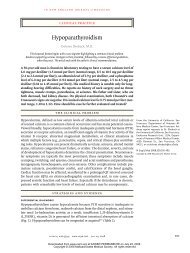
<strong>Contrast</strong> agents<br />
Ionic<br />
MONOMERS<br />
DIMERS<br />
I I I I I I<br />
I<br />
COO – Na + - - -R- - -<br />
COO – Na +<br />
Iothalamate *<br />
Ioxaglate †<br />
Diatrizoate *<br />
I<br />
I<br />
Metrizoate *<br />
Nonionic<br />
I I I I I I<br />
I<br />
- - -R- - -<br />
Iohexol †<br />
Iodixanol ‡<br />
Ioversol †<br />
I<br />
I<br />
Ioparnidol †<br />
*High-osmolar agents<br />
†Low-osmolar agents<br />
‡Iso-osmolar agent<br />
ADAPTED FROM RUDNICK MR. THE ROLE OF OSMOLALITY IN CONTRAST-ASSOCIATED NEPHROTOXICITY. APPLICATIONS IN IMAGING—<br />
CARDIAC INTERVENTIONS. SCOTCH PLAINS, NY: ANDERSON PUBLISHING, 2003.<br />
The newest<br />
contrast agent<br />
is a nonionic<br />
dimer and is<br />
iso-osmolar<br />
FIGURE 1<br />
Moreover, an enormous number of<br />
patients receive contrast agents. For example,<br />
in 2000, approximately 1,318,000 diagnostic<br />
cardiac catheterizations and 561,000 percutaneous<br />
transluminal coronary angioplasty procedures<br />
were performed, which are just two of<br />
the many procedures in which contrast is<br />
used. 2<br />
■ TYPES OF CONTRAST MEDIA<br />
The earliest contrast agents were ionic, containing<br />
a sodium a<strong>to</strong>m that dissociated from<br />
the molecule in aqueous solution. Each molecule<br />
of the agent carried three iodine a<strong>to</strong>ms.<br />
Therefore, these agents required two osmotically<br />
active particles <strong>to</strong> deliver three iodine<br />
a<strong>to</strong>ms, and they had extremely high osmolal<strong>it</strong>ies<br />
(about 2,000 mOsm/L). These agents,<br />
termed high-osmolar or ionic, were the predominant<br />
ones used until the 1980s (FIGURE 1).<br />
The next generation, introduced in the<br />
1980s and still the predominant contrast<br />
media in use, are nonionic. 3 Since they therefore<br />
need only one osmotically active particle<br />
<strong>to</strong> deliver three iodine a<strong>to</strong>ms, their osmolal<strong>it</strong>y<br />
is only about 600 <strong>to</strong> 900 mOsm/L, and they<br />
are termed low-osmolar.<br />
Both types of agents are monomers, w<strong>it</strong>h<br />
one benzene ring and three iodine a<strong>to</strong>ms.<br />
Dimer molecules consisting of two joined benzene<br />
rings contain a <strong>to</strong>tal of six iodine a<strong>to</strong>ms<br />
per molecule. There is one ionic dimer,<br />
ioxaglate, which has a 6:2 or 3:1 ratio of<br />
iodine a<strong>to</strong>ms <strong>to</strong> osmotically active particles<br />
and has an osmolal<strong>it</strong>y of 600 mOsm/L, similar<br />
<strong>to</strong> other low-osmolar contrast agents.<br />
The newest contrast agent, iodixanol, is a<br />
nonionic dimer. The chemical structure of this<br />
agent allows six iodine a<strong>to</strong>ms <strong>to</strong> be attached <strong>to</strong><br />
one osmotically active particle, resulting in an<br />
osmolal<strong>it</strong>y of 300 mOsm/L, which is iso-osmolar<br />
w<strong>it</strong>h normal plasma.<br />
■ DOES CONTRAST NEPHROPATHY<br />
INCREASE MORTALITY?<br />
Patients undergoing percutaneous coronary<br />
interventions have a higher mortal<strong>it</strong>y rate if<br />
<strong>nephropathy</strong> <strong>develops</strong>. 4,5 The risk of dying is<br />
greatest in patients who require dialytic support<br />
because of the <strong>nephropathy</strong>. For example,<br />
76 CLEVELAND CLINIC JOURNAL OF MEDICINE VOLUME 73 • NUMBER 1 JANUARY 2006
McCullough et al 5 found that in-hosp<strong>it</strong>al<br />
mortal<strong>it</strong>y rates were 1.1% for patients w<strong>it</strong>h no<br />
contrast-<strong>induced</strong> <strong>nephropathy</strong> compared w<strong>it</strong>h<br />
7.1% for those w<strong>it</strong>h <strong>nephropathy</strong> alone, and<br />
up <strong>to</strong> 35.7% for those w<strong>it</strong>h <strong>nephropathy</strong><br />
requiring dialysis.<br />
In this and other studies, patients in<br />
whom <strong>nephropathy</strong> developed had a higher<br />
prevalence of preexisting cond<strong>it</strong>ions and<br />
periprocedural complications than those in<br />
whom <strong>it</strong> did not develop.<br />
The comorbid<strong>it</strong>ies complicate the analysis,<br />
as one cannot determine w<strong>it</strong>h certainty<br />
whether contrast-<strong>induced</strong> <strong>nephropathy</strong> contributes<br />
directly <strong>to</strong> mortal<strong>it</strong>y in this population,<br />
whether this complication simply selects out a<br />
subgroup of patients at significantly greater risk<br />
of dying, or if both possibil<strong>it</strong>ies are correct.<br />
Multivariate regression analyses demonstrated<br />
that contrast-<strong>induced</strong> <strong>nephropathy</strong> was an<br />
independent predic<strong>to</strong>r of death, but this type of<br />
analysis does not prove a cause-and-effect relationship.<br />
The question of whether contrast<strong>induced</strong><br />
<strong>nephropathy</strong> directly contributes <strong>to</strong><br />
mortal<strong>it</strong>y is further confounded by recent<br />
studies demonstrating an increased risk of<br />
death in cardiac patients w<strong>it</strong>h preexisting<br />
renal insufficiency undergoing coronary<br />
revascularization. 6,7 Since most patients<br />
who develop contrast-<strong>induced</strong> <strong>nephropathy</strong><br />
have preexisting renal insufficiency, the specific<br />
contribution of contrast-<strong>induced</strong><br />
<strong>nephropathy</strong> alone <strong>to</strong> increased mortal<strong>it</strong>y is<br />
unclear.<br />
■ FEW PATIENTS NEED DIALYSIS<br />
In most cases of contrast-<strong>induced</strong> <strong>nephropathy</strong>,<br />
serum creatinine begins <strong>to</strong> rise w<strong>it</strong>hin 24<br />
<strong>to</strong> 48 hours after exposure, reaches a peak<br />
w<strong>it</strong>hin 3 <strong>to</strong> 5 days, and then returns <strong>to</strong> baseline<br />
levels w<strong>it</strong>hin 7 <strong>to</strong> 10 days. 8 In more severe<br />
cases, the creatinine concentration may not<br />
peak until 5 <strong>to</strong> 10 days, and the increase may<br />
be associated w<strong>it</strong>h oliguria. 8,9<br />
Fortunately, few patients need acute<br />
hemodialysis. Diaabetic patients who take<br />
insulin and have advanced renal insufficiency<br />
are more susceptible <strong>to</strong> prolonged acute renal<br />
failure, often w<strong>it</strong>h oliguria or the need for<br />
hemodialysis.<br />
Findings on urinalysis in patients w<strong>it</strong>h<br />
contrast-<strong>induced</strong> <strong>nephropathy</strong> are similar <strong>to</strong><br />
those in patients w<strong>it</strong>h other causes of acute<br />
tubular necrosis. Typical findings are coarse<br />
granular casts, renal tubular ep<strong>it</strong>helial cells,<br />
and amorphous debris.<br />
■ RISK FACTORS<br />
Preexisting renal insufficiency is the single<br />
greatest risk fac<strong>to</strong>r. 8,9 In one comprehensive<br />
review, an estimated 60% of patients w<strong>it</strong>h<br />
contrast-<strong>induced</strong> <strong>nephropathy</strong> had preexisting<br />
renal insufficiency. 9<br />
The more severe the baseline renal insufficiency,<br />
the greater the risk. 8,9 Although the<br />
risk of contrast-<strong>induced</strong> <strong>nephropathy</strong> for a<br />
given serum creatinine value can vary widely,<br />
one can roughly estimate the percent risk by<br />
multiplying the serum creatinine concentration<br />
in milligrams per decil<strong>it</strong>er by 10.<br />
Diabetes mell<strong>it</strong>us is often c<strong>it</strong>ed as a risk<br />
fac<strong>to</strong>r for contrast-<strong>induced</strong> <strong>nephropathy</strong>, 9,10<br />
but the risk ascribed <strong>to</strong> <strong>it</strong> is probably due <strong>to</strong><br />
coexisting renal insufficiency, usually diabetic<br />
<strong>nephropathy</strong>, rather than <strong>to</strong> the diabetes per<br />
se. 9,10 In recent prospective studies, the incidence<br />
in patients w<strong>it</strong>h diabetes and normal<br />
renal function was similar <strong>to</strong> that in nondiabetic<br />
patients w<strong>it</strong>h normal renal function. 10,11<br />
On the other hand, patients w<strong>it</strong>h diabetes<br />
and preexisting renal insufficiency have a<br />
greater risk for contrast-<strong>induced</strong> <strong>nephropathy</strong><br />
than nondiabetic patients w<strong>it</strong>h similar levels of<br />
preexisting renal insufficiency. 10,11 Moreover,<br />
when patients in this high-risk group develop<br />
<strong>nephropathy</strong>, they more often develop oliguria<br />
and need dialysis. 12 As w<strong>it</strong>h patients w<strong>it</strong>hout<br />
diabetes, the risk of contrast-<strong>induced</strong> <strong>nephropathy</strong><br />
is directly proportional <strong>to</strong> the sever<strong>it</strong>y of<br />
preexisting renal insufficiency.<br />
Volume of contrast media. Some studies<br />
found a correlation between the volume of<br />
contrast given and the risk of <strong>nephropathy</strong>,<br />
11–13 whereas other studies did not. 14<br />
Cigarroa et al 13 used a predetermined formula<br />
based on body weight and baseline renal<br />
function <strong>to</strong> lim<strong>it</strong> the volume of contrast media<br />
in patients undergoing coronary angiography.<br />
The lim<strong>it</strong> was 5 mL of contrast per kg of body<br />
weight up <strong>to</strong> a maximum of 300 mL, divided<br />
by the serum creatinine concentration in mil-<br />
To calculate<br />
the risk of<br />
contrast<br />
<strong>nephropathy</strong>,<br />
multiply the<br />
creatinine level<br />
by 10<br />
CLEVELAND CLINIC JOURNAL OF MEDICINE VOLUME 73 • NUMBER 1 JANUARY 2006 77
CONTRAST-INDUCED NEPHROPATHY<br />
RUDNICK AND COLLEAGUES<br />
Even under<br />
normal<br />
cond<strong>it</strong>ions,<br />
the renal<br />
medulla is<br />
poorly<br />
oxygenated<br />
ligrams per decil<strong>it</strong>er. Nephropathy developed<br />
in 21% of the patients in whom the <strong>to</strong>tal volume<br />
of contrast exceeded the formula amount,<br />
compared w<strong>it</strong>h 2% (P < .001) of patients in<br />
whom the contrast volume fell w<strong>it</strong>hin the prescribed<br />
lim<strong>it</strong>.<br />
Multiple myeloma has trad<strong>it</strong>ionally been<br />
considered a risk fac<strong>to</strong>r for contrast-<strong>induced</strong><br />
<strong>nephropathy</strong>. 9,10 <strong>How</strong>ever, McCarthy and<br />
Becker 15 reviewed several retrospective studies<br />
of contrast use in patients w<strong>it</strong>h myeloma<br />
and found an incidence of <strong>nephropathy</strong> of<br />
only 0.6% <strong>to</strong> 1.25%, indicating that this group<br />
is not at increased risk w<strong>it</strong>h modern contrast<br />
agents, provided that volume expansion is<br />
achieved at the time of exposure.<br />
Even though multiple myeloma should<br />
not be an absolute contraindication for contrast<br />
use, clinical prudence warrants performing<br />
radiologic studies w<strong>it</strong>h contrast only if<br />
necessary and avoiding dehydration in these<br />
patients.<br />
■ HOW DO CONTRAST AGENTS<br />
CAUSE NEPHROPATHY?<br />
The primary pathways by which contrast<br />
agents cause <strong>nephropathy</strong> are by renal<br />
ischemia (by reducing blood flow or increasing<br />
oxygen demand) and, possibly, by direct <strong>to</strong>xic<strong>it</strong>y<br />
<strong>to</strong> tubular ep<strong>it</strong>helial cells.<br />
Renal ischemia<br />
After contrast is injected, renal blood flow<br />
transiently increases and then decreases over a<br />
longer time, suggesting that renal ischemia is a<br />
major fac<strong>to</strong>r in the pathogenesis of contrast<strong>induced</strong><br />
<strong>nephropathy</strong>. 16<br />
In experimental studies of contrast-<strong>induced</strong><br />
<strong>nephropathy</strong>, the kidneys s<strong>how</strong> pathologic<br />
ischemic changes—necrosis of the medullary<br />
thick ascending limbs as well as tubular collapse<br />
and casts—primarily in the outer medullary<br />
area of the kidney. 17 Moreover, contrast agents<br />
cause a marked decrease in medullary oxygenation<br />
that can be directly measured w<strong>it</strong>h oxygen<br />
microelectrodes. 18<br />
Based on these observations, the following<br />
mechanism for acute renal failure <strong>induced</strong> by<br />
contrast agents has been proposed. 19,20<br />
Even under normal cond<strong>it</strong>ions, the renal<br />
medulla is poorly oxygenated, making <strong>it</strong> particularly<br />
susceptible <strong>to</strong> hypoxic injury. The<br />
oxygen tension in the medulla is 10 <strong>to</strong> 20 mm<br />
Hg compared w<strong>it</strong>h 50 mm Hg in the cortex.<br />
Reasons for the low oxygen tension are countercurrent<br />
exchange of oxygen between the<br />
vasa recta and oxygen use by active transport<br />
of sodium in the ascending limb of the loop of<br />
Henle. 19<br />
<strong>Contrast</strong> agents reduce the oxygen tension<br />
in both the cortex and the medulla. 18<br />
This effect may be due <strong>to</strong> increased work of<br />
active transport in response <strong>to</strong> an osmotic<br />
diuresis from hyperosmolar agents, as well as<br />
the release of vasoconstrictive compounds<br />
such as endothelin (see below). Furthermore,<br />
blockade of vasodila<strong>to</strong>ry compounds such as<br />
n<strong>it</strong>rous oxide and prostaglandins appears <strong>to</strong><br />
markedly exacerbate contrast-<strong>induced</strong> medullary<br />
hypoxic injury. 19<br />
Vasoconstriction<br />
Many substances may mediate renal vasoconstriction<br />
and subsequent hypoxic injury. Of<br />
note, adrenergic stimulation and activation of<br />
the renin-angiotensin system do not seem <strong>to</strong><br />
be involved in contrast-<strong>induced</strong> vasoconstriction.<br />
16,17 Prostaglandins w<strong>it</strong>h vasodila<strong>to</strong>ry<br />
properties may counter the vasoconstriction<br />
<strong>induced</strong> by contrast media, since pretreatment<br />
w<strong>it</strong>h indomethacin is necessary <strong>to</strong> induce<br />
experimental contrast-<strong>induced</strong> renal injury. 18<br />
Endothelin. Multiple experimental observations<br />
suggest that endothelin, a potent renal<br />
vasoconstric<strong>to</strong>r, may play a cr<strong>it</strong>ical role in<br />
contrast-mediated vasoconstriction. 8<br />
These observations led <strong>to</strong> a clinical trial in<br />
which patients w<strong>it</strong>h chronic kidney disease<br />
undergoing cardiac angiography were randomized<br />
<strong>to</strong> receive e<strong>it</strong>her the endothelin recep<strong>to</strong>r<br />
antagonist SB 290670 or placebo. 21 Surprisingly,<br />
the incidence of contrast-<strong>induced</strong> <strong>nephropathy</strong><br />
was higher in the treatment group (56%) than in<br />
the placebo group (29%; P = .002).<br />
Adenosine. The role of adenosine in the<br />
pathogenesis of contrast-<strong>induced</strong> <strong>nephropathy</strong><br />
is described in detail in an excellent review by<br />
Pflueger et al. 22 Adenosine causes vasodilatation<br />
through A2 stimulation of the efferent<br />
arteriole and medullary capillaries, and <strong>it</strong> also<br />
causes vasoconstriction through A1 stimulation<br />
of the afferent arterioles. <strong>How</strong>ever, renal<br />
vasoconstriction dominates, explaining why<br />
78 CLEVELAND CLINIC JOURNAL OF MEDICINE VOLUME 73 • NUMBER 1 JANUARY 2006
intrarenal adenosine infusion results in a<br />
decrease in renal blood flow. 22<br />
In experimental studies, theophylline, a<br />
nonselective adenosine recep<strong>to</strong>r antagonist,<br />
inhib<strong>it</strong>ed contrast-media <strong>induced</strong> renal vasoconstriction.<br />
22<br />
Role of osmolal<strong>it</strong>y<br />
Several clinical and experimental observations<br />
suggest that the hyperosmolal<strong>it</strong>y of contrast<br />
media may play a role in the pathogenesis<br />
of contrast-<strong>induced</strong> <strong>nephropathy</strong>. Clinical<br />
studies demonstrated that low-osmolar contrast<br />
agents cause less nephro<strong>to</strong>xic<strong>it</strong>y than<br />
high osmolar agents. 11,23 Furthermore, in one<br />
study, 24 the incidence of contrast-<strong>induced</strong><br />
<strong>nephropathy</strong> was lower w<strong>it</strong>h an iso-osmolar<br />
contrast agent than w<strong>it</strong>h a low-osmolar agent.<br />
In experimental studies, hyper<strong>to</strong>nic solutions<br />
of saline or mann<strong>it</strong>ol reduce the<br />
glomerular filtration rate and renal blood flow<br />
and increase enzymuria similarly <strong>to</strong> highosmolar<br />
contrast media but w<strong>it</strong>h a lesser magn<strong>it</strong>ude.<br />
16,25 A theory <strong>to</strong> account for these<br />
nonspecific adverse effects is that hyperosmolal<strong>it</strong>y<br />
activates tubuloglomerular feedback or<br />
causes an increase in tubular hydrostatic pressures,<br />
e<strong>it</strong>her of which could lead <strong>to</strong> a decrease<br />
in glomerular filtration. In add<strong>it</strong>ion, the<br />
osmotic diuresis produced by contrast media<br />
may result in increased active transport of<br />
sodium in the thick ascending limb and also<br />
in vasoconstriction, and both of these could<br />
lead <strong>to</strong> worsened medullary hypoxemia. 18–20<br />
On the other hand, most studies in animals<br />
specifically comparing iso-osmolar contrast<br />
agents (iodixanol and iotrolan) w<strong>it</strong>h<br />
high-osmolar and low-osmolar contrast agents<br />
have not demonstrated any lower rate of renal<br />
abnormal<strong>it</strong>ies w<strong>it</strong>h the iso-osmolar agents. 26,27<br />
The reason may be that the iso-osmolar agents<br />
are more viscous, which could increase red<br />
blood cell aggregation and decrease renal<br />
blood flow, offsetting any reduction in<br />
medullary hypoxemia from their lower osmolal<strong>it</strong>y.<br />
Reactive oxygen species<br />
Reactive oxygen species formed as a result of<br />
postischemic oxidative stress can lead <strong>to</strong> acute<br />
renal failure through their direct effects on<br />
renal endothelial cells, which include apop<strong>to</strong>tic<br />
cell death. Adenosine’s role in the<br />
pathogenesis of contrast-<strong>induced</strong> <strong>nephropathy</strong><br />
may be due <strong>to</strong> this molecule’s abil<strong>it</strong>y <strong>to</strong><br />
increase generation of oxygen-free radicals. 28<br />
The possible benef<strong>it</strong> of N-acetylcysteine and<br />
sodium bicarbonate in <strong>prevent</strong>ing contrast<strong>induced</strong><br />
<strong>nephropathy</strong> (see below) is hypothesized<br />
<strong>to</strong> be due <strong>to</strong> the abil<strong>it</strong>y of these compounds<br />
<strong>to</strong> m<strong>it</strong>igate oxidative injury.<br />
Direct cellular <strong>to</strong>xic<strong>it</strong>y<br />
A number of experimental observations suggest<br />
that contrast agents are directly <strong>to</strong>xic <strong>to</strong><br />
kidney cells, causing proximal cell vacuolization,<br />
interst<strong>it</strong>ial inflammation, cellular necrosis,<br />
and enzymuria. 8,17 Furthermore, suspensions<br />
of proximal tubular segments exposed <strong>to</strong><br />
contrast media s<strong>how</strong>ed abnormal<strong>it</strong>ies in several<br />
markers of cellular injury, that were potentiated<br />
by hypoxia and were more pronounced<br />
w<strong>it</strong>h high-osmolar agents than w<strong>it</strong>h lowosmolar<br />
agents. 29<br />
■ PREVENTIVE MEASURES:<br />
MANY TRIED, FEW SUCCEEDED<br />
Currently, there is no treatment <strong>to</strong> reverse or<br />
ameliorate contrast-<strong>induced</strong> <strong>nephropathy</strong><br />
once <strong>it</strong> occurs, but prophylaxis is possible.<br />
Many <strong>prevent</strong>ive measures have been<br />
tried that may interfere w<strong>it</strong>h one or more of<br />
the currently accepted pathogenetic mechanisms<br />
for contrast-<strong>induced</strong> <strong>nephropathy</strong><br />
(TABLE 1). <strong>How</strong>ever, many of these measures<br />
later failed <strong>to</strong> s<strong>how</strong> benef<strong>it</strong>s in well-designed,<br />
prospective, randomized, double-blinded trials.<br />
Among this group are diuretics, 30 mann<strong>it</strong>ol,<br />
30 dopamine, 31 atrial natriuretic peptide,<br />
32 endothelin recep<strong>to</strong>r antagonists, 21<br />
and fenoldopam. 33<br />
This review will focus only on the strategies<br />
that have possible or proven prophylactic<br />
value.<br />
Hydration is indicated,<br />
but what kind, <strong>how</strong> much?<br />
Hydration is the primary intervention for <strong>prevent</strong>ing<br />
contrast <strong>nephropathy</strong>. 34<br />
The theoretical rationale for hydration is<br />
that <strong>it</strong> should decrease the activ<strong>it</strong>y of the<br />
renin-angiotensin system, reduce the levels of<br />
other vasoconstrictive hormones such as<br />
More contrast<br />
<strong>nephropathy</strong><br />
occurred w<strong>it</strong>h<br />
saline plus<br />
mann<strong>it</strong>ol or<br />
furosemide<br />
than w<strong>it</strong>h<br />
saline alone<br />
CLEVELAND CLINIC JOURNAL OF MEDICINE VOLUME 73 • NUMBER 1 JANUARY 2006 79
CONTRAST-INDUCED NEPHROPATHY<br />
RUDNICK AND COLLEAGUES<br />
TABLE 1<br />
Strategies for <strong>prevent</strong>ing<br />
contrast-<strong>induced</strong> <strong>nephropathy</strong><br />
Strategies that do not work<br />
Mann<strong>it</strong>ol<br />
Furosemide<br />
Dopamine<br />
Atrial natriuretic fac<strong>to</strong>r<br />
Fenoldopam<br />
Hemodialysis<br />
Strategies that may work<br />
Calcium channel blockers<br />
Theophylline<br />
Iso-osmolar contrast media<br />
N-acetylcysteine<br />
Hemofiltration<br />
Sodium bicarbonate<br />
Ascorbic acid<br />
Prostaglandins<br />
Currently recommended strategies<br />
Employ noniodinated contrast studies<br />
Avoid nonsteroidal anti-inflamma<strong>to</strong>ry drugs<br />
Provide adequate time between contrast procedures<br />
Minimize contrast volume<br />
Parenteral hydration<br />
Low-osmolar or iso-osmolar contrast media<br />
endothelin, increase sodium diuresis, decrease<br />
tubuloglomerular feedback, <strong>prevent</strong> tubular<br />
obstruction, protect against reactive oxygen<br />
species, and dilute the contrast media in the<br />
tubule, thus decreasing any direct nephro<strong>to</strong>xic<br />
effect of the contrast agent on the tubular<br />
ep<strong>it</strong>helium. 34<br />
Several studies in animals demonstrated<br />
hydration w<strong>it</strong>h saline infusions <strong>to</strong> be beneficial<br />
in <strong>prevent</strong>ing contrast-<strong>induced</strong> <strong>nephropathy</strong>. 35<br />
Early clinical studies used his<strong>to</strong>rical controls<br />
for comparison and also suggested that<br />
hydration is beneficial. Subsequently, intravenous<br />
hydration became the standard<br />
method <strong>to</strong> <strong>prevent</strong> contrast-<strong>induced</strong> <strong>nephropathy</strong>.<br />
8,9<br />
There have been a few prospective randomized<br />
studies comparing saline alone vs<br />
other therapies as prophylactic strategies.<br />
21,30–33<br />
Solomon et al 30 randomized patients w<strong>it</strong>h<br />
chronic kidney disease undergoing cardiac<br />
angiography <strong>to</strong> receive e<strong>it</strong>her saline alone,<br />
saline and mann<strong>it</strong>ol, or saline and furosemide.<br />
All three groups received 0.45% saline intravenously<br />
at 1 mL/kg/hour for 12 hours before<br />
and 12 hours after receiving contrast. Nephropathy<br />
occurred in 11% of patients receiving<br />
saline alone vs 28% who received saline and<br />
mann<strong>it</strong>ol and 40% who received saline and<br />
furosemide.<br />
Different regimens of saline hydration<br />
have been used, but no one regimen has<br />
demonstrated clear superior<strong>it</strong>y.<br />
Trivedi et al 36 prospectively randomized<br />
patients undergoing cardiac angiography <strong>to</strong><br />
receive e<strong>it</strong>her intravenous saline for 12 hours<br />
both before and after catheterization or oral<br />
fluids only, taken as desired. <strong>Contrast</strong>-<strong>induced</strong><br />
<strong>nephropathy</strong> occurred in 3.7% of those who<br />
received intravenous saline vs 34.6% of those<br />
who received only oral fluids.<br />
In contrast, the Preparation for Angiography<br />
in Renal Dysfunction (PREPARED)<br />
trial 37 s<strong>how</strong>ed that, in patients w<strong>it</strong>h chronic<br />
kidney disease undergoing coronary angiography,<br />
hydration on an outpatient basis before<br />
catheterization, coupled w<strong>it</strong>h a brief period of<br />
intravenous hydration, was equivalent <strong>to</strong><br />
overnight intravenous hydration.<br />
Bader et al 38 randomized patients undergoing<br />
computed <strong>to</strong>mography or dig<strong>it</strong>al angiography<br />
<strong>to</strong> receive e<strong>it</strong>her 2,000 mL of intravenous<br />
fluid over 24 hours (12 hours before<br />
and 12 hours after contrast) or 300 mL of<br />
intravenous fluid during the radiologic procedure.<br />
The glomerular filtration rate fell by 18.3<br />
mL/minute in the continuous infusion group<br />
compared w<strong>it</strong>h a 34.6 mL/minute fall in the<br />
bolus infusion group (P < .05), suggesting that<br />
slow hydration is superior <strong>to</strong> bolus expansion<br />
during the procedure.<br />
The Prevention of Radiocontrast Induced<br />
Nephropathy Clinical Evaluation (PRINCE)<br />
study 39 tested the hypothesis that forced<br />
diuresis w<strong>it</strong>h maintenance of intravascular<br />
volume would result in less contrast-<strong>induced</strong><br />
renal injury. Although no difference in the<br />
incidence of contrast-<strong>induced</strong> <strong>nephropathy</strong><br />
was observed between patients who underwent<br />
forced diuresis and those who did not,<br />
the incidence in participants w<strong>it</strong>h urine flow<br />
rates greater than 150 mL/hour was 21.6% vs<br />
45.9% in those w<strong>it</strong>h lower urine flow rates (P<br />
= .03).<br />
80 CLEVELAND CLINIC JOURNAL OF MEDICINE VOLUME 73 • NUMBER 1 JANUARY 2006
Mueller et al 40 compared the use of iso<strong>to</strong>nic<br />
(0.9%) saline (n = 685) vs half-iso<strong>to</strong>nic<br />
(0.45%) saline (n = 698) in patients undergoing<br />
coronary angioplasty. Both groups<br />
received about 2,000 mL of intravenous fluid.<br />
The incidence of contrast-<strong>induced</strong> <strong>nephropathy</strong><br />
was significantly lower w<strong>it</strong>h iso<strong>to</strong>nic<br />
saline (0.7%) than w<strong>it</strong>h half-iso<strong>to</strong>nic saline<br />
(2%, P = .04).<br />
Comment. These experimental and clinical<br />
studies support the use of intravenous<br />
hydration <strong>to</strong> <strong>prevent</strong> contrast-<strong>induced</strong><br />
<strong>nephropathy</strong>, especially in patients w<strong>it</strong>h<br />
azotemia at high risk. As yet, no sufficiently<br />
powered, controlled, prospective trials have<br />
examined the minimally effective length of<br />
time, optimal rate, and fluid compos<strong>it</strong>ion of<br />
intravenous hydration required before and<br />
after contrast administration in high-risk<br />
azotemic patients.<br />
N-acetylcysteine: Data are conflicting<br />
N-acetylcysteine has been s<strong>how</strong>n in experiments<br />
in animals <strong>to</strong> ameliorate renal injuries<br />
from ischemia and nephro<strong>to</strong>xins. 41 Potential<br />
mechanisms include antioxidation (e<strong>it</strong>her<br />
directly as a free radical oxygen scavenger or<br />
indirectly through glutathione production), 41<br />
<strong>prevent</strong>ing apop<strong>to</strong>tic cell death mediated by<br />
the generation of oxygen free radicals, and<br />
vasodilation. 42<br />
Tepel et al 43 first found N-acetylcysteine<br />
beneficial in a study of 83 patients w<strong>it</strong>h<br />
chronic renal failure (32.5% had diabetic<br />
<strong>nephropathy</strong>) undergoing computed <strong>to</strong>mography<br />
w<strong>it</strong>h contrast. Patients received intravenous<br />
saline for 12 hours before and after<br />
receiving the contrast and were prospectively<br />
randomized <strong>to</strong> receive e<strong>it</strong>her N-acetylcysteine<br />
600 mg by mouth twice daily 1 day before and<br />
on the day of the study (<strong>to</strong>tal of four doses<br />
over 2 days) or placebo. <strong>Contrast</strong>-<strong>induced</strong><br />
<strong>nephropathy</strong> occurred in 2% of the N-acetylcysteine<br />
group vs 21% of the placebo group (P<br />
= .01; relative risk 0.1).<br />
Several subsequent studies confirmed the<br />
value of N-acetylcysteine in <strong>prevent</strong>ing contrast-<strong>induced</strong><br />
<strong>nephropathy</strong>, all in patients<br />
undergoing cardiac angiography. 41 Based on<br />
these data, N-acetylcysteine became widely<br />
accepted as a prophylactic therapy.<br />
Because N-acetylcysteine undergoes significant<br />
first-pass metabolism and <strong>it</strong>s oral<br />
administration poses logistical problems, Baker<br />
et al 44 evaluated <strong>it</strong>s intravenous use. acetylcysteine<br />
was given intravenously at a dose of 150<br />
mg/kg in 500 mL of normal saline solution<br />
over 30 minutes immediately before contrast<br />
exposure and then 50 mg/kg in 500 mL of normal<br />
saline solution over the next 4 hours.<br />
<strong>Contrast</strong>-<strong>induced</strong> <strong>nephropathy</strong> occurred in<br />
5% of the N-acetylcysteine group compared<br />
w<strong>it</strong>h 21% of the saline-alone group (relative<br />
risk 0.28, P = .045). These results suggest that<br />
prolonged use of N-acetylcysteine before contrast<br />
exposure may not be necessary.<br />
On the other hand, many other studies<br />
did not demonstrate a prophylactic value for<br />
N-acetylcysteine. 41 For example, Durham et<br />
al 45 studied 79 patients w<strong>it</strong>h chronic kidney<br />
disease who underwent diagnostic cardiac<br />
catheterization, percutaneous coronary intervention,<br />
or both. The patients were randomly<br />
assigned <strong>to</strong> receive oral acetylcysteine or<br />
placebo. All patients received hydration w<strong>it</strong>h<br />
0.45% saline for up <strong>to</strong> 12 hours before and<br />
after catheterization. There was no significant<br />
difference in the incidence of contrast<strong>induced</strong><br />
<strong>nephropathy</strong> between the two groups:<br />
26.3% in the acetylcysteine group and 22% in<br />
the control group.<br />
Nallamothu et al 46 performed a metaanalysis<br />
of 20 studies involving 2,195 patients<br />
and calculated that the relative risk of contrast<br />
<strong>nephropathy</strong> in patients who received N-<br />
acetylcysteine was 0.73 (95% confidence<br />
interval 0.52–1.0; P = .08). Pannu et al 47 performed<br />
another meta-analysis of 15 studies<br />
involving 1,776 patients and calculated the<br />
relative risk at 0.65 (95% confidence interval<br />
0.43–1.00). Both groups of investiga<strong>to</strong>rs were<br />
cautious in their conclusions, pointing out<br />
that the individual studies s<strong>how</strong>ed substantial<br />
heterogene<strong>it</strong>y in design and results and calling<br />
for defin<strong>it</strong>ive studies. Some of the variables<br />
that differed among the studies published <strong>to</strong><br />
date include the sever<strong>it</strong>y of baseline renal<br />
insufficiency, the percentage of diabetic<br />
patients, the type and amount of contrast<br />
used, the amount and timing of N-acetylcysteine<br />
administration, and the amount of<br />
hydration.<br />
Conclusions. Although N-acetylcysteine<br />
is safe, easy <strong>to</strong> use, and inexpensive, <strong>it</strong>s value<br />
Studies of<br />
N-acetylcysteine<br />
differed<br />
in design and<br />
results<br />
CLEVELAND CLINIC JOURNAL OF MEDICINE VOLUME 73 • NUMBER 1 JANUARY 2006 83
CONTRAST-INDUCED NEPHROPATHY<br />
RUDNICK AND COLLEAGUES<br />
Low-osmolar<br />
agents have<br />
become the<br />
standard<br />
in <strong>prevent</strong>ing contrast-<strong>induced</strong> <strong>nephropathy</strong><br />
remains controversial.<br />
Theophylline:<br />
Not recommended at this time<br />
Several reports suggest that theophylline, an<br />
adenosine antagonist, <strong>prevent</strong>s contrast<strong>induced</strong><br />
<strong>nephropathy</strong>. 22,48<br />
Erley et al 48 randomized 39 patients who<br />
received contrast media <strong>to</strong> receive e<strong>it</strong>her intravenous<br />
theophylline or placebo. Although no<br />
patient in e<strong>it</strong>her group developed contrast<strong>induced</strong><br />
<strong>nephropathy</strong>, the glomerular filtration<br />
rate decreased in the placebo group from 88<br />
mL/minute at baseline <strong>to</strong> 75 mL/minute 4<br />
hours after contrast administration; <strong>it</strong> remained<br />
unchanged in the theophylline group.<br />
In several other placebo-controlled studies,<br />
theophylline (given orally or intravenously)<br />
<strong>prevent</strong>ed contrast-<strong>induced</strong> falls in creatinine<br />
clearance, but all the studies were in lowrisk<br />
patients, and contrast-<strong>induced</strong> <strong>nephropathy</strong><br />
was not seen in any groups.<br />
Theophylline has potential risks, including<br />
ventricular arrhythmias, seizures, and<br />
shock—all of which may be potentiated by a<br />
variety of other drugs. 22<br />
Conclusions. The data regarding theophylline<br />
are mixed. Favorable studies were<br />
lim<strong>it</strong>ed by small numbers, absence of high-risk<br />
patients, and a failure <strong>to</strong> demonstrate differences<br />
in the incidence of contrast-<strong>induced</strong><br />
<strong>nephropathy</strong>. Therefore, theophylline cannot<br />
be recommended as standard prophylaxis<br />
against contrast-<strong>induced</strong> <strong>nephropathy</strong> at this<br />
time.<br />
Low-osmolar agents<br />
are better than high osmolar agents<br />
Introduced in the 1980s, nonionic low-osmolar<br />
contrast agents have replaced ionic highosmolar<br />
agents as the standard intravascular<br />
contrast media because of their lower incidence<br />
of adverse effects. 3<br />
Studies in animals have demonstrated<br />
that, compared w<strong>it</strong>h high-osmolar agents,<br />
low-osmolar agents result in less nephro<strong>to</strong>xic<strong>it</strong>y<br />
but still can cause <strong>nephropathy</strong>. 8 In<strong>it</strong>ial<br />
clinical studies comparing high-osmolar vs<br />
low-osmolar contrast agents failed <strong>to</strong> demonstrate<br />
a difference between these two types of<br />
agents but were underpowered in respect <strong>to</strong><br />
high-risk azotemic patients. 8<br />
In 1995, Rudnick et al 11 performed a<br />
prospective, randomized, double-blind study<br />
comparing the high-osmolar contrast agent diatrizoate<br />
w<strong>it</strong>h the low-osmolar contrast agent<br />
iohexol in 1,196 patients, including 509<br />
azotemic patients, of whom 213 had diabetes.<br />
In patients w<strong>it</strong>hout azotemia, the incidence of<br />
contrast-<strong>induced</strong> <strong>nephropathy</strong> was negligible<br />
w<strong>it</strong>h e<strong>it</strong>her agent, regardless of whether or not<br />
the patient had diabetes. <strong>How</strong>ever, in patients<br />
w<strong>it</strong>h azotemia but w<strong>it</strong>hout diabetes, the incidence<br />
of contrast-<strong>induced</strong> <strong>nephropathy</strong> was 7%<br />
w<strong>it</strong>h the high-osmolar agent vs 4% w<strong>it</strong>h the<br />
low-osmolar agent. In patients w<strong>it</strong>h azotemia<br />
and diabetes, the differences were even more<br />
pronounced: 27% w<strong>it</strong>h the high- osmolar agent<br />
and 12% w<strong>it</strong>h the low-osmolar agent.<br />
A subsequent meta-analysis indicated that<br />
low-osmolar agents reduced the incidence of<br />
contrast-<strong>induced</strong> <strong>nephropathy</strong> by 50%. 23<br />
Are iso-osmolar agents<br />
better than low-osmolar agents?<br />
The iso-osmolar contrast agents have undergone<br />
experimental and clinical studies comparing<br />
their nephro<strong>to</strong>xic<strong>it</strong>y w<strong>it</strong>h that of the<br />
currently popular low-osmolar agents. As discussed<br />
above, these third-generation agents<br />
have not demonstrated less nephro<strong>to</strong>xic<strong>it</strong>y<br />
than low-osmolar agents in studies in animals.<br />
Furthermore, only a few clinical studies have<br />
compared the incidence of contrast-<strong>induced</strong><br />
<strong>nephropathy</strong> w<strong>it</strong>h the two types of agents. 24,49<br />
Aspelin et al 24 performed a prospective,<br />
randomized, double-blind, multicenter trial in<br />
129 patients w<strong>it</strong>h azotemia and diabetes.<br />
Patients were randomly assigned <strong>to</strong> receive<br />
e<strong>it</strong>her iohexol (a low-osmolar agent) or iodixanol<br />
(an iso-osmolar agent). The incidence of<br />
contrast-<strong>induced</strong> <strong>nephropathy</strong> was 3% w<strong>it</strong>h<br />
the iso-osmolar agent vs 26% w<strong>it</strong>h the lowosmolar<br />
agent (odds ratio 0.09, P = .002).<br />
Larger randomized trials will be needed <strong>to</strong><br />
verify these encouraging results, especially<br />
w<strong>it</strong>h comparisons <strong>to</strong> other low-osmolar contrast<br />
agents.<br />
Hemodialysis and hemofiltration<br />
Numerous studies have demonstrated that 2 <strong>to</strong><br />
3 hours of hemodialysis effectively removes<br />
60% <strong>to</strong> 90% of contrast medium. 50 Several<br />
84 CLEVELAND CLINIC JOURNAL OF MEDICINE VOLUME 73 • NUMBER 1 JANUARY 2006
studies explored the prophylactic value of<br />
hemodialysis in high-risk patients, but most<br />
failed <strong>to</strong> demonstrate a reduced incidence of<br />
contrast-<strong>induced</strong> <strong>nephropathy</strong>. 50<br />
On the other hand, Marenzi et al 51 recently<br />
found that hemofiltration significantly<br />
reduced contrast-<strong>induced</strong> <strong>nephropathy</strong> in<br />
patients at high risk. In this study, patients<br />
w<strong>it</strong>h chronic kidney disease undergoing coronary<br />
angiography were randomized <strong>to</strong> undergo<br />
e<strong>it</strong>her hemofiltration in an intensive care un<strong>it</strong><br />
or parenteral saline hydration. Hemofiltration<br />
was started 4 <strong>to</strong> 6 hours before contrast administration,<br />
s<strong>to</strong>pped for coronary angiography,<br />
then resumed for an add<strong>it</strong>ional 18 <strong>to</strong> 24 hours.<br />
Iso<strong>to</strong>nic saline was used as replacement fluid<br />
and was given at a rate of 1 L per hour, which<br />
matched the ultrafiltration rate so that no net<br />
fluid loss resulted. In the control group, iso<strong>to</strong>nic<br />
saline was given at 1 mL/kg/hour for 6 <strong>to</strong><br />
8 hours before and 24 hours after angiography.<br />
The incidence of contrast-<strong>induced</strong><br />
<strong>nephropathy</strong> was 5% in the hemofiltration<br />
group compared w<strong>it</strong>h 50% in the control<br />
group (P < .001). The in-hosp<strong>it</strong>al mortal<strong>it</strong>y<br />
rate was 2% in the hemofiltration group compared<br />
w<strong>it</strong>h 14% in the control group (P = .02).<br />
Desp<strong>it</strong>e these impressive results, the conclusions<br />
of this study should be viewed w<strong>it</strong>h<br />
some caution. Removal of creatinine by<br />
hemofiltration per se could result in a lower<br />
incidence of contrast-<strong>induced</strong> <strong>nephropathy</strong>,<br />
although this alone would not account for differences<br />
in mortal<strong>it</strong>y. Moreover, the mortal<strong>it</strong>y<br />
rate in the control group was inordinately high,<br />
suggesting that <strong>it</strong> was not a good representative<br />
cohort. Both groups received an extraordinary<br />
volume of contrast (approximately 250 mL) for<br />
patients w<strong>it</strong>h moderately severe chronic kidney<br />
disease (their baseline mean creatinine concentration<br />
was 3.0 mg/dL).<br />
Conclusions. Given these reservations,<br />
due <strong>to</strong> the logistical effort and high cost associated<br />
w<strong>it</strong>h hemofiltration, larger randomized<br />
trials should be performed before this technique<br />
can be recommended as standard prophylaxis<br />
against contrast-<strong>induced</strong> <strong>nephropathy</strong><br />
in high-risk patients.<br />
Somewhat related is the not-infrequent<br />
clinical question of when <strong>to</strong> perform the next<br />
hemodialysis treatment in a patient undergoing<br />
chronic hemodialysis who receives<br />
intravascular contrast media. Although this<br />
question has not been extensively investigated<br />
in clinical trials, there is evidence that<br />
most patients can safely wa<strong>it</strong> 24 <strong>to</strong> 36 hours<br />
after contrast exposure until their next<br />
hemodialysis treatment.<br />
Sodium bicarbonate: Data are preliminary<br />
Merten et al, 52 in a randomized controlled<br />
trial at a single center, compared hydration<br />
w<strong>it</strong>h sodium bicarbonate vs sodium chloride<br />
<strong>to</strong> <strong>prevent</strong> contrast-<strong>induced</strong> <strong>nephropathy</strong> in<br />
azotemic patients receiving low-osmolar<br />
contrast agents. Both infusions contained<br />
154 mEq of e<strong>it</strong>her sodium chloride or sodium<br />
bicarbonate in 1 L of 5% dextrose and water.<br />
A close approximation of the sodium bicarbonate<br />
solution can be achieved by adding 3<br />
ampules (150 mEq) of sodium bicarbonate <strong>to</strong><br />
1 L of 5% dextrose in water: the final sodium<br />
bicarbonate concentration is 130 mEq/L.<br />
The infusion rate for e<strong>it</strong>her fluid was 3<br />
mL/kg/hour for 1 hour before contrast<br />
administration, followed by 1 mL/kg/hour<br />
during contrast administration and then for<br />
6 hours afterward.<br />
<strong>Contrast</strong>-<strong>induced</strong> <strong>nephropathy</strong> occurred<br />
in 1.7% of patients who received sodium<br />
bicarbonate compared w<strong>it</strong>h 13.6% of patients<br />
who received sodium chloride (P = .02).<br />
The benef<strong>it</strong> of sodium bicarbonate in <strong>prevent</strong>ing<br />
contrast-<strong>induced</strong> <strong>nephropathy</strong> is<br />
probably not simply due <strong>to</strong> volume expansion,<br />
which was similar between treatment groups.<br />
The authors postulate instead that sodium<br />
bicarbonate may reduce the formation of oxygen<br />
free radicals (a pH-dependent reaction),<br />
previously reported <strong>to</strong> play a pathogenetic role<br />
in contrast-<strong>induced</strong> <strong>nephropathy</strong>. 52<br />
Conclusions. We agree w<strong>it</strong>h the authors<br />
that sodium bicarbonate infusion may provide a<br />
simple, safe, and inexpensive method <strong>to</strong> <strong>prevent</strong><br />
contrast-<strong>induced</strong> <strong>nephropathy</strong> but the results of<br />
this study need <strong>to</strong> be confirmed in a larger, multicenter,<br />
prospective randomized trial.<br />
■ CURRENT RECOMMENDATIONS<br />
The use of intravascular contrast in patients at<br />
risk for contrast-<strong>induced</strong> <strong>nephropathy</strong> should be<br />
considered only when alternative imaging tests<br />
that do not use iodinated contrast cannot pro-<br />
Start saline<br />
hydration 2–4<br />
hours before<br />
the procedure<br />
and continue<br />
4–6 hours after<br />
CLEVELAND CLINIC JOURNAL OF MEDICINE VOLUME 73 • NUMBER 1 JANUARY 2006 85
CONTRAST-INDUCED NEPHROPATHY<br />
RUDNICK AND COLLEAGUES<br />
vide the necessary clinical information. In many<br />
cases, ultrasonography, nuclear medicine, magnetic<br />
resonance, and unenhanced computed<br />
<strong>to</strong>mography can provide sufficient data w<strong>it</strong>hout<br />
exposing the patient <strong>to</strong> iodinated contrast media<br />
and the risk of contrast-<strong>induced</strong> <strong>nephropathy</strong>.<br />
When exposure <strong>to</strong> iodinated contrast<br />
media is unavoidable, we recommend the following<br />
approach.<br />
• Nonsteroidal anti-inflamma<strong>to</strong>ry drugs<br />
should be discontinued before contrast exposure.<br />
• Patients should receive hydration w<strong>it</strong>h<br />
intravenous normal saline starting 2 <strong>to</strong> 4 hours<br />
before receiving the contrast, during the radiographic<br />
procedure, and continuing 4 <strong>to</strong> 6 hours<br />
afterward. The duration of the saline infusion<br />
should be longer w<strong>it</strong>h more severe chronic kidney<br />
disease or if underlying diabetic <strong>nephropathy</strong><br />
is present.<br />
• The radiologist or cardiologist should use<br />
the smallest volume of contrast needed <strong>to</strong><br />
obtain the cr<strong>it</strong>ical imaging.<br />
• Based on currently available data, there<br />
may be an advantage in using iso-osmolar contrast<br />
media.<br />
• Hypotension in the peri-imaging period<br />
should be avoided if possible.<br />
• N-acetylcysteine and bicarbonate hydration<br />
can be used since they are safe and inexpensive,<br />
although their use is somewhat controversial.<br />
• Serum creatinine should be measured<br />
approximately 48 hours after contrast exposure<br />
<strong>to</strong> determine if contrast-<strong>induced</strong> <strong>nephropathy</strong><br />
has occurred.<br />
■ REFERENCES<br />
1. Hou SH, Bushinsky DA, Wish JB, Cohen JJ, Harring<strong>to</strong>n JT. Hosp<strong>it</strong>alacquired<br />
renal insufficiency: a prospective study. Am J Med 1983;<br />
74:243–248.<br />
2. American Heart Association. Heart Disease and Stroke Statistics—<br />
2003 Update, pp 38–39.<br />
3. McClennan BL. Low-osmolal<strong>it</strong>y contrast media: premises and promises.<br />
Radiology 1987; 162:1–8.<br />
4. Levy EM, Viscoli CM, Horw<strong>it</strong>z RI. The effect of acute renal failure on<br />
mortal<strong>it</strong>y. A cohort analysis. JAMA 1996; 275:1489–1494.<br />
5. McCullough PA, Wolyn R, Rocher LL, Levin RN, O’Neill WW. Acute<br />
renal failure after coronary intervention: incidence, risk fac<strong>to</strong>rs, and<br />
relationship <strong>to</strong> mortal<strong>it</strong>y. Am J Med 1997; 103:368–375.<br />
6. Szczech LA, Reddan DN, Owen WF Jr, et al. Differential survival after<br />
coronary revascularization procedures among patients w<strong>it</strong>h renal<br />
insufficiency. Kidney Int 2001; 60:292–299.<br />
7. Reinecke H, Trey T, Matzkies F, Fobker M, Bre<strong>it</strong>hardt G, Schaefer RM.<br />
Grade of chronic renal failure, and acute and long-term outcome<br />
after percutaneous coronary interventions. Kidney Int 2003;<br />
63:696–701.<br />
8. Rudnick MR, Berns JS, Cohen RM, Goldfarb S. <strong>Contrast</strong> media-associated<br />
nephro<strong>to</strong>xic<strong>it</strong>y. Semin Nephrol 1997; 17:15–26.<br />
9. Berns AS. Nephro<strong>to</strong>xic<strong>it</strong>y of contrast media. Kidney Int 1989;<br />
36:730–740.<br />
10. Rudnick MR, Berns JS, Cohen RM, Goldfarb S. Nephro<strong>to</strong>xic risks of<br />
renal angiography: contrast media-associated nephro<strong>to</strong>xic<strong>it</strong>y and<br />
atheroembolism—a cr<strong>it</strong>ical review. Am J Kidney Dis 1994;<br />
24:713–727.<br />
11. Rudnick MR, Goldfarb S, Wexler L, et al. The Iohexol Cooperative<br />
Study. Nephro<strong>to</strong>xic<strong>it</strong>y of ionic and nonionic contrast media in 1196<br />
patients: a randomized trial. Kidney Int 1995; 47:254–261.<br />
12. Manske CL, Sprafka JM, Strony JT, Wang Y. <strong>Contrast</strong> <strong>nephropathy</strong> in<br />
azotemic diabetic patients undergoing coronary angiography. Am J<br />
Med 1990; 89:615–620.<br />
13. Cigarroa RG, Lange RA, Williams RH, Hillis LD. Dosing of contrast<br />
material <strong>to</strong> <strong>prevent</strong> contrast <strong>nephropathy</strong> in patients w<strong>it</strong>h renal disease.<br />
Am J Med 1989; 86(6 Pt 1):649–652.<br />
14. Barrett BJ, Parfrey PS, Vavasour HM, et al. <strong>Contrast</strong> <strong>nephropathy</strong> in<br />
patients w<strong>it</strong>h impaired renal function: high versus low osmolar<br />
media. Kidney Int 1992; 41:1274–1279.<br />
15. McCarthy CS, Becker JA. Multiple myeloma and contrast media.<br />
Radiology 1992; 183:519–522.<br />
16. Katzberg RW, Morris TW, Burgener FA, Kamm DE, Fischer HW. Renal<br />
renin and hemodynamic responses <strong>to</strong> selective renal artery catheterization<br />
and angiography. Invest Radiol 1977; 12:381–388.<br />
17. Heyman SN, Brezis M, Reubinoff CA, et al. Acute renal failure w<strong>it</strong>h<br />
selective medullary injury in the rat. J Clin Invest 1988; 82:401–412.<br />
18. Heyman SN, Brezis M, Epstein FH, Spokes K, Silva P, Rosen S. Early<br />
renal medullary hypoxic injury from radiocontrast and<br />
indomethacin. Kidney Int 1991; 40:632–642.<br />
19. Heyman SN, Rosen S, Brezis M. Radiocontrast <strong>nephropathy</strong>: a paradigm<br />
for the synergism between <strong>to</strong>xic and hypoxic insults in the kidney.<br />
Exp Nephrol 1994; 2:153–157.<br />
20. Brezis M, Rosen S. Hypoxia of the renal medulla—<strong>it</strong>s implications for<br />
disease. N Engl J Med 1995; 332:647–655.<br />
21. Wang A, Holcslaw T, Bashore TM, et al. Exacerbation of radiocontrast<br />
nephro<strong>to</strong>xic<strong>it</strong>y by endothelin recep<strong>to</strong>r antagonism. Kidney Int<br />
2000; 57:1675–1680.<br />
22. Pflueger A, Larson TS, Nath KA, King BF, Gross JM, Knox FG. Role of<br />
adenosine in contrast media-<strong>induced</strong> acute renal failure in diabetes<br />
mell<strong>it</strong>us. Mayo Clin Proc 2000; 75:1275–1283.<br />
23. Barrett BJ, Carlisle EJ. Meta-analysis of the relative nephro<strong>to</strong>xic<strong>it</strong>y of<br />
high- and low-osmolal<strong>it</strong>y iodinated contrast media. Radiology 1993;<br />
188:171–178.<br />
24. Aspelin P, Aubry P, Fransson SG, Strasser R, Willenbrock R, Berg KJ;<br />
Nephro<strong>to</strong>xic<strong>it</strong>y in High-Risk Patients Study of Iso-Osmolar and Low-<br />
Osmolar Non-Ionic <strong>Contrast</strong> Media Study Investiga<strong>to</strong>rs. Nephro<strong>to</strong>xic<br />
effects in high-risk patients undergoing angiography. N Engl J Med<br />
2003; 348:491–499.<br />
25. Talner LB, Rushmer HN, Coel MN. The effect of renal artery injection<br />
of contrast material on urinary enzyme excretion. Invest Radiol 1972;<br />
7:311–322.<br />
26. Liss P, Nygren A, Erikson U, Ulfendahl HR. Injection of low and isoosmolar<br />
contrast medium decreases oxygen tension in the renal<br />
medulla. Kidney Int 1998; 53:698–702.<br />
27. Lancelot E, Idee JM, Lacledere C, Santus R, Corot C. Effects of two<br />
dimeric iodinated contrast media on renal medullary blood perfusion<br />
and oxygenation in dogs. Invest Radiol 2002; 37:368–375.<br />
28. Bakris GL, Lass N, Gaber AO, Jones JD, Burnett JC Jr. Radiocontrast<br />
medium-<strong>induced</strong> declines in renal function: a role for oxygen free<br />
radicals. Am J Physiol 1990;258(1 Pt 2):F111–F120.<br />
29. Messana JM, Cieslinski DA, Nguyen VD, Humes HD. Comparison of<br />
the <strong>to</strong>xic<strong>it</strong>y of the radiocontrast agents, iopamidol and diatrizoate,<br />
<strong>to</strong> rabb<strong>it</strong> renal proximal tubule cells in v<strong>it</strong>ro. J Pharmacol Exp Ther<br />
1988; 244:1139–1144.<br />
86 CLEVELAND CLINIC JOURNAL OF MEDICINE VOLUME 73 • NUMBER 1 JANUARY 2006
30. Solomon R, Werner C, Mann D, D’Elia J, Silva P. Effects of saline, mann<strong>it</strong>ol,<br />
and furosemide <strong>to</strong> <strong>prevent</strong> acute decreases in renal function<br />
<strong>induced</strong> by radiocontrast agents. N Engl J Med 1994; 331:1416–1420.<br />
31. Weisberg LS, Kurnik PB, Kurnik BR. Risk of radiocontrast <strong>nephropathy</strong> in<br />
patients w<strong>it</strong>h and w<strong>it</strong>hout diabetes mell<strong>it</strong>us. Kidney Int 1994;<br />
45:259–265.<br />
32. Kurnik BR, Allgren RL, Genter FC, Solomon RJ, Bates ER, Weisberg LS.<br />
Prospective study of atrial natriuretic peptide for the <strong>prevent</strong>ion of<br />
radiocontrast-<strong>induced</strong> <strong>nephropathy</strong>. Am J Kidney Dis 1998; 31:674–680.<br />
33. S<strong>to</strong>ne GW, McCullough PA, Tumlin JA, et al, for the CONTRAST<br />
Investiga<strong>to</strong>rs. Fenoldopam mesylate for the <strong>prevent</strong>ion of contrast<strong>induced</strong><br />
<strong>nephropathy</strong>: a randomized controlled trial. JAMA 2003;<br />
290:2284–2291.<br />
34. Erley CM. Does hydration <strong>prevent</strong> radiocontrast-<strong>induced</strong> acute renal failure?<br />
Nephrol Dial Transplant 1999; 14:1064–1066.<br />
35. Vari RC, Natarajan LA, Wh<strong>it</strong>escarver SA, Jackson BA, Ott CE. Induction,<br />
<strong>prevent</strong>ion and mechanisms of contrast media-<strong>induced</strong> acute renal failure.<br />
Kidney Int 1988; 33:699–707.<br />
36. Trivedi HS, Moore H, Nasr H, et al. A randomized prospective trial <strong>to</strong><br />
assess the role of saline hydration on the development of contrast<br />
nephro<strong>to</strong>xic<strong>it</strong>y. Nephron Clin Pract 2003; 93:C29–C34.<br />
37. Taylor AJ, Hotchkiss D, Morse RW, McCabe J. PREPARED: Preparation for<br />
Angiography in Renal Dysfunction: a randomized trial of inpatient vs<br />
outpatient hydration pro<strong>to</strong>cols for cardiac catheterization in mild-<strong>to</strong>moderate<br />
renal dysfunction. Chest 1998; 114:1570–1574.<br />
38. Bader BD, Berger ED, Heede MB, et al. What is the best hydration regimen<br />
<strong>to</strong> <strong>prevent</strong> contrast media-<strong>induced</strong> nephro<strong>to</strong>xic<strong>it</strong>y? Clin Nephrol<br />
2004; 62:1–7.<br />
39. Stevens MA, McCullough PA, Tobin KJ, et al. A prospective randomized<br />
trial of <strong>prevent</strong>ion measures in patients at high risk for contrast<br />
<strong>nephropathy</strong>: results of the P.R.I.N.C.E. Study. Prevention of<br />
Radiocontrast Induced Nephropathy Clinical Evaluation. J Am Coll<br />
Cardiol 1999; 33:403–411.<br />
40. Mueller C, Buerkle G, Buettner HJ, et al. Prevention of contrast mediaassociated<br />
<strong>nephropathy</strong>: randomized comparison of 2 hydration regimens<br />
in 1,620 patients undergoing coronary angioplasty. Arch Intern<br />
Med 2002; 162:329–336.<br />
41. Fishbane S, Durham JH, Marzo K, Rudnick M. N-Acetylcysteine in the<br />
<strong>prevent</strong>ion of radiocontrast-<strong>induced</strong> <strong>nephropathy</strong>. J Am Soc Nephrol<br />
2004; 15:251–260.<br />
42. Heyman SN, Goldfarb M, Shina A, Karmeli F, Rosen S.<br />
N-Acetylcysteine ameliorates renal microcirculation: studies in rats.<br />
Kidney Int 2003; 63:634–641.<br />
43. Tepel M, van der Giet M, Schwarzfeld C, Laufer U, Liermann D, Zidek W.<br />
Prevention of radiographic-contrast-agent-<strong>induced</strong> reductions in renal<br />
function by acetylcysteine. N Engl J Med 2000; 343:180–184.<br />
44. Baker CS, Wragg A, Kumar S, De Palma R, Baker LR, Knight CJ. A rapid<br />
pro<strong>to</strong>col for the <strong>prevent</strong>ion of contrast-<strong>induced</strong> renal dysfunction: the<br />
RAPPID study. J Am Coll Cardiol 2003; 41:2114–2118.<br />
45. Durham JD, Capu<strong>to</strong> C, Dokko J, et al. A randomized controlled trial of<br />
acetylcysteine <strong>to</strong> <strong>prevent</strong> contrast <strong>nephropathy</strong> in cardiac angiography.<br />
Kidney Int 2002; 62:2202–2207.<br />
46. Nallamothu BK, Shojania KG, Saint S, et al. Is acetylcysteine effective in<br />
<strong>prevent</strong>ing contrast-related <strong>nephropathy</strong>? A meta-analysis. Am J Med<br />
2004; 117:938–947.<br />
47. Pannu N, Manns B, Lee H, Tonelli M. Systematic review of the impact of<br />
acetylcysteine on contrast <strong>nephropathy</strong>. Kidney Int 2004; 65:1366–1374.<br />
48. Erley CM, Duda SH, Schlepckow S, et al. Adenosine antagonist theophylline<br />
<strong>prevent</strong>s the reduction of glomerular filtration after contrast<br />
media application. Kidney Int 1994; 45:1425–1431.<br />
49. Chalmers N, Jackson RW. Comparison of iodixanol and iohexol in renal<br />
impairment. Br J Radiol 1999; 72:701–703.<br />
50. Sterner G, Frennby B, Kurkus K, Nyman U. Does post-angiographic<br />
hemodialysis reduce the risk of contrast medium <strong>nephropathy</strong>? Scand J<br />
Urol Nephrol 2000; 34:323–326.<br />
51. Marenzi G, Marana I, Lauri G, et al. The <strong>prevent</strong>ion of radiocontrastagent-<strong>induced</strong><br />
<strong>nephropathy</strong> by hemofiltration. N Engl J Med 2003;<br />
349:1333–1340.<br />
52. Merten GJ, Burgess WP, Gray LV, et al. Prevention of contrast-<strong>induced</strong><br />
<strong>nephropathy</strong> w<strong>it</strong>h sodium bicarbonate: a randomized controlled trial.<br />
JAMA 2004; 291:2328–2334.<br />
ADDRESS: Michael Rudnick, MD, Presbyterian Medical Center, Medical<br />
Office Bldg., Su<strong>it</strong>e 240, 39th and Market Streets, Philadelphia, PA 19104;<br />
e-mail rudnickm@uphs.upenn.edu.<br />
For sales inquiries, contact<br />
sales@infopoems.com<br />
or call 877-MED-POEM<br />
www.infopoems.com<br />
CLEVELAND CLINIC JOURNAL OF MEDICINE VOLUME 73 • NUMBER 1 JANUARY 2006 87