Hypoparathyroidism - Q-Notes for Adult Medicine
Hypoparathyroidism - Q-Notes for Adult Medicine
Hypoparathyroidism - Q-Notes for Adult Medicine

Create successful ePaper yourself
Turn your PDF publications into a flip-book with our unique Google optimized e-Paper software.
The new england journal of medicine<br />
clinical practice<br />
<strong>Hypoparathyroidism</strong><br />
Dolores Shoback, M.D.<br />
This Journal feature begins with a case vignette highlighting a common clinical problem.<br />
Evidence supporting various strategies is then presented, followed by a review of <strong>for</strong>mal guidelines,<br />
when they exist. The article ends with the author’s clinical recommendations.<br />
A 58-year-old man is found on laboratory testing to have a serum calcium level of<br />
6.0 mg per deciliter (1.5 mmol per liter) (normal range, 8.5 to 10.5 mg per deciliter<br />
[2.1 to 2.6 mmol per liter]), an albumin level of 3.9 g per deciliter, and a phosphorus<br />
level of 6.0 mg per deciliter (1.94 mmol per liter) (normal range, 2.5 to 4.5 mg per<br />
deciliter [0.81 to 1.45 mmol per liter]). His medical history is notable only <strong>for</strong> longstanding<br />
hearing difficulties. He reports no history of neck surgery and no throat<br />
tightness, muscle cramps, paresthesias, or seizures. His father and sister, who are<br />
both deceased, had kidney disease. On physical examination, both Chvostek’s and<br />
Trousseau’s signs are negative. His ionized calcium level is 0.75 mmol per liter (normal<br />
range, 1.10 to 1.32). How should his case be further evaluated and treated?<br />
The Clinical Problem<br />
Hypocalcemia, defined as low serum levels of albumin-corrected total calcium or<br />
of ionized calcium, is a common clinical occurrence and has many potential causes.<br />
Viewed broadly, hypocalcemia results from inadequate parathyroid hormone (PTH)<br />
secretion or receptor activation, an insufficient supply of vitamin D or activity of the<br />
vitamin D receptor, abnormal magnesium metabolism, or clinical situations in<br />
which multiple factors (e.g., pancreatitis, sepsis, and critical illness) play contributing<br />
roles. Hypocalcemia can present dramatically as tetany, seizures, altered mental<br />
status, refractory congestive heart failure, or stridor. The duration, severity, and rate<br />
of development of hypocalcemia determine the clinical presentation. Neuromuscular<br />
symptoms are typically the most prominent; these symptoms include muscle<br />
cramping, twitching, and spasms; circumoral and acral numbness and paresthesias;<br />
laryngospasm; bronchospasm; and even seizures. Other complications include premature<br />
cataracts, pseudotumor cerebri, and calcifications of the basal ganglia.<br />
Cardiac function may be affected, manifested by a prolonged QT interval corrected<br />
<strong>for</strong> heart rate (QTc) on electrocardiographic examination and, in rare cases, depressed<br />
systolic function and heart failure. Especially if the disturbance is chronic,<br />
patients with remarkably low levels of ionized calcium may be asymptomatic.<br />
From the University of Cali<strong>for</strong>nia, San<br />
Francisco, Department of Veterans Affairs<br />
Medical Center, San Francisco. Address<br />
reprint requests to Dr. Shoback at<br />
the University of Cali<strong>for</strong>nia, San Francisco,<br />
Endocrine Research Unit, 111N, Department<br />
of Veterans Affairs Medical Center,<br />
4150 Clement St., San Francisco, CA<br />
94121, or at dolores.shoback@ucsf.edu.<br />
N Engl J Med 2008;359:391-403.<br />
Copyright © 2008 Massachusetts Medical Society.<br />
Strategies and Evidence<br />
Differential Diagnosis<br />
<strong>Hypoparathyroidism</strong> causes hypocalcemia because PTH secretion is inadequate to<br />
mobilize calcium from bone, reabsorb calcium from the distal nephron, and stimulate<br />
renal 1α-hydroxylase activity; as a result, insufficient 1,25-dihydroxyvitamin D<br />
(1,25[OH] 2 vitamin D) is generated <strong>for</strong> efficient intestinal absorption of calcium<br />
(Fig. 1). <strong>Hypoparathyroidism</strong> can be congenital or acquired 1-3 (Table 1).<br />
n engl j med 359;4 www.nejm.org july 24, 2008 391<br />
Downloaded from www.nejm.org at KAISER PERMANENTE on July 23, 2008 .<br />
Copyright © 2008 Massachusetts Medical Society. All rights reserved.
The new england journal of medicine<br />
Parathyroid<br />
cell<br />
Bone<br />
resorption<br />
Bone<br />
CaSR<br />
Low<br />
Ca 2+<br />
PTH<br />
+<br />
+<br />
Increased<br />
calcium<br />
reabsorption,<br />
decreased PO 4<br />
3−<br />
reabsorption<br />
PTH<br />
Low serum Ca 2+<br />
sensed through<br />
CaSRs<br />
25(OH)D<br />
1,25(OH) 2 D<br />
Kidney<br />
Parathyroid<br />
glands<br />
Intestine<br />
Increased Ca 2+<br />
and PO 4<br />
3−<br />
Increased Ca 2+<br />
and PO 4<br />
3− into serum<br />
(normal range restored)<br />
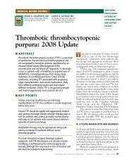
Figure 1. Control of Mineral Metabolism by Parathyroid Hormone.<br />
Levels of serum ionized calcium (Ca 2+ ) are tightly controlled through<br />
the action of parathyroid hormone (PTH) and 1,25-dihydroxyvitamin D<br />
(1,25[OH] 2 D). Both the rate and magnitude of changes in the serum ionized<br />
Ca 2+ concentration are detected by extracellular calcium-sensing receptors<br />
(CaSRs) expressed on parathyroid cells. When ionized Ca 2+ levels<br />
COLOR FIGURE<br />
decrease, the release of PTH secretion is triggered. Conversely, when ionized<br />
Ca 2+ levels increase, PTH secretion is suppressed. PTH stimulates<br />
Version 5 07/3/08<br />
Author Shoback<br />
bone resorption, which delivers calcium and phosphorus (PO 3− Fig # 1<br />
4 ) into the<br />
circulation. In the kidney, PTH Title stimulates <strong>Hypoparathyroidism</strong> renal reabsorption of calcium<br />
and promotes phosphate ME excretion. PTH also enhances the conversion<br />
DE Solomon<br />
of 25-hydroxyvitamin D (25[OH]D) to the active vitamin D metabolite<br />
Artist TV<br />
1,25(OH) 2 D, which increases the<br />
AUTHOR<br />
transepithelial<br />
PLEASE NOTE:<br />
transport of calcium and<br />
Figure has been redrawn and type has been reset<br />
PO 3− 4 through actions in intestinal cells. In concert, these steps restore<br />
Please check carefully<br />
ionized Ca 2+ levels to the normal<br />
Issue date<br />
range,<br />
07/24/08and, through the actions of PTH<br />
and other factors (e.g., fibroblast-derived growth factor 23) in the kidney,<br />
reset the level of serum PO 3− 4 within the normal range. When the actions<br />
of PTH are reduced or lost, all subsequent steps in the maintenance of<br />
homeostasis are impaired, resulting in hypocalcemia, hyperphosphatemia,<br />
and hypercalciuria.<br />
+<br />
Acquired hypoparathyroidism is most commonly<br />
the result of inadvertent removal or irreversible<br />
damage to the glands, usually to their<br />
blood supply, during thyroidectomy, parathyroidectomy,<br />
or radical neck dissection. Definitions<br />
of permanent postsurgical hypoparathyroidism<br />
vary, but the definition is generally accepted to<br />
be insufficient PTH to maintain normocalcemia<br />
+<br />
6 months after surgery. <strong>Hypoparathyroidism</strong> is<br />
estimated to occur after approximately 0.5 to 6.6%<br />
of total thyroidectomies; the rates of this complication<br />
are even higher in some case series,<br />
whereas reported rates at endocrine surgical centers<br />
with high volumes are 0.9 to 1.6%. 25-28 The<br />
occurrence of hypoparathyroidism depends on<br />
the surgeon’s experience, extent of thyroid resection<br />
and nodal dissection <strong>for</strong> cancer, and underlying<br />
thyroid disease, with the presence of substernal<br />
goiter, cancer, or Graves’ disease increasing<br />
the risk. The risk is also greater if one or more<br />
parathyroid glands are not identified intraoperatively<br />
and if the procedure is a reoperation.<br />
Parathyroid secretory reserve is ample, so considerable<br />
damage must occur be<strong>for</strong>e hypoparathyroidism<br />
develops. It is estimated that one normal<br />
gland is sufficient <strong>for</strong> maintaining PTH levels and<br />
serum calcium homeostasis. Immune-mediated<br />
destruction of the parathyroid glands can be either<br />
isolated or part of autoimmune polyendocrine<br />
syndrome type 1 (APS-1). <strong>Hypoparathyroidism</strong><br />
may also be caused by accumulation in the<br />
parathyroid glands of iron (hemochromatosis or<br />
transfusion-dependent thalas semia) 5-7 or copper<br />
(Wilson’s disease) 8 or (in rare cases) by iodine-<br />
131 therapy <strong>for</strong> thyroid diseases 29 or metastatic<br />
infiltration of the parathyroid glands by tumor 30<br />
(Tables 1 and 2).<br />
Magnesium depletion or excess may cause<br />
hypocalcemia by inducing functional hypoparathyroidism.<br />
9,44-46 Magnesium is essential <strong>for</strong> PTH<br />
secretion and activation of the PTH receptor by<br />
ligand. In hypomagnesemia, PTH levels are inappropriately<br />
low or in the lower portion of the<br />
normal range, in the presence of usually mild<br />
hypocalcemia, because the parathyroid is unable<br />
to secrete sufficient hormone, and renal and skeletal<br />
responses to PTH are blunted. In rare instances,<br />
when magnesium is administered parenterally<br />
(e.g., in tocolytic therapy) or accumulates<br />
because of renal insufficiency and serum magnesium<br />
levels rise, PTH secretion is inhibited. 44,45<br />
Magnesium, like calcium, can activate extracellular<br />
calcium-sensing receptors and suppress<br />
PTH release. 47 Once the magnesium status is corrected,<br />
the capacity to secrete PTH and respond<br />
to it resumes.<br />
Genetic disorders must also be considered as<br />
a possible cause of hypocalcemia. The DiGeorge,<br />
or velocardiofacial, syndrome, due to microdele-<br />
392<br />
n engl j med 359;4 www.nejm.org july 24, 2008<br />
Downloaded from www.nejm.org at KAISER PERMANENTE on July 23, 2008 .<br />
Copyright © 2008 Massachusetts Medical Society. All rights reserved.
clinical practice<br />
tions of chromosome 22q11.2, affects 1 in 4000<br />
to 5000 live births. 39-41 Activating mutations in<br />
the extracellular calcium-sensing receptor are<br />
also frequently identified in patients with inherited<br />
hypoparathyroidism and are manifested as<br />
autosomal dominant hypocalcemia at any age<br />
(Tables 1 and 2). 10-13 Mutations in the pre-proPTH<br />
gene and in transcription factors that control<br />
parathyroid gland development are rare but shed<br />
light on basic developmental mechanisms. 2,15-18<br />
Familial hypoparathyroidism due to dysgenesis of<br />
the parathyroid glands results from mutations in<br />
the transcription factors GCMB (glial cells missing<br />
B) or GCM2 (glial cells missing 2) 19-21 and<br />
GATA3 2,3,22,23 and possibly the transcription factor<br />
SOX3 (Sry-box 3) (Tables 1 and 2). 31 Other<br />
disorders that include hypoparathyroidism are<br />
the hypoparathyroidism, retardation, and dysmorphism<br />
syndrome and disorders due to mitochondrial-gene<br />
defects (Table 2). 2,3,36-38,42,43<br />
Evaluation<br />
Review of the patient’s medical and family histories<br />
may suggest the cause of hypocalcemia.<br />
A history of neck surgery suggests that parathyroid<br />
function may have been compromised by the<br />
surgical procedure. A family history of hypocalcemia<br />
suggests a genetic cause (Table 2). The<br />
presence of other autoimmune endocrinopathies<br />
(e.g., adrenal insufficiency) or candidiasis prompts<br />
consideration of autoimmune polyendocrine syndrome<br />
type 1. 32-34 Immunodeficiency and other<br />
congenital defects point to the DiGeorge syndrome<br />
(Table 2). 39-41<br />
Physical examination should include an assessment<br />
of neuromuscular irritability by testing <strong>for</strong><br />
Chvostek’s and Trousseau’s signs. Chvostek’s sign<br />
is elicited by tapping the cheek (2 cm anterior to<br />
the earlobe below the zygomatic process) over the<br />
path of the facial nerve. A positive sign is ipsilateral<br />
twitching of the upper lip. Trousseau’s sign<br />
is elicited by inflating a sphygmomanometer<br />
placed on the upper arm to a level above the<br />
systolic blood pressure <strong>for</strong> 3 minutes. A positive<br />
sign is the occurrence of a painful carpal spasm.<br />
The skin should be examined carefully <strong>for</strong> a neck<br />
scar (which suggests a postsurgical cause of hypocalcemia);<br />
<strong>for</strong> candidiasis and vitiligo (which are<br />
suggestive of APS-1); and <strong>for</strong> generalized bronzing<br />
and signs of liver disease (which are suggestive<br />
of hemochromatosis). Features such as growth<br />
failure, congenital anomalies, hearing loss, or<br />
retardation point to the possibility of genetic<br />
disease.<br />
Laboratory testing should include measurements<br />
of serum total and ionized calcium, albumin,<br />
phosphorus, magnesium, creatinine, intact<br />
PTH, and 25-hydroxyvitamin D (25[OH] vitamin<br />
D) levels. Albumin-corrected total calcium is calculated<br />
as follows:<br />
Corrected total calcium = measured total calcium<br />
+ 0.8 (4.0 − serum albumin),<br />
where calcium is measured in milligrams per<br />
deciliter and albumin is measured in grams per<br />
deciliter. <strong>Hypoparathyroidism</strong> is diagnosed when<br />
the intact PTH level is normal or inappropriately<br />
low in a patient with subnormal serum albumin<br />
corrected total or ionized calcium values, after<br />
hypomagnesemia has been ruled out. Serum<br />
phosphorus levels are usually high or at the high<br />
end of the normal range. Patients with pseudohypoparathyroidism<br />
have a laboratory profile that<br />
resembles that in patients with hypoparathyroidism<br />
(i.e., low calcium and high phosphorus levels),<br />
but they have elevated PTH levels (Table 1).<br />
It may be difficult to rule out hypomagnesemia<br />
as the cause of or a contributor to hypocalcemia<br />
because the serum magnesium level may be normal,<br />
even when intracellular magnesium stores<br />
are reduced. Once magnesium depletion progresses,<br />
however, serum levels decrease to subnormal<br />
levels. In general, if the primary disturbance is<br />
magnesium depletion, serum calcium levels are<br />
only slightly decreased. Intact PTH is often detectable<br />
but inappropriately low.<br />
Measurement of 25(OH) vitamin D levels is<br />
essential to rule out vitamin D deficiency as a<br />
contributor to or cause of hypocalcemia. In classic<br />
vitamin D deficiency, intact PTH levels are<br />
elevated, and serum phosphorus levels are low<br />
or at the low end of the normal range, in marked<br />
contrast to the high levels in hypoparathyroidism.<br />
Measurement of 1,25(OH) 2 vitamin D levels is<br />
generally not necessary in the initial evaluation<br />
of patients with hypoparathyroidism.<br />
Measurements of urinary calcium, magnesium,<br />
and creatinine in a 24-hour collection can<br />
also be helpful in the diagnosis of hypoparathyroidism.<br />
Low urinary calcium levels may be present<br />
in both severe hypocalcemia due to hypo-<br />
n engl j med 359;4 www.nejm.org july 24, 2008 393<br />
Downloaded from www.nejm.org at KAISER PERMANENTE on July 23, 2008 .<br />
Copyright © 2008 Massachusetts Medical Society. All rights reserved.
The new england journal of medicine<br />
Table 1. Pathophysiological Features of Disorders Considered in the Differential Diagnosis of <strong>Hypoparathyroidism</strong>.*<br />
Mechanism and Disorder Clinical Observations Reference<br />
Destruction or removal of parathyroid tissue, with inadequate secretory reserve remaining<br />
Postsurgical hypoparathyroidism Most common <strong>for</strong>m of hypoparathyroidism; can present years after surgery† Winer et al. 4<br />
Autoimmune hypoparathyroidism May be either isolated deficiency or combined with multiple endocrine deficiencies<br />
Radiation-induced destruction of parathyroid<br />
Very rare complication<br />
tissue<br />
Metastatic infiltration of the parathyroid<br />
glands<br />
Deposition of heavy metals in parathyroid<br />
tissue<br />
Several documented cases due to a variety of underlying primary tumors, but generally rare site <strong>for</strong> metastases<br />
Occurs as a result of excess iron in ≥10% of patients with thalassemia, usually in second decade of life,<br />
when other end-organ complications (liver and heart disease, diabetes, hypogonadism, and hypothyroidism)<br />
are present, and correlates with extent of iron overload; less frequent complication of hemochromatosis<br />
and very rare complication of copper accumulation in Wilson’s disease<br />
Reversible impairment of PTH secretion or PTH action with intact underlying secretory function<br />
Severe magnesium depletion Associated with chronic conditions such as alcoholism, malnutrition, malabsorption, diarrhea, diabetes;<br />
drugs (e.g., diuretics, cisplatinum, aminoglycoside antibiotics, amphotericin B, and cyclo sporine); metabolic<br />
acidosis; and renal disorders leading to magnesium wasting (chronic pyelonephritis, postobstructive<br />
nephropathy, renal tubular acidosis, primary renal magnesium wasting, and diuretic stage of<br />
acute tubular necrosis)<br />
Hypermagnesemia May occur in patients receiving tocolytic therapy or in patients with chronic kidney disease receiving magnesium<br />
supplements, antacids, or laxatives<br />
Constitutively active CaSRs Most commonly caused by mutations and rarely caused by acquired antibodies that stimulate the CaSR;<br />
appears to be among the most common causes of hypoparathyroidism<br />
Angelopoulos et al., 5 Toumba<br />
et al., 6 de Sèze et al., 7<br />
Carpenter et al. 8<br />
Tong and Rude 9<br />
Winer et al., 4 Brown, 10 Egbuna and<br />
Brown, 11 Yamamoto et al., 12<br />
Lien hardt et al., 13 Ki<strong>for</strong> et al. 14<br />
Genetic disorders of PTH biosynthesis and parathyroid gland development<br />
PTH gene mutations Responsible <strong>for</strong> isolated hypoparathyroidism Arnold et al., 15 Parkinson and<br />
Thakker, 16 Sunthornthepvarakul<br />
et al., 17 Datta et al. 18<br />
Mutations or deletions in transcription<br />
factors and other regulators of<br />
the development of the parathyroid<br />
glands<br />
May present as either isolated hypoparathyroidism (e.g., GCMB or GCM2 mutations) or as part of complex<br />
genetic syndromes (e.g., GATA3 mutations)<br />
Mutations in mitochondrial DNA May be manifested as hypoparathyroidism plus other metabolic disturbances and congenital anomalies<br />
Ding et al., 19 Baumber et al., 20<br />
Thomée et al., 21 Van Esch<br />
et al., 22 Ali et al. 23<br />
394<br />
n engl j med 359;4 www.nejm.org july 24, 2008<br />
Downloaded from www.nejm.org at KAISER PERMANENTE on July 23, 2008 .<br />
Copyright © 2008 Massachusetts Medical Society. All rights reserved.
clinical practice<br />
Resistance to PTH action‡<br />
Pseudohypoparathyroidism Bastepe 24<br />
Type 1a Clinical features include AHO (round facies, mental retardation, frontal bossing, short stature, obesity,<br />
brachydactyly, ectopic ossifications), hypocalcemia, hyperphosphatemia, and elevated PTH levels; hypothyroidism<br />
develops in a majority of patients because of thyrotropin resistance and less frequently<br />
hypogonadism occurs as a result of gonadotropin resistance; autosomal dominant inheritance pattern<br />
with maternal transmission of the biochemical phenotype; blunted urinary cyclic AMP response to administration<br />
of PTH; a majority of patients have heterozygous inactivating mutations in the gene encoding<br />
the G s -α subunit protein (the GNAS gene)<br />
Type 1b No features of AHO, but same biochemical features as pseudohypoparathyroidism type 1a, including<br />
blunted urinary cyclic AMP response to administration of PTH; caused by selective resistance to PTH<br />
(not to other hormones) and by imprinting defects in GNAS<br />
Type 2 Less common than pseudohypoparathyroidism type 1a or 1b but with same biochemical profile; inherited<br />
or sporadic occurrence; cause of PTH resistance is unclear; patients have normal urinary cyclic AMP<br />
but no phosphaturic responses to PTH<br />
* AHO denotes Albright’s hereditary osteodystrophy, CaSR extracellular calcium-sensing receptor, GCMB glial cells missing B, GCM2 glial cells missing 2, and PTH parathyroid hormone.<br />
† The frequency of each of the diagnoses is difficult to establish because there are few large contemporary series of patients. The three most common causes appear to be postsurgical<br />
hypoparathyroidism, autoimmune polyendocrine syndrome type 1, and autosomal dominant hypocalcemia due to activating CaSR mutations. 4<br />
‡ Resistance lies in the pathway that couples receptor activation to the effector adenylate cyclase. These disorders must be considered in the initial evaluation of patients with hypocalcemia,<br />
especially if hyperphosphatemia is present. Once the intact PTH value is shown to be elevated and vitamin D deficiency or resistance is ruled out, the diagnosis of resistance to<br />
PTH action (not impaired PTH secretion) is established.<br />
parathyroidism and in vitamin D deficiency. In<br />
patients with hypocalcemia due to activating mutations<br />
in the extracellular calcium-sensing receptor,<br />
the ratio of 24-hour urinary calcium to creatinine<br />
has been reported to be substantially<br />
higher than in patients with other types of hypoparathyroidism<br />
(mean value in one report, 0.362<br />
vs. 0.093) and more like that in controls with<br />
normocalcemia (mean value, 0.331). 12<br />
If magnesium deficiency is detected, it is useful<br />
to measure the 24-hour urinary magnesium<br />
level be<strong>for</strong>e repletion is initiated. Elevated or<br />
even detectable urinary levels of magnesium suggest<br />
renal losses as the cause of magnesium depletion,<br />
since the kidney should conserve magnesium<br />
when body stores are depleted (Table 1).<br />
Specialized testing (available in hospital or<br />
reference laboratories) may be warranted to establish<br />
the cause of hypoparathyroidism. This<br />
testing may include gene sequencing <strong>for</strong> the extracellular<br />
calcium-sensing receptor, GATA3, or<br />
the autoimmune regulator protein; microarray<br />
studies or fluorescence in situ hybridization to<br />
diagnose the DiGeorge syndrome; and other hormone<br />
measurements to diagnose autoimmune<br />
polyendocrine syndrome type 1. In many cases,<br />
referral to a pediatric or adult endocrinologist or<br />
geneticist is indicated.<br />
Treatment and Clinical Monitoring<br />
The goals of therapy are to control symptoms<br />
while minimizing complications. The urgent care<br />
of patients with hypocalcemia should be guided<br />
by the nature and severity of the symptoms and<br />
the level of serum calcium. 46,48-51 Severe symptoms<br />
(e.g., seizures, laryngospasm, bronchospasm,<br />
cardiac failure, and altered mental status)<br />
warrant intravenous calcium therapy, even if the<br />
serum calcium level is only mildly reduced (e.g.,<br />
7.0 to 8.0 mg per deciliter [1.75 to 2.00 mmol per<br />
liter]). In such cases, the decrease in the serum<br />
calcium level may have precipitated the symptoms,<br />
and patients usually have immediate, substantial<br />
relief of symptoms with intravenous therapy<br />
(Table 3). Patients with congestive heart failure<br />
due to chronic hypocalcemia require additional<br />
medical treatment (e.g., supplemental oxygen and<br />
diuretics). Intravenous calcium therapy is also recommended<br />
in such patients, even though cardiac<br />
symptoms may resolve more slowly.<br />
Intravenous calcium injections raise the level<br />
of serum calcium transiently; continuous infu-<br />
n engl j med 359;4 www.nejm.org july 24, 2008 395<br />
Downloaded from www.nejm.org at KAISER PERMANENTE on July 23, 2008 .<br />
Copyright © 2008 Massachusetts Medical Society. All rights reserved.
The new england journal of medicine<br />
Table 2. Genetic Syndromes and Other Inherited Forms of <strong>Hypoparathyroidism</strong>.*<br />
Disorder<br />
Familial hypocalcemia<br />
with hypercalciuria†<br />
Familial isolated<br />
hypoparathyroidism<br />
X-linked hypoparathyroidism<br />
Responsible<br />
Locus or<br />
Gene Inheritance Pathogenic Mechanism Comments Reference<br />
3q13,CaSR Autosomal<br />
dominant<br />
11p15, preproPTH<br />
11p15, preproPTH<br />
6p23-p24;<br />
GCMB or<br />
GCM2<br />
APS-1‡ 21q22.3<br />
AIRE<br />
Autosomal<br />
recessive<br />
Autosomal<br />
dominant<br />
Autosomal<br />
recessive<br />
Xq26-27 X-linked<br />
recessive<br />
Autosomal<br />
recessive<br />
Heterozygous gain-of-function mutations in the<br />
CaSR that cause generally mild hypocalcemia<br />
and hypomagnesemia and hypercalciuria; mutant<br />
receptors cause a left-shifted set point <strong>for</strong><br />
PTH secretion, defined as the extracellular calcium<br />
level required <strong>for</strong> half-maximal suppression<br />
of secretion; the altered set point causes<br />
inappropriately normal or low PTH levels even<br />
at low serum calcium levels<br />
Mutations in the signal peptide, disrupting PTH<br />
secretion, or in a donor-splice site of the PTH<br />
gene, leading to skipping of PTH exon 2, which<br />
contains the initiation codon and signal peptide<br />
Point mutation in the signal sequence of pre-proPTH<br />
that prevents processing and translocation of<br />
PTH across endoplasmic reticulum and membrane<br />
<strong>for</strong> exocytosis<br />
Large deletion of GCMB with loss-of-function or<br />
point mutations in the DNA-binding domain of<br />
GCMB, abolishing its transactivation capacity<br />
Deletion and insertion involving genetic material<br />
from chromosomes 2p25.3 and Xq27.1, causing<br />
a position effect on possible regulatory elements<br />
controlling SOX3 transcription<br />
Loss-of-function mutations in AIRE, a zinc-finger<br />
transcription factor present in thymus and<br />
lymph nodes and critical <strong>for</strong> mediating central<br />
tolerance by the thymus; NALP5, an intracellular<br />
signaling molecule strongly expressed in<br />
the parathyroid, may be a specific parathyroid<br />
autoantigen in patients with APS-1; antibodies<br />
to NALP5 were identified in 49% of patients<br />
with APS-1 and hypoparathyroidism<br />
Phenotype caused by >40 mutations, mainly in extracellular<br />
and transmembrane domains of CaSR; Bartter’s<br />
syndrome (salt wasting, hypokalemia, metabolic alkalosis,<br />
elevated renin and aldosterone levels) subtype<br />
V also described in a subgroup of patients; constitutive<br />
CaSR activation described in two patients with<br />
acquired antibodies that activate the CaSR along with<br />
other autoimmune conditions<br />
Homozygous mutations in the pre-proPTH gene cause<br />
very low or undetectable levels of PTH and symptomatic<br />
hypocalcemia<br />
Mutant PTH is trapped within the endoplasmic reticulum<br />
inside cells; resulting stress in the endoplasmic<br />
reticulum is thought to predispose cells to undergo<br />
apoptosis<br />
GCMB is highly expressed in parathyroid tissue and<br />
controls embryologic development of parathyroid<br />
glands<br />
Parathyroid agenesis; transcription factor SOX3 is<br />
thought to be expressed in the developing parathyroid<br />
glands<br />
Cases are concentrated in Fin nish, Iranian Jewish, and<br />
Sardinian populations with >58 known mutations<br />
causing variable clinical presentations; classic triad<br />
is mucocutaneous candidiasis, adrenal insufficiency,<br />
and hypoparathyroidism (any two of these conditions<br />
are sufficient to establish the diagnosis); other features<br />
include hypogonadism, type 1 diabetes mellitus,<br />
hypothyroidism, vitiligo, alopecia, keratoconjunctivitis,<br />
hepatitis, pernicious anemia, and malabsorption‡;<br />
>80% of patients with APS-1 have hypoparathyroidism,<br />
which may be the sole endocrinopathy;<br />
presentation in childhood or adolescence<br />
is typical, but patients with only one disease<br />
manifestation should be followed long-term <strong>for</strong> the<br />
appearance of other signs of disease; occasional cases<br />
with autosomal dominant pattern of inheritance reported<br />
Brown, 10 Egbuna<br />
and Brown, 11<br />
Yamamoto et<br />
al., 12 Lienhardt<br />
et al., 13 Ki<strong>for</strong><br />
et al. 14<br />
Parkinson and<br />
Thakker, 16 Sunthornthepvarakul<br />
et al. 17<br />
Arnold et al., 15<br />
Datta et al. 18<br />
Thakker, 2 Ding et al., 19<br />
Baumber et al., 20<br />
Thomée et al. 21<br />
Bowl et al. 31<br />
Dittmar and<br />
Kahaly, 32<br />
Eisenbarth<br />
and Gottlieb, 33<br />
Perheentupa, 34<br />
Alimohammadi<br />
et al. 35<br />
396<br />
n engl j med 359;4 www.nejm.org july 24, 2008<br />
Downloaded from www.nejm.org at KAISER PERMANENTE on July 23, 2008 .<br />
Copyright © 2008 Massachusetts Medical Society. All rights reserved.
clinical practice<br />
Syndrome of hypopara<br />
thyroidism,<br />
deafness, and<br />
renal anomalies<br />
Syndrome of hypoparathyroidism,<br />
growth<br />
and mental retardation,<br />
and<br />
dysmorphism<br />
DiGeorge, or velocardiofacial,<br />
syndrome<br />
10p14-10-<br />
pter,<br />
GATA3<br />
1q42-q43,<br />
TBCE<br />
22q11.2,<br />
TBX1<br />
Mitochondrial<br />
disorders with<br />
hypoparathyroidism<br />
Mitochondrial<br />
gene<br />
defects<br />
Autosomal<br />
dominant<br />
Autosomal<br />
recessive<br />
Heterozygous<br />
deletions of<br />
chromosome<br />
22q11.2 occurring<br />
mostly<br />
through<br />
new mutations<br />
Mutations or deletions that interfere with the ability<br />
of the transcription factor GATA3 to bind to DNA<br />
or interact with proteins that alter the expression<br />
of GATA3, a factor critical <strong>for</strong> parathyroid, kidney,<br />
and otic-vesicle development<br />
Mutations in TBCE causing loss of function and<br />
probably altered microtubule assembly in<br />
affected tissues<br />
Loss of function of genes on chromosome 22q11,<br />
most notably TBX1, a transcription factor responsible<br />
<strong>for</strong> regulating expression of other transcription<br />
and growth factors important in development<br />
of thymus and parathyroid glands; parathyroid<br />
and thymic defects are caused by abnormal<br />
development in third and fourth branchial<br />
pouches<br />
Maternal Deletions of varying size, mutations, rearrangements,<br />
and duplications in the mitochondrial<br />
genome<br />
Clinical features include hypoparathyroidism, bilateral<br />
sensorineural deafness (mild to profound), and renal<br />
anomalies or dysfunction<br />
Includes the Kenny–Caffey syndrome (short stature, osteosclerosis,<br />
cortical bone thickening, calcification of<br />
basal ganglia, ocular abnormalities, and hypoparathyroidism<br />
that is probably due to agenesis of the glands)<br />
and the Sanjad–Sakati syndrome (parathyroid aplasia,<br />
growth failure, ocular mal<strong>for</strong>mations, microencephaly,<br />
and retardation)<br />
Wide phenotypic spectrum; may include conotruncal cardiac<br />
defects, parathyroid and thymic hypoplasia, neurocognitive<br />
problems, and palatal, renal, ocular, and<br />
skeletal anomalies; hypocalcemia (in 50–60% of patients)<br />
can be transient or permanent and can develop<br />
in adulthood; microarray analysis per<strong>for</strong>med as an initial<br />
diagnostic screening test, with the deletion confirmed<br />
by FISH<br />
Syndromes include the Kearns–Sayre syndrome (progressive<br />
external ophthalmoplegia, pigmentary retinopathy,<br />
heart block or cardiomyopathy, diabetes, and hypoparathyroidism);<br />
MELAS with diabetes and hypoparathyroidism;<br />
and MTPDS, a disorder of fattyacid<br />
oxidation associated with peripheral neuropathy,<br />
retinopathy, acute fatty liver in pregnancy, and hypoparathyroidism<br />
Thakker, 2 Goltzman<br />
and Cole, 3 Van<br />
Esch et al., 22<br />
Ali et al. 23<br />
Thakker, 2 Parvari<br />
et al., 36,37 Sanjad<br />
et al. 38<br />
Kobrynski and<br />
Sullivan, 39 Zweier<br />
et al., 40 Goldmuntz<br />
41<br />
Thakker, 2 Cassandrini<br />
et al., 42<br />
Labarthe et al. 43<br />
* AIRE denotes autoimmune regulator protein, APS-1 autoimmune polyendocrine syndrome type 1, CaSR extracellular calcium-sensing receptor, FISH fluorescence in situ hybridization,<br />
GCMB glial cells missing B, MELAS mitochondrial encephalopathy, lactic acidosis, and strokelike episodes, MTPDS mitochondrial trifunctional protein deficiency syndrome, NALP5<br />
NACHT leucine-rich-repeat protein 5, pre-proPTH pre-proparathyroid hormone, PTH parathyroid hormone, SOX3 Sry-box 3, TBCE tubulin chaperone E, and TBX1 T-box transcription<br />
factor 1.<br />
† Cases that include Bartter’s syndrome are caused by certain activating CaSR mutations (K29E, L125P, C131W, and A843E). Data are from Egbuna and Brown. 11 These mutant CaSRs<br />
are thought to inhibit the activity of a renal outer medullary potassium channel responsible <strong>for</strong> maintaining the transepithelial voltage gradient in the loop of Henle.<br />
‡ Manifestations in a large Finnish cohort of patients included candidiasis (median age at onset, 5.4 years [range, 0.2 to 31.0]), hypoparathyroidism (6 years [1.6–43.0]), and adrenal insufficiency<br />
(10 years [3.5 to 41.0]); data are from Perheentupa. 34<br />
n engl j med 359;4 www.nejm.org july 24, 2008 397<br />
Downloaded from www.nejm.org at KAISER PERMANENTE on July 23, 2008 .<br />
Copyright © 2008 Massachusetts Medical Society. All rights reserved.
The new england journal of medicine<br />
Table 3. Treatment of <strong>Hypoparathyroidism</strong> and Monitoring.<br />
Agent Formulation and Dose Comments References<br />
For short-term management<br />
Calcium gluconate 1 g of calcium gluconate (93 mg of elemental calcium); infuse 1 or 2 g<br />
slowly, each over a period of 10 min, with electrocardiographic and<br />
clinical monitoring of the patient; this initial dose will increase the serum<br />
calcium level <strong>for</strong> only 2 or 3 hr, so it should be followed by a slow<br />
infusion of calcium; 10 g of calcium gluconate in 1 liter of 5% dextrose<br />
in water, infused at a rate of 1–3 mg/kg of body weight/hr in adults<br />
For long-term management<br />
Calcium-containing solutions can be irritating<br />
to surrounding tissues if extravasated,<br />
so a central venous catheter is<br />
preferred; therapy should be individualized<br />
and guided by frequent measurements<br />
of serum ionized calcium<br />
levels<br />
Cooper and Gottoes, 46<br />
Tohme and Bilezikian,<br />
49 Brick man, 50<br />
Rude 51<br />
Calcium salts*<br />
Calcium carbonate 40% elemental calcium by weight; begin with 500–1000 mg of elemental<br />
calcium (three times per day) and adjust the dose to control symptoms<br />
and achieve the targeted calcium level; at least 1–2 g of elemental<br />
calcium (three times daily) generally required and more frequent<br />
dosing sometimes needed<br />
(Titralac) 420-mg tablet (168 mg of elemental calcium), 750-mg tablet (300 mg)<br />
Constipation is a common side effect; calcium<br />
carbonate is best absorbed with<br />
meals and with acid present in the<br />
stomach<br />
(Os-Cal) 650-mg tablet (260 mg of elemental calcium), 1.25-g tablet (500 mg)<br />
(Tums) 500-mg tablet (200 mg of elemental calcium), 750-mg tablet (300 mg),<br />
1000-mg tablet (400 mg), 1.2-g tablet (480 mg)<br />
(Caltrate) 1.5-g tablet (600 mg of elemental calcium)<br />
Calcium citrate 21% elemental calcium by weight Recommended in patients who<br />
have achlorhydria or who<br />
are taking a proton-pump<br />
inhibitor, in order to achieve<br />
sufficient absorption of calcium<br />
(Citracal) 950-mg tablet (200 mg of elemental calcium)<br />
398<br />
n engl j med 359;4 www.nejm.org july 24, 2008<br />
Downloaded from www.nejm.org at KAISER PERMANENTE on July 23, 2008 .<br />
Copyright © 2008 Massachusetts Medical Society. All rights reserved.
clinical practice<br />
Vitamin D metabolites†<br />
Vitamin D 2 (ergocalciferol)<br />
or vitamin D 3 (cholecalciferol)<br />
1,25-dihydroxyvitamin D 3<br />
(calcitriol)<br />
25,000–100,000 IU once daily; time to onset of action, 10–14 days; time to<br />
offset of action, 14–75 days<br />
0.25–1.0 μg once or twice daily; time to onset of action, 1–2 days; time to<br />
offset of action, 2–3 days<br />
Vitamin D 3 is more potent than D 2 but<br />
may be more difficult to obtain at the<br />
doses needed. Because of the long<br />
half-life of vitamin D 2 and D 3 , dosing<br />
can be adjusted and serum levels of<br />
calcium, albumin, phosphorus, and<br />
creatinine determined every 4 weeks,<br />
once symptoms have been controlled.<br />
Most active metabolite of vitamin D at the<br />
vitamin D receptor in vivo<br />
Cooper and Gottoes, 46<br />
Tohme and Bilezikian,<br />
49 Brickman, 50<br />
Rude, 51 Okano et<br />
al., 52 Halabe et al. 53<br />
1α-hydroxyvitamin D 3<br />
(alfacalcidiol)<br />
0.5–3.0 μg (occasionally up to 5.0 μg) daily; time to onset of action, 1–2<br />
days; time to offset of action, 5–7 days<br />
Dihydrotachysterol 0.2–1.0 mg once daily; time to onset of action, 4–7 days; time to offset of<br />
action, 7–21 days<br />
This metabolite is rapidly converted to<br />
1,25-dihydroxyvitamin D 3 in vivo; its<br />
duration of action closely resembles<br />
that of 1,25-dihydroxyvitamin D 3<br />
Thiazide diuretics Added to prevent or control hypercalciuria;<br />
should be combined with a low-salt<br />
diet (80–100 mmol of sodium per day)<br />
to promote calcium retention; doses<br />
are increased as tolerated; adverse<br />
events include hypokalemia and hyponatremia<br />
Hydrochlorothiazide 25–100 mg per day Doses at high end of these ranges are usually<br />
needed to achieve substantial lowering<br />
of urinary calcium<br />
Chlorthalidone 25–100 mg per day<br />
Porter et al. 54<br />
Amiloride and hydrochlorothiazide<br />
(Moduretic)<br />
50 mg of hydrochlorothiazide combined with 5 mg of amiloride once daily Potassium-sparing diuretics may be used to<br />
prevent hypokalemia<br />
* The list of calcium preparations is not comprehensive. Of the calcium preparations available, only the carbonate and citrate salts contain sufficient elemental calcium (per tablet) <strong>for</strong><br />
the efficient treatment of most patients with hypoparathyroidism. Other preparations may be used in patients who cannot tolerate citrate and carbonate salts. The percentage of elemental<br />
calcium is lower in these other preparations: calcium lactate (13%), calcium gluconate (9%), and calcium glubionate (6.6%); thus, larger numbers of tablets must be given.<br />
† Vitamin D toxicity is an important concern and may occur at any time. Manifestations may include altered mental status, fatigue, thirst, dehydration, reduced renal function, nephrolithiasis,<br />
and constipation. Treatment involves discontinuation of the vitamin D preparation and the calcium salt. Depending on the severity, and especially if the toxic effects are from<br />
vitamin D metabolites with long half-lives, intravenous saline hydration and possibly oral glucocorticoids may be warranted to antagonize vitamin D action and more rapidly restore<br />
normocalcemia. Levels of 25-hydroxyvitamin D should be monitored, even in patients receiving calcitrol and alfacalcidiol to prevent vitamin D insufficiency. The target 25-hydroxyvitamin<br />
D level is 30 ng/ml or more.<br />
n engl j med 359;4 www.nejm.org july 24, 2008 399<br />
Downloaded from www.nejm.org at KAISER PERMANENTE on July 23, 2008 .<br />
Copyright © 2008 Massachusetts Medical Society. All rights reserved.
The new england journal of medicine<br />
sions should follow to fully control symptoms<br />
and achieve safe and stable ionized calcium levels,<br />
usually above 1.0 mmol per liter. In the short<br />
term, the serum ionized calcium level should be<br />
measured frequently in order to monitor therapy<br />
(e.g., every 1 to 2 hours initially, while the infusion<br />
rate is being adjusted and until the patient’s<br />
condition has stabilized, and then every 4 to<br />
6 hours). The recurrence of symptoms caused<br />
by hypocalcemia may indicate the need to increase<br />
the infusion rate and should be correlated<br />
with a simultaneous ionized calcium value to assess<br />
the progress of treatment. Oral calcium and<br />
vitamin D therapy should be initiated as soon as<br />
practical (Table 3). Intravenous infusions are<br />
generally tapered slowly (over a period of 24 to<br />
48 hours or longer) while oral therapy is adjusted.<br />
Patients with low calcium levels (e.g., total<br />
calcium,
clinical practice<br />
ments maintained the serum calcium level within<br />
or slightly below the normal range (7.6 to 8.8 mg<br />
per deciliter [1.9 to 2.2 mmol per liter]), 4 but the<br />
use of PTH resulted in less urinary calcium excretion.<br />
Although PTH significantly increased biochemical<br />
markers of bone turnover (as compared<br />
with no significant change with calcitriol), there<br />
were no differences in bone mineral density between<br />
the groups. 4 Creatinine clearances did not<br />
differ significantly between the groups, and they<br />
were stable in both groups during the study. PTH<br />
(1–34) is not approved by the Food and Drug Administration<br />
<strong>for</strong> this indication.<br />
Limited data suggest that the quality of life<br />
may be compromised in patients with hypoparathyroidism<br />
despite treatment to optimize their<br />
biochemical values. In one study involving 25<br />
women at a university center who were treated<br />
with vitamin D and calcium <strong>for</strong> postsurgical<br />
hypoparathyroidism, scores <strong>for</strong> somatization,<br />
depression, anxiety, and phobic anxiety were<br />
significantly higher than among age- and sexmatched<br />
controls with intact parathyroid function<br />
after thyroidectomy. 56 The effect of PTH<br />
replacement on quality-of-life measures is not<br />
known.<br />
Whereas most patients with mutations in<br />
the extracellular calcium-sensing receptor have<br />
mild hypocalcemia <strong>for</strong> which no treatment is required,<br />
10,11 some have severe, symptomatic hypocalcemia<br />
necessitating therapy. Because treatment<br />
with calcium and vitamin D in these patients may<br />
exacerbate baseline hypercalciuria and result in<br />
nephrocalcinosis and renal insufficiency, PTH<br />
therapy may warrant particular consideration in<br />
these patients. Several patients have been treated<br />
successfully with PTH therapy, averting hypercalciuria<br />
and reduced renal function. 4 More data,<br />
however, are needed be<strong>for</strong>e its use can be recommended<br />
in practice. In the future, drugs that<br />
antagonize the extracellular calcium-sensing receptor<br />
(i.e., calcilytic agents), which are in development,<br />
might be used to stimulate endogenous<br />
PTH in such patients.<br />
Guidelines<br />
There are no <strong>for</strong>mal guidelines <strong>for</strong> the management<br />
of hypoparathyroidism.<br />
Conclusions and<br />
Recommendations<br />
The initial evaluation of a patient with hypocalcemia<br />
should include a detailed family history<br />
(which may suggest a genetic cause) and relevant<br />
medical history (particularly regarding neck surgery<br />
and autoimmune disease). Laboratory testing<br />
should include measurements of serum total<br />
and ionized calcium, albumin, phosphorus, magnesium,<br />
and intact PTH levels. If the patient has<br />
severe symptoms, therapy with intravenous calcium<br />
should be initiated immediately, and the<br />
diagnosis pursued after the patient’s condition<br />
has been stabilized. In the patient described in<br />
the vignette, the hearing deficits and family history<br />
of renal disease suggest the diagnosis of the<br />
syndrome of hypoparathyroidism, deafness, and<br />
renal anomalies. The expectation is that the intact<br />
PTH level would be detectable but low. Given<br />
the absence of symptoms in this patient, outpatient<br />
treatment with calcium carbonate three<br />
times daily and calcitriol once or twice daily would<br />
be appropriate, with adjustment as needed to<br />
maintain a target level of albumin-corrected serum<br />
calcium at the lower end of the normal range<br />
(approximately 8.0 to 8.5 mg per deciliter [2.00 to<br />
2.12 mmol per liter]), a 24-hour urinary calcium<br />
level well below 300 mg, and a calcium–phosphate<br />
product below 55.<br />
No potential conflict of interest relevant to this article was<br />
reported.<br />
An audio version of this article is available at www.nejm.org.<br />
References<br />
1. Marx SJ. Hyperparathyroid and hypoparathyroid<br />
disorders. N Engl J Med<br />
2000;343:1863-75. [Errata, N Engl J Med<br />
2001;344:240, 696.]<br />
2. Thakker RV. Genetics of endocrine<br />
and metabolic disorders: parathyroid. Rev<br />
Endocr Metab Disord 2004;5:37-51.<br />
3. Goltzman D, Cole DEC. <strong>Hypoparathyroidism</strong>.<br />
In: Favus MJ, ed. Primer on the<br />
metabolic bone diseases and disorders of<br />
mineral metabolism. 6th ed. Washington,<br />
DC: American Society <strong>for</strong> Bone and Mineral<br />
Research, 2006:216-9.<br />
4. Winer KK, Ko CW, Reynolds JC, et al.<br />
Long-term treatment of hypoparathyroidism:<br />
a randomized controlled study comparing<br />
parathyroid hormone (1-34) versus<br />
calcitriol and calcium. J Clin Endocrinol<br />
Metab 2003;88:4214-20.<br />
5. Angelopoulos NG, Goula A, Rombopoulos<br />
G, et al. <strong>Hypoparathyroidism</strong> in<br />
n engl j med 359;4 www.nejm.org july 24, 2008 401<br />
Downloaded from www.nejm.org at KAISER PERMANENTE on July 23, 2008 .<br />
Copyright © 2008 Massachusetts Medical Society. All rights reserved.
The new england journal of medicine<br />
transfusion-dependent patients with<br />
β-thalassemia. J Bone Miner Metab 2006;<br />
24:138-45.<br />
6. Toumba M, Sergis A, Kanaris C,<br />
Skordis N. Endocrine complications in<br />
patients with thalassaemia major. Pediatr<br />
Endocrinol Rev 2007;5:642-8.<br />
7. de Sèze S, Solnica J, Mitrovic D,<br />
Miravet L, Dorfmann H. Joint and bone<br />
disorders and hypoparathyroidism in hemochromatosis.<br />
Semin Arthritis Rheum<br />
1972;2:71-94.<br />
8. Carpenter TO, Carnes DL Jr, Anast CS.<br />
<strong>Hypoparathyroidism</strong> in Wilson’s disease.<br />
N Engl J Med 1983;309:873-7.<br />
9. Tong GM, Rude RK. Magnesium deficiency<br />
in critical illness. J Intensive Care<br />
Med 2005;20:3-17.<br />
10. Brown EM. Clinical lessons from the<br />
calcium-sensing receptor. Nat Clin Pract<br />
Endocrinol Metab 2007;3:122-33.<br />
11. Egbuna OI, Brown EM. Hypercalcaemic<br />
and hypocalcaemic conditions due to<br />
calcium-sensing receptor mutations. Best<br />
Pract Res Clin Rheumatol 2008;22:129-48.<br />
12. Yamamoto M, Akatsu T, Nagase T,<br />
Ogata E. Comparison of hypocalcemic hypercalciuria<br />
between patients with idiopathic<br />
hypoparathyroidism and those<br />
with gain-of-function mutations in the<br />
calcium-sensing receptor: is it possible to<br />
differentiate the two disorders? J Clin Endocrinol<br />
Metab 2000;85:4583-91.<br />
13. Lienhardt A, Bai M, Lagarde J-P, et al.<br />
Activating mutations of the calciumsensing<br />
receptor: management of hypocalcemia.<br />
J Clin Endocrinol Metab 2001;<br />
86:5313-23.<br />
14. Ki<strong>for</strong> O, McElduff A, LeBoff MS, et al.<br />
Activating antibodies to the calcium-sensing<br />
receptor in two patients with autoimmune<br />
hypoparathyroidism. J Clin Endocrinol<br />
Metab 2004;89:548-56.<br />
15. Arnold A, Horst SA, Gardella TJ, Baba<br />
H, Levine MA, Kronenberg HM. Mutation<br />
of the signal peptide-encoding region of<br />
the preproparathyroid hormone gene in<br />
familial isolated hypoparathyroidism.<br />
J Clin Invest 1990;86:1084-7.<br />
16. Parkinson DB, Thakker RV. A donor<br />
splice site mutation in the parathyroid<br />
hormone gene is associated with autosomal<br />
recessive hypoparathyroidism. Nat<br />
Genet 1992;1:149-52.<br />
17. Sunthornthepvarakul T, Churesigaew<br />
S, Ngowngarmratana S. A novel mutation<br />
of the signal peptide of the preproparathyroid<br />
hormone gene associated with<br />
autosomal recessive familial isolated hypoparathyroidism.<br />
J Clin Endocrinol Metab<br />
1999;84:3792-6.<br />
18. Datta R, Waheed A, Shah GN, Sly WS.<br />
Signal sequence mutation in autosomal<br />
dominant <strong>for</strong>m of hypoparathyroidism<br />
induces apoptosis that is corrected by a<br />
chemical chaperone. Proc Natl Acad Sci<br />
U S A 2007;104:19989-94.<br />
19. Ding C, Buckingham B, Levine MA.<br />
Familial isolated hypoparathyroidism<br />
caused by a mutation in the gene <strong>for</strong> the<br />
transcription factor GCMB. J Clin Invest<br />
2001;108:1215-20.<br />
20. Baumber L, Tufarelli C, Patel S, et al.<br />
Identification of a novel mutation disrupting<br />
the DNA binding activity of GCM2 in<br />
autosomal recessive familial isolated hypoparathyroidism.<br />
J Med Genet 2005;42:<br />
443-8.<br />
21. Thomée C, Schubert SW, Parma J, et<br />
al. GCMB mutation in familial isolated<br />
hypoparathyroidism with residual secretion<br />
of parathyroid hormone. J Clin Endocrinol<br />
Metab 2005;90:2487-92.<br />
22. Van Esch H, Groenen P, Nesbit MA, et<br />
al. GATA3 haplo-insufficiency causes human<br />
HDR syndrome. Nature 2000;406:<br />
419-22.<br />
23. Ali A, Christine PT, Grigorieva IV, et<br />
al. Functional characterization of GATA3<br />
mutations causing the hypoparathyroidism-deafness-renal<br />
(HDR) dysplasia syndrome:<br />
insight into mechanisms of DNA<br />
binding by the GATA3 transcription factor.<br />
Hum Mol Genet 2007;16:265-75.<br />
24. Bastepe M. The GNAS locus and<br />
pseudohypoparathyroidism. Adv Exp Med<br />
Biol 2008;626:27-40.<br />
25. Thomusch O, Machens A, Sekulla C,<br />
Ukkat J, Brauckhoff M, Dralle H. The impact<br />
of surgical technique on postoperative<br />
hypoparathyroidism in bilateral thyroid<br />
surgery: a multivariate analysis of<br />
5846 consecutive patients. Surgery 2003;<br />
133:180-5.<br />
26. Zarnegar R, Brunaud L, Clark OH.<br />
Prevention, evaluation, and management<br />
of complications following thyroidectomy<br />
<strong>for</strong> thyroid carcinoma. Endocrinol Metab<br />
Clin North Am 2003;32:483-502.<br />
27. Page C, Strunski V. Parathyroid risk in<br />
total thyroidectomy <strong>for</strong> bilateral, benign,<br />
multinodular goitre: report of 351 surgical<br />
cases. J Laryngol Otol 2007;121:237-41.<br />
28. Asari R, Passler C, Kaczirek K, Scheuba<br />
C, Niederle B. <strong>Hypoparathyroidism</strong><br />
after total thyroidectomy: a prospective<br />
study. Arch Surg 2008;143:132-7.<br />
29. Winslow CP, Meyers AD. Hypocalcemia<br />
as a complication of radioiodine<br />
therapy. Am J Otolaryngol 1998;19:401-3.<br />
30. Goddard CJ, Mbewu A, Evanson JM.<br />
Symptomatic hypocalcaemia associated<br />
with metastatic invasion of the parathyroid<br />
glands. Br J Hosp Med 1990;43:72.<br />
31. Bowl MR, Nesbit MA, Harding B, et<br />
al. An interstitial deletion-insertion involving<br />
chromosomes 2p25.3 and Xq27.1,<br />
near SOX3, causes X-linked recessive hypoparathyroidism.<br />
J Clin Invest 2005;115:<br />
2822-31.<br />
32. Dittmar M, Kahaly GJ. Polyglandular<br />
autoimmune syndromes: immunogenetics<br />
and long-term follow-up. J Clin Endocrinol<br />
Metab 2003;88:2983-92.<br />
33. Eisenbarth GS, Gottlieb PA. Autoim-<br />
mune polyendocrine syndromes. N Engl J<br />
Med 2004;350:2068-79.<br />
34. Perheentupa J. Autoimmune polyendo-<br />
crinopathy-candidiasis-ectodermal dystrophy.<br />
J Clin Endocrinol Metab 2006;91:<br />
2843-50.<br />
35. Alimohammadi M, Björklund P, Hallgren<br />
Å, et al. Autoimmune polyendocrine<br />
syndrome type 1 and NALP5, a parathyroid<br />
autoantigen. N Engl J Med 2008;358:<br />
1018-28.<br />
36. Parvari R, Diaz GA, Hershkovitz E.<br />
Parathyroid development and the role of<br />
tubulin chaperone E. Horm Res 2007;67:<br />
12-21.<br />
37. Parvari R, Hershkovitz E, Grossman<br />
N, et al. Mutation of TBCE causes hypoparathyroidism-retardation-dysmorphism<br />
and autosomal recessive Kenny-Caffey<br />
syndrome. Nat Genet 2002;32:448-52.<br />
38. Sanjad SA, Sakati NA, Abu-Osba YK,<br />
Kaddoura R, Milner RDG. A new syndrome<br />
of congenital hypoparathyroidism,<br />
severe growth failure, and dysmorphic<br />
features. Arch Dis Child 1991;66:193-6.<br />
39. Kobrynski LJ, Sullivan KE. Velocardiofacial<br />
syndrome, DiGeorge syndrome: the<br />
chromosome 22q11.2 deletion syndromes.<br />
Lancet 2007;370:1443-52.<br />
40. Zweier C, Sticht H, Aydin-Yaylagül I,<br />
Campbell CE, Rauch A. Human TBX1<br />
missense mutations cause gain of function<br />
resulting in the same phenotype as<br />
22q11.2 deletions. Am J Hum Genet<br />
2007;80:510-7.<br />
41. Goldmuntz E. DiGeorge syndrome: new<br />
insights. Clin Perinatol 2005;32:963-78.<br />
42. Cassandrini D, Savasta S, Bozzola M,<br />
et al. Mitochondrial DNA deletion in a<br />
child with mitochondrial encephalomyopathy,<br />
growth hormone deficiency, and<br />
hypoparathyroidism. J Child Neurol 2006;<br />
21:983-5.<br />
43. Labarthe F, Benoist JF, Brivet M, Vianey-Saban<br />
C, Despert F, de Baulny HO.<br />
Partial hypoparathyroidism associated<br />
with mitochondrial trifunctional protein<br />
deficiency. Eur J Pediatr 2006;165:389-91.<br />
44. Cholst IN, Steinberg SF, Tropper PJ,<br />
Fox HE, Segre GV, Bilezikian JP. The influence<br />
of hypermagnesemia on serum<br />
calcium and parathyroid hormone levels<br />
in human subjects. N Engl J Med 1984;<br />
310:1221-5.<br />
45. Koontz SL, Friedman SA, Schwartz<br />
ML. Symptomatic hypocalcemia after tocolytic<br />
therapy with magnesium sulfate<br />
and nifedipine. Am J Obstet Gynecol<br />
2004;190:1773-6.<br />
46. Cooper MS, Gottoes NJ. Diagnosis<br />
and management of hypocalcemia. BMJ<br />
2008;336:1298-302.<br />
47. Hofer AM, Brown EM. Extracellular<br />
calcium sensing and signalling. Nat Rev<br />
Mol Cell Biol 2003;4:530-8.<br />
48. Lebowitz MR, Moses AM. Hypocalcemia.<br />
Semin Nephrol 1992;12:146-58.<br />
49. Tohme JF, Bilezikian JP. Hypocalcemic<br />
emergencies. Endocrinol Metab Clin<br />
North Am 1993;22:363-75.<br />
50. Brickman AS. Disorders of calcitropic<br />
hormones in adults. In: Manual of endo-<br />
402<br />
n engl j med 359;4 www.nejm.org july 24, 2008<br />
Downloaded from www.nejm.org at KAISER PERMANENTE on July 23, 2008 .<br />
Copyright © 2008 Massachusetts Medical Society. All rights reserved.
clinical practice<br />
crinology and metabolism. 3rd ed. Philadelphia:<br />
Lippincott Williams & Wilkins,<br />
2002:293-324.<br />
51. Rude RK. Hypocalcemia and hypoparathyroidism.<br />
Curr Ther Endocrinol<br />
Metab 1997;6:546-51.<br />
52. Okano K, Furukawa Y, Morii H, Fujita<br />
T. Comparative efficacy of various vitamin<br />
D metabolites in the treatment of various<br />
types of hypoparathyroidism. J Clin Endocrinol<br />
Metab 1982;55:238-43.<br />
53. Halabe A, Arie R, Mimran D, Samuel<br />
R, Liberman UA. <strong>Hypoparathyroidism</strong> —<br />
a long-term follow-up experience with<br />
1α-vitamin D3 therapy. Clin Endocrinol<br />
(Oxf ) 1994;40:303-7.<br />
54. Porter RH, Cox BG, Heaney D,<br />
Hostetter TH, Stinebaugh BJ, Suki WN.<br />
Treatment of hypoparathyroid patients<br />
with chlorthalidone. N Engl J Med 1978;<br />
298:577-81.<br />
55. Winer KK, Yanovski JA, Cutler GB Jr.<br />
Synthetic human parathyroid hormone<br />
1-34 vs calcitriol and calcium in the treatment<br />
of hypoparathyroidism. JAMA 1996;<br />
276:631-6.<br />
56. Arlt W, Fremerey C, Callies F, et al.<br />
Well-being, mood and calcium homeostasis<br />
in patients with hypoparathyroidism<br />
receiving standard treatment with calcium<br />
and vitamin D. Eur J Endocrinol 2002;<br />
146:215-22.<br />
Copyright © 2008 Massachusetts Medical Society.<br />
c o l l e c t i o n s o f a r t i c l e s o n t h e j o u r n a l’s w e b s i t e<br />
The Journal’s Web site (www.nejm.org) sorts published articles into<br />
more than 50 distinct clinical collections, which can be used as convenient<br />
entry points to clinical content. In each collection, articles are cited in reverse<br />
chronologic order, with the most recent first.<br />
n engl j med 359;4 www.nejm.org july 24, 2008 403<br />
Downloaded from www.nejm.org at KAISER PERMANENTE on July 23, 2008 .<br />
Copyright © 2008 Massachusetts Medical Society. All rights reserved.