Technical Aspects of Cardiac CT - ImPACT CT Scanner Evaluation ...
Technical Aspects of Cardiac CT - ImPACT CT Scanner Evaluation ...
Technical Aspects of Cardiac CT - ImPACT CT Scanner Evaluation ...

Create successful ePaper yourself
Turn your PDF publications into a flip-book with our unique Google optimized e-Paper software.
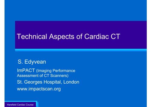
<strong>Technical</strong> <strong>Aspects</strong> <strong>of</strong> <strong>Cardiac</strong> <strong>CT</strong><br />
S. Edyvean<br />
ImPA<strong>CT</strong> (Imaging Performance<br />
Assessment <strong>of</strong> <strong>CT</strong> <strong>Scanner</strong>s)<br />
St. Georges Hospital, London<br />
www.impactscan.org<br />
Harefield <strong>Cardiac</strong> Course
<strong>Technical</strong> <strong>Aspects</strong> <strong>of</strong> <strong>Cardiac</strong> <strong>CT</strong><br />
• Introduction<br />
• Multi-slice <strong>CT</strong> (MS<strong>CT</strong>)<br />
• Scanning the heart with MS<strong>CT</strong><br />
• Improving<br />
– Temporal resolution<br />
– Volume coverage<br />
– Spatial resolution<br />
Harefield <strong>Cardiac</strong> Course
<strong>Cardiac</strong> <strong>CT</strong><br />
• Godfrey Hounsfield, inventor <strong>of</strong> clinical <strong>CT</strong>, 1971<br />
– 1979 Nobel prize<br />
–1 st Oct 1971 – 1 st patient scanned<br />
1919 – 2004<br />
Harefield <strong>Cardiac</strong> Course
Godfrey Hounsfield – Nobel Speech 1979<br />
A further promising field may be the detection <strong>of</strong> the coronary arteries.<br />
It may be possible to detect these under special conditions <strong>of</strong> scanning.<br />
Harefield <strong>Cardiac</strong> Course
Applications <strong>of</strong> cardiac <strong>CT</strong><br />
• Calcium scoring<br />
– calcified plaque<br />
• Coronary <strong>CT</strong> angiography (<strong>CT</strong>A)<br />
– Coronary artery anatomy<br />
– Stenosis<br />
– Stent viability<br />
– Graft anatomy and patency<br />
• Functional imaging<br />
Harefield <strong>Cardiac</strong> Course
<strong>Cardiac</strong> <strong>CT</strong><br />
• 1990’s: Electron beam <strong>CT</strong> (EB<strong>CT</strong>)<br />
– Calcium scoring (Agatston score)<br />
Harefield <strong>Cardiac</strong> Course<br />
6
Modern multi-slice scanners<br />
• 1998 (4 slice), 2001(16 slice), 2004 (64 slice), ...<br />
Harefield <strong>Cardiac</strong> Course
The scanner<br />
Cables<br />
Tube<br />
Aperture /<br />
bore<br />
Fan<br />
beam<br />
Y<br />
X<br />
Detectors ~ 1000<br />
Harefield <strong>Cardiac</strong> Course
The scanner<br />
Y<br />
Y<br />
X<br />
X<br />
Z<br />
Z<br />
Typical detector length ~ 40 mm<br />
Harefield <strong>Cardiac</strong> Course<br />
(20 - 160 mm )<br />
Picture courtesy <strong>of</strong> K. Gelijns, Leiden
The scanner<br />
64 x 0.5 = 32 mm<br />
Aquilion 64<br />
64 X 0.5 mm<br />
Z-axis<br />
Y<br />
X<br />
z-<br />
axis<br />
Z<br />
Depending on scanner:<br />
4, 16, 64, 128, 320 rows (slices <strong>of</strong> data)<br />
min size <strong>of</strong> detector element ~ 0.5, 0.6 mm<br />
Harefield <strong>Cardiac</strong> Course<br />
Picture courtesy <strong>of</strong> K. Gelijns, Leiden
Beam width, detectors and slices<br />
• GE LightSpeed 64<br />
– 64 x 0.625 mm detectors<br />
Beam = 40 mm<br />
64 x 0.63 mm<br />
32 x 1.25 mm<br />
16 x 2.5 mm<br />
40 mm<br />
z-axis<br />
Harefield <strong>Cardiac</strong> Course<br />
64 x 0.625 mm<br />
Beam = 20 mm<br />
8 x 2.5 mm
Multi-slice <strong>CT</strong> - coverage<br />
10 20 40 80 160 mm<br />
Harefield <strong>Cardiac</strong> Course<br />
z-axis
<strong>Scanner</strong> rotation speeds<br />
0.3 Second rot timea.MPG<br />
Harefield <strong>Cardiac</strong> Course<br />
Typical fastest rotation speeds < 0.5 sec/rot<br />
(0.5, 0.4, 0.3, 0.27 sec/rot)
Axial scanning – ‘step and shoot’<br />
– Also known as sequential scanning<br />
Detector array<br />
x-axis<br />
Harefield <strong>Cardiac</strong> Course<br />
z<br />
Sub-mm detectors<br />
Full extent <strong>of</strong> detector matrix<br />
z-axis
Helical (spiral) scanning<br />
• Continuous gantry rotation + continuous table feed<br />
• Scan data traces a helical path - or ‘spiral’ - around<br />
patient<br />
– data used to form axial images<br />
Detector array<br />
x-axis<br />
Sub-mm detectors<br />
Full extent <strong>of</strong> detector matrix<br />
z-axis<br />
xy plane<br />
Harefield <strong>Cardiac</strong> Course<br />
z
Helical (spiral) scanning - pitch<br />
Pitch =<br />
table travel / rotation<br />
X-ray beam width<br />
z<br />
Harefield <strong>Cardiac</strong> Course
Helical (spiral) scanning - pitch<br />
pitch 2<br />
T = 80<br />
Table travel/rot = 80 mm<br />
Beam width = 40 mm<br />
X = 40<br />
z-axis<br />
pitch 1<br />
pitch 0.5<br />
Harefield <strong>Cardiac</strong> Course
Image reconstruction<br />
• Attenuation pr<strong>of</strong>iles through every angle<br />
– ~1000 detector elements<br />
– ~1000 angular projections<br />
detector elements<br />
90°<br />
Harefield <strong>Cardiac</strong> Course<br />
attenuation<br />
attenuation<br />
detector elements<br />
0°
Harefield <strong>Cardiac</strong> Course
Harefield <strong>Cardiac</strong> Course
• Analytical techniques<br />
Image reconstruction<br />
– 2-D Filtered back projection (slices up to ~ 12)<br />
– Techniques to overcome cone beam artefacts (slices > 12)<br />
• 3-D approximations (Tilted slice, Feldkamp)<br />
– Cone beam reconstruction<br />
• Iterative reconstruction<br />
– ASIR, MBIR (VEO), IRIS, SAFFIR, AIDR, iDOSE ….<br />
Harefield <strong>Cardiac</strong> Course<br />
standard<br />
iterative<br />
Courtesy Philips
<strong>CT</strong> Image<br />
• Pixel value (<strong>CT</strong> number)<br />
– Represents average attenuation <strong>of</strong> the 3-D volume element<br />
512<br />
pixels<br />
slice<br />
width<br />
voxel<br />
• Pixel size = fov / matrix<br />
– eg 350 / 512 = 0.68 mm, If 1024 matrix = 0.34 mm<br />
Harefield <strong>Cardiac</strong> Course
• Volume set <strong>of</strong> data<br />
Image presentation<br />
– that can be reconstructed in any direction by a variety <strong>of</strong><br />
techniques<br />
axial<br />
0.3 x 0.3 x 0.3 mm<br />
Harefield <strong>Cardiac</strong> Course
<strong>Technical</strong> <strong>Aspects</strong> <strong>of</strong> <strong>Cardiac</strong> <strong>CT</strong><br />
• Introduction<br />
• Multi-slice <strong>CT</strong> (MS<strong>CT</strong>)<br />
• Scanning the heart with MS<strong>CT</strong><br />
• Improving<br />
– Temporal resolution<br />
– Volume coverage<br />
– Spatial resolution<br />
Harefield <strong>Cardiac</strong> Course
• Heart rate<br />
The heart<br />
– Average 60 bpm (1 beat per sec) (40 bpm – 120 bpm)<br />
– Vessels move at different speeds<br />
• Length ~ 120 mm<br />
• Very fine vessels < 1mm<br />
• Plaque<br />
– calcium, fatty, s<strong>of</strong>t, fibrous<br />
Conventional angiography<br />
Harefield <strong>Cardiac</strong> Course
<strong>Cardiac</strong> <strong>CT</strong> - ECG signal<br />
• Acquisition and reconstruction linked to ECG<br />
Harefield <strong>Cardiac</strong> Course
<strong>Cardiac</strong> <strong>CT</strong> – ECG phases<br />
• To ‘freeze’ cardiac motion:<br />
– Image during phase <strong>of</strong> least cardiac motion<br />
– Phase given as percentage <strong>of</strong> R-R interval (eg 70%)<br />
– Ideal width at least 10% <strong>of</strong> R-R interval<br />
Phase position<br />
R<br />
R R R<br />
ECG<br />
t<br />
<strong>Cardiac</strong><br />
motion<br />
Harefield <strong>Cardiac</strong> Course
<strong>Cardiac</strong> <strong>CT</strong> – ECG phases<br />
• To ‘freeze’ cardiac motion:<br />
– Image during phase <strong>of</strong> least cardiac motion<br />
– Phase given as percentage <strong>of</strong> R-R interval<br />
– Ideal width at least 10% <strong>of</strong> R-R interval:<br />
Phase position<br />
width<br />
60 bpm (1 bps) 100 ms<br />
R<br />
R R R<br />
ECG<br />
t<br />
<strong>Cardiac</strong><br />
motion<br />
Harefield <strong>Cardiac</strong> Course<br />
Ideal imaging window ~ 10%
<strong>Cardiac</strong> <strong>CT</strong> – ECG phases<br />
• 2 definitions <strong>of</strong> phase position<br />
60% R-R<br />
Reconstruction phase<br />
R<br />
R<br />
70% R-R<br />
– Beginning <strong>of</strong> phase window (eg 60%)<br />
– Middle <strong>of</strong> phase window (eg 70%)<br />
Harefield <strong>Cardiac</strong> Course
• Optimal phase for reconstruction for <strong>CT</strong>A<br />
–~ 70 %(<br />
<strong>Cardiac</strong> <strong>CT</strong> – ECG phases<br />
Optimal reconstruction phase<br />
70% R-R<br />
Eg. 50 60<br />
70<br />
80<br />
Harefield <strong>Cardiac</strong> Course
<strong>Cardiac</strong> <strong>CT</strong> – ECG phases<br />
• For higher heart rates<br />
– ~ 30 – 40% phase position (also for RCA)<br />
– This region doesn’t shorten as much as the 70% region<br />
Reconstruction phase<br />
40% R-R<br />
Some flexibility <strong>of</strong> reconstruction<br />
phase position required<br />
Harefield <strong>Cardiac</strong> Course
Data acquisition – how much data do you need?<br />
• Opposing projections provide the same information<br />
– To reconstruct images only 180° <strong>of</strong> scan data is required<br />
• Image time = rotation / 2<br />
0°<br />
attenuation<br />
300 ms rotation<br />
150 ms<br />
attenuation<br />
180°<br />
one z-axis position<br />
Harefield <strong>Cardiac</strong> Course
<strong>Cardiac</strong> <strong>CT</strong> - scan modes<br />
Scan<br />
Axial<br />
Helical<br />
<strong>Cardiac</strong><br />
Prospective triggering<br />
Retrospective gating<br />
Harefield <strong>Cardiac</strong> Course
<strong>Cardiac</strong> <strong>CT</strong>- axial scanning<br />
• R wave recognised - scan triggered<br />
Radiation on<br />
Harefield <strong>Cardiac</strong> Course
<strong>Cardiac</strong> <strong>CT</strong>- axial scanning<br />
• Images reconstructed<br />
Radiation on<br />
Required data<br />
Harefield <strong>Cardiac</strong> Course
<strong>Cardiac</strong> <strong>CT</strong> - axial scanning with padding<br />
• Axial scanning with ‘padding’<br />
• More flexibility with reconstructed phase position<br />
Radiation on<br />
‘padding’ for<br />
<strong>CT</strong>A<br />
70<br />
Required data<br />
Harefield <strong>Cardiac</strong> Course
<strong>Cardiac</strong> <strong>CT</strong> - axial scanning with padding<br />
• Axial scanning with ‘padding’<br />
• More flexibility with reconstructed phase position<br />
Radiation on<br />
‘padding’ for<br />
<strong>CT</strong>A<br />
60<br />
Required data<br />
Harefield <strong>Cardiac</strong> Course
<strong>Cardiac</strong> <strong>CT</strong> – helical scanning<br />
• Scan with overlapping pitch ~ 0.2<br />
• Image reconstruction selected retrospectively<br />
Harefield <strong>Cardiac</strong> Course<br />
X-rays on
<strong>Cardiac</strong> <strong>CT</strong> – helical scanning<br />
• Scan with overlapping pitch ~ 0.2<br />
• Image reconstruction selected retrospectively<br />
– Choose best phase for cardiac <strong>CT</strong>A<br />
– Multiple phases for functional studies<br />
Harefield <strong>Cardiac</strong> Course<br />
X-rays on
0% 5% 10% 15%<br />
20%<br />
25% 30% 35% 40% 45%<br />
50% 55% 60% 65% 70%<br />
41<br />
Harefield <strong>Cardiac</strong> Course<br />
75%<br />
80% 85%<br />
90%<br />
95%
0% 5% 10% 15% 20%<br />
25% 30% 35% 40%<br />
45%<br />
50% 55% 60% 65%<br />
70%<br />
42<br />
Harefield <strong>Cardiac</strong> Course<br />
75% 80% 85%<br />
90%<br />
95%
Functional Imaging<br />
• Using all phases in cine loop<br />
phase<br />
Harefield <strong>Cardiac</strong> Course
Helical cardiac <strong>CT</strong>– ECG dose modulation<br />
• Tube current (mA) decreased to a prescribed<br />
minimum value outside phase region <strong>of</strong> interest<br />
– eg 20%, 4% <strong>of</strong> maximum dose<br />
• Full dose at required phase region, with a margin<br />
• Other phases can still be used for functional study<br />
Radiation on<br />
Reconstruction phase<br />
Available for image recon.<br />
100%<br />
100%<br />
20%<br />
4%<br />
Harefield <strong>Cardiac</strong> Course
Harefield <strong>Cardiac</strong> Course<br />
Helical pitch in cardiac scanning
Helical cardiac <strong>CT</strong> - pitch<br />
• Gantry rotates faster than heart rate. Eg. :<br />
– 0.3 sec scan = 3 rotations / second<br />
– Heart rate: @ 60 bpm = 1 beat per second<br />
• If Pitch =1, gaps in cardiac anatomy<br />
3 rotations<br />
per heart beat<br />
~300ms<br />
<strong>Scanner</strong> rotations<br />
Harefield <strong>Cardiac</strong> Course
Helical cardiac <strong>CT</strong> - pitch<br />
• Require an overlapping pitch<br />
– ~0.2 – 0.3 to eliminate gaps in coverage<br />
Harefield <strong>Cardiac</strong> Course
Helical cardiac <strong>CT</strong> - pitch<br />
• Require an overlapping pitch<br />
– ~0.2 – 0.3 to eliminate gaps in coverage<br />
Example:<br />
pitch 0.25<br />
Example:<br />
pitch 0.33<br />
Image position<br />
Harefield <strong>Cardiac</strong> Course
<strong>Cardiac</strong> <strong>CT</strong> – scan modes<br />
Scanning mode <strong>Cardiac</strong> gating Features<br />
Axial/Sequence Prospective triggering Padding<br />
Helical Retrospective gating ECG modulation<br />
Harefield <strong>Cardiac</strong> Course
<strong>Technical</strong> <strong>Aspects</strong> <strong>of</strong> <strong>Cardiac</strong> <strong>CT</strong><br />
• Introduction<br />
• Multi-slice <strong>CT</strong> (MS<strong>CT</strong>)<br />
• Scanning the heart with MS<strong>CT</strong><br />
• Improving<br />
– Temporal resolution<br />
– Speed <strong>of</strong> volume coverage<br />
–Spatial resolution<br />
Harefield <strong>Cardiac</strong> Course
Heart rates and required imaging times<br />
Heart rate<br />
(Beats per min.)<br />
Heart rate<br />
(Beats per<br />
sec.)<br />
Time for one<br />
beat (R-R)<br />
(sec.)<br />
Useful ‘still’ time<br />
~ 10% <strong>of</strong> (R-R)<br />
40 0.7 1.5 sec 150 ms<br />
60 1 1 sec 100 ms<br />
120 2 0.5 sec 50 ms<br />
Harefield <strong>Cardiac</strong> Course<br />
Typical<br />
scanners:<br />
shortest<br />
rotation<br />
times<br />
Rotation times (sec) Half rot. time (ms)<br />
0.27 135 ms<br />
0.33 165 ms<br />
0.4 200 ms<br />
0.5 250 ms
Techniques to improve temporal resolution<br />
• Patient<br />
– Aim for a slow and regular heart rate (beta blockers)<br />
Harefield <strong>Cardiac</strong> Course
Techniques to improve temporal resolution<br />
• <strong>Scanner</strong> - shorten imaging time (‘shutter speed’)<br />
– Shorter rotation times<br />
– Multi-sector reconstruction (all manufacturers)<br />
– Two tubes (Siemens)<br />
Harefield <strong>Cardiac</strong> Course
Multi-sector reconstruction<br />
• Used in helical^ scanning – sectors <strong>of</strong> data taken<br />
from different rotations<br />
^<br />
Except Toshiba Aquilion One where multi-sector axial scanning is possible<br />
Harefield <strong>Cardiac</strong> Course
Multi-sector reconstruction<br />
• Single sector<br />
– Single sector <strong>of</strong> 180 ° eg sector time = 150 ms<br />
– Each image uses data from one heart beat<br />
300 ms rotation<br />
150 ms<br />
Time<br />
one z-axis position<br />
Harefield <strong>Cardiac</strong> Course
Multi-sector reconstruction<br />
• Two sector<br />
– Two sectors each <strong>of</strong> 90 ° eg. Sector time = 75 ms<br />
– Each z-axis image uses data from two heart beats<br />
300 ms rotation<br />
Time for 3 ¼ rotations<br />
75 ms<br />
~1 s<br />
75 ms<br />
Time<br />
same z-axis position<br />
Harefield <strong>Cardiac</strong> Course
Multi-sector reconstruction<br />
• 3-sector (~38 ms)<br />
38 ms<br />
• 4-sector (~19 ms)<br />
19 ms<br />
Harefield <strong>Cardiac</strong> Course
Multi-sector reconstruction<br />
2 sectors 3sectors<br />
Harefield <strong>Cardiac</strong> Course<br />
Courtesy Philips
Multi-sector reconstruction - issues<br />
• In theory good for fast heart rates but…<br />
– Require steady heart rate for good registration <strong>of</strong> sectors<br />
<br />
x<br />
Harefield <strong>Cardiac</strong> Course
Multi-sector reconstruction - issues<br />
• Temporal resolution optimised only for specific heart rates<br />
• Worst case when heart rate in synchrony with tube rotation<br />
900 ms = 67 bpm<br />
300 ms rotation<br />
75 ms<br />
Time for 3 rotations<br />
same z-axis position<br />
Harefield <strong>Cardiac</strong> Course
Multi-sector reconstruction - issues<br />
• Temporal resolution optimised only for specific heart rates<br />
• Worst case when heart rate in synchrony with tube rotation<br />
• In this instance reconstruction reverts to single sector<br />
300 ms rotation<br />
150 ms<br />
Harefield <strong>Cardiac</strong> Course
Multi-sector reconstruction - issues<br />
• Complex relationship between heart rate, rotation<br />
time, pitch and effect on temporal resolution<br />
Temporal Sector resolution window (ms) (ms)<br />
Use <strong>of</strong> multi-sector<br />
Points <strong>of</strong><br />
synchrony<br />
Increasing number<br />
<strong>of</strong> sectors<br />
Heart rate<br />
Harefield <strong>Cardiac</strong> Course
• Manufacturers<br />
Multi-sector reconstruction<br />
– different number <strong>of</strong> sector options<br />
– Automatic selection to varying degrees<br />
IGE^ Philips Siemens<br />
(1 tube)<br />
Siemens<br />
(2 tube)<br />
Toshiba<br />
No <strong>of</strong><br />
sectors<br />
1, 2, 4 Up to 5 1 or 2 1 or 2 Up to 5<br />
360° plus sector θ<br />
Harefield <strong>Cardiac</strong> Course<br />
^snapshot, snapshot burst, snapshot burst plus
Two tubes - Siemens Dual Source<br />
• Acquires 2 sectors <strong>of</strong> data simultaneously - in ¼ rotation<br />
– Definition Classic - 83 ms resolution (for 0.33 sec rotation)<br />
– Definition Flash – 75 ms (0.285 s rotation)<br />
Harefield <strong>Cardiac</strong> Course<br />
Courtesy Siemens
Two tubes - Siemens Dual Source<br />
• Acquires 2 sectors <strong>of</strong> data simultaneously - in ¼ rotation<br />
– Definition Classic - 83 ms resolution (for 0.33 sec rotation)<br />
– Definition Flash – 75 ms (0.285 s rotation)<br />
• From one heart beat – acquired 2 sectors simultaneously<br />
Tube A<br />
75 ms<br />
75 ms<br />
Harefield <strong>Cardiac</strong> Course<br />
Courtesy Siemens
Challenges in imaging the heart - volume coverage<br />
Harefield <strong>Cardiac</strong> Course
Volume coverage<br />
• Scan length: ~ 120 – 140^ mm<br />
z-axis<br />
Harefield <strong>Cardiac</strong> Course<br />
^Haulseiter, JAMA 2009 301(5), pp 500 - 507
Volume coverage<br />
• <strong>Scanner</strong> detector lengths^<br />
‘Slices’ Typical lengths<br />
4 < 20 mm<br />
16 20 – 32 mm<br />
’64’ ~ 30 – 40 mm<br />
> 64 40 – 160 mm<br />
Improved<br />
detector<br />
coverage<br />
Harefield <strong>Cardiac</strong> Course<br />
^ For thin slices sometimes shorter detector length used
Volume coverage<br />
• Motion needs to be repeatable – regular heart rate<br />
– reduce potential for mis-registration<br />
i<br />
i<br />
i<br />
i<br />
i<br />
i<br />
i<br />
i<br />
i<br />
i<br />
i<br />
i<br />
i<br />
i<br />
i<br />
ECG<br />
Harefield <strong>Cardiac</strong> Course
Volume coverage<br />
• Motion needs to be repeatable – regular heart rate<br />
– reduce potential for mis-registration<br />
i<br />
i<br />
i<br />
i<br />
i<br />
i<br />
i<br />
i<br />
i<br />
i<br />
i<br />
i<br />
i<br />
i<br />
i<br />
Harefield <strong>Cardiac</strong> Course
Challenges in imaging the heart - volume coverage<br />
Harefield <strong>Cardiac</strong> Course
• Heart rate<br />
The heart<br />
– Average 60 bpm (1 beat per sec) (40 bpm – 120 bpm)<br />
– Vessels move at different speeds<br />
• Not necessarily regular<br />
– Rate increases with breath hold<br />
– Arrhythmia, ectopic beats<br />
• Length ~ 120 mm<br />
• Very fine vessels < 1mm<br />
• Plaque<br />
– calcium, fatty, s<strong>of</strong>t, fibrous<br />
Conventional angiography<br />
Harefield <strong>Cardiac</strong> Course
Volume coverage – helical scan<br />
• Breath hold issues with 4 slice scanner<br />
• Time to cover heart (number <strong>of</strong> beats) decreases with larger<br />
detector array<br />
4 x 1 mm slice<br />
16 x 1mm slices 64 x 0.5 mm slices<br />
4 mm<br />
16 mm 32 mm<br />
~48 sec ~12 sec ~6 sec<br />
Harefield <strong>Cardiac</strong> Course<br />
0.5 s rotation, 0.33 pitch
Volume coverage – axial scan<br />
• Number <strong>of</strong> heart beats depends on detector coverage<br />
40 mm<br />
‘padding’ for<br />
<strong>CT</strong>A<br />
Radiation on<br />
Required data<br />
Harefield <strong>Cardiac</strong> Course
Volume coverage – axial scan<br />
• Number <strong>of</strong> beats decreases with larger detector array<br />
80 mm 160 mm<br />
Harefield <strong>Cardiac</strong> Course
Philips Brilliance i<strong>CT</strong><br />
8 cm coverage<br />
One<br />
rotation<br />
Nano-Panel<br />
Harefield <strong>Cardiac</strong> Course<br />
RSNA 2005<br />
128 x 0.6 mm<br />
Courtesy <strong>of</strong> Philips
Volume coverage – overlap<br />
• Small overlap with larger (>40 mm) detector coverage<br />
Nominal detector<br />
array width defined at<br />
iso-centre<br />
Harefield <strong>Cardiac</strong> Course
Volume coverage – overlap<br />
• Small overlap with larger (>40 mm) detector coverage<br />
Imaged<br />
volume<br />
overlap<br />
Harefield <strong>Cardiac</strong> Course
Volume coverage – single beat<br />
• Single heart beat coverage achieved in two ways:<br />
– full organ coverage (axial) – high helical pitch<br />
Toshiba Aquilion One<br />
Siemens Flash<br />
160 mm<br />
38.8 mm<br />
Harefield <strong>Cardiac</strong> Course
Volume coverage – single beat<br />
• Single heart beat coverage achieved in two ways:<br />
– full organ coverage (axial) – high helical pitch<br />
Toshiba Aquilion One<br />
Siemens Flash<br />
Dual source<br />
Flash mode (Pitch 3.4)<br />
< 1 sec<br />
160 mm<br />
Tube 1 Tube 2<br />
Harefield <strong>Cardiac</strong> Course
Volume coverage – single beat<br />
• Toshiba Aquilion One<br />
– 320 x 0.5 mm = 160 mm coverage (axial)<br />
– (Helical up to 80 mm, but not needed for cardiac)<br />
Harefield <strong>Cardiac</strong> Course
Volume coverage – single beat high pitch<br />
• Siemens Definition Flash<br />
– 2 tubes, data treated separately, one heart beat<br />
– ‘Prospectively triggered’ helical<br />
~300 ms<br />
Flash mode (Pitch 3.4)<br />
Courtesy Siemens<br />
Tube 1<br />
Tube 2<br />
Harefield <strong>Cardiac</strong> Course<br />
85
Volume coverage – single beat high pitch<br />
• Siemens Definition Flash<br />
– high pitch helical (pitch 3.4), each image 75 ms<br />
– phase difference between first and last ~ 300 ms<br />
– only suitable for regular heart rates < 65 bpm<br />
~300 ms<br />
Flash mode (Pitch 3.4)<br />
< 1 sec<br />
Radiation on<br />
Acquired slices<br />
(each at 75 ms)<br />
R<br />
R<br />
Tube 1 Tube 2<br />
Harefield <strong>Cardiac</strong> Course
• Improved temporal resolution<br />
– Fast scan speeds, multi-sector reconstruction, dual tube<br />
• Fast volume coverage<br />
<strong>Cardiac</strong> <strong>CT</strong><br />
– Larger detector arrays<br />
– High pitch scanning (‘Flash’)<br />
Harefield <strong>Cardiac</strong> Course
<strong>Technical</strong> <strong>Aspects</strong> <strong>of</strong> <strong>Cardiac</strong> <strong>CT</strong><br />
• Introduction<br />
• Multi-slice <strong>CT</strong> (MS<strong>CT</strong>)<br />
• Scanning the heart with MS<strong>CT</strong><br />
• Improving<br />
– Temporal resolution<br />
– Volume coverage<br />
– Spatial resolution<br />
Harefield <strong>Cardiac</strong> Course
Image quality issues - spatial resolution<br />
• Ideally isotropic spatial resolution < 1 mm<br />
– equal resolution in all planes<br />
Voxel size: x= y = z<br />
Harefield <strong>Cardiac</strong> Course
MS<strong>CT</strong> technology – spatial resolution<br />
Scan plane<br />
X-ray tube<br />
Z-axis<br />
Focal spot size<br />
Focal spot size<br />
Detector size<br />
~ 0.5 mm<br />
Detector size (slice)<br />
~ 0.5 mm<br />
Number <strong>of</strong> samples<br />
Recon algorithm<br />
x-axis<br />
Detector array<br />
Sub-mm detectors<br />
Full extent <strong>of</strong> detector matrix<br />
Dynamic focal spot –<br />
doubles samples<br />
Recon techniques<br />
z-axis<br />
Harefield <strong>Cardiac</strong> Course
Double sampling – Z-axis<br />
• 32 detectors – 64 ‘slices’<br />
– Double sampling in z-axis<br />
– Improved resolution in 3-D reconstructions<br />
0,6 mm<br />
0.3 mm<br />
0,6 mm<br />
X-ray tube<br />
Sampling<br />
distance<br />
0.3 mm<br />
Detector array<br />
x-axis<br />
Sub-mm detectors<br />
Full extent <strong>of</strong> detector matrix<br />
z-axis<br />
32 x 0.6 mm<br />
Harefield <strong>Cardiac</strong> Course<br />
32 Slice Detector - 64 Slice DAS
Spatial resolution – Z-axis<br />
• Minimum slice thickness - detector acquisition width<br />
• Acquire thick – recon thick<br />
– eg 4 x 5mm will produce >= 5 mm slices<br />
• Acquire thin – recon thick or thin<br />
– eg 8 x 2.5 mm will give 2.5 mm or 5 mm slices<br />
Harefield <strong>Cardiac</strong> Course
Spatial resolution – Z-axis<br />
• Minimum slice thickness - detector acquisition width<br />
• Acquire thick – recon thick<br />
– eg 4 x 5mm will produce >= 5 mm slices<br />
• Acquire thin – recon thick or thin<br />
– eg 8 x 2.5 mm will give 2.5 mm or 5 mm slices<br />
Harefield <strong>Cardiac</strong> Course<br />
Applies in axial and helical
Spatial resolution – Z-axis<br />
• Helical scanning - ‘Overlapping’ reconstructions<br />
– better z-axis resolution in 3-D reconstructions<br />
Object<br />
MPR<br />
contiguous<br />
Helical,<br />
overlapping<br />
MPR <strong>of</strong> skull<br />
from 5mm slices<br />
MPR <strong>of</strong> skull<br />
from 5mm slices<br />
recon every 2.5 mm<br />
Harefield <strong>Cardiac</strong> Course
Spatial resolution – display<br />
• Optimise pixel size (pixel size = fov / matrix)<br />
Fov (mm)<br />
Pixel size (mm)<br />
350 350 / 512 = 0.68<br />
250 250 / 512 = 0.5<br />
100 0.2<br />
Pixel 0.68 mm<br />
Pixel 0.5 mm<br />
Pixel 0.2 mm<br />
350 mm<br />
Harefield <strong>Cardiac</strong> Course<br />
250 mm<br />
100 mm
Blooming Artefact<br />
• Blooming artefact – calcium/stent obscures vessel<br />
• Improvement with better spatial resolution<br />
Improved spatial<br />
resolution<br />
and display<br />
(recon alg., fov)<br />
Harefield <strong>Cardiac</strong> Course<br />
96
<strong>Cardiac</strong> <strong>CT</strong> - scan modes<br />
Scanning mode <strong>Cardiac</strong> scanning mode Features<br />
Axial /<br />
Prospective triggering (gating) Padding<br />
Sequence<br />
Helical Retrospective gating ECG<br />
modulation<br />
Helical (Flash) Prospective triggering (High pitch)<br />
Harefield <strong>Cardiac</strong> Course
• Improved temporal resolution<br />
– Fast scan speeds, multi-sector reconstruction, dual tube<br />
• Fast volume coverage<br />
– Larger detector arrays<br />
– High pitch scanning (‘Flash’)<br />
– Spatial resolution<br />
<strong>Cardiac</strong> <strong>CT</strong><br />
– Acquired image width<br />
–Fov<br />
– Overlapping recons<br />
– Improved bloomng artefacts<br />
Harefield <strong>Cardiac</strong> Course
What do you need on a cardiac scanner?<br />
• Good temporal resolution<br />
– to ‘freeze’ cardiac motion<br />
• Fast volume coverage<br />
– to minimise breathing and mis-registration artefacts<br />
– to minimise chance <strong>of</strong> ectopic beats<br />
Harefield <strong>Cardiac</strong> Course
What do you need on a cardiac scanner?<br />
• Good 3-D high contrast spatial resolution<br />
– to image narrow, tortuous arteries<br />
• Reduced artefacts from calcium and stents<br />
• High dose efficiency<br />
– for low dose scans with good image quality<br />
Harefield <strong>Cardiac</strong> Course
• This talk and others<br />
– www.impactscan.org<br />
• <strong>CT</strong>ISUS.org<br />
Teaching material<br />
Harefield <strong>Cardiac</strong> Course
Report on <strong>Cardiac</strong> <strong>CT</strong><br />
Market review: Advanced <strong>CT</strong> scanners for coronary angiography<br />
CEP10043, March 2010<br />
http://www.impactscan.org/reports/CEP10043.htm<br />
•<br />
Harefield <strong>Cardiac</strong> Course
<strong>Technical</strong> <strong>Aspects</strong> <strong>of</strong> <strong>Cardiac</strong> <strong>CT</strong><br />
S. Edyvean<br />
Imaging Performance Assessment <strong>of</strong> <strong>CT</strong><br />
<strong>Scanner</strong>s<br />
St. Georges Hospital<br />
www.impactscan.org<br />
Harefield <strong>Cardiac</strong> Course
<strong>Cardiac</strong> scanner spatial resolution<br />
• <strong>Scanner</strong> limiting spatial resolution:<br />
– Scan plane: up to 25 lp/cm (0.2 mm)<br />
– Z-axis: up to 15 lp/cm (0.33 mm)<br />
• Sharpest filters not utilised in cardiac <strong>CT</strong>A<br />
– high noise<br />
Line pair<br />
(object plus<br />
gap)<br />
10 lp/cm ≡ 0.5 mm<br />
Harefield <strong>Cardiac</strong> Course<br />
Images from Lin, EC et al; http://emedicine.medscape.com/article/1603072-overview
<strong>Cardiac</strong> scanner spatial resolution<br />
• For standard cardiac scans<br />
– Scan plane: ~ 8 lp/cm (0.6 mm)<br />
– Z-axis: ~ 13 lp/cm (0.4 mm)<br />
• For reduced ‘blooming’ e.g. stents, calcium<br />
– sharper filters may be used scan plane ~ 10 lp/cm (0.5 mm)<br />
Courtesy Siemens<br />
Harefield <strong>Cardiac</strong> Course<br />
Images from Lin, EC et al; http://emedicine.medscape.com/article/1603072-overview