accessed here - ImPACT CT Scanner Evaluation Centre
accessed here - ImPACT CT Scanner Evaluation Centre
accessed here - ImPACT CT Scanner Evaluation Centre

You also want an ePaper? Increase the reach of your titles
YUMPU automatically turns print PDFs into web optimized ePapers that Google loves.
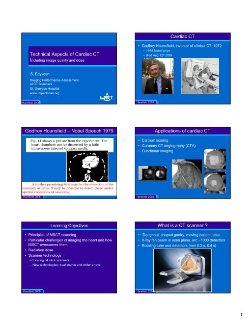
Cardiac <strong>CT</strong><br />
Technical Aspects of Cardiac <strong>CT</strong><br />
Including image quality and dose<br />
• Godfrey Hounsfield, inventor of clinical <strong>CT</strong>, 1972<br />
– 1979 Nobel prize<br />
– died Aug 12 th 2004<br />
S. Edyvean<br />
Imaging Performance Assessment<br />
of <strong>CT</strong> <strong>Scanner</strong>s<br />
St. Georges Hospital<br />
www.impactscan.org<br />
Harefield 2008<br />
<strong>ImPA<strong>CT</strong></strong><br />
Harefield 2008<br />
Godfrey Hounsfield – Nobel Speech 1979<br />
Fig. 14 shows a picture from the experiment. The<br />
heart chambers can be discerned by a little<br />
intravenous injected contrast media.<br />
Applications of cardiac <strong>CT</strong><br />
• Calcium scoring<br />
• Coronary <strong>CT</strong> angiography (<strong>CT</strong>A)<br />
• Functional imaging<br />
A further promising field may be the detection of the<br />
coronary arteries. It may be possible to detect these under<br />
special conditions of scanning.<br />
Harefield 2008<br />
Harefield 2008<br />
Learning Objectives<br />
• Principles of MS<strong>CT</strong> scanning<br />
• Particular challenges of imaging the heart and how<br />
MS<strong>CT</strong> overcomes them<br />
• Radiation dose<br />
• <strong>Scanner</strong> technology<br />
– Existing 64 slice scanners<br />
– New technologies: dual source and wider arrays<br />
What is a <strong>CT</strong> scanner ?<br />
• ‘Doughnut’ shaped gantry, moving patient table<br />
• X-fay fan beam in scan plane, arc ~1000 detectors<br />
• Rotating tube and detectors (min 0.3 s, 0.4 s)<br />
Harefield 2008<br />
Harefield 2008<br />
1
Side View of <strong>CT</strong> <strong>Scanner</strong><br />
• Narrow wider in z-direction<br />
Side View of <strong>CT</strong> <strong>Scanner</strong><br />
• Narrow wider in z-direction<br />
X-ray tube<br />
Detector array<br />
x-axis<br />
Sub-mm detectors<br />
Full extent of detector matrix<br />
z-axis<br />
Harefield 2008<br />
Harefield 2008<br />
40 mm<br />
Projections<br />
• measure attenuation<br />
– Each detector, each rotation angle<br />
• Image reconstructed<br />
– Filtered backprojection<br />
90°<br />
– More complex<br />
• 16, 64 slice<br />
detector position<br />
<strong>CT</strong> imaging requirements<br />
• Opposing projections provide the same information<br />
• To reconstruct images<br />
– Only 180° (+ fan angle) of scan data is required<br />
0°<br />
attenuation<br />
Harefield 2008<br />
attenuation<br />
~1000 positions<br />
~1000 projections<br />
attenuation<br />
360°<br />
0°<br />
detector position<br />
attenuation<br />
Harefield 2008<br />
360°<br />
Power<br />
Data<br />
Multi-slice <strong>CT</strong> <strong>Scanner</strong>s<br />
• Axial acquisition (‘step and shoot’)<br />
• Many simultaneous images<br />
Multi-Slice <strong>CT</strong><br />
• Helical scanning – volume set of data<br />
• Attenuation data interpolated to image slice position required<br />
40 mm<br />
40 mm<br />
Harefield 2008<br />
Harefield 2008<br />
2
Helical MS<strong>CT</strong> Scanning<br />
• Projection data interpolated to required image position<br />
• A number of detectors contribute to an image<br />
• Complex 3-D reconstruction for MS<strong>CT</strong><br />
Multi-Slice <strong>CT</strong> scanners<br />
• Beam width switched<br />
• Detector signals grouped<br />
– eg GE LightSpeed 16<br />
16 x 0.625 mm<br />
16 x 1.25 mm<br />
z-axis<br />
4 x 5 mm<br />
4 x 1.25 16 x 0.625 4 x 1.25<br />
Harefield 2008<br />
Recon position<br />
Harefield 2008<br />
20 mm<br />
Definition of Pitch<br />
Learning Objectives<br />
Pitch x = table travel in one rotation<br />
total acquisition width<br />
Pitch x = D rot = 1.25<br />
X<br />
x<br />
D rot<br />
• Principles of MS<strong>CT</strong> scanning<br />
• Particular challenges of imaging the heart and how<br />
MS<strong>CT</strong> overcomes them<br />
• Radiation dose<br />
• <strong>Scanner</strong> technology<br />
– Existing 64 slice scanners<br />
– New technologies: dual source and wider arrays<br />
In cardiac <strong>CT</strong> use a pitch ~0.2<br />
x<br />
Harefield 2008<br />
Harefield 2008<br />
Challenges of the heart<br />
• Spatial resolution<br />
• Low contrast resolution<br />
• Coverage<br />
– In one breath hold<br />
• Temporal resolution<br />
– Freezing the motion of the heart<br />
– Gating techniques for registration<br />
– Sector reconstruction<br />
The Heart – Spatial Resolution<br />
• Tortuous vessels narrowing to < 1 mm<br />
• Require good 3-D spatial resolution
<strong>CT</strong> Image<br />
• Grey level represents the average attenuation of<br />
the 3-D volume element<br />
• 3-D resolution of image<br />
– in-plane<br />
– slice thickness<br />
slice<br />
width<br />
MS<strong>CT</strong><strong>Scanner</strong>s - Spatial Resolution<br />
• 3-D resolution (scan plane, z) determined by<br />
– Detector size<br />
– Sampling<br />
– Reconstruction parameters<br />
• For isotropic resolution - limit is in the z-axis<br />
In plane resolution<br />
= ~0.3 mm<br />
Y<br />
X<br />
In plane resolution = ~0.3 mm<br />
Harefield 2008<br />
Harefield 2008<br />
Z<br />
MS<strong>CT</strong><strong>Scanner</strong>s - Spatial Resolution<br />
• Many thin slices of data<br />
– eg 4, 16, 32, 64 slices x 0.5, 0.625 mm<br />
Z-axis Spatial Resolution – Sampling<br />
• Improved z-sampling with optimised pitches<br />
– By interleaving opposing projections<br />
X-ray tube<br />
Detector array<br />
Pitch = 1 Pitch = 0.875<br />
x-axis<br />
Harefield 2008<br />
Sub-mm detectors<br />
Full extent of detector matrix<br />
z-axis<br />
Harefield 2008<br />
Z-axis Spatial Resolution – Sampling<br />
• 64-Slice <strong>CT</strong>: double z-Sampling: Overlap doubles information<br />
3D Spatial Resolution<br />
• Isotropic resolution: equal in x, y & z<br />
0,6 mm<br />
Oversampling<br />
0,6 mm<br />
Z<br />
0.3 x 0.3 x 0.3 mm<br />
Sampling distance 0.3 mm<br />
Harefield 2008<br />
32 Slice Detector<br />
64 Slice DAS<br />
Courtesy Siemens<br />
Harefield 2008<br />
Courtesy Siemens Medical Solutions<br />
4
The Heart – Low Contrast Resolution<br />
• Low contrast resolution (non-calcified plaque)<br />
– require noise levels equivalent to current <strong>CT</strong> imaging<br />
Higher mAs<br />
Challenges of the heart<br />
• Spatial resolution<br />
• Low contrast resolution<br />
• Coverage<br />
– In one breath hold<br />
• Temporal resolution<br />
– Freezing the motion of the heart<br />
– Gating techniques for registration<br />
– Sector reconstruction<br />
L – Vessel Lumen<br />
N – Non-calcified plaque<br />
C – Calcified plaque<br />
Harefield 2008<br />
Lower noise<br />
Harefield 2008<br />
The Heart<br />
• Approximately 12 cm in length<br />
– Existing technology<br />
• 4 slice < 20 mm<br />
• 16 slice 20 – 32 mm<br />
• 64 slice ~30 – 40 mm<br />
• Coverage single breath hold<br />
– Limited in four slice scanners<br />
– Heart rate not so stable with longer breath holds<br />
• For cardiac <strong>CT</strong>A need overlapping pitch (0.2 - 0.3)<br />
64 slice<br />
Volume coverage<br />
16 x 0.625 mm 4 x 1.25 mm<br />
Harefield 2008<br />
10 mm 5 mm Harefield 2008<br />
Volume coverage – in one breath hold<br />
• Time to cover heart<br />
4 x 1 mm slice 16 x 1mm slices 64 x 0.5 mm slices<br />
The Heart<br />
• Beating at 60 – 120 bpm (1 – 2 bps)<br />
– Freeze motion<br />
– Registration of sections<br />
– Not necessarily repeatable – irregular heart rate<br />
– Image in as few beats as possible<br />
120 mm<br />
~48 sec ~12 sec ~6 sec<br />
0.5 s, 0.33 pitch<br />
ECG<br />
Harefield 2008<br />
Harefield 2008<br />
5
Cardiac Motion<br />
Cardiac Motion and the ECG<br />
• Imaging 'window' during period of least cardiac motion<br />
– Eg ~ 70% of R-R<br />
– Width between 10-20% of R-R<br />
70%<br />
phase<br />
R<br />
R<br />
ECG<br />
width 15%<br />
t<br />
Cardiac<br />
motion<br />
Harefield 2008<br />
Harefield 2008<br />
Cardiac Motion - Temporal resolution<br />
ECG Gating Techniques<br />
• Heart rate 60 -120 bpm ie 1 -2 bps<br />
• Temporal resolution required ~10 % of R-R interval<br />
– eg 60 bpm = 100 ms, 120 bpm = 60 ms<br />
• Half rotation of data needed for image reconstruction<br />
– Temporal resolution = ½ rotation time<br />
300 ms rotation R – R<br />
~ 1000 ms<br />
required<br />
temporal<br />
resolution<br />
150 ms<br />
Harefield 2008<br />
60 bpm<br />
~ 100 ms<br />
Harefield 2008<br />
Cardiac Motion - Registration<br />
• Need gating techniques to avoid misregistration<br />
64 slice<br />
64 slice<br />
ECG Gating Techniques<br />
• Prospective ECG gating (of irradiation)<br />
– Sequential (‘step and shoot’) scanning<br />
– Calcium scoring, <strong>CT</strong>A (newer scanners)<br />
• Retrospective ECG gating (of acquired data)<br />
– Helical scanning<br />
– <strong>CT</strong>A, Functional imaging<br />
axial scans,<br />
prospective gating<br />
of irradiation<br />
helical scan<br />
retrospective<br />
gating of data<br />
Sequential, ‘step and shoot’<br />
prospective gating<br />
helical<br />
retrospective gating<br />
Z<br />
Harefield 2008<br />
Harefield 2008<br />
6
Prospectively gated cardiac <strong>CT</strong><br />
• Axial - ‘step and shoot’<br />
Prospective Gating<br />
• Unsuitable for high heart rates<br />
– Limited to ½ rotation time<br />
• Niche for calcium scoring – Agatston, mass<br />
• Beginning to be used for <strong>CT</strong>A<br />
• Better coverage with 64 narrow slice scanners<br />
Harefield 2008<br />
Harefield 2008<br />
Retrospective gating<br />
• Helical scan<br />
– Volume set of data acquired<br />
– Reconstructions can be made at any phase<br />
Z<br />
R – R<br />
~ 1000 ms<br />
Retrospective Gating<br />
• ECG-gated images reconstructed<br />
– choose optimal cardiac phase for coronary angiography<br />
• Diastole, systole<br />
– multiple phases for functional studies<br />
• Temporal resolution limited by rotation time<br />
– Multi-sector reconstruction improves this<br />
400 ms rotation<br />
200 ms<br />
R – R<br />
~ 1000 ms<br />
‘temporal<br />
resolution’<br />
Harefield 2008<br />
Harefield 2008<br />
Single-sector reconstruction<br />
• Single sector<br />
– Data used from 180°<br />
– Sector time window = ½ rotation time<br />
• eg 0.5 sec rotation (500 ms), sector = 0.25 s (250 ms)<br />
Multi-sector reconstruction<br />
• Two sector<br />
– Two sectors each of 90 °<br />
• Sector time = ¼ rotation, eg = 125 ms<br />
– data from 1 ½ rotations, in two heart beats<br />
250 ms<br />
~250 ms<br />
125 ms<br />
‘temporal<br />
resolution’<br />
Harefield 2008<br />
acquisition<br />
Harefield 2008<br />
Time for 1 rotation + 1 sector<br />
Time<br />
same z-axis position<br />
7
• 3-sector (83 ms)<br />
Multi-sector reconstruction<br />
Multi-sector reconstruction<br />
• If heart R-R matches scan time<br />
• Synchronisation<br />
– Two sector<br />
360° plus sector θ<br />
• 4-sector (~63 ms)<br />
Time for 1 rotation<br />
Harefield 2008<br />
360° plus sector θ<br />
Harefield 2008<br />
Time<br />
Pitch<br />
• Require an overlapping pitch (~0.2)<br />
– To ensure detectors cover image<br />
position for every sector<br />
Pitch<br />
• More sectors the lower the pitch<br />
Harefield 2008<br />
Harefield 2008<br />
time<br />
Multi-sector reconstruction<br />
2 Cycles 3 Cycles<br />
Multi-sector reconstruction<br />
• Require steady heart beat for good registration<br />
Harefield 2008<br />
Courtesy Philips<br />
Harefield 2008<br />
8
Multi-sector reconstruction<br />
• Minimum rotation time important<br />
• Sector time = ‘temporal resolution’<br />
– eg Two sectors halve the time window<br />
• More sectors require more beats<br />
– lower pitch<br />
– Require steady heart beat for good registration<br />
• Avoid synchronisation<br />
Temporal resolution graph – multi-sector<br />
• Complex relationship of heart rate, temporal<br />
resolution, and scan time<br />
Sector window (ms)<br />
Mulitple sectors of<br />
equal length<br />
single sector time window<br />
Harefield 2008<br />
Harefield 2008<br />
Heart rate<br />
Number of segments used<br />
Learning Objectives<br />
• Number of sectors available<br />
• Automatic selection to varying degrees<br />
No of<br />
sectors<br />
IGE<br />
1, 2, 4<br />
Philips<br />
Up to 5<br />
Siemens(<br />
1 tube)<br />
1 or 2<br />
Siemens<br />
(2 tube)<br />
1 or 2<br />
Toshiba<br />
Up to 5<br />
• Principles of MS<strong>CT</strong> scanning<br />
• Particular challenges of imaging the heart and how<br />
MS<strong>CT</strong> overcomes them<br />
• Radiation dose<br />
• <strong>Scanner</strong> technology<br />
– Existing 64 slice scanners<br />
– New technologies: dual source and wider arrays<br />
Harefield 2008<br />
360° plus sector<br />
θ<br />
Harefield 2008<br />
Dosimetry<br />
Dosimetry<br />
• Dose parameters used in <strong>CT</strong><br />
– Effective dose<br />
– <strong>CT</strong>DI and DLP<br />
• Typical dose values<br />
– Comparison with other examinations and modalities<br />
• Dose Saving techniques<br />
– Increased pitch<br />
– ECG dose modulation<br />
– Special beam shaping filters<br />
Harefield 2008<br />
absorbed dose:<br />
organ dose :<br />
effective dose:<br />
Harefield 2008<br />
f 1 + f 2 + f 3<br />
- localised dose (mGy)<br />
- localised dose averaged over the<br />
organ (mGy)<br />
- sum of all the average organ doses<br />
- modified by tissue radio-sensitivity<br />
factors<br />
- describes risk (mSieverts)<br />
- Measure of total amount<br />
of radiation given<br />
9
• Pitch doubled<br />
– Organ dose halved<br />
– Effective dose halved<br />
Dose<br />
Cardiac Scanning<br />
• High dose<br />
– overlapping pitch<br />
– lot of wasted information if only reconstructing one phase<br />
x<br />
x<br />
Harefield 2008<br />
Harefield 2008<br />
Dose parameters used in <strong>CT</strong><br />
• <strong>CT</strong>DI vol and DLP are indicated on scanner<br />
Dose parameters used in <strong>CT</strong><br />
• <strong>CT</strong>DIvol<br />
– Average dose to perspex phantom of standard size<br />
– 32 cm (body), 16 cm (head)<br />
– Indication of effect of exposure settings (kV, mAs, pitch)<br />
– Rough indication of average organ dose<br />
– ‘Standard size patient’<br />
average<br />
dose<br />
<strong>CT</strong>DI vol<br />
2.28mGy<br />
(not a cardiac scan)<br />
Harefield 2008<br />
Harefield 2008<br />
Dose parameters used in <strong>CT</strong><br />
Dose parameters used in <strong>CT</strong><br />
• DLP = <strong>CT</strong>DI x L<br />
– Average dose x scan length<br />
– Includes the effect of the length irradiated (total irradiation)<br />
– Rough indication of effective dose<br />
– Can use a conversion factor to convert to ED<br />
average<br />
dose<br />
<strong>CT</strong>DI vol (mGy)<br />
• Typical <strong>CT</strong>DI<br />
– From manufacturers suggested protocols<br />
60<br />
50<br />
40<br />
30<br />
20<br />
10<br />
52<br />
3<br />
19<br />
56<br />
7<br />
17<br />
55<br />
3<br />
13<br />
16<br />
<strong>CT</strong>A<br />
55<br />
Ca Score (ax)<br />
Ca score (hel)<br />
Abdo<br />
18<br />
9<br />
Exam <strong>CT</strong>DI (mGy)<br />
Abdo<br />
54<br />
Ca (axial)<br />
Mean Values<br />
15<br />
3<br />
Ca (helical) 13<br />
<strong>CT</strong>A 54<br />
15<br />
13<br />
3<br />
Harefield 2008<br />
L<br />
0<br />
Harefield 2008 A B C D mean<br />
10
Effective Doses<br />
• From manufacturers suggested protocols<br />
Abdo<br />
Ca score<br />
(hel)<br />
Ca Score (ax)<br />
<strong>CT</strong>A<br />
0.8<br />
4.5<br />
5.5<br />
16.6<br />
0.0 5.0 10.0 15.0 20.0 25.0<br />
Effective dose (mSv)<br />
Effective Doses<br />
• Cardiac <strong>CT</strong> radiation doses are relatively high.<br />
• Ball park figures (dependent on technique etc)<br />
• In practice cardiac <strong>CT</strong>A figures often higher<br />
Technique<br />
<strong>CT</strong> angiography<br />
planar coronary angiography<br />
SPE<strong>CT</strong><br />
Approximate Dose<br />
(mSv)<br />
10 – 15 +<br />
PET 82 Rb<br />
5<br />
PET 13 NH 3 2<br />
5<br />
10<br />
Harefield 2008<br />
Harefield 2008<br />
ECG Tube Current Modulation<br />
• mA reduced outside of required recon. phase by ~ 80%<br />
– Claim net dose savings ~ 50%<br />
• Best for regular heart rates. Better for slow.<br />
• Can be automatically deactivated if ECG changes<br />
• Can only image one phase<br />
• Can’t use normal dose modulation systems with ECG mod.<br />
Beam Shaping Filters<br />
• Beam shaping filters more appropriate for small fov<br />
reconstruction within a larger fov<br />
– Eg Toshiba use ‘small fov’ filter, GE use ‘head’ filter<br />
• Can reduce mA by ~ 15 – 20%<br />
100%<br />
20%<br />
Harefield 2008<br />
Harefield 2008<br />
Dynamic Collimation<br />
• In helical scanning extra rotations are needed at<br />
end of imaged volume<br />
– Significant extra dose: wide beam widths and short scans<br />
• Dynamic collimation - collimator blades open and<br />
close asymetrically at start and end of scan<br />
Prospective Gating of <strong>CT</strong>A<br />
• Prospectively gated coronary <strong>CT</strong> angiography<br />
– GE: SnapShot Pulse<br />
– Philips: Step and Shoot<br />
• Possible with scanners large, thin slice coverage<br />
Scan range<br />
Scan range<br />
Harefield 2008<br />
Conventional technology<br />
without Dose Shield<br />
The SOMATOM Definition AS is pending 510(k) review and is not yet commercially available in the US.<br />
AS+ is a configuration of the SOMATOM Definition AS<br />
SOMATOM Definition AS+ with<br />
Adaptive Dose Shield<br />
Courtesy Siemens Medical Systems Harefield 2008<br />
Courtesy GE Healthcare<br />
11
Retrospective Gating of <strong>CT</strong>A<br />
• Retrospectively gated C<strong>CT</strong>A<br />
~15 mSv<br />
Prospective Gating of <strong>CT</strong>A<br />
• Prospectively gated C<strong>CT</strong>A<br />
~ 1 mSv<br />
100%<br />
Harefield 2008<br />
20%<br />
Harefield 2008<br />
Learning Objectives<br />
Current 64 Slice <strong>CT</strong> <strong>Scanner</strong>s<br />
• Principles of MS<strong>CT</strong> scanning<br />
• Particular challenges of imaging the heart and how<br />
MS<strong>CT</strong> overcomes them<br />
• Radiation dose<br />
• <strong>Scanner</strong> technology<br />
– Existing 64 slice scanners<br />
– New technologies: dual source and wider arrays<br />
• Coverage<br />
– Beam widths 20 – 40 mm<br />
• Narrow slices<br />
– 0.5 – 0.625 mm detectors<br />
• Speed of rotation<br />
– 0.3 – 0.4s<br />
• Ability to do multi-sector reconstruction<br />
– 2-5 sectors<br />
Z<br />
Harefield 2008<br />
Harefield 2008<br />
PowerData<br />
Harefield 2008<br />
64 Slice Systems<br />
• GE LightSpeed 32, 64<br />
– 64 x 0.625 (40 mm total)<br />
• Philips Brilliance 40, 64<br />
– 64 x 0.625 (40 mm total)<br />
• Siemens Sensation 64<br />
– 32 x 0.6 (19.2 mm) and 8 x 1.2 (28.8 mm total) (double sampled)<br />
• Toshiba Aquilion 32, 64<br />
– 64 x 0.5, 32 mm<br />
GE, Philips<br />
Siemens<br />
Toshiba<br />
40 mm<br />
28.8 mm<br />
32 mm<br />
• Depends on<br />
– pitch, rotation time, detector acquisition length<br />
64 slice scanners<br />
Acquisition width<br />
Min rotation<br />
times (s)<br />
Detector length<br />
(mm)<br />
Time to cover<br />
120 mm ^ (s)<br />
Harefield 2008<br />
Time to cover heart<br />
IGE<br />
0.625<br />
0.35<br />
40<br />
5.3<br />
Philips<br />
0.625<br />
0.42<br />
40<br />
6.3<br />
^assume pitch 0.2<br />
Siemens<br />
(1 tube)<br />
0.6<br />
0.33<br />
19.2<br />
10.3<br />
Siemens<br />
(2 tube)<br />
0.6<br />
0.33<br />
19.2<br />
5.1<br />
Toshiba<br />
0.5<br />
0.4<br />
32<br />
7.5<br />
12
Number of segments used<br />
New Technologies<br />
• Number of sectors available<br />
• Automatic selection to varying degrees<br />
IGE<br />
Philips<br />
Siemens(<br />
1 tube)<br />
Siemens<br />
(2 tube)<br />
Toshiba<br />
• Siemens Dual Tube<br />
• Toshiba Aquilion One (160 mm coverage)<br />
• Philips Brilliance i<strong>CT</strong> (80 mm coverage)<br />
No of<br />
sectors<br />
1, 2, 4<br />
Up to 5<br />
1 or 2<br />
1 or 2<br />
Up to 5<br />
One<br />
rotation<br />
Harefield 2008<br />
360° plus sector<br />
θ<br />
Harefield 2008<br />
Siemens Definition Dual Tube<br />
• Siemens Definition launched RSNA '05<br />
• Two tubes at 90°<br />
– 2 x 1/4 scans in 83 ms<br />
Siemens Definition Dual Tube<br />
Temporal resolution of 83 ms<br />
Harefield 2008<br />
Temporal Resolution =<br />
Rotation Time<br />
4<br />
Harefield 2008<br />
= 83 ms<br />
Courtesy Siemens<br />
Siemens Definition Dual Tube<br />
• Half temporal resolution (½ of ½ rotation time)<br />
– Single segment (83 ms) or dual segment (42 ms)<br />
• Array<br />
– 28.8 mm for 1.2 mm slices<br />
– 19.2 mm for 0.6 mm (z-axis flying fs => 0.3 mm)<br />
• Claim no beta blockers and any heart rate<br />
– Applies to single segment recon<br />
• Faster table speeds possible<br />
– only need to ‘hang around’ for one or two segments<br />
• Dual energy imaging<br />
Toshiba Aquilion One<br />
• 320 x 0.5 mm = 160 mm coverage<br />
Harefield 2008<br />
Harefield 2008<br />
Courtesy of Toshiba<br />
13
Toshiba AquilionOne<br />
• Allow whole organ coverage in single rotation<br />
256 row detector (256 x 0.5)<br />
8 cm coverage<br />
Philips Brilliance i<strong>CT</strong><br />
One<br />
rotation<br />
Z - axis<br />
Aquilion 64 detector<br />
Harefield 2008<br />
Detector mock-ups courtesy of Toshiba<br />
Nano-Panel<br />
Harefield 2008<br />
RSNA 2005<br />
Philips MD<strong>CT</strong><br />
Courtesy of Philips<br />
Larger Detector Arrays<br />
Larger Detector Arrays<br />
• Cover more of heart (or all) in one go<br />
• Axial rotation ‘step and shoot’<br />
• Single segment ‘one shot’<br />
– fastest image acquisition = ~half rotation time<br />
• Dual segment<br />
– Better temporal resolution (with stable beat)<br />
– Two rotations -> higher dose<br />
• Much lower dose than helical, retrospective gating<br />
– 3 – 5 mSv<br />
• Only one phase of heart cycle (+/- 1/3 rotation time)<br />
– Can’t do functional unless irradiate many times<br />
<strong>Scanner</strong><br />
Slices<br />
Array length<br />
(Coverage)<br />
Rotation time<br />
Temporal<br />
resolution for 1<br />
segment (ms)<br />
Aquilion<br />
One<br />
320<br />
160 mm<br />
(0.5 mm)<br />
0.35<br />
180<br />
Philips<br />
Brilliance<br />
i<strong>CT</strong><br />
256 (128)<br />
80 mm<br />
(0.625<br />
mm)<br />
0.27<br />
130<br />
Siemens<br />
Definition<br />
AS+<br />
(128 slices)<br />
128 (64)<br />
38 mm<br />
(64 x 0.6 mm)<br />
0.3<br />
160<br />
Siemens Definition<br />
Dual tube<br />
64 (32), 24<br />
19. 2 mm (0.6 mm)<br />
28.8 mm (1.2 mm)<br />
0.33<br />
83<br />
Harefield 2008<br />
Harefield 2008<br />
Learning Objectives<br />
• Principles of MS<strong>CT</strong> scanning<br />
• Particular challenges of imaging the heart and how<br />
MS<strong>CT</strong> overcomes them<br />
• Radiation dose<br />
• <strong>Scanner</strong> technology<br />
– Existing 64 slice scanners<br />
– New technologies: dual source and wider arrays<br />
Harefield 2008<br />
Technical aspects of Cardiac <strong>CT</strong><br />
S. Edyvean<br />
Imaging Performance Assessment<br />
of <strong>CT</strong> <strong>Scanner</strong>s<br />
St. Georges Hospital<br />
www.impactscan.org<br />
Harefield 2008<br />
<strong>ImPA<strong>CT</strong></strong><br />
14
<strong>Scanner</strong> options to improve image quality<br />
• Before the scan<br />
– Monitor pre-scan heart beat<br />
– Automatic determination of pitch, rotation time, number of<br />
sectors<br />
• After the scan<br />
– ECG editing<br />
– Motion maps to determine optimum phase<br />
Harefield 2008<br />
15