Evaluation of the Antimicrobial and Cytotoxic Activity of Epiphany ...
Evaluation of the Antimicrobial and Cytotoxic Activity of Epiphany ...
Evaluation of the Antimicrobial and Cytotoxic Activity of Epiphany ...

Create successful ePaper yourself
Turn your PDF publications into a flip-book with our unique Google optimized e-Paper software.
Egyptian Journal <strong>of</strong> Medical Microbiology, January 2007 Vol. 16, No. 1<br />
<strong>Evaluation</strong> <strong>of</strong> <strong>the</strong> <strong>Antimicrobial</strong> <strong>and</strong> <strong>Cytotoxic</strong> <strong>Activity</strong> <strong>of</strong> <strong>Epiphany</strong><br />
Root Canal Sealer in Vitro<br />
Amany E. Badr 1 , Amro A Abdul Razek 2 , Nesrene S. Omar 3 , <strong>and</strong> Farid A.Badria 4<br />
1 Lecturer <strong>of</strong> Endodontics, Conservative Dentistry Department, 2 Assistant Pr<strong>of</strong>essor <strong>of</strong> Endodontics,<br />
Conservative Dentistry Department, 3 Associate Pr<strong>of</strong>essor <strong>of</strong> Medical Microbiology <strong>and</strong> Immunology,<br />
Faculty <strong>of</strong> Medicine, Mansoura University, 4 Pr<strong>of</strong>essor <strong>of</strong> Pharmacognosy , Faculty <strong>of</strong> Pharmacy,<br />
Mansoura University<br />
This study evaluated <strong>the</strong> antimicrobial <strong>and</strong> cytotoxic effect <strong>of</strong> <strong>Epiphany</strong> root canal sealer<br />
versus commonly used sealers; EndoFill, Apexit, Ketac-Endo, <strong>and</strong> AH-26. The first portion <strong>of</strong> this<br />
study used four different microorganisms; Enterococcus faecalis (E.faecalis), staphylococcus aureus<br />
(S. aureus), Ecshericia coli (E.coli) <strong>and</strong> C<strong>and</strong>ida albican (C. albican) to determine <strong>the</strong> antimicrobial<br />
effect using <strong>the</strong> agar-well diffusion method. Secondly, In vitro <strong>Cytotoxic</strong>ity assay using human<br />
periodontal ligament cell culture was used to evaluate <strong>the</strong> cytotoxicity <strong>of</strong> <strong>Epiphany</strong> sealer compared<br />
to <strong>the</strong> o<strong>the</strong>r four sealers. The cell cultures were incubated for 24h with freshly mixed material <strong>and</strong><br />
after 24 h <strong>of</strong> setting. The viability <strong>of</strong> <strong>the</strong> fibroblast cells was determined using trypan blue. The<br />
results <strong>of</strong> this comparative study indicated that <strong>Epiphany</strong> sealer exhibited a potential antibacterial<br />
activity with no antic<strong>and</strong>ida effect. The cytotoxicity <strong>of</strong> <strong>Epiphany</strong> was comparable to that <strong>of</strong><br />
EndoFill <strong>and</strong> Apexit, however it was significantly less toxic than Ketac-Endo, <strong>and</strong> AH-26.<br />
INTRODUCTION<br />
The role <strong>of</strong> bacteria <strong>and</strong> <strong>the</strong>ir byproducts<br />
in <strong>the</strong> initiation <strong>and</strong> perpetuation <strong>of</strong><br />
pulp <strong>and</strong> periapical diseases has been well<br />
established. Most infecting bacteria, toge<strong>the</strong>r<br />
with <strong>the</strong>ir principal substrate <strong>of</strong> necrotic pulp<br />
debris, may be removed by routine<br />
endodontic procedures such as<br />
instrumentation, irrigation, <strong>and</strong> <strong>the</strong> use <strong>of</strong> an<br />
intracanal medicament with antimicrobial<br />
activity 1 . However, infection may persist in<br />
canals with high anatomic complexity, with a<br />
great number <strong>of</strong> bacteria, especially<br />
facultative anaerobic bacteria 2 . In addition,<br />
several studies have associated <strong>the</strong> presence<br />
<strong>of</strong> fungi with <strong>the</strong>rapy-resistant endodontic<br />
infections 3 .<br />
After <strong>the</strong> microbial control phase <strong>of</strong><br />
endodontic <strong>the</strong>rapy, a root canal filling is<br />
placed to seal <strong>the</strong> root canal system from <strong>the</strong><br />
external environment 3. The st<strong>and</strong>ard method<br />
<strong>of</strong> obturation <strong>of</strong> <strong>the</strong> root canal space is by<br />
using a core material in combination with a<br />
root canal sealer 4 . <strong>Antimicrobial</strong> activity<br />
plays an important role in <strong>the</strong> efficacy <strong>of</strong> an<br />
endodontic sealer during root canal filling,<br />
<strong>and</strong> for this reason many studies have dealt<br />
with <strong>the</strong> antibacterial activity <strong>of</strong> endodontic<br />
sealers 5 .<br />
Facultative microorganisms such as<br />
E. faecalis, E.coli <strong>and</strong> S.aureus <strong>and</strong> even<br />
C.albicans have been considered to be <strong>the</strong><br />
most resistant species in <strong>the</strong> oral cavity <strong>and</strong><br />
possible cause <strong>of</strong> failure <strong>of</strong> root canal<br />
treatment 6 . The agar diffusion method has<br />
been widely used to test <strong>the</strong> antimicrobial<br />
activity <strong>of</strong> dental materials <strong>and</strong> medications,<br />
<strong>the</strong> advantage <strong>of</strong> this method is that it allows<br />
direct comparisons <strong>of</strong> root canal sealers<br />
against <strong>the</strong> test microorganisms, indicating<br />
which sealer has <strong>the</strong> potential to eliminate<br />
bacteria in <strong>the</strong> local microenvironment <strong>of</strong> <strong>the</strong><br />
root canal system 7 .<br />
Good tissue compatibility is<br />
decisive for root canal sealers because <strong>the</strong>y<br />
may come into direct contact with tissues<br />
specially when extruded to <strong>the</strong> apical area 8 .<br />
Toxicity testing <strong>of</strong> dental materials can be<br />
assessed ei<strong>the</strong>r in vitro or in vivo. Using cell<br />
lines is a common method <strong>of</strong> testing dental<br />
materials that allows for a simple,<br />
reproducible result that can be controlled in a<br />
laboratory setting. In vitro testing also allows<br />
for <strong>the</strong> comparison between several materials<br />
using <strong>the</strong> same cells under <strong>the</strong> same<br />
conditions 9 .<br />
Improvements in adhesive<br />
technology have fostered attempts to reduce<br />
apical <strong>and</strong> coronal leakage by bonding to<br />
canal walls 10 . These improvements rely on<br />
<strong>the</strong> incorporation <strong>of</strong> resin monomers into <strong>the</strong><br />
sealer or application <strong>of</strong> resins during a distinct<br />
conditioning step. O<strong>the</strong>r strategies have<br />
focused on substitutes for gutta percha that<br />
bond to <strong>the</strong> root dentin, <strong>the</strong>reby establishing a<br />
so called monoblock-obturation 11 .<br />
95
Egyptian Journal <strong>of</strong> Medical Microbiology, January 2007 Vol. 16, No. 1<br />
The <strong>Epiphany</strong> obturating system<br />
(Pentron Clinical Technologies, Wallingford,<br />
CT) which is a dual curable dental resin<br />
composite sealer uses Resilon points <strong>and</strong> is<br />
bonded to <strong>the</strong> root dentin 12 . Resilon (Resilon<br />
Research LLC, Madison, CT), a <strong>the</strong>rmoplastic<br />
syn<strong>the</strong>tic polymer based root canal filling<br />
material, has been developed that performs<br />
like gutta-percha, has <strong>the</strong> same h<strong>and</strong>ling<br />
properties, <strong>and</strong> for retreatment purposes may<br />
be s<strong>of</strong>tened with heat or dissolved with<br />
solvents like chlor<strong>of</strong>orm. Based on polymers<br />
<strong>of</strong> polyester, Resilon contains bioactive glass,<br />
bismuth oxychloride <strong>and</strong> barium sulfate 12 .<br />
Resilon associated with <strong>Epiphany</strong> has<br />
recently been introduced. Some <strong>of</strong> its<br />
mechanical <strong>and</strong> chemical properties have<br />
been evaluated with controversial results 11, 13 .<br />
In contrast, <strong>the</strong> biological properties <strong>of</strong><br />
Resilon <strong>and</strong> <strong>Epiphany</strong> are not well<br />
documented 14 .<br />
The properties <strong>of</strong> root canal<br />
cements can be divided into physicochemical,<br />
antimicrobial <strong>and</strong> biological 15 . Therefore, <strong>the</strong><br />
aim <strong>of</strong> this study was to in vitro evaluate <strong>the</strong><br />
antimicrobial <strong>and</strong> cytotoxic properties <strong>of</strong><br />
<strong>Epiphany</strong> sealer in comparison with <strong>the</strong><br />
commonly used root canal sealers.<br />
MATERIALS AND METHODS<br />
The various root canal sealers that<br />
were tested in <strong>the</strong> present study are <strong>the</strong><br />
adhesive resin root canal sealer (<strong>Epiphany</strong>)†,<br />
ZOE based sealer (EndoFill)‡, calcium<br />
hydroxide based sealer (Apexit)*, glass<br />
ionomer based sealer (Ketac-Endo)§ <strong>and</strong><br />
Resin sealer (AH-26) ‡.<br />
†Pentron clinical technologies<br />
‡DentSply<br />
*Ivoclar Vivadent<br />
§3M ESPE<br />
<strong>Antimicrobial</strong> test<br />
Agar-well diffusion method (AWDM):<br />
Cultures <strong>of</strong> E. faecalis , S. aureus,<br />
E.coli <strong>and</strong> C. albican were obtained from<br />
endodontic isolates. All microorganisms<br />
were previously subcultured in appropriate<br />
culture plates <strong>and</strong> under gaseous conditions<br />
to confirm <strong>the</strong>ir purity. From <strong>the</strong> broth<br />
culture suspensions <strong>of</strong> <strong>the</strong> tested<br />
microorganism 0.2 ml were prepared<br />
adjusted to No. 0.5 McFarl<strong>and</strong> scale were<br />
spread on four Petri dishes containing<br />
Mueller-Hinton Agar medium. Five wells <strong>of</strong><br />
5mm depth <strong>and</strong> 4mm diameter were<br />
punched in <strong>the</strong> agar plates <strong>and</strong> filled with <strong>the</strong><br />
test sealers (mixed according to<br />
manufacturer instructions). The plates were<br />
incubated aerobically at 37Cº for 48h. The<br />
diameters <strong>of</strong> <strong>the</strong> zones <strong>of</strong> microbial<br />
inhibition around each well were measured<br />
in millimeters after <strong>the</strong> incubation period.<br />
The inhibitory zone was considered to be <strong>the</strong><br />
shortest diameter from <strong>the</strong> outer margin <strong>of</strong><br />
<strong>the</strong> well to <strong>the</strong> initial point <strong>of</strong> <strong>the</strong> microbial<br />
growth.<br />
Three replicates were measured for<br />
each microorganism <strong>and</strong> a mean diameter was<br />
determined for each sealer. Greater diameters<br />
<strong>of</strong> zones <strong>of</strong> inhibition were interpreted to<br />
indicate greater antimicrobial activity <strong>of</strong> <strong>the</strong><br />
involved sealers 16, 17 .<br />
<strong>Cytotoxic</strong>ity assay<br />
Test materials <strong>and</strong> specimen preparation:<br />
The tested materials were mixed<br />
according to <strong>the</strong> manufacturer's instructions.<br />
Freshly mixed materials were filled in<br />
polyethylene rings <strong>of</strong> 5mm inside diameter<br />
<strong>and</strong> 4mm in height. The test was performed<br />
on fresh mix <strong>and</strong> 24h set mix. To prevent<br />
contamination <strong>of</strong> <strong>the</strong> set samples <strong>the</strong>y were<br />
exposed to UV light for 2h before performing<br />
<strong>the</strong> test. In a pilot study made previously 18 ,<br />
this period was sufficient to sterilize<br />
experimental materials.<br />
Cell Culture:<br />
Periodontal ligament cells (PDL)<br />
were obtained from teeth extracted for<br />
orthodontic purposes. Fibroblasts were grown<br />
in Dulbecco's modified Eagles medium<br />
(DMEM) supplemented with 10% fetal<br />
bovine serum <strong>and</strong> antibiotics (10,000 units <strong>of</strong><br />
penicillin-G/ml, 10 mg <strong>of</strong> streptomycin/ml<br />
<strong>and</strong> 20 mM <strong>of</strong> L-glutamine) 19 .<br />
Growth Measurement:<br />
The fibroblast cells were plated at a<br />
density <strong>of</strong> 3-4 x10 4 cells/ml <strong>and</strong> dispensed<br />
onto 96-well culture plate with 1ml <strong>of</strong><br />
medium per well <strong>and</strong> incubated at 37Cº<br />
supplemented with 5% CO 2 for 48h to allow<br />
96
Egyptian Journal <strong>of</strong> Medical Microbiology, January 2007 Vol. 16, No. 1<br />
attachment <strong>of</strong> <strong>the</strong> fibroblasts to <strong>the</strong> bottom <strong>of</strong><br />
<strong>the</strong> wells 20 . Specimens <strong>of</strong> different sealers<br />
were placed onto <strong>the</strong> wells <strong>of</strong> <strong>the</strong> culture plate<br />
<strong>and</strong> each specimen was covered by 100µl<br />
suspension <strong>of</strong> fibroblasts <strong>and</strong> incubated at<br />
37Cº with 5% CO 2 for 48h. At <strong>the</strong> end <strong>of</strong> <strong>the</strong><br />
incubation period, <strong>the</strong> culture medium was<br />
aspirated <strong>and</strong> 100µl <strong>of</strong> formaldehyde<br />
phosphate buffered saline (FPBS) was added<br />
to each culture well for 2h. FPBS was <strong>the</strong>n<br />
removed <strong>and</strong> each well was thoroughly rinsed<br />
with distilled water <strong>and</strong> 100µl <strong>of</strong> 0.25% <strong>of</strong><br />
trypan blue (wt/vol) was added to each well to<br />
stain <strong>the</strong> nonviable cells. After 3h, <strong>the</strong> trypan<br />
blue was removed <strong>and</strong> <strong>the</strong> cells were<br />
thoroughly rinsed with distilled water. The<br />
number <strong>of</strong> viable cells was expressed by<br />
averaging <strong>of</strong> six readings using an inverted<br />
microscope (Olympus, 1X71, Japan) 19 .<br />
Statistical analysis was performed by one way<br />
ANOVA, <strong>and</strong> Bonferroni tests.<br />
RESULTS<br />
Table 1 shows <strong>the</strong> mean <strong>of</strong> <strong>the</strong><br />
zones <strong>of</strong> inhibition <strong>of</strong> microbial growth <strong>of</strong><br />
each sealer against <strong>the</strong> microorganisms tested<br />
measured in mm <strong>and</strong> <strong>the</strong> average values <strong>of</strong> <strong>the</strong><br />
antimicrobial activity <strong>of</strong> each sealer against<br />
all microorganisms. EndoFill produced <strong>the</strong><br />
largest inhibitory zone followed by <strong>Epiphany</strong><br />
(by average values). On <strong>the</strong> o<strong>the</strong>r h<strong>and</strong>,<br />
Apexit produced <strong>the</strong> smallest inhibitory zones<br />
against <strong>the</strong> tested microorganisms (by average<br />
values). It appears that C<strong>and</strong>ida albican was<br />
<strong>the</strong> most resistant organism to <strong>the</strong> effect <strong>of</strong> <strong>the</strong><br />
sealers in this experiment. <strong>Epiphany</strong> had <strong>the</strong><br />
largest inhibitory zone on E.faecalis followed<br />
by S. aureus <strong>and</strong> E.coli. However, <strong>Epiphany</strong><br />
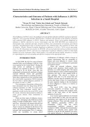
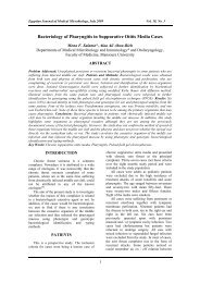
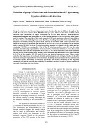
had no effect on C. albican. Figures 1-4 show<br />
<strong>the</strong> zones <strong>of</strong> inhibition <strong>of</strong> <strong>the</strong> tested sealers<br />
with <strong>the</strong> different microorganisms.<br />
Table 1: Mean values <strong>of</strong> antimicrobial activity <strong>of</strong> root canal sealers against microorganisms<br />
tested (mm):<br />
E. faecalis<br />
S. aureus<br />
E. coli<br />
C. albicans<br />
Average inhibition zone <strong>of</strong><br />
sealers for all<br />
microorganisms<br />
<strong>Epiphany</strong><br />
9<br />
6<br />
5<br />
0<br />
5<br />
EndoFill<br />
7<br />
9<br />
6<br />
7<br />
7.25<br />
Apexit<br />
1.3<br />
5<br />
0.66<br />
0<br />
1.74<br />
Ketac‐<br />
Endo<br />
0<br />
7<br />
4<br />
0<br />
2.75<br />
AH‐26<br />
6<br />
5<br />
4<br />
0<br />
3.75<br />
97
Egyptian Journal <strong>of</strong> Medical Microbiology, January 2007 Vol. 16, No. 1<br />
1 2<br />
2<br />
1<br />
3<br />
3<br />
5<br />
4<br />
5<br />
4<br />
tested sealers with E.faecalis<br />
Fig. 1: Inhibition zones <strong>of</strong> <strong>the</strong><br />
Fig. 2: Inhibition zones <strong>of</strong> <strong>the</strong> tested<br />
sealers with S. aureus<br />
1<br />
2<br />
1<br />
2<br />
5<br />
3<br />
5<br />
3<br />
4<br />
4<br />
Fig. 3: Inhibition zones <strong>of</strong> <strong>the</strong><br />
tested sealers with E.coli.<br />
١<br />
1)<strong>Epiphany</strong> 2)EndoFill 3)Apexit 4)Ketac-Endo<br />
٢<br />
5)AH-26<br />
Fig. 4: Inhibition zones <strong>of</strong> <strong>the</strong> tested sealers<br />
with C. albican.<br />
٢<br />
١<br />
٣<br />
٥<br />
٥<br />
٤<br />
٣<br />
98
Egyptian Journal <strong>of</strong> Medical Microbiology, January 2007 Vol. 16, No. 1<br />
Table 2: Mean <strong>and</strong> st<strong>and</strong>ard deviation <strong>of</strong> viable cells after exposure to different sealers at 0-h (fresh) <strong>and</strong> 24-h.<br />
0-h 24-h<br />
Material mean value SD mean value SD<br />
<strong>Epiphany</strong> 28.1 a 0.4 30.5 a 0.83<br />
EndoFill 27.3 a 1.36 29.3 a 1.36<br />
Apexit 29.8 a 2.71 32.6 a 1.63<br />
Ketac-Endo 18.8 c 0.752 22.3 c 3.26<br />
AH-26 19.3 c 1.21 25.6 c 0 .516<br />
---------------------------------------------------------------------------------------------------------------<br />
Values are means ± SD. Different superscript letters (a-c) indicate groups that are significantly<br />
different (p≤0.05). Groups identified with <strong>the</strong> same superscript letters are not significantly<br />
different (p>0.05).<br />
35<br />
30<br />
25<br />
% <strong>of</strong> viability.<br />
20<br />
15<br />
10<br />
0 h<br />
24 h<br />
5<br />
0<br />
<strong>Epiphany</strong> EndoFill Apexit Ketac-<br />
Endo<br />
Root canal Sealers<br />
AH-26<br />
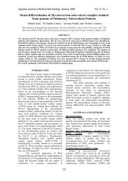
Fig. 5: Histogram representing <strong>the</strong> Percentage <strong>of</strong> fibroblast cells viability after exposure to<br />
different sealers<br />
99
Egyptian Journal <strong>of</strong> Medical Microbiology, January 2007 Vol. 16, No. 1<br />
The results <strong>of</strong> cytotoxic effect<br />
evaluation <strong>of</strong> <strong>the</strong> examined sealers on<br />
fibroblast cells are shown in Table 2 <strong>and</strong><br />
Figure 5. Although every material tested<br />
showed some degree <strong>of</strong> toxicity, <strong>the</strong><br />
percentage <strong>of</strong> cell viability was greater with<br />
<strong>Epiphany</strong>, EndoFill <strong>and</strong> Apexit than with<br />
o<strong>the</strong>r sealers with no statistically significant<br />
difference between <strong>the</strong>se three sealers in both<br />
time periods (fresh <strong>and</strong> 24 h). O<strong>the</strong>r materials<br />
(Ketac-Endo, AH-26) showed significantly<br />
lower percentage <strong>of</strong> cell viability. All sealers<br />
showed increased number <strong>of</strong> viable cells after<br />
24 h.<br />
DISCUSSION<br />
A number <strong>of</strong> approaches have been<br />
used to evaluate <strong>the</strong> antimicrobial<br />
effectiveness in <strong>the</strong> laboratory. These include;<br />
incubation <strong>of</strong> broth cultures <strong>of</strong> <strong>the</strong> selected<br />
microorganisms with <strong>the</strong> agent under <strong>the</strong> test,<br />
growth <strong>of</strong> <strong>the</strong> selected microorganisms as<br />
lawns on agar surfaces using <strong>the</strong> disc<br />
diffusion method, <strong>and</strong> <strong>the</strong> artificial infection<br />
<strong>of</strong> extracted teeth with <strong>the</strong> selected organisms<br />
<strong>and</strong> irrigation with <strong>the</strong> test agent 21 .<br />
The agar diffusion method used in<br />
this study is one <strong>of</strong> <strong>the</strong> most <strong>of</strong>ten used<br />
methods for <strong>the</strong> antimicrobial activity<br />
assessment. The results <strong>of</strong> this method do not<br />
depend only on <strong>the</strong> toxicity <strong>of</strong> <strong>the</strong> material for<br />
a particular microorganism, but also on <strong>the</strong><br />
diffusibility <strong>of</strong> <strong>the</strong> material across <strong>the</strong><br />
medium. However, great care has been taken<br />
in this study to keep <strong>the</strong> plates for 2h at room<br />
temperature to allow <strong>the</strong> diffusion <strong>of</strong> <strong>the</strong><br />
agent through <strong>the</strong> agar <strong>and</strong> <strong>the</strong>n incubated at<br />
37ºC under appropriate gaseous conditions.<br />
The present study tested <strong>the</strong> antimicrobial<br />
activity <strong>of</strong> different sealers against<br />
microorganisms considered to be resistant to<br />
endodontic treatment. Therefore, if a sealer is<br />
effective against <strong>the</strong>se microorganisms, it will<br />
probably be effective against <strong>the</strong> most<br />
susceptible ones 22 .<br />
In this study, <strong>Epiphany</strong> produced a<br />
clear antibacterial effect against <strong>the</strong> tested<br />
bacteria especially <strong>the</strong> most resistant one<br />
(E.faecalis). On <strong>the</strong> contrary, Bodrumlu <strong>and</strong><br />
Semiz 23 showed that <strong>Epiphany</strong> root canal<br />
sealers had little effect on E.faecalis 23 . This<br />
can be explained by a research conducted by<br />
Gomes et al. 5 , who determined that <strong>the</strong><br />
antimicrobial activity depends on <strong>the</strong><br />
sensitivity <strong>of</strong> <strong>the</strong> drug, bacterial source (wild<br />
strains or collection species), number <strong>of</strong><br />
bacteria inoculated, pH <strong>of</strong> <strong>the</strong> substrates in<br />
plates or tubes, agar viscosity, storage<br />
conditions <strong>of</strong> <strong>the</strong> agar plates, incubation time<br />
<strong>and</strong> <strong>the</strong> metabolic activity <strong>of</strong> <strong>the</strong><br />
microorganisms.<br />
The results <strong>of</strong> this study showed<br />
that <strong>Epiphany</strong> had no effect on C.albicans.<br />
Moderate inhibition was produced by<br />
<strong>Epiphany</strong> on E.coli <strong>and</strong> S. aureus isolates.<br />
EndoFill had <strong>the</strong> maximum average<br />
zones <strong>of</strong> inhibition as compared to o<strong>the</strong>r<br />
tested sealers. However, it comes next to<br />
<strong>Epiphany</strong> regarding <strong>the</strong> effect against<br />
E.faecalis. On <strong>the</strong> o<strong>the</strong>r h<strong>and</strong>, this material<br />
was <strong>the</strong> only effective sealer on C.albican.<br />
Previous studies indicated that root canal<br />
sealers containing ZOE have a strong<br />
antibacterial effect 24 . This effect was due to<br />
<strong>the</strong> action <strong>of</strong> eugenol 25 . Kaplan <strong>and</strong> o<strong>the</strong>rs<br />
have stated that <strong>the</strong> most effective<br />
antimicrobial sealers contain eugenol <strong>and</strong><br />
formaldehyde 26 .<br />
O<strong>the</strong>r endodontic sealers tested<br />
(Apexit, AH-26 <strong>and</strong> Ketac-Endo) were less<br />
effective than <strong>Epiphany</strong> in killing <strong>the</strong><br />
microorganisms. The antimicrobial activity <strong>of</strong><br />
Apexit may be based on its content <strong>of</strong> calcium<br />
hydroxide. Root canal sealers integrated with<br />
calcium hydroxide have enhanced<br />
antimicrobial activity 23, 27. The<br />
antimicrobial effect <strong>of</strong> this sealer is produced<br />
by <strong>the</strong> release <strong>of</strong> hydroxyl ions which<br />
increases <strong>the</strong> pH above 12.5 23. In this<br />
experiment Apexit had <strong>the</strong> lowest average<br />
diameter <strong>of</strong> inhibition zones. Bystrom <strong>and</strong><br />
Sundqvist 28 also found that for a calcium<br />
hydroxide based sealer to be an effective<br />
antimicrobial agent, it should maintain a pH<br />
level greater than 12.5. As <strong>the</strong> calcium<br />
hydroxide sealer sets, <strong>the</strong> pH declines rapidly<br />
to 9.4 causing loss <strong>of</strong> effectiveness.<br />
Some endodontic sealers consists <strong>of</strong><br />
polymer materials such as AH-26 which<br />
release small amount <strong>of</strong> formaldehyde during<br />
<strong>the</strong> polymerization process, <strong>and</strong> it is this agent<br />
that gives <strong>the</strong> resin based sealer its<br />
antimicrobial effect 29. Bodrumlu <strong>and</strong> Semiz<br />
23 showed that AH-26 had <strong>the</strong> lowest<br />
antibacterial effect against E.faecalis, <strong>and</strong><br />
attributed this effect to <strong>the</strong> release <strong>of</strong> a small<br />
amount <strong>of</strong> formaldehyde over a brief period<br />
<strong>of</strong> time. In this study AH-26 had a minor<br />
effect against <strong>the</strong> tested organisms <strong>and</strong> in<br />
100
Egyptian Journal <strong>of</strong> Medical Microbiology, January 2007 Vol. 16, No. 1<br />
particular, it had a moderate effect on<br />
E.faecalis.<br />
The antimicrobial activity <strong>of</strong> Ketac-<br />
Endo is assumed to rely on fluoride ion<br />
release, but <strong>the</strong> quantities <strong>of</strong> fluoride released<br />
may not be sufficient to reach a concentration<br />
that effectively kills microorganisms 30. A<br />
weak antimicrobial activity <strong>of</strong> Ketac-Endo<br />
has been reported by Heling <strong>and</strong> Ch<strong>and</strong>ler 31.<br />
<strong>Antimicrobial</strong> activity <strong>of</strong> root<br />
canal sealers helps destroy <strong>the</strong> remaining<br />
bacteria. On <strong>the</strong> o<strong>the</strong>r h<strong>and</strong>, severe toxicity <strong>of</strong><br />
a filling material may be a reason for damage<br />
<strong>of</strong> <strong>the</strong> periapical tissue <strong>the</strong>reby abolishing <strong>the</strong><br />
beneficial effects <strong>of</strong> <strong>the</strong> antimicrobial<br />
properties <strong>of</strong> <strong>the</strong> material<br />
22 . The<br />
antimicrobial components <strong>of</strong> <strong>the</strong> sealer do not<br />
have selective toxicity against<br />
microorganisms; <strong>the</strong>y also exert toxic effects<br />
on host cells 31 .<br />
Azar, et al 32 thought that in general,<br />
<strong>the</strong> toxicity <strong>of</strong> newly developed materials<br />
should be assessed using <strong>the</strong> three steps<br />
approach. A first step is to screen a c<strong>and</strong>idate<br />
material using a series <strong>of</strong> in vitro cytotoxicity<br />
assays using cell cultures. Then, if <strong>the</strong><br />
material is determined not to be cytotoxic in<br />
vitro, it can be implanted in subcutaneous<br />
tissue or muscle <strong>and</strong> <strong>the</strong> local tissue reaction<br />
evaluated. Finally, <strong>the</strong> in vivo reaction <strong>of</strong> <strong>the</strong><br />
target tissue versus <strong>the</strong> test material must be<br />
evaluated in human subjects or animals.<br />
Therefore, if <strong>the</strong> test material induces a strong<br />
cytotoxic reaction in cell culture tests, it is<br />
very likely also to exert toxicity in living<br />
tissue. A reduction in <strong>the</strong> number <strong>of</strong> animal<br />
tests <strong>and</strong> <strong>the</strong> resulting expenses might be an<br />
additional benefit <strong>of</strong> such a screening<br />
approach.<br />
In this study, <strong>the</strong> cytotoxicity was<br />
evaluated by measuring <strong>the</strong> cell growth via<br />
cell counting. This method was chosen<br />
because it is easy, not requiring complicated<br />
or expensive equipments.<br />
In this present study, an in vitro<br />
cytotoxicity tests was done by using primary<br />
cells, mainly oral fibroblasts, to test <strong>the</strong> dental<br />
materials. Primary cell lines have a<br />
predetermined life span <strong>and</strong> will eventually<br />
reach a plateau <strong>of</strong> growth <strong>and</strong> <strong>the</strong>n die even if<br />
<strong>the</strong> conditions for growth are acceptable.<br />
Because <strong>the</strong> materials tested would more<br />
likely come into contact with human<br />
fibroblasts in vivo, <strong>the</strong>y were chosen to more<br />
closely represent clinical conditions 33<br />
This study showed that <strong>the</strong><br />
<strong>Epiphany</strong> root canal sealer was less toxic than<br />
AH-26 <strong>and</strong> Ketac-Endo, <strong>the</strong> values <strong>of</strong> <strong>the</strong><br />
cytotoxicity <strong>of</strong> <strong>Epiphany</strong> were not significant<br />
when compared to EndoFill <strong>and</strong> Apexit.<br />
These results are in agreement with Souza et<br />
al34 who showed that <strong>Epiphany</strong> root canal<br />
sealer was <strong>the</strong> only material that presented<br />
intraosseous biocompatibility compared to<br />
AH Plus <strong>and</strong> EndoRez.<br />
On <strong>the</strong> contrary, Susini14 et al<br />
showed that <strong>Epiphany</strong> + Resilon were <strong>the</strong><br />
most cytotoxic material at 1 <strong>and</strong> 2 days <strong>and</strong><br />
this toxicity decreases with time until become<br />
nontoxic at 7 days. They explained this<br />
toxicity to be due to leaching <strong>of</strong> uncured<br />
monomers from <strong>the</strong> bulk <strong>of</strong> <strong>the</strong> resin because<br />
<strong>Epiphany</strong> set under anaerobic conditions <strong>and</strong><br />
no curing system leads to a 100% <strong>of</strong><br />
conversion 35.<br />
The results <strong>of</strong> this study concerning<br />
<strong>the</strong> ZOE sealer were similar to previous<br />
findings that showed that this material is<br />
moderately toxic <strong>and</strong> this toxicity is attributed<br />
to <strong>the</strong> release <strong>of</strong> free eugenol which is <strong>the</strong><br />
main liquid components <strong>of</strong> ZOE based<br />
sealers 36, 37. It is interesting to know that<br />
when zinc oxide eugenolate is formed, most<br />
<strong>of</strong> <strong>the</strong> free eugenol is bound which account<br />
for <strong>the</strong> relatively low toxicity <strong>of</strong> ZOE based<br />
sealers in <strong>the</strong> initial setting reaction 38 .<br />
However, after 48h enough free eugenol<br />
escaped to cause <strong>the</strong> reaction to become<br />
predominantly severe 39 .<br />
This study indicated that AH-26<br />
had a moderate to severe cytotoxic reaction<br />
immediately after mixing <strong>and</strong> this toxicity<br />
decreased after 24h. These effects may be due<br />
to formaldehyde, which is released during <strong>the</strong><br />
initial setting reaction. It has been<br />
demonstrated that AH-26 is relatively<br />
38-40<br />
cytotoxic sealer as it releases<br />
formaldehyde during <strong>and</strong> after setting 39 .<br />
Periapical inflammatory reactions were<br />
detected when AH-26 was used as sealer 42 .<br />
Ketac-Endo showed a pronounced<br />
cytotoxic effect in <strong>the</strong> early phase <strong>of</strong> <strong>the</strong><br />
setting reaction <strong>and</strong> this toxicity decreased<br />
after 24h. This observation is in agreement<br />
with a previous study which showed strong<br />
inflammatory reactions caused by Ketac-Endo<br />
40 . Schwarze 36 et al. explained this toxicity by<br />
<strong>the</strong> high water solubility <strong>of</strong> <strong>the</strong> glass ionomer<br />
cement in <strong>the</strong> early phase <strong>of</strong> <strong>the</strong> setting<br />
101
Egyptian Journal <strong>of</strong> Medical Microbiology, January 2007 Vol. 16, No. 1<br />
reaction leading to <strong>the</strong> elution <strong>of</strong> cytotoxic<br />
substances.<br />
In conclusion, <strong>Epiphany</strong> sealer demonstrates<br />
a clear antibacterial activity, particularly<br />
against E.faecalis which is <strong>the</strong> one <strong>of</strong> <strong>the</strong><br />
most resistant endodontic flora. However it<br />
had no activity against fungi (C.albicans), so<br />
<strong>the</strong> emergence <strong>of</strong> yeast with resistance to<br />
<strong>Epiphany</strong> sealer warrants continued, prudent<br />
monitoring .<br />
The cytotoxicity <strong>of</strong> <strong>Epiphany</strong> was<br />
comparable to that <strong>of</strong> EndoFill <strong>and</strong> Apexit, on<br />
<strong>the</strong> o<strong>the</strong>r h<strong>and</strong> it was less toxic than Ketac-<br />
Endo <strong>and</strong> AH-26.<br />
Fur<strong>the</strong>r in vivo studies are needed<br />
to confirm <strong>the</strong> value <strong>of</strong> <strong>Epiphany</strong> sealer using<br />
a root model.<br />
REFERENCES<br />
1. Gomes BP, Lilley JD <strong>and</strong> Drucker<br />
DB.(1996): Clinical significance <strong>of</strong><br />
dental root canal micr<strong>of</strong>lora. J Dent ; 24:<br />
47-55.<br />
2. Siren EK, Haapasalo NP, Ranta K,<br />
Salmi P <strong>and</strong> Kerosuo<br />
EN.(1997):Microbiological findings <strong>and</strong><br />
clinical procedures in endodontic cases<br />
selected for microbiological<br />
investigation. Int Endod J; 30: 91-5.<br />
3. Sundqvist G, Figdor D, Persson S <strong>and</strong><br />
Sjogren U.(1998): Microbiological<br />
analysis <strong>of</strong> teeth with failed endodontic<br />
treatment <strong>and</strong> <strong>the</strong> outcome <strong>of</strong><br />
conservative re-treatment. Oral Surg<br />
Oral Med Oral Pathol;85:86-93.<br />
4. Leonardo MR, da Silva LA, Tanomaru<br />
Filho M, Bonifácio KC <strong>and</strong> Ito IY.<br />
(2000):In vitro evaluation <strong>of</strong><br />
antimicrobial activity <strong>of</strong> sealers <strong>and</strong><br />
pastes used in endodontics. J Endod; 26:<br />
391-4.<br />
5. Gomes BP, Ferraz CC, Vianna ME,<br />
Rosalen PL, Zaia AA, et al.(2002): In<br />
vitro antimicrobial activity <strong>of</strong> calcium<br />
hydroxide pastes <strong>and</strong> <strong>the</strong>ir vehicles<br />
against selected microorganisms.Braz<br />
Dent J ;13: 155-61.<br />
6. Siqueira JF <strong>and</strong> Gonçalves RB.<br />
(1996):Antibacterial activities <strong>of</strong> root<br />
canal sealers against selected anaerobic<br />
bacteria.J Endod; 22: 79-80.<br />
7. Saleh IM, Ruyter IE, Haapasalo MP, <strong>and</strong><br />
Orstavik D.(2003):Adhesion <strong>of</strong><br />
endodontic sealers: scanning electron<br />
microscopy <strong>and</strong> energy dispersive<br />
spectroscopy. J Endod ; 29: 595-601.<br />
8. Willershausen B, Briseno B, Schafer D<br />
<strong>and</strong> Schulze R.(2002): <strong>Cytotoxic</strong>ity <strong>of</strong><br />
root canal filling materials to three<br />
different human cell lines. J Endod; 26:<br />
703-7.<br />
9. Cohen S, Burns C.(2002): Pathways <strong>of</strong> <strong>the</strong><br />
pulp, 8th ed.; 398.<br />
10. Tay F, Loushine R, Weller N <strong>and</strong><br />
Kimbrougb A(2005): Ultrastructural<br />
evaluation <strong>of</strong> <strong>the</strong> apical seal in roots<br />
filled with a polycaprolactone based root<br />
canal filling material. J Endod; 31: 514-<br />
9.<br />
11. Teixeira F, Teixiera E, Thompson J <strong>and</strong><br />
Trope M.(2004):Fracture resistance <strong>of</strong><br />
endodontically treated roots using a new<br />
type <strong>of</strong> resin filling material. J Am Dent<br />
Assoc; 135: 646- 52.<br />
12. Shipper G, Teixeira FB, Arnold RR<br />
<strong>and</strong> Trope M.(2005):Periapical<br />
inflammation after coronal microbial<br />
inoculation <strong>of</strong> dog roots filled with gutta<br />
percha or resilon. J Endod; 31: 91-6.<br />
13. Gesi A, Raffaelli O, Gorracci C,<br />
Pashley DH, et al.(2005): Interfacial<br />
strength <strong>of</strong> Resilon <strong>and</strong> gutta-percha to<br />
intraradicular dentin. J Endod 2005; 31:<br />
809-13.<br />
14. Susini G, About I, Hung L <strong>and</strong> Camps<br />
J.(20026): <strong>Cytotoxic</strong>ity <strong>of</strong> <strong>Epiphany</strong> <strong>and</strong><br />
Resilon with a root model. Int Endod J ;<br />
39: 940-4.<br />
15. Carvalho-Junior JR, Guimaraes LF,<br />
Correr-Sobrinho L, et al.(2003):<br />
<strong>Evaluation</strong> <strong>of</strong> solubility, disintegration,<br />
<strong>and</strong> dimensional alterations <strong>of</strong> a glass<br />
ionomer root canal sealer. Braz Dent J;<br />
14: 114-8.<br />
16. Al-Khatib ZZ, Baum RH, Morse ER<br />
<strong>and</strong> Yesilsoy C. (1990):The<br />
antimicrobial effects <strong>of</strong> various<br />
endodontic sealers. Oral Surg Oral Med<br />
Oral Path ; 70: 784-90.<br />
102
Egyptian Journal <strong>of</strong> Medical Microbiology, January 2007 Vol. 16, No. 1<br />
17. Gomes BP, Ferraz CC, Garrido FO, et<br />
al.(2002): Microbial susceptibility to<br />
calcium hydroxide pastes <strong>and</strong> <strong>the</strong>ir<br />
vehicles. J Endod ; 28: 758-61.<br />
18. Miletic I, Anie I <strong>and</strong> Karlovic Z.<br />
(2000):<strong>Cytotoxic</strong> effects <strong>of</strong> four root<br />
filling materials. Endod Dent Traumatol<br />
;16: 287-90.<br />
19. Kuo-Wei T <strong>and</strong> Yu-Chao C.(2000):<br />
<strong>Cytotoxic</strong>ity evaluation <strong>of</strong> perforation<br />
repair materials on human periodontal<br />
ligament cells in vitro. J Endod ; 26:395-<br />
7.<br />
20. Bouillaguet S, Wataha JC, Lockwood<br />
PE, et al.(2004): <strong>Cytotoxic</strong>ity <strong>and</strong><br />
sealing properties <strong>of</strong> four classes <strong>of</strong><br />
endodontic sealers evaluated by succinic<br />
dehydrogenase activity <strong>and</strong> confocal<br />
laser scanning microscopy. Eur J Oral<br />
Sci ; 112: 182-7.<br />
21. Ayhan H, Sultan N, Cirak M, Ruhi MZ<br />
<strong>and</strong> Bodur H.(1999): <strong>Antimicrobial</strong><br />
effects <strong>of</strong> various endodontic irrigants on<br />
selected microorganisms. Int Endod J;<br />
32: 99-102.<br />
22. Gomes BP, Pinheiro ET, Gade Neto<br />
CR, et al.(2004):Microbiological<br />
examination <strong>of</strong> infected dental root<br />
canals. Oral Microbiol Immunol; 19: 71-<br />
6.<br />
23. Bodrumlu E <strong>and</strong> Semiz M.(2006):<br />
Antibacterial activity <strong>of</strong> a new<br />
endodontic sealer against Enterococcus<br />
faecalis. J Can Dent Assoc; 72: 637-7.<br />
24. Pupo J, Biral RR, Benatti O, Abe A <strong>and</strong><br />
Vadrighi L.(1983):<strong>Antimicrobial</strong> effects<br />
<strong>of</strong> endodontic filling cements on<br />
microorganisms from root canal. Oral<br />
Surg Oral Med Oral Pathol ; 55: 622-7.<br />
25. Mickel Ak, Nguyen TH <strong>and</strong> Chogle<br />
S.(2003): <strong>Antimicrobial</strong> activity <strong>of</strong><br />
endodontic sealers on Enterococcus<br />
faecalis. J Endod; 29: 257-8.<br />
26. Kaplan AE, Picca M, Gonzalez MI,<br />
Macchi RL, et al.,(1999):<strong>Antimicrobial</strong><br />
effects <strong>of</strong> six endodontic sealers: an in<br />
vitro evaluation. Endod Dent Traumatol;<br />
15: 42-5.<br />
27. Sjogren U, Figdor D, Spangberg L <strong>and</strong><br />
Sundqvist G.(1991): The antimicrobial<br />
effects <strong>of</strong> calcium hydroxide as a short<br />
term intracanal dressing .Int Endod J ;<br />
24: 119-25.<br />
28. Bystrom A <strong>and</strong> Sundqvist G.(1985):The<br />
antibacterial action <strong>of</strong> sodium<br />
hypochlorite <strong>and</strong> EDTA in 60 cases <strong>of</strong><br />
endodontic <strong>the</strong>rapy. Int Endod J ; 18: 35-<br />
40.<br />
29. Leonardo MR, Bezerra da Silva LA, et<br />
al.,(1999):Release <strong>of</strong> formaldehyde by 4<br />
endodontic sealers. Oral Surg Oral Med<br />
Oral Pathol; 88: 221-5.<br />
30. Saleh IM, Ruyter IE, Haapasalo M <strong>and</strong><br />
Orstavik D.(2004):Survival <strong>of</strong><br />
Enterococcus faecalis in infected dentinal<br />
tubules after root canal filling with<br />
different root canal sealers in vitro. Int<br />
Endod J; 37: 193- 8.<br />
31. Heling I <strong>and</strong> Ch<strong>and</strong>ler NP.(1996): The<br />
antimicrobial effects within dentinal<br />
tubules <strong>of</strong> four root canal sealers. J<br />
Endod ; 22: 257-9.<br />
32. Azar NG, Heidari M, Bahrami ZS <strong>and</strong><br />
Shokri F.(2000):In vitro cytotoxicity <strong>of</strong><br />
a new epoxy resin root canal sealer. J<br />
Endod; 26: 462-5.<br />
33. Key JE, Rahemtulla FG, Eleazer<br />
PD.(2006): <strong>Cytotoxic</strong>ity <strong>of</strong> a new root<br />
canal filling material on human<br />
gingival<br />
fibroblasts.J<br />
Endod;32(8):756-8.<br />
34. Souza CJ, Montes CR, Pascon EA,<br />
Loyola AM <strong>and</strong> Versiani MA.(2006):<br />
Comparison <strong>of</strong> <strong>the</strong> intraosseous<br />
biocompatibility <strong>of</strong> AH Plus, EndoRez<br />
<strong>and</strong> <strong>Epiphany</strong> root canal sealers. J<br />
Endod; 32: 656-62.<br />
35. Peutzfeld A.(1997): Resin composites in<br />
dentistry: <strong>the</strong> monomer system. Euro J<br />
Oral Sci 1997; 105: 97-116.<br />
36. Schwarze T, Fiedler I, Leyhausen G <strong>and</strong><br />
Geurtsen W.(2002): The cellular<br />
compatibility <strong>of</strong> five endodontic sealers<br />
during <strong>the</strong> setting periods. J Endod ; 28:<br />
784-6.<br />
37. Yoshimine Y, Yamamoto M,<br />
Ogasawara T, et al.(2003):In vitro<br />
evaluation <strong>of</strong> <strong>the</strong> cytocompatibility <strong>of</strong> a<br />
glass ionomer cement sealer. J Endod<br />
;29(7):453-5.<br />
38. Morse R, Bayardo M, ,et al.(1984):A<br />
comparative tissue toxicity evaluation <strong>of</strong><br />
103
Egyptian Journal <strong>of</strong> Medical Microbiology, January 2007 Vol. 16, No. 1<br />
gutta percha root canal sealers part II,<br />
forty eight hours findings. J Endod ; 10:<br />
484-6.<br />
39. Gerosa R, Menegazzi G, Borin M <strong>and</strong><br />
Cavalleri G.(1995): <strong>Cytotoxic</strong>ity<br />
evaluation <strong>of</strong> six root canal sealers. J<br />
Endod ; 21:446-8.<br />
40. Leyhausen G, Heil J, Reifferscheid G,<br />
Waldman P <strong>and</strong> Geurtsen W.(1999):<br />
Genotoxicity <strong>and</strong> cytotoxicity <strong>of</strong> <strong>the</strong><br />
epoxy resin based root canal sealer AH<br />
Plus. J Endod; 25:109-13.<br />
41. Willerhausen B, Briseno B, Schafer D<br />
<strong>and</strong> Schuze R.(2000): <strong>Cytotoxic</strong>ity <strong>of</strong><br />
root canal filling materials to three<br />
different human cell lines. J<br />
Endod;26(12):703-7.<br />
42. Huang FM, Tai KW, Chou MY <strong>and</strong><br />
Chang YC.(2002): <strong>Cytotoxic</strong>ity <strong>of</strong> resin,<br />
zinc oxide eugenol, <strong>and</strong> calcium<br />
hydroxide based root canal sealers on<br />
human periodontal ligament cells <strong>and</strong><br />
permanent V79 cells. Int Endod J ; 35:<br />
153-8.<br />
104
Egyptian Journal <strong>of</strong> Medical Microbiology, January 2007 Vol. 16, No. 1<br />
دراسه معمليه لتقييم النشاط المضاد للميكروبات و التسمم الخلوي لمادة الابيفاني الغالقة<br />
لقنوات جذور الأسنان<br />
امانى بدر-عمرو عبد الرازق-نسرين عمر و فريد بدرية<br />
هذه الدراسة قيمت<br />
معمليا النشاط المضاد للميكروبات و التسمم الخلوي لمادة الابيفاني<br />
الغالقة لقنوات الجذور مقارنة ببعض مواد غالقة أخري معتاد استعمالها و هي الاندوفل،<br />
ابكسيت ، كتاك-اندو، و<br />
ايه اتش<br />
٢٦ في الجزء الأول من هذه الرسالة تم استخدام عدة<br />
ميكروبات وهم انتيروكوكس فيكالز،<br />
ستافيلوكوكس ايوريس،<br />
اشريشيا كولاي،<br />
و كانديدا البيكان<br />
لمعرفة التأثير المضاد للميكروبات وذلك باستخدام طريقة انتشار المواد المستخدمة في مادة<br />
الاجار.<br />
وقد استخدمت مزرعة من خلايا<br />
الاربطه المحيطه بالخذر<br />
الادمي.<br />
في التقيم الخارجي<br />
للتسمم الخلوي لمادة الابيفاني وذلك لعد الخلايا الحية بعد حضانة المزرعة مع الابيفاني المخلوط<br />
حديثا و أخر بعد<br />
٢٤ ساعة. وقد أسفرت نتائج هذه الدراسة بان مادة الابيفاني لها تاثير فعال في<br />
قتل البكتيريا وليس<br />
الكنديدا<br />
وجد أن التسمم الخلوي لمادة الابيفاني<br />
متقاربا مع مادة الاندوفل و<br />
.<br />
الابكسيت و لكنه اقل سميه من مادتي الكيتاك-اندو و<br />
٢٦. ايه اتش<br />
105