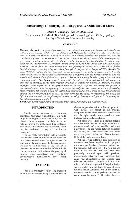
Bacteriology of Pharyngitis in Suppurative Otitis Media Cases
Bacteriology of Pharyngitis in Suppurative Otitis Media Cases
Bacteriology of Pharyngitis in Suppurative Otitis Media Cases

You also want an ePaper? Increase the reach of your titles
YUMPU automatically turns print PDFs into web optimized ePapers that Google loves.
Egyptian Journal <strong>of</strong> Medical Microbiology, July 2009 Vol. 18, No. 3<br />
<strong>Bacteriology</strong> <strong>of</strong> <strong>Pharyngitis</strong> <strong>in</strong> <strong>Suppurative</strong> <strong>Otitis</strong> <strong>Media</strong> <strong>Cases</strong><br />
Mona F. Salama*, Alaa Ali Abou-Bieh<br />
Departments <strong>of</strong> Medical Microbiology and Immunology* and Otolaryngology,<br />
Faculty <strong>of</strong> Medic<strong>in</strong>e, Mansoura University<br />
ABSTRACT<br />
Problem Addressed: Unexpla<strong>in</strong>ed persistent or recurrent bacterial pharyngitis <strong>in</strong> some patients who are<br />
suffer<strong>in</strong>g from <strong>in</strong>fected middle ear cleft. Patients and Methods: Bacteriological swabs were obta<strong>in</strong>ed<br />
from both ears and pharynx <strong>of</strong> thirty-seven cases with chronic otorrhea and perforation, who are<br />
compla<strong>in</strong><strong>in</strong>g <strong>of</strong> recurrent or persistent sore throat. Isolation and identification <strong>of</strong> the micro-organisms<br />
were done. Isolated Gram-negative bacilli were subjected to further identification by biochemical<br />
reactions and antimicrobial susceptibility test<strong>in</strong>g us<strong>in</strong>g modified Kirby Bauer disk diffusion method.<br />
Identical isolates from the same patient (ear and pharyngeal swabs) were subjected to further<br />
identification by genotyp<strong>in</strong>g us<strong>in</strong>g the pulsed field gel electrophoresis technique (PFGE). Results: Six<br />
cases (16%) showed identity <strong>in</strong> both phenotypes and genotypes for ear and pharyngeal samples from the<br />
same patient. Four <strong>of</strong> the isolates were Pseudomonas aerug<strong>in</strong>osa, one was Proteus mirabilis, and one<br />
was Escherichia coli. None <strong>of</strong> these three species is known to be among the primary organisms that may<br />
cause pharyngitis. Conclusion: Bacterial pharyngitis <strong>in</strong> patients with chronically <strong>in</strong>fected middle ear<br />
cleft may be attributed to the same organism <strong>in</strong>vad<strong>in</strong>g the middle ear mucosa. In addition, this study<br />
highlights some organisms as pharyngeal <strong>in</strong>vaders although they are not among the previously<br />
documented causes <strong>of</strong> bacterial pharyngitis. However, the study does not confirm the method <strong>of</strong> spread <strong>of</strong><br />
these organisms between the middle ear cleft and the pharynx and does not prove whether the spread was<br />
directly via the eustachian tube, or not. The study correlates the causative organism <strong>of</strong> the middle ear<br />
<strong>in</strong>fection and that <strong>in</strong>fected the pharyngeal mucosa by us<strong>in</strong>g phenotypic and genotypic bacteriological<br />
identification and typ<strong>in</strong>g methods.<br />
Key Words: Chronic suppurative otitis media, <strong>Pharyngitis</strong>, Pulsed field gel electrophoresis.<br />
INTRODUCTION<br />
Chronic throat soreness is a common<br />
compla<strong>in</strong>t. Nowadays it is attributed to a wide<br />
range <strong>of</strong> etiologies. It was noteworthy that the<br />
chronic throat soreness compla<strong>in</strong>t <strong>of</strong> some<br />
patients, whom are at the same time suffer<strong>in</strong>g<br />
from chronically <strong>in</strong>fected middle ear cleft, could<br />
not be attributed to any <strong>of</strong> the known<br />
etiologies (1) .<br />
The aim <strong>of</strong> the present study is to <strong>in</strong>vestigate<br />
whether the reason <strong>of</strong> this compla<strong>in</strong>t is related<br />
to a bacteriological causative agent or not. In<br />
addition, if the cause would be bacteriological,<br />
we aim to f<strong>in</strong>d if there is any correlation<br />
between the causative organism <strong>of</strong> the middle<br />
ear <strong>in</strong>fection and that <strong>in</strong>fected the pharyngeal<br />
mucosa us<strong>in</strong>g phenotypic and genotypic<br />
bacteriological identification and typ<strong>in</strong>g<br />
methods.<br />
PATIENTS<br />
This is a prospective study. The <strong>in</strong>clusion<br />
criteria for cases were be<strong>in</strong>g a known case <strong>of</strong><br />
chronic suppurative otitis media and presented<br />
with chronic sore throat as the pr<strong>in</strong>cipal<br />
compla<strong>in</strong>t. Thirty-seven cases met these criteria<br />
over the eight months study period and were<br />
<strong>in</strong>cluded <strong>in</strong> the study population.<br />
All cases were adults as pediatric patients<br />
were excluded due to the high <strong>in</strong>cidence <strong>of</strong><br />
recurrent attacks <strong>of</strong> acute tonsillitis and acute<br />
pharyngitis. The age ranged between seventeen<br />
and seventy-two with mean fifty-four. There<br />
were twenty-two males and fifteen females.<br />
Eight <strong>of</strong> the males were smokers.<br />
All cases were suffer<strong>in</strong>g from persistent or<br />
recurrent otorrhoea together with tympanic<br />
membrane perforation. N<strong>in</strong>eteen patients had<br />
the disease <strong>in</strong> both ears, ten <strong>in</strong> the right ear and<br />
eight <strong>in</strong> the left ear. Six ears have been<br />
previously operated upon with persistent<br />
mastoid and / or tympanic cavity <strong>in</strong>fection. Two<br />
cases had cholesteatoma but patients were<br />
reluctant to undergo surgery. All ears with<br />
mixed <strong>in</strong>fection or superadded fungal <strong>in</strong>fection<br />
were excluded from the study. In most <strong>of</strong> cases,<br />
the sore throat compla<strong>in</strong>t was <strong>in</strong> the form <strong>of</strong><br />
recurrent attacks. Some patients noticed or<br />
could remember an on / <strong>of</strong>f relation between<br />
1
Egyptian Journal <strong>of</strong> Medical Microbiology, July 2009 Vol. 18, No. 3<br />
some soreness attacks and otorrhea bouts. Other<br />
patients compla<strong>in</strong>ed <strong>of</strong> persistent chronic<br />
soreness.<br />
Other etiologies for chronic throat soreness<br />
<strong>in</strong> adults were assessed <strong>in</strong> these patients. Any<br />
form <strong>of</strong> upper aero-digestive tract neoplasia was<br />
excluded. Although this would not exclude<br />
gastroesophageal reflux disease (GERD) or<br />
laryngopharyngeal reflux (LPR), but none <strong>of</strong> the<br />
patients showed any hiatal hernia, reflux, or<br />
delayed gastric empty<strong>in</strong>g <strong>in</strong> the barium swallow<br />
/ upper GI series. Two patients also were<br />
subjected to upper gastro<strong>in</strong>test<strong>in</strong>al endoscopy<br />
(one was prior to his <strong>in</strong>clusion <strong>in</strong> the study).<br />
Dur<strong>in</strong>g the time <strong>of</strong> study five patients were on<br />
antireflux treatment <strong>in</strong>cluded even proton pump<br />
<strong>in</strong>hibitors with m<strong>in</strong>imal or no pharyngeal<br />
symptoms relieve. None <strong>of</strong> the patients had<br />
history <strong>of</strong> previous deep neck spaces<br />
suppuration, any foreign body <strong>in</strong>gestion or<br />
recent neck trauma. In addition, autoimmune<br />
and hematological conditions were excluded.<br />
Blood iron was assessed <strong>in</strong> all females. Two<br />
patients had frequent cement exposure and one<br />
patient with smoke exposure due to their<br />
occupation. N<strong>in</strong>e patients had their tonsils<br />
removed (all more than ten years before the<br />
study time). Eleven patients had postnasal drip<br />
on exam<strong>in</strong>ation, four <strong>of</strong> them showed chronic<br />
s<strong>in</strong>usitis on radiological evaluation (all patients<br />
had bacteriological swabs from their nasal<br />
cavities on further step dur<strong>in</strong>g the study). Five<br />
<strong>of</strong> the eleven patients had used local nasal<br />
steroids before. One patient with cleft palates<br />
and lip had them surgically repaired eighteen<br />
years ago. Two were diabetic patients and four<br />
were hypertensive, two <strong>of</strong> them were on ACE<br />
<strong>in</strong>hibitors and another one was on B blocker.<br />
One patient was asthmatic and on Fluticasone<br />
propionate <strong>in</strong>haler.<br />
All patients were asked to stop topical and<br />
systemic antibiotics ten days at least prior to<br />
samples collection. All patients were <strong>in</strong>formed<br />
about their <strong>in</strong>clusion <strong>in</strong> the study. A written<br />
consent was obta<strong>in</strong>ed from all <strong>of</strong> them.<br />
METHODS<br />
Specimens:<br />
Simultaneous swabs from the ear discharge,<br />
the posterior pharyngeal wall and bilaterally<br />
from the nasal cavities from each patient were<br />
obta<strong>in</strong>ed. This was done at three different<br />
successive sett<strong>in</strong>gs. MDIC ® sterile swabs (Ajax,<br />
Ontario, Canada) were used. Swabs were taken<br />
under proper precautions. They were taken<br />
under direct visualization or endoscopically<br />
guided, after proper external ear canal suction<br />
and were collected as deep as possible, us<strong>in</strong>g<br />
sterile <strong>in</strong>struments.<br />
Isolation and identification <strong>of</strong> microbial<br />
isolates:<br />
Direct Gram sta<strong>in</strong>ed films for all specimens<br />
were prepared and studied first. Then all<br />
specimens were <strong>in</strong>oculated on proper culture<br />
media with <strong>in</strong>cubation at 37 o C for 24 - 48 hours<br />
aerobically. These media were blood agar and<br />
MacConkey’s agar. Then all the isolated<br />
organisms were identified accord<strong>in</strong>g to standard<br />
microbiological methods and, where necessary,<br />
the API (Analytical Pr<strong>of</strong>ile Index) system<br />
(bioMerieux, France) was used. Antimicrobial<br />
susceptibility test (Antibiotyp<strong>in</strong>g) for aerobic<br />
bacteria was performed. This was done by us<strong>in</strong>g<br />
modified Kirby - Bauer disk diffusion method (2) ,<br />
and accord<strong>in</strong>g to the Cl<strong>in</strong>ical and Laboratory<br />
Standards Institute/NCCLS breakpo<strong>in</strong>ts for<br />
<strong>in</strong>terpretation <strong>of</strong> results (3) , us<strong>in</strong>g the follow<strong>in</strong>g<br />
antimicrobial discs and concentrations:<br />
Amikac<strong>in</strong> ® (AK), 30 mg,<br />
Amoxacill<strong>in</strong>/Clavulanic ® acid (AMC), 30 mg,<br />
Aztereonam ® (ATM), 30 mg, Cefotaxime ®<br />
(CTX), 30 mg, Cephrad<strong>in</strong>e ® (CE), 30 mg,<br />
Cipr<strong>of</strong>loxac<strong>in</strong> ® (CIP) 5 mg, Cl<strong>in</strong>damyc<strong>in</strong> ®<br />
(DA), 2 mg, Erythromyc<strong>in</strong> ® (E), 15 mg,<br />
Imipenum ® (IPM), 10 mg, Trimethoprim -<br />
Sulphamethoxazole ® (SXT), 25 mg,<br />
Tetracycl<strong>in</strong> ® (TE), 30 mg and Ceftazidime ®<br />
(CAZ), 30 mg. Apart from the standard<br />
antibiotics, test<strong>in</strong>g was also done specifically for<br />
Gentamic<strong>in</strong> ® (CN), 10 mg and<br />
Chloramphenicol ® (C), 30 mg which are<br />
available locally as topical antibiotic eardrops.<br />
Isolates were considered to belong to different<br />
antibiotypes if at least one difference <strong>in</strong><br />
antibiotic susceptibility was observed (4) . Isolates<br />
from the same patient (ear and pharyngeal<br />
swabs) which showed the same antibiotype<br />
were suspected to be related to the same stra<strong>in</strong>,<br />
and were subjected to further identification for<br />
confirmation by genotyp<strong>in</strong>g us<strong>in</strong>g pulsed field<br />
gel electrophoresis (PFGE).<br />
The genotyp<strong>in</strong>g by pulsed field gel<br />
electrophoresis was done us<strong>in</strong>g the technique<br />
described by Tenover et al. (5) to evaluate the<br />
relatedness <strong>of</strong> the isolates. First, organisms were<br />
grown overnight to plateau phase were<br />
embedded <strong>in</strong> low-melt agarose and digested for<br />
24 hours each, first with lysozyme and then with<br />
prote<strong>in</strong>ase K, both adjusted to 1 mg/mL. The<br />
resultant, which was exposed bacterial DNA,<br />
was then digested with selected endonuclease<br />
2
Egyptian Journal <strong>of</strong> Medical Microbiology, July 2009 Vol. 18, No. 3<br />
restriction enzymes designed to yield ten to<br />
twenty-five large fragments. These<br />
endonuclease restriction enzymes were: XbaI<br />
for Enterobacter, Klebsiella, Citrobacter, and<br />
Stenotrophomonas; NotI for Escherichia coli;<br />
SpeI for Pseudomonas and Serratia; and SmaI<br />
for Ac<strong>in</strong>etobacter. DNA fragments then were<br />
separated on 1% agarose gels us<strong>in</strong>g<br />
preprogrammed protocols as recommended by<br />
the manufacturer <strong>of</strong> the apparatus (Biorad<br />
CHEF GenePath System, Hercules, CA).<br />
Accord<strong>in</strong>g to the number <strong>of</strong> band differences<br />
isolates were divided <strong>in</strong>to: 1) <strong>in</strong>dist<strong>in</strong>guishable,<br />
if their restriction patterns have the same<br />
numbers <strong>of</strong> bands, 2) closely related, if their<br />
restriction patterns result <strong>in</strong> two to three bands<br />
difference, 3) possibly related, if their restriction<br />
patterns result <strong>in</strong> four to six bands difference<br />
and 4) unrelated, if their restriction patterns<br />
result <strong>in</strong> seven or more bands difference.<br />
Organisms were def<strong>in</strong>ed as concordant when<br />
they differed by three or fewer bands after<br />
visual <strong>in</strong>spection <strong>of</strong> the restriction endonuclease<br />
pattern. Isolates that were proven to have the<br />
same antibiotype, and were closely related by<br />
PFGE were considered to belong to same<br />
stra<strong>in</strong>s (6) .<br />
RESULTS<br />
Table (1) shows the bacteriology <strong>of</strong> the<br />
eighteen cases with unilaterally <strong>in</strong>fected middle<br />
ear cleft. Pseudomonas aerug<strong>in</strong>osa was the<br />
most commonly detected organism (n<strong>in</strong>e cases),<br />
while Proteus mirabilis was the next<br />
encountered organism (five cases). The situation<br />
was nearly close <strong>in</strong> the n<strong>in</strong>eteen cases with<br />
bilaterally <strong>in</strong>fected middle ear clefts (table 2).<br />
Proteus mirabilis was detected <strong>in</strong> seven cases <strong>in</strong><br />
both ears and <strong>in</strong> one ear only <strong>in</strong> two cases.<br />
Pseudomonas aerug<strong>in</strong>osa was detected <strong>in</strong> both<br />
ears <strong>in</strong> six cases and <strong>in</strong> one ear <strong>in</strong> the two cases<br />
with Proteus <strong>in</strong> the opposite side. Gram-positive<br />
organisms were only detected <strong>in</strong> two cases<br />
where middle ear was unilaterally <strong>in</strong>volved.<br />
Bacteriological pharyngeal swabs f<strong>in</strong>d<strong>in</strong>gs were<br />
significant <strong>in</strong> n<strong>in</strong>eteen cases (table 3). Group A-<br />
β hemolytic Streptococcus pyogenes was the<br />
most commonly encountered species (n<strong>in</strong>e<br />
cases), while four other cases showed α -<br />
hemolytic Streptococci and Staphylococci.<br />
Gram-negative species could be detected <strong>in</strong> six<br />
cases, four <strong>of</strong> them were Pseudomonas<br />
aerug<strong>in</strong>osa, one was Proteus mirabilis and the<br />
last was Escherichia coli. Nasal cavities isolates<br />
were n<strong>in</strong>e cases with Streptococcal pneumoniae<br />
and one case with Klebsiella isolate.<br />
Into a further step <strong>of</strong> organisms<br />
identification, and as mentioned above, isolates<br />
from the same patient (ear and pharyngeal<br />
swabs) which showed the same phenotyp<strong>in</strong>g<br />
and were identical <strong>in</strong> antibiotyp<strong>in</strong>g (Fig 1) were<br />
suspected to be related to the same stra<strong>in</strong>, and<br />
were subjected to further identification for<br />
confirmation by genotyp<strong>in</strong>g us<strong>in</strong>g pulsed field<br />
gel electrophoresis (PFGE). Then after the<br />
results <strong>of</strong> PFGE (Fig 2), isolates that were<br />
proven to have the same antibiotype, and were<br />
closely related by PFGE were considered to<br />
belong to same stra<strong>in</strong>s. By follow<strong>in</strong>g this<br />
stepwise technique, the study could recognize<br />
identity <strong>in</strong> both phenotypes and genotypes for<br />
different samples from the same patient (ear and<br />
pharynx) <strong>in</strong> all the six cases with Gram-negative<br />
isolates from pharyngeal swabs (Tables 4 & 5).<br />
These cases were represent<strong>in</strong>g sixteen percent<br />
<strong>of</strong> the study cases.<br />
3
Egyptian Journal <strong>of</strong> Medical Microbiology, July 2009 Vol. 18, No. 3<br />
Table (1): <strong>Bacteriology</strong> <strong>of</strong> <strong>in</strong>fected middle ear cleft <strong>in</strong> unilaterally <strong>in</strong>volved cases<br />
Bacteria<br />
No. <strong>of</strong> <strong>Cases</strong><br />
Pseudomonas aerug<strong>in</strong>osa 9<br />
Proteus mirabilis 5<br />
Escherichia coli 1<br />
Klebsiella sp. 1<br />
Streptococcus sp. 1<br />
Staphylococcus epidermidis 1<br />
- Total number <strong>of</strong> cases 18 - sp. = species<br />
Table (2): <strong>Bacteriology</strong> <strong>of</strong> <strong>in</strong>fected middle ear cleft <strong>in</strong> bilaterally <strong>in</strong>volved cases<br />
Bacteria<br />
No. <strong>of</strong> <strong>Cases</strong><br />
Proteus mirabilis (<strong>in</strong> both ears) 7<br />
Pseudomonas aerug<strong>in</strong>osa (<strong>in</strong> both ears) 6<br />
Proteus mirabilis (<strong>in</strong> one ear) & Pseudomonas aerug<strong>in</strong>osa (<strong>in</strong> other ear) 2<br />
Escherichia coli (<strong>in</strong> both ears) 2<br />
Klebsiella sp. (<strong>in</strong> both ears) 1<br />
Enterobacter sp. (<strong>in</strong> both ears) 1<br />
- Total number <strong>of</strong> cases 19 - sp. = species<br />
Table (3): <strong>Bacteriology</strong> <strong>of</strong> pharyngeal swabs <strong>in</strong> all study cases<br />
Bacteria<br />
No. <strong>of</strong> <strong>Cases</strong><br />
Pseudomonas aerug<strong>in</strong>osa 4<br />
Proteus mirabilis 1<br />
Escherichia coli 1<br />
Group A-β hemolytic Streptococcus pyogenes 9<br />
α - hemolytic Streptococci 2<br />
Staphylococcus sp. 2<br />
No significant growth <strong>in</strong> 3 swabs 18<br />
- Total number <strong>of</strong> cases 37 - sp. = species<br />
Table (4): Detected identity <strong>in</strong> both phenotypes and genotypes for different samples from the same<br />
patient<br />
Bacteria<br />
No. <strong>of</strong> <strong>Cases</strong><br />
Pseudomonas aerug<strong>in</strong>osa 4<br />
Proteus mirabilis 1<br />
Escherichia coli 1<br />
- Total number <strong>of</strong> cases 6<br />
Table (5): Detected identity <strong>in</strong> both phenotypes and genotypes for different samples from the same<br />
patient<br />
Bacteria<br />
No. <strong>of</strong> <strong>Cases</strong><br />
Pseudomonas aerug<strong>in</strong>osa (<strong>in</strong> one ear) 3<br />
Pseudomonas aerug<strong>in</strong>osa (<strong>in</strong> both ears) 1<br />
Proteus mirabilis (<strong>in</strong> both ears) 1<br />
Escherichia coli (<strong>in</strong> both ears) 1<br />
- Total number <strong>of</strong> cases 6<br />
4
Egyptian Journal <strong>of</strong> Medical Microbiology, July 2009 Vol. 18, No. 3<br />
Right Ear<br />
Left ear<br />
Pharynx<br />
Fig 1: Isolates from the same patient (both ears and pharyngeal swabs) show<strong>in</strong>g the same antibiotype<br />
5
Egyptian Journal <strong>of</strong> Medical Microbiology, July 2009 Vol. 18, No. 3<br />
Fig 2: Detected identity <strong>in</strong> genotypes for different samples from the same patient (Arrow 1: Right Ear,<br />
Arrow 2: Left Ear, Arrow 3: Pharynx, Black Arrow: Marker)<br />
DISCUSSION<br />
The study idea came from an observation for<br />
some cases <strong>of</strong> chronic suppurative otitis media<br />
<strong>in</strong> the outpatient department. Those cases,<br />
although very few, drew the attention to some<br />
serious results. Chronic suppurative otitis media<br />
is a condition which necessitates patient to<br />
follow up for a long period either pre or<br />
postoperatively. Usually the symptoms and<br />
reasons that oblige the patient to seek medical<br />
advice are related to the otorrhea, hear<strong>in</strong>g loss<br />
or that related to sequelae or complications <strong>of</strong><br />
the disease. For those cases who triggered this<br />
study, the ma<strong>in</strong> reason for seek<strong>in</strong>g medical<br />
advice was a long history <strong>of</strong> throat soreness. In<br />
some <strong>of</strong> them, it was so annoy<strong>in</strong>g and<br />
<strong>in</strong>capacitat<strong>in</strong>g for a normal daily life. Even one<br />
<strong>of</strong> the patients was already gett<strong>in</strong>g a psychiatric<br />
counsel<strong>in</strong>g when he jo<strong>in</strong>ed the study. Also, most<br />
<strong>of</strong> the cases when jo<strong>in</strong>ed were already<br />
<strong>in</strong>vestigated for the throat compla<strong>in</strong>t either <strong>in</strong><br />
our hospital or <strong>in</strong> other places. These<br />
<strong>in</strong>vestigations revealed either no clear reason for<br />
the throat compla<strong>in</strong>t or when the reason they<br />
revealed were treated, very limited<br />
improvement could be achieved. Some special<br />
data highlighted a questionable relation between<br />
the otologic problem and the pharyngeal<br />
problem. An on / <strong>of</strong>f relation between the sore<br />
throat and the otorrhea was the first special<br />
symptom demonstrated from the first patient<br />
trigged the study. After several years <strong>of</strong><br />
recurrent attacks <strong>of</strong> otorrhea, the patient could<br />
realize that shortly after the start <strong>of</strong> the otorrhea<br />
attack he usually develop a period <strong>of</strong> severe<br />
soreness <strong>in</strong> his throat. This is usually without<br />
any other systematic manifestation apart from<br />
marked malaise and halitosis locally. This<br />
on/<strong>of</strong>f relation could be demonstrated <strong>in</strong> other<br />
cases jo<strong>in</strong>ed the study later, although some<br />
others noticed that this soreness is persistent <strong>in</strong><br />
its nature. Another remark by some patients was<br />
that only certa<strong>in</strong> antibiotics would be effective<br />
with the pharyngeal problem. As mostly the<br />
otorrhea was treated by local treatment, patients<br />
noticed that their throat soreness usually not<br />
improve by usual antibiotics and most <strong>of</strong> them<br />
6
Egyptian Journal <strong>of</strong> Medical Microbiology, July 2009 Vol. 18, No. 3<br />
knew that they need some different antibiotic to<br />
improve (mostly was from the Qu<strong>in</strong>olones<br />
group). In addition, <strong>in</strong> one <strong>of</strong> the early study<br />
cases and dur<strong>in</strong>g an attack <strong>of</strong> otorrhea, a swab<br />
was taken from his pharynx, then we got an<br />
early note from the microbiology laboratory<br />
about a Gram-negative very similar to that<br />
caused the patient’s otorrhea could be<br />
demonstrated <strong>in</strong> the pharyngeal swab.<br />
Accord<strong>in</strong>g to this, we proceeded to <strong>in</strong>vestigate<br />
the case and a phenotype identity could be<br />
assured between ear and pharynx samples <strong>in</strong> this<br />
patient. So this study was designed to<br />
understand this f<strong>in</strong>d<strong>in</strong>gs and try to f<strong>in</strong>d if this<br />
was an isolated situation or it can be<br />
demonstrated <strong>in</strong> other cases (repeatable and<br />
demonstrable).<br />
The protocol for the study was to <strong>in</strong>vestigate<br />
any case met the case selection criteria<br />
(mentioned above <strong>in</strong> the patients section) and<br />
try to f<strong>in</strong>d if its throat compla<strong>in</strong>t is due to a<br />
bacteriological causative agent or not and<br />
whether this agent is related to the same one<br />
<strong>in</strong>volved <strong>in</strong> the ear problem. To ensure<br />
achiev<strong>in</strong>g this, we tried to demonstrate this both<br />
phenotypically by the regular microbiological<br />
methods and genotypically by a more accurate<br />
method. This is because it is known that the<br />
goal <strong>of</strong> stra<strong>in</strong> typ<strong>in</strong>g studies is to provide<br />
laboratory evidence that phenotypically related<br />
isolates are also genetically related and thus<br />
represent the same stra<strong>in</strong> (Tenover et al. 1995)<br />
and subtyp<strong>in</strong>g <strong>of</strong> isolates to the stra<strong>in</strong> level has<br />
been <strong>in</strong>creas<strong>in</strong>gly important <strong>in</strong> order to verify if<br />
organisms are clonally related, i.e. have a<br />
common orig<strong>in</strong> (7) . Several approaches have been<br />
used for subtyp<strong>in</strong>g <strong>of</strong> bacterial isolates,<br />
<strong>in</strong>clud<strong>in</strong>g both phenotyp<strong>in</strong>g and genotyp<strong>in</strong>g so<br />
called f<strong>in</strong>gerpr<strong>in</strong>t<strong>in</strong>g methods; flagell<strong>in</strong> typ<strong>in</strong>g<br />
(Fla), random amplified polymorphic DNA<br />
(RAPD), amplified fragment length<br />
polymorphism (AFLP), multilocus enzyme<br />
electrophoresis (MEE), nucleotide sequenc<strong>in</strong>g<br />
(NS), polymerase cha<strong>in</strong> reaction (PCR) and<br />
pulse field gel electrophoresis (PFGE) (8) .<br />
Genotyp<strong>in</strong>g methods may be compared based<br />
on a range <strong>of</strong> criteria, <strong>in</strong>clud<strong>in</strong>g sensitivity,<br />
availability, reproducibility, rapidity, ease <strong>of</strong><br />
use, and cost. One <strong>of</strong> the most important<br />
characteristic is discrim<strong>in</strong>atory power <strong>of</strong> the<br />
technique (8) . The genotyp<strong>in</strong>g by the pulsed field<br />
gel electrophoresis technique was chosen<br />
because it is a genomic-based method that has<br />
been successfully used for the subtyp<strong>in</strong>g <strong>of</strong><br />
several bacterial species (9,10) . Except for the long<br />
time need (the whole procedure can take as long<br />
as four days) and be<strong>in</strong>g an expensive method<br />
that requires sophisticated equipment, pulsed<br />
field gel electrophoresis is the genotyp<strong>in</strong>g<br />
method that proven to be superior to most other<br />
biochemical and molecular typ<strong>in</strong>g methods with<br />
its high discrim<strong>in</strong>atory powers regard<strong>in</strong>g<br />
different bacterial stra<strong>in</strong>s (11) .<br />
Besides detect<strong>in</strong>g <strong>of</strong> a bacteriological<br />
causative agent for pharyngitis <strong>in</strong> sixteen<br />
percent <strong>of</strong> the study population whom were<br />
suffer<strong>in</strong>g for years without known etiology, the<br />
results <strong>in</strong> general presented another two ma<strong>in</strong><br />
end-po<strong>in</strong>ts. First, is recogniz<strong>in</strong>g identity <strong>in</strong> both<br />
phenotypes and genotypes for ear and pharynx<br />
samples <strong>of</strong> the same patient. Second, is<br />
demonstrat<strong>in</strong>g Gram-negative isolates from<br />
pharyngeal swabs that are not known to be<br />
among the primary organisms which may cause<br />
pharyngitis.<br />
On review<strong>in</strong>g the previously published<br />
studies, the primary causative bacterial<br />
organisms <strong>of</strong> chronic suppurative otitis media<br />
may be aerobic <strong>in</strong>clud<strong>in</strong>g Pseudomonas<br />
aerug<strong>in</strong>osa, Escherichia coli, Staphylococcus<br />
aureus, Streptococcus pyogenes, Proteus<br />
mirabilis and Klebsiella species or anaerobic<br />
<strong>in</strong>clud<strong>in</strong>g Bacteroides, Peptostreptococcus and<br />
Proprionibacterium. Bacteria that may lead to<br />
acute suppurative otitis media on top <strong>of</strong> chronic<br />
suppurative otitis media <strong>in</strong>clude Streptococcus<br />
pneumoniae, Staphylococcus aureus,<br />
Haemophilus <strong>in</strong>fluenzae and Micrococcus<br />
catarrhalis. These are respiratory pathogens that<br />
may have been <strong>in</strong>sufflated from the<br />
nasopharynx <strong>in</strong>to the middle ear through the<br />
Eustachian tube dur<strong>in</strong>g bouts <strong>of</strong> upper<br />
respiratory <strong>in</strong>fections (12) . Pseudomonas<br />
aerug<strong>in</strong>osa has been particularly blamed for the<br />
deep-seated and progressive destruction <strong>of</strong><br />
middle ear and mastoid structures through its<br />
tox<strong>in</strong>s and enzymes (13) . Our present study<br />
f<strong>in</strong>d<strong>in</strong>gs regard the bacterial causatives <strong>of</strong><br />
chronic ear active <strong>in</strong>fection (or even if it was<br />
acute on top <strong>of</strong> chronic <strong>in</strong>fection) are almost the<br />
same as these published literatures (Tables 1 &<br />
2).<br />
Meanwhile the published studies about<br />
primary causative organisms <strong>of</strong> bacterial<br />
pharyngitis showed that bacterial pharyngitis is<br />
less common than viral and its s<strong>in</strong>gle most<br />
frequent cause is Group A-β hemolytic<br />
Streptococcus pyogenes. They found other<br />
bacterial causes <strong>in</strong>clude Group C, G, F<br />
streptococci, Arcanobacterium haemolyticum,<br />
Neisseria gonorrhoeae, Treponema pallidum,<br />
Chlamydia pneumoniae, Mycoplasma<br />
7
Egyptian Journal <strong>of</strong> Medical Microbiology, July 2009 Vol. 18, No. 3<br />
pneumoniae, Mycobacterium tuberculosis<br />
Francisella tularensis, Corynebacterium<br />
diphtheriae, Yers<strong>in</strong>ia enterocolitica and<br />
Yers<strong>in</strong>ia pestis (14,15,16) . In addition, the normal<br />
throat <strong>in</strong>habitants are Staphylococcus aureus,<br />
Streptococcus viridans, Bacteroids,<br />
Haemophilus <strong>in</strong>fluenzae, Haemophilus<br />
para<strong>in</strong>fluenzae, Escherichia coli and<br />
Enterobacteriaceae (17) . In our present work also,<br />
the ma<strong>in</strong> organism detected was the Group A-β<br />
hemolytic Streptococcus pyogenes (Table 3).<br />
The difference between our results and the other<br />
published studies is the detection <strong>of</strong> three types<br />
<strong>of</strong> organisms <strong>in</strong> sixteen percent <strong>of</strong> cases that are<br />
not mentioned <strong>in</strong> the literature as primary<br />
causative <strong>of</strong> bacterial pharyngitis. These are<br />
four Pseudomonas aerug<strong>in</strong>osa cases, one<br />
Proteus mirabilis case and one Escherichia coli<br />
case (Tables 4 & 5). This f<strong>in</strong>d<strong>in</strong>g together with<br />
hav<strong>in</strong>g the study could recognize identity <strong>in</strong><br />
both phenotypes and genotypes for different<br />
samples from the same patient (ear and<br />
pharynx) <strong>in</strong> all <strong>of</strong> these six cases, would prove<br />
that bacterial pharyngitis <strong>in</strong> patients with<br />
chronically <strong>in</strong>fected middle ear cleft may be<br />
attributed to the same organism <strong>in</strong>vaded the<br />
middle ear mucosa. This also would expla<strong>in</strong> the<br />
chronological relation between the patients’<br />
otorrhea and throat symptoms and these<br />
symptoms improvement with antibiotics that<br />
affects Gram-negative organisms either before<br />
or dur<strong>in</strong>g the study. Moreover, this would<br />
expla<strong>in</strong> the severity <strong>of</strong> symptoms <strong>in</strong> some cases<br />
as these organisms are known by produc<strong>in</strong>g<br />
destructive tox<strong>in</strong>s and enzymes and may lead to<br />
pharyngeal muscles and structures <strong>in</strong>flammation<br />
rather than the simple mucosal <strong>in</strong>flammation<br />
caused by the usual pathogens (18) .<br />
The study could not confirm the method and<br />
the route <strong>of</strong> spread <strong>of</strong> these organisms between<br />
the middle ear cleft and the pharyngeal area and<br />
if this might be through the eustachian tube. In<br />
addition, it could not expla<strong>in</strong> why these<br />
organisms were not detected <strong>in</strong> the results <strong>of</strong> the<br />
nasal swabs, which were taken simultaneously<br />
with the pharyngeal swabs. Although we<br />
suggest that, this may be due to the action <strong>of</strong> the<br />
nasal mucociliary transport<strong>in</strong>g function and its<br />
direction toward the pharynx and away from the<br />
nasal cavities (19) . Another item worth enough to<br />
be noticed, negative pharyngeal swabs <strong>in</strong><br />
suspected cases like those <strong>in</strong>cluded <strong>in</strong> this study,<br />
should not be consider conclusive. As we<br />
noticed dur<strong>in</strong>g our work, the tim<strong>in</strong>g <strong>of</strong><br />
pharyngeal swab <strong>in</strong> relation to the onset <strong>of</strong><br />
otorrhea is very important.<br />
CONCLUSION<br />
Bacterial pharyngitis <strong>in</strong> patients with<br />
chronically <strong>in</strong>fected middle ear cleft may be<br />
attributed to the same organism <strong>in</strong>vaded the<br />
middle ear mucosa. In addition, this study<br />
highlights some organisms as pharyngeal<br />
<strong>in</strong>vaders (under special circumstances) although<br />
they are not among the previously documented<br />
causatives <strong>of</strong> bacterial pharyngitis.<br />
REFERENCES<br />
1. Acu<strong>in</strong> J. <strong>Bacteriology</strong>. In: Chronic<br />
suppurative otitis media: Burden <strong>of</strong> Illness<br />
and Management Options. Geneva: WHO<br />
2004. p. 10 - 1.<br />
2. Bauer AW, Kirby WM, Sherris JC,<br />
Turck M. Antibiotic susceptibility test<strong>in</strong>g<br />
by a standardised s<strong>in</strong>gle disc method. Am J<br />
Cl<strong>in</strong> Pathol 1966; 45: 493-6.<br />
3. Cl<strong>in</strong>ical and Laboratory Standards<br />
Institute /NCCLS: Methods for<br />
determ<strong>in</strong><strong>in</strong>g bactericidal activity <strong>of</strong><br />
antimicrobial agents: approved guidel<strong>in</strong>e<br />
M26-A, Wayne, Pa, 1999, CLSI.<br />
4. Struelens MJ, Deplano A, Godard C,<br />
Maes N, Serruys E. Epidemiologic typ<strong>in</strong>g<br />
and del<strong>in</strong>eation <strong>of</strong> genetic relatedness <strong>of</strong><br />
methicill<strong>in</strong>-resistant Staphylococcus aureus<br />
by macrorestriction analysis <strong>of</strong> genomic<br />
DNA by us<strong>in</strong>g pulsed-field gel<br />
electrophoresis. J Cl<strong>in</strong> Microbiol 1992;<br />
30(10):2599 - 605.<br />
5. Tenover FC., Arbeit RD, Goer<strong>in</strong>g RV,<br />
Mickelsen PA, Murray BE, Pers<strong>in</strong>g DH,<br />
Swam<strong>in</strong>athan B. Guest Commentary:<br />
Interpret<strong>in</strong>g chromosomal DNA restriction<br />
patterns produced by pulsed-field gel<br />
electrophoresis: criteria for bacterial stra<strong>in</strong><br />
typ<strong>in</strong>g. J. Cl<strong>in</strong>. Microbiol 1995; 33: 2233 –<br />
9.<br />
6. O'Neill GL, Murchan S, Gil-Setas A,<br />
Aucken HM. Identification and<br />
characterization <strong>of</strong> phage variants <strong>of</strong> a<br />
stra<strong>in</strong> <strong>of</strong> epidemic methicill<strong>in</strong>-resistant<br />
Staphylococcus aureus (EMRSA-15). J<br />
Cl<strong>in</strong> Microbiol 2001; 39: 1540 - 8.<br />
7. Adamsson I, Edlund C, Seensalu R,<br />
Engstrand L. The use <strong>of</strong> AP-PCR and<br />
flaA-RFLP typ<strong>in</strong>g <strong>in</strong>vestigate treatment<br />
failure <strong>in</strong> Helicobacter pylori <strong>in</strong>fection. Cl<strong>in</strong><br />
Micribiol and Infect 2002; 6: 265 -7.<br />
8. Wassenaar TM, Newell DG. M<strong>in</strong>ireview,<br />
Genotyp<strong>in</strong>g <strong>of</strong> Campylobacter spp. Ap. and<br />
Env. Microbiol 2000; 66: 1 - 9.<br />
8
Egyptian Journal <strong>of</strong> Medical Microbiology, July 2009 Vol. 18, No. 3<br />
9. Murray B, S<strong>in</strong>gh K, Don Heath J,<br />
Sharma B, We<strong>in</strong>stock G. Comparison <strong>of</strong><br />
genomic DNAs <strong>of</strong> different enterococcal<br />
isolates us<strong>in</strong>g restriction endonucleases<br />
with <strong>in</strong>frequent recognition sites. J Cl<strong>in</strong><br />
Microbiol 1990; 9: 2059 – 63.<br />
10. Nass T, Oxacelay C, Nordmann P.<br />
Identification <strong>of</strong> CTXM-type Extended-<br />
Spectrum beta –Lactamase genes us<strong>in</strong>g<br />
real-time PCR and pyrosequenc<strong>in</strong>g.<br />
Antimicrobial Agents and Chemotherapy<br />
2007; 51: 223 - 30.<br />
11. Olive DM, Bean P. Pr<strong>in</strong>ciples and<br />
applications <strong>of</strong> methods for DNA-based<br />
typ<strong>in</strong>g <strong>of</strong> microbial organisms. J Cl<strong>in</strong><br />
Microbiol 1999; 37: 1661 – 9.<br />
12. Brook I, Frazier E. Microbial dynamics <strong>of</strong><br />
persistent purulent otitis media <strong>in</strong> children.<br />
J Pediatrics 1996: 128: 237-240.<br />
13. Mawson S, Pollack M. Special role <strong>of</strong><br />
Pseudomonas aerug<strong>in</strong>osa <strong>in</strong> chronic<br />
suppurative otitis media. Ann Otol Rh<strong>in</strong>ol<br />
Laryngol H N Surg 1988: 97 (Suppl 130):<br />
10 - 13.<br />
14. Pichichero ME. Group A streptococcal<br />
tonsillopharyngitis: cost-effective diagnosis<br />
and treatment. Ann Emerg Med 1995; 25:<br />
390 - 403.<br />
15. L<strong>in</strong>der JA, Stafford RS. Antibiotic<br />
treatment <strong>of</strong> adults with sore throat by<br />
community primary care physicians: a<br />
national survey, 1989-1999. JAMA 2001;<br />
286:1181 - 6.<br />
16. CDC. Manual for the Surveillance <strong>of</strong><br />
Vacc<strong>in</strong>e-Preventable Diseases. 2008; 4 th<br />
Edition.<br />
17. Bonilla JA, Bluestone CD. <strong>Pharyngitis</strong>.<br />
When is aggressive treatment warranted?<br />
Postgrad Med 1995; 97: 61 - 2, 65 - 9.<br />
18. Rafailidis PI, Kapaskelis A, Falagas ME.<br />
Multifocal Pseudomonas aerug<strong>in</strong>osa<br />
myositis <strong>in</strong> an apparently healthy adult. Eur<br />
J Cl<strong>in</strong> Microbiol Infect Dis 2008; 27: 159 -<br />
61.<br />
19. Baroody FM. Mucociliary transport <strong>in</strong><br />
chronic rh<strong>in</strong>os<strong>in</strong>usitis. Cl<strong>in</strong> Allergy<br />
Immunol 2007; 20: 103 - 19.<br />
9
Egyptian Journal <strong>of</strong> Medical Microbiology, July 2009 Vol. 18, No. 3<br />
البكتريا المسببة لإلتهاب الحل ق في حالات التهاب الأذن الوسطى التقيحي<br />
*<br />
-<br />
د. منى فودة سلامة د. علاء علي أبو بيه<br />
من قسمي الميكروبيولوجيا والمناعة الطبية و الأذن والانف والحنجرة*<br />
– آلية طب المنصورة<br />
،<br />
يعاني بعض مرضى التهابات الأذن الوسطى من التهاب دائم او مرتجع بالحلق ، وتهدف هذه الدراسة إلى معرفة ما إذا آان<br />
مسبب هذا الإلتهاب بكتيري أم لا آما تهدف لمعرفة وجود أي ارتباط بين البكتيريا المسببة لالتهاب الاذن الوسطى وتلك<br />
الموجودة بالحلق وذلك عن طريق طرق التصنيف الميكروبي الظاهري والجيني.<br />
وقد اشتملت هذه الدراسة على سبع وثلاثين حالة تعاني من الإصابة بالتهاب صديدي مزمن بالأذن الوسطى وتعاني في الوقت<br />
نفسه من التهاب مزمن بالحلق. وقد تم أخذ مسحات بكتيرية متزامنة ومتعددة من الاذنين و الحلق والبلعوم وتم عزل<br />
الميكروبات الموجودة بالاذن والحلق وتم فحصها عن طريق أفلام مصبوغة بالجرام من المسحات مباشرة قبل وبعد زراعتها<br />
على الأوساط البكتيرية. ثم خضعت الميكروبات سالبة الجرام المسببة للإلتهاب للتصنيف الظاهري عن طريق اختبار<br />
الحساسية للمضادات الحيوية والإختبارات الكيميائية ثم التصنيف الجيني باستخدام الفصل الكهربائي متعدد المجال.<br />
وقد أثبتت الدراسة وجود تطابق ظاهري وجيني للسلالات الميكروبية المسببة لالتهاب الأذن الوسطى والبلعوم في ١٦% من<br />
الحالات ، وقد عزل ميكروب الزائفة الزنجارية aerug<strong>in</strong>osa) (Pseudomonas من حلق أربع حالات وميكروب<br />
الإشريكية القولونية من حالة وميكروب المتقلبة الرائعة mirabilis) (Proteus من حالة أخرى. و المعروف ان<br />
أي من هذه الميكروبات الثلاث غير مصنف من الميكروبات الأولية المسببة لالتهاب الحلق.<br />
وقد خلصت هذه الدراسة إلى ان التهاب الحلق والبلعوم في بعض المرضى المصابين بالتهاب الأذن الوسطى التقيحي يمكن أن<br />
يعزى لنفس الميكروب الموجود بالأذن الوسطى آما اثبتت ان بعض الميكروبات التي لم تكن مصنفة من قبل آمسببات اولية<br />
لالتهاب البلعوم يمكن ان تتسبب في التهاب الحلق والبلعوم تحت شروط خاصة.<br />
(E.coli)<br />
10