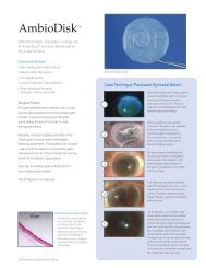
Glaucoma drainage device and patch grafts - IOP Inc
Glaucoma drainage device and patch grafts - IOP Inc
Glaucoma drainage device and patch grafts - IOP Inc

Create successful ePaper yourself
Turn your PDF publications into a flip-book with our unique Google optimized e-Paper software.
G L A U CO M A<br />
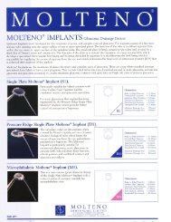
<strong>Glaucoma</strong> <strong>drainage</strong> <strong>device</strong> <strong>and</strong> <strong>patch</strong> <strong>grafts</strong>
New Molteno3 is the result of 40 years<br />
of clinical expertise with the tube <strong>and</strong><br />
plate principle pioneered by Professor<br />
Anthony Molteno. The newest, 2012<br />
enhancements, have demonstrated<br />
improved surgical utility.<br />
Low-profile pressure ridge for<br />
staged bleb management<br />
Implantation of a new Molten 3 for long-term <strong>Glaucoma</strong> treatment<br />
1<br />
The unique low-profile pressure ridge used<br />
together with tube ligation is intended to<br />
assist the process of bleb management. The<br />
pressure ridge is positioned under posterior<br />
Tenon’s capsule. The purpose is to establish<br />
the bleb system in two stages, thus tempering<br />
the effects of the subsequent hypotensive<br />
<strong>and</strong> hypertensive stages of bleb formation.<br />
Suture holes<br />
repositioned 1.0mm<br />
more anterior <strong>and</strong><br />
adjacent to the tube.<br />
Pressure ridge is<br />
set back 1.2mm for<br />
improved closure <strong>and</strong><br />
capsule formation.<br />
2<br />
3<br />
An absorbable suture is used to ligate the tube.<br />
Temporary <strong>drainage</strong> is achieved by venting<br />
the tube with “Sherwood” slits. This allows<br />
aqueous to drain under the <strong>patch</strong> graft <strong>and</strong><br />
facilitates the development of a preformed<br />
tissue capsule forming around the implant. In<br />
5 to 6 weeks the absorbable ligating suture<br />
dissolves or is released manually <strong>and</strong> the tube<br />
opens. Aqueous releases into the preformed<br />
bleb system.<br />
The pressure ridge is designed to confine<br />
aqueous to the primary <strong>drainage</strong> space. This<br />
space has a volume approximately equal to<br />
10-15% of aqueous from the anterior chamber.<br />
The primary <strong>drainage</strong> area is is intended to<br />
function like a drain with a small surface area.<br />
This enables the short-term benefit of reduced<br />
complications from excessive filtration.<br />
Improved contour<br />
Improved contour<br />
<strong>and</strong> edge detail aids<br />
insertion.<br />
Profile reduced<br />
.55mm from 1.5mm<br />
to 0.95 total height.<br />
4<br />
Aqueous begins filtering through the virgin<br />
bleb lining in the area above the pressure<br />
ridge until hypertensive resistance is induced<br />
by inflammatory components of the aqueous.<br />
The gradual increase in pressure elevates<br />
the tissue over the ridge <strong>and</strong> allows the<br />
aqueous to flow into the secondary bleb<br />
system established by the remaining surface<br />
area of the implant.<br />
5<br />
A fully functioning bleb will be visible over<br />
the entire plate surface. The plate incorporates<br />
two peripheral fenestrations. These fenestrations<br />
are intended to secure the implant in<br />
place with fibrous tissue <strong>and</strong> limit the potential<br />
for the bleb to effect rectus muscle function.<br />
MEET OUR CONSULTANTS<br />
Our surgical consultants are experts on the<br />
latest ophthalmic techniques <strong>and</strong> related<br />
technologies. They can guide you through<br />
a series of educational resources <strong>and</strong> O.R.<br />
support. Our consultants are certified<br />
<strong>and</strong> trained to support you on every level.<br />
For more information regarding<br />
staged bleb management please visit:<br />
www.iopinc.com/train
Patching glaucoma stent with<br />
processed Tutoplast <strong>patch</strong> graft<br />
Tutoplast Sclera used to <strong>patch</strong> a Molteno3.<br />
1<br />
A section of Tutoplast processed<br />
sclera is soaked in antibiotic fluid <strong>and</strong><br />
laid over the tube. It is trimmed to an<br />
appropriate size <strong>and</strong> shape with sharp<br />
Wesstcot Scissors. The <strong>patch</strong> should<br />
not extend to the Molteno3 plate<br />
posteriorly or the limbus anteriorly.<br />
• High profile collagen with a<br />
nominal thickness of 1 mm.<br />
• Multidirectional matrix for<br />
superior surgical h<strong>and</strong>ling <strong>and</strong><br />
suture utility.<br />
2<br />
The interior edge of the graft is beveled<br />
to create a partial-thickness slope.<br />
Trimming the anterior temporal<br />
corner of the <strong>patch</strong> graft also reduces<br />
the potential visibility of the <strong>patch</strong>,<br />
improving cosmesis.<br />
5mm x 8 mm<br />
3<br />
The two anterior corners of the <strong>patch</strong><br />
graft are secured with sutures. Don’t<br />
suture the two posterior corners so the<br />
tube remains uncompressed <strong>and</strong> the effectiveness<br />
of the venting slits<br />
is maintained.<br />
• High profile collagen with a<br />
nominal thickness of 1 mm.<br />
• Multidirectional matrix for<br />
superior surgical h<strong>and</strong>ling <strong>and</strong><br />
suture utility.<br />
1 cm x 1 cm<br />
4<br />
Conjunctiva <strong>and</strong> tenons are grasped<br />
together with non-toothed Pierce Hoskins<br />
tissue forceps <strong>and</strong> pulled forward to<br />
the limbus.<br />
• Bioengineered lamellar <strong>patch</strong><br />
graft consists of submucosa<br />
membrane that has been<br />
decellurized <strong>and</strong> processed to<br />
implantable <strong>device</strong> st<strong>and</strong>ards.<br />
• Clear graft option retaining<br />
strength & maximizing<br />
translucency.<br />
1 cm x 1.5 cm<br />
Conjunctiva is closed with a running<br />
9-0 Vicryl suture.<br />
5<br />
To view more techniques regarding<br />
the application of our <strong>patch</strong> <strong>grafts</strong> please visit:<br />
www.iopinc.com/train<br />
Contact your local<br />
surgical consultant.<br />
www.iopinc.com/consult<br />
Our tools. Your skills.
CODE DESCRIPTION DIMENSIONS<br />
M3-185 Molteno3 185 sq. mm<br />
M3-245 Molteno3 245 sq. mm<br />
CODE DESCRIPTION DIMENSIONS<br />
68333 Tutoplast Sclera 0.5 x 0.8 cm<br />
68337 Tutoplast Sclera 0.6 x 1.0 cm<br />
68334 Tutoplast Sclera 1 x 3 cm<br />
CODE DESCRIPTION DIMENSIONS<br />
68250 Tutoplast Pericardium 1.5 x 1.5 cm<br />
68260 Tutoplast Pericardium 2 x 3 cm<br />
68252 Tutoplast Pericardium 4 x 5 cm<br />
68257 Tutoplast Pericardium 5 x 5 cm<br />
68254 Tutoplast Pericardium 6 x 12 cm<br />
68255 Tutoplast Pericardium 2 x 5 cm<br />
68256 Tutoplast Pericardium 1.0 x 2.5 cm<br />
CODE DESCRIPTION DIMENSIONS<br />
TK-41015 keraSys Patch Graft 1cm x 1.5cm<br />
3184-B Airway Avenue<br />
Costa Mesa, CA 92626 USA<br />
Tel 714.549.1185<br />
Fax 714.549.0557<br />
molteno3@iopinc.com<br />
Customer Service<br />
800.535.3545<br />
iopinc.com<br />
Molteno3 is a registered trademark of Molteno Ophthalmic Ltd. <strong>and</strong> is used with permission. Tutoplast is a registered trademark or RTI Biologics<br />
<strong>and</strong> is used with permission. <strong>IOP</strong>atch <strong>and</strong> keraSys are trademarks of <strong>IOP</strong> Ophthalmics. © 2012 <strong>IOP</strong>, <strong>Inc</strong>. <strong>IOP</strong>-1076.00