WET LAB DNA Barcoding: From Samples to Sequences - Northwest ...
WET LAB DNA Barcoding: From Samples to Sequences - Northwest ... WET LAB DNA Barcoding: From Samples to Sequences - Northwest ...
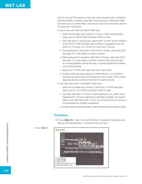
WET LAB load 10 µl of the PCR reaction on their gel, which should contain a sufficient quantity of DNA to visualize using either staining protocol. Alternative DNA gel stains (such as Carolina Blue) could also be used. For more details, see the manufacturer’s instructions. ‣ Quick Stain with 100X Fast Blast DNA stain: i. Dilute the Fast Blast stock solution 1:5 (final is 100X) with deionized water, such as 100 ml 500X Fast Blast in 400 ml water. ii. Stain each gel in a staining tray, Tupperware ® , or other similar container in 40–50 ml of 100X Fast Blast stain (enough to completely cover the gel) for 2–3 minutes. Do not stain for more than 3 minutes. iii. Rinse each gel for 10 seconds in 500–700 ml of clean, warm (40–55°C) tap water in a 1-liter beaker or similar container. iv. Wash each gel for 5 minutes in 500–700 ml of clean, warm (40–55°C) tap water in a 1-liter beaker or similar container. Place gel and water on a rocking platform during the wash, or gently agitate the container every 30–60 seconds. v. Repeat the 5 minute wash step with fresh, warm water. vi. Visualize results by placing gels on a white light box, or in sandwichsized zip-top plastic bags and holding them up to a light. Gels in plastic bags may also be scanned and printed for student records. ‣ Overnight Stain with 1X Fast Blast DNA stain: i. Dilute the Fast Blast stock solution 1:500 (final is 1X) with deionized water, such as 1 ml of 500X Fast Blast in 499 ml water. ii. Stain gels overnight in 1X stain in a gel staining tray (i.e., plastic tub or Tupperware ® ), rocking or agitating occasionally if possible. You should begin to see DNA bands after 2 hours, but at least 8 hours of staining is recommended for complete visualization. iii. Visualize results as described above under the Quick Stain procedure, Step vi. Procedure Using Bioinformatics: Genetic Research Wet Lab: Slide #11 19. Show Slide #11, “Lab 3: Did Your PCR Work? Analyzing PCR Results with Agarose Gel Electrophoresis” as students enter the room. 334 ©Northwest Association for Biomedical Research—Updated October 2012
WET LAB 20. Show Slide #12, and remind students that their overall goal is to obtain DNA sequence data from the species they are studying. In the last experiment, students copied, or amplified, the barcoding gene from their purified DNA samples. Today, they will use agarose gel electrophoresis to determine whether their PCR was successful. Wet Lab: Slide #12 21. Show Slide #13, and tell students that agarose gel electrophoresis is a method used by scientists to separate DNA molecules by size. DNA is placed at the top of the gel, which is a solid, porous material. An electric current is passed through the gel, and the negatively-charged DNA at the top of the gel moves toward the positively-charged electrode at the bottom of the gel. Wet Lab: Slide #13 22. Walk through the gel image on Slide #13 and highlight these key points with students: a. Each DNA sample is in a different vertical lane. b. Every DNA gel includes a molecular weight standard, which contains DNA bands of known size that scientists use to estimate the size of the DNA bands in their sample. In this gel, the molecular weight standard is in Lane 1. Molecular weight standard: Sometimes called a “molecular weight marker” or a “DNA ladder” and abbreviated “MW, this is a mixture of DNA fragments of known size used to identify the approximate size of a molecule run on a gel, using the principle that molecular weight is inversely proportional to migration rate through a gel matrix. Therefore, when used in gel electrophoresis, standards effectively provide a logarithmic scale by which to estimate the size of the other fragments (providing the fragment sizes of the standard are known). Standards are loaded in lanes adjacent to sample lanes before the run commences. Wet Lab – DNA Barcoding: From Samples to Sequences 335 ©Northwest Association for Biomedical Research—Updated October 2012
- Page 1 and 2: WET LAB DNA Barcoding: From Samples
- Page 3 and 4: WET LAB Lab 1: DNA Purification for
- Page 5 and 6: WET LAB Lab 4: Preparation of PCR S
- Page 7 and 8: WET LAB • Teachers may wish to ha
- Page 9 and 10: WET LAB Wet Lab: Slide #3 Lysis or
- Page 11 and 12: WET LAB • If students are not fam
- Page 13 and 14: WET LAB Wet Lab: Slide #9 14. Show
- Page 15: WET LAB Lab 3: Analyzing PCR Result
- Page 19 and 20: WET LAB Wet Lab: Slide #15 26. Tell
- Page 21 and 22: WET LAB Wet Lab: Slide #18 Wet Lab:
- Page 23 and 24: WET LAB 34. Optional Graphing Exten
- Page 25 and 26: WET LAB e. Deoxynucleotides (dNTPs)
- Page 27 and 28: WET LAB Glossary Agarose gel electr
- Page 29 and 30: WET LAB PCR beads: Lyophilized or
- Page 31 and 32: WET LAB CLASS SET Lab 1: DNA Purifi
- Page 33 and 34: WET LAB CLASS SET Day 2 Procedure:
- Page 35 and 36: WET LAB CLASS SET 14. Place the spi
- Page 37 and 38: WET LAB CLASS SET Lab 2: Copying th
- Page 39 and 40: WET LAB CLASS SET 9. Add each of th
- Page 41 and 42: WET LAB CLASS SET Lab 3: Analyzing
- Page 43 and 44: WET LAB CLASS SET 11. To prepare yo
- Page 45 and 46: WET LAB CLASS SET Lab 4: Preparatio
- Page 47 and 48: WET LAB CLASS SET On your separate
- Page 49 and 50: WET LAB KEY Lab 1: DNA Purification
- Page 51 and 52: WET LAB KEY Lab 2: Copying the DNA
- Page 53 and 54: WET LAB KEY Lab 4: Preparation of P
- Page 55 and 56: WET LAB RESOURCE Aliquoting DNA Bar
<strong>WET</strong> <strong>LAB</strong><br />
load 10 µl of the PCR reaction on their gel, which should contain a sufficient<br />
quantity of <strong>DNA</strong> <strong>to</strong> visualize using either staining pro<strong>to</strong>col. Alternative <strong>DNA</strong><br />
gel stains (such as Carolina Blue) could also be used. For more details, see the<br />
manufacturer’s instructions.<br />
‣ Quick Stain with 100X Fast Blast <strong>DNA</strong> stain:<br />
i. Dilute the Fast Blast s<strong>to</strong>ck solution 1:5 (final is 100X) with deionized<br />
water, such as 100 ml 500X Fast Blast in 400 ml water.<br />
ii. Stain each gel in a staining tray, Tupperware ® , or other similar container<br />
in 40–50 ml of 100X Fast Blast stain (enough <strong>to</strong> completely cover the<br />
gel) for 2–3 minutes. Do not stain for more than 3 minutes.<br />
iii. Rinse each gel for 10 seconds in 500–700 ml of clean, warm (40–55°C)<br />
tap water in a 1-liter beaker or similar container.<br />
iv. Wash each gel for 5 minutes in 500–700 ml of clean, warm (40–55°C)<br />
tap water in a 1-liter beaker or similar container. Place gel and water<br />
on a rocking platform during the wash, or gently agitate the container<br />
every 30–60 seconds.<br />
v. Repeat the 5 minute wash step with fresh, warm water.<br />
vi. Visualize results by placing gels on a white light box, or in sandwichsized<br />
zip-<strong>to</strong>p plastic bags and holding them up <strong>to</strong> a light. Gels in plastic<br />
bags may also be scanned and printed for student records.<br />
‣ Overnight Stain with 1X Fast Blast <strong>DNA</strong> stain:<br />
i. Dilute the Fast Blast s<strong>to</strong>ck solution 1:500 (final is 1X) with deionized<br />
water, such as 1 ml of 500X Fast Blast in 499 ml water.<br />
ii. Stain gels overnight in 1X stain in a gel staining tray (i.e., plastic tub or<br />
Tupperware ® ), rocking or agitating occasionally if possible. You should<br />
begin <strong>to</strong> see <strong>DNA</strong> bands after 2 hours, but at least 8 hours of staining is<br />
recommended for complete visualization.<br />
iii. Visualize results as described above under the Quick Stain procedure, Step vi.<br />
Procedure<br />
Using Bioinformatics: Genetic Research<br />
Wet Lab: Slide #11<br />
19. Show Slide #11, “Lab 3: Did Your PCR Work? Analyzing PCR Results with<br />
Agarose Gel Electrophoresis” as students enter the room.<br />
334<br />
©<strong>Northwest</strong> Association for Biomedical Research—Updated Oc<strong>to</strong>ber 2012


