Cnideria and Ctenophora
Cnideria and Ctenophora
Cnideria and Ctenophora

Create successful ePaper yourself
Turn your PDF publications into a flip-book with our unique Google optimized e-Paper software.
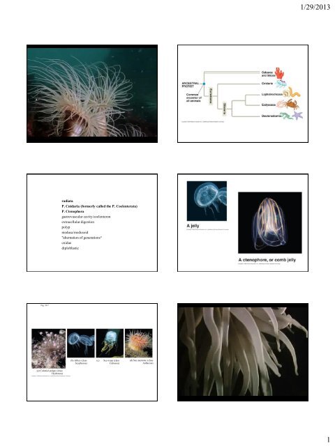
1/29/2013<br />
radiata<br />
P. Cnidaria (formerly called the P. Coelenterata)<br />
P. <strong>Ctenophora</strong><br />
gastrovascular cavity/coelenteron<br />
extracellular digestion<br />
polyp<br />
medusa/medusoid<br />
"alternation of generations“<br />
cnidae<br />
diploblastic<br />
Fig. 33-7<br />
(b) Jellies (class<br />
Scyphozoa)<br />
(c)<br />
Sea wasp (class<br />
Cubozoa)<br />
(d) Sea anemone (class<br />
Anthozoa)<br />
(a) Colonial polyps (class<br />
Hydrozoa)<br />
1
1/29/2013<br />
Fig. 13.2<br />
radiata<br />
P. Cnidaria (formerly called the P. Coelenterata)<br />
P. <strong>Ctenophora</strong><br />
gastrovascular cavity/coelenteron<br />
extracellular digestion<br />
polyp<br />
medusa/medusoid<br />
"alternation of generations“<br />
cnidae<br />
diploblastic<br />
Fig. 13.9 Figure 06_01<br />
Hydra<br />
-fresh water<br />
-no medusa<br />
-not colonial<br />
2
1/29/2013<br />
Fig. 13.3<br />
epidermis<br />
epitheliomuscular cell<br />
myofibrils<br />
interstitial cell<br />
cnidocyte<br />
cnidocil<br />
nematocyst<br />
operculum<br />
mucus-secreting cell (especially on basal disc)<br />
receptor/sensory cell<br />
nerve cell/neuron (form nerve nets)<br />
gastrodermis (mucus secreting cell, nerve cell/net,<br />
<strong>and</strong>…)<br />
nutritive-muscular cell<br />
gl<strong>and</strong> cell<br />
Figure 06_03<br />
3
1/29/2013<br />
Figure 06_02<br />
Fig. 13.7<br />
Dioecious/gonochoristic with “direct”<br />
development<br />
4
1/29/2013<br />
C. Hydrozoa<br />
hydroid colony<br />
Obelia<br />
monopodial<br />
sympodial<br />
polymorphism<br />
gastrozooid<br />
gonozooid<br />
Physalia<br />
gonophore<br />
dactylozooid<br />
hydromedusa<br />
velum<br />
manubrium<br />
planula<br />
actinula<br />
medusoid suppression<br />
hydrocorals<br />
Obelia, a<br />
colonial hydroid<br />
Figure 06_14 Fig. 13.9<br />
5
1/29/2013<br />
C. Hydrozoa<br />
hydroid colony<br />
Obelia<br />
monopodial<br />
sympodial<br />
polymorphism<br />
gastrozooid<br />
gonozooid<br />
Physalia<br />
gonophore<br />
dactylozooid<br />
hydromedusa<br />
velum<br />
manubrium<br />
planula<br />
actinula<br />
medusoid suppression<br />
hydrocorals<br />
Figure 06_16cont<br />
Figure 06_15<br />
C. Hydrozoa<br />
hydroid colony<br />
Obelia<br />
monopodial<br />
sympodial<br />
polymorphism<br />
gastrozooid<br />
gonozooid<br />
Physalia<br />
gonophore<br />
dactylozooid<br />
hydromedusa<br />
velum<br />
manubrium<br />
planula<br />
actinula<br />
medusoid suppression<br />
hydrocorals<br />
6
1/29/2013<br />
Figure 06_11<br />
planula larvae<br />
“medusoid suppression”<br />
hydrocorals<br />
7
1/29/2013<br />
Figure 06_06<br />
Figure 06_05<br />
C. Scyphozoa<br />
jellyfishes<br />
scyphomedusa<br />
no velum<br />
manubrium stomach gastric pouches<br />
manubrium with oral arms<br />
gastrovascular canals<br />
rhopalium<br />
statocyst<br />
ocelli<br />
planula scyphistoma strobila ephyrae<br />
scyphomedusa<br />
8
1/29/2013<br />
Figure 06_07 Figure 06_08<br />
Figure 06_10<br />
Class Cubozoa, a cuboidal medusa<br />
9
1/29/2013<br />
Figure 06_18<br />
C. Anthozoa<br />
sea anemone<br />
siphonoglyph<br />
biradial symmetry<br />
acrorhagi<br />
corals<br />
10
1/29/2013<br />
Figure 06_20<br />
Acrorhagi used in defense against other anemones<br />
Figure 06_22<br />
Figure 06_23<br />
11
1/29/2013<br />
Text Art 06_02<br />
The demise of a phylum of protists: phylogeny of Myxozoa <strong>and</strong> other<br />
parasitic cnidaria. Siddall ME, Martin DS, Bridge D, Desser SS, Cone DK<br />
(1995)<br />
Abstract<br />
The notion that members of the phylum Myxozoa Grassé, 1970 do not properly belong in<br />
classifications of protists has frequently been suggested because the infective spores of these<br />
parasites are not unicellular. Systematists have failed to be decisive about myxozoan phylogenetic<br />
affinities, either finding the suggestion of a cnidarian connection to be preposterous or considering<br />
the recent suggestion of a relationship with nematodes to be an obvious failure of molecular<br />
phylogenetics. Thus, the group has remained in classifications as a protistan phylum in its own right.<br />
The ultrastructure of the development of myxozoans was critically re-examined in order to more<br />
fully explore the possibility of morphological synapomorphies with metazoan taxa. These<br />
morphological characters, in combination with small ribosomal subunit gene sequences, were used in<br />
a phylogenetic analysis in order to assess myxozoan origins. The results unequivocally support the<br />
inclusion of myxozoans as a clade of highly derived parasitic cnidarians, <strong>and</strong> as sister taxon to the<br />
narcomedusan Polypodium hydriforme. Reassessment of myxozoans as metazoans reveals terminal<br />
differentiation, typical metazoan cellular junctions, <strong>and</strong> collagen production. Their "polar capsules"<br />
are redescribed as typical nematocysts bearing atrichous isorhiza. Insofar as taxa cannot be<br />
contained within other taxa of equal rank, the phylum Myxozoa is ab<strong>and</strong>oned <strong>and</strong> it is recommended<br />
that the group as a whole be removed from all protistan classifications <strong>and</strong> placed in a more<br />
comprehensive cnidarian system.<br />
12
1/29/2013<br />
Metazoan characteristics of the myxozoan Thelohanellus nikolskii<br />
A. Intercellular<br />
cytoplasmic<br />
communication seen<br />
between arrows.<br />
Scale bar = 1 um<br />
B. Septate (sj) <strong>and</strong><br />
desmosomal (dj)<br />
junctions with<br />
characteristic<br />
tonofilaments (t)<br />
bewteen adjacent<br />
sporogenic cells (s).<br />
Scale bar = 1 um<br />
Cnidarian characteristics of the myxozoan Thelohanellus nikolskii<br />
C. Extracellular<br />
production of crosslinked<br />
collagen (cf)<br />
by a pansporoblast<br />
(PS), arrow indicates<br />
active exocytosis of<br />
tropocollagen (tc).<br />
Scale bar = 1 um<br />
Structures seen in the development of<br />
the so-called "polar capsule" of<br />
Thelohanellus nikolskii labeled in<br />
accordance with the homologous<br />
structure in cnidarian nematocysts.<br />
Figures A through D represent the<br />
earliest stage in cellular ontogeny<br />
through to that of the mature<br />
nematocyst respectively.<br />
c, cnidocyte; pt, preinverted tubule; np,<br />
nematocyst primordium;<br />
s, differentiating sporogenic cells;<br />
th, tubular thread<br />
Scale bar = 1 um<br />
c, cnidocyte; np, nematocyst<br />
primordium; it, inverted tubule;<br />
s, differentiating sporogenic cells.<br />
ai, atrichous izorhiza, sh, shaft of<br />
atrichous izorhiza; op, opercular<br />
cap of atrichous izorhiza; N,<br />
mature nematocyst Scale bar = 1<br />
um.<br />
From combined morphological <strong>and</strong> 18S rDNA characters<br />
13
1/29/2013<br />
Text Art 06_02 Fig. 13.38<br />
Figure 07_06<br />
Chapter opener 07<br />
P. <strong>Ctenophora</strong><br />
comb jelly<br />
comb rows<br />
colloblast<br />
anal pores<br />
apical sense organ<br />
modified medusa?<br />
14
1/29/2013<br />
Figure 07_04 Figure 07_01<br />
Figure 07_02<br />
Table 07_01 Figure 07_05<br />
15