Epstein-Barr Virus VCA IFA IgG (English) - Focus Diagnostics
Epstein-Barr Virus VCA IFA IgG (English) - Focus Diagnostics
Epstein-Barr Virus VCA IFA IgG (English) - Focus Diagnostics
You also want an ePaper? Increase the reach of your titles
YUMPU automatically turns print PDFs into web optimized ePapers that Google loves.
phosphate buffered saline which removes unbound serum antibodies. In the<br />
second stage, each antigen well is overlaid with fluorescein-labeled antibody<br />
to human <strong>IgG</strong>. The slide is incubated allowing antigen-antibody complexes to<br />
react with the fluorescein labeled anti-human <strong>IgG</strong>. After the slide is washed,<br />
dried, and mounted, it is examined using fluorescence microscopy. Positive<br />
reactions appear as cells exhibiting bright apple-green cytoplasmic<br />
fluorescence against a background of <strong>VCA</strong> negative control cells. Semiquantitative<br />
endpoint titers are obtained by testing serial dilutions of positive<br />
specimens.<br />
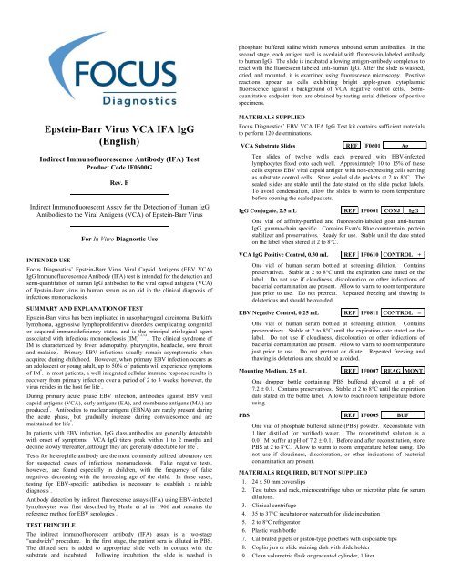
<strong>Epstein</strong>-<strong>Barr</strong> <strong>Virus</strong> <strong>VCA</strong> <strong>IFA</strong> <strong>IgG</strong><br />
(<strong>English</strong>)<br />
Indirect Immunofluorescence Antibody (<strong>IFA</strong>) Test<br />
Product Code IF0600G<br />
Rev. E<br />
Indirect Immunofluorescent Assay for the Detection of Human <strong>IgG</strong><br />
Antibodies to the Viral Antigens (<strong>VCA</strong>) of <strong>Epstein</strong>-<strong>Barr</strong> <strong>Virus</strong><br />
For In Vitro Diagnostic Use<br />
INTENDED USE<br />
<strong>Focus</strong> <strong>Diagnostics</strong>’ <strong>Epstein</strong>-<strong>Barr</strong> <strong>Virus</strong> Viral Capsid Antigens (EBV <strong>VCA</strong>)<br />
<strong>IgG</strong> Immunofluorescence Antibody (<strong>IFA</strong>) test is intended for the detection and<br />
semi-quantitation of human <strong>IgG</strong> antibodies to the viral capsid antigens (<strong>VCA</strong>)<br />
of <strong>Epstein</strong>-<strong>Barr</strong> virus in human serum as an aid in the clinical diagnosis of<br />
infectious mononucleosis.<br />
SUMMARY AND EXPLANATION OF TEST<br />
<strong>Epstein</strong>-<strong>Barr</strong> virus has been implicated in nasopharyngeal carcinoma, Burkitt's<br />
lymphoma, aggressive lymphoproliferative disorders complicating congenital<br />
or acquired immunodeficiency states, and is the principal etiological agent<br />
associated with infectious mononucleosis (IM) 1,2,3,4 . The clinical syndrome of<br />
IM is characterized by fever, adenopathy, pharyngitis, headache, sore throat<br />
and malaise 5 . Primary EBV infections usually remain asymptomatic when<br />
acquired during childhood. However, when primary EBV infection occurs as<br />
an adolescent or young adult, up to 50% of patients will experience symptoms<br />
of IM 6 . In most patients, a well integrated cellular immune response results in<br />
recovery from primary infection over a period of 2 to 3 weeks; however, the<br />
virus resides in the host for life 6 .<br />
During primary acute phase EBV infection, antibodies against EBV viral<br />
capsid antigens (<strong>VCA</strong>), early antigens (EA), and membrane antigens (MA) are<br />
produced 6 . Antibodies to nuclear antigens (EBNA) are rarely present during<br />
the acute phase, but gradually increase during convalescence and are<br />
maintained for life 6 .<br />
In patients with EBV infection, <strong>IgG</strong> class antibodies are generally detectable<br />
with onset of symptoms. <strong>VCA</strong> <strong>IgG</strong> titers peak within 1 to 2 months and<br />
decline slowly thereafter, although they are generally detectable for life 7 .<br />
Tests for heterophile antibody are the most commonly utilized laboratory test<br />
for suspected cases of infectious mononucleosis. False negative tests,<br />
however, are found especially in children, with the frequency of false<br />
negatives decreasing with the increasing age of the child. In these cases,<br />
testing for EBV-specific antibodies is necessary to establish a reliable<br />
diagnosis 8 .<br />
Antibody detection by indirect fluorescence assays (<strong>IFA</strong>) using EBV-infected<br />
lymphocytes was first described by Henle et al in 1966 and remains the<br />
reference method for EBV serologies 9 .<br />
TEST PRINCIPLE<br />
The indirect immunofluorescent antibody (<strong>IFA</strong>) assay is a two-stage<br />
"sandwich" procedure. In the first stage, the patient sera is diluted in PBS.<br />
The diluted sera is added to appropriate slide wells in contact with the<br />
substrate and incubated. Following incubation, the slide is washed in<br />
MATERIALS SUPPLIED<br />
<strong>Focus</strong> <strong>Diagnostics</strong>’ EBV <strong>VCA</strong> <strong>IFA</strong> <strong>IgG</strong> Test kit contains sufficient materials<br />
to perform 120 determinations.<br />
<strong>VCA</strong> Substrate Slides REF IF0601 Ag<br />
Ten slides of twelve wells each prepared with EBV-infected<br />
lymphocytes fixed onto each well. Approximately 10 to 15% of these<br />
cells express EBV viral capsid antigen with non-expressing cells serving<br />
as substrate control cells. Store sealed slide packets at 2 to 8°C. The<br />
sealed slides are stable until the date stated on the slide packet labels.<br />
To avoid condensation, allow the slides to warm to room temperature<br />
before opening the sealed packets.<br />
<strong>IgG</strong> Conjugate, 2.5 mL REF IF0001 CONJ <strong>IgG</strong><br />
One vial of affinity-purified and fluorescein-labeled goat anti-human<br />
<strong>IgG</strong>, gamma-chain specific. Contains Evan's Blue counterstain, protein<br />
stabilizer and preservatives. Ready for use. Stable until the date stated<br />
on the label when stored at 2 to 8°C.<br />
<strong>VCA</strong> <strong>IgG</strong> Positive Control, 0.30 mL REF IF0610 CONTROL +<br />
One vial of human serum bottled at screening dilution. Contains<br />
preservatives. Stable at 2 to 8°C until the expiration date stated on the<br />
label. Do not use if cloudiness, discoloration or other indications of<br />
bacterial contamination are present. Allow to warm to room temperature<br />
just prior to use. Do not pretreat. Repeated freezing and thawing is<br />
deleterious and should be avoided.<br />
EBV Negative Control, 0.25 mL REF IF0811 CONTROL −<br />
One vial of human serum bottled at screening dilution. Contains<br />
preservatives. Stable at 2 to 8°C until the expiration date stated on the<br />
label. Do not use if cloudiness, discoloration or other indications of<br />
bacterial contamination are present. Allow to warm to room temperature<br />
just prior to use. Do not pretreat or dilute. Repeated freezing and<br />
thawing is deleterious and should be avoided.<br />
Mounting Medium, 2.5 mL REF IF0007 REAG MONT<br />
One dropper bottle containing PBS buffered glycerol at a pH of<br />
7.2 ± 0.1. Contains preservatives. Stable at 2 to 8°C until the expiration<br />
date stated on the bottle label. Allow to reach room temperature before<br />
using.<br />
PBS REF IF0005 BUF<br />
One vial of phosphate buffered saline (PBS) powder. Reconstitute with<br />
1 liter distilled (or purified) water. The reconstituted solution is a<br />
0.01 M buffer at pH of 7.2 ± 0.1. Before and after reconstitution, store<br />
PBS at 2 to 8°C. Allow to warm to room temperature before using. Do<br />
not use if cloudiness, discoloration, or other indications of bacterial<br />
contamination are present.<br />
MATERIALS REQUIRED, BUT NOT SUPPLIED<br />
1. 24 x 50 mm coverslips<br />
2. Test tubes and rack, microcentrifuge tubes or microtiter plate for serum<br />
dilutions.<br />
3. Clinical centrifuge<br />
4. 35 to 37°C incubator or waterbath for slide incubation<br />
5. 2 to 8°C refrigerator<br />
6. Plastic wash bottle<br />
7. Calibrated pipets or piston-type pipettors with disposable tips<br />
8. Coplin jars or slide staining dish with slide holder<br />
9. Clean volumetric flask or graduated cylinder, 1 liter
FOCUS <strong>Diagnostics</strong><br />
10. Humid chamber for incubation of slides<br />
11. Distilled or purified water<br />
12. Timer<br />
13. Absorbant paper for blotting slides<br />
14. Fluorescence microscope, recommended parameters<br />
Excitation Filter 470–490nm<br />
<strong>Barr</strong>ier Filter 520–560nm<br />
Light Source HBO 100W, mercury<br />
Objective 20–40X, fluorescence quality, high dry<br />
SHELF LIFE AND HANDLING<br />
1. Kits are stable through the end of the month indicated in the expiration<br />
date when stored at 2 to 8°C.<br />
2. Do not use test kit or reagents beyond their expiration dates.<br />
3. Do not expose reagents to strong light during storage or incubation.<br />
WARNINGS AND PRECAUTIONS:<br />
1. This kit is for in vitro diagnostic use only.<br />
2. All blood products should be treated as potentially infectious. Source<br />
materials from which this product (including controls) was derived have<br />
been screened for Hepatitis B surface antigen, Hepatitis C antibody and<br />
HIV-1/2 (AIDS) antibody by FDA-approved methods and found to be<br />
negative. However, as no known test methods can offer 100% assurance<br />
that products derived from human blood will not transmit these or other<br />
infectious agents, all controls, serum specimens and equipment coming<br />
into contact with these specimens should be considered potentially<br />
infectious and decontaminated or disposed of using proper biohazard<br />
precautions.<br />
3. Evan's Blue is a carcinogen. Avoid contact with skin or eyes.<br />
4. Do not substitute or mix reagents from different kit lots or from other<br />
manufacturers.<br />
5. Use only protocols described in this insert. Incubation times or<br />
temperatures other than those specified may give erroneous results.<br />
6. Cross-contamination of patient specimens on a slide can cause erroneous<br />
results. Add patient specimens and handle slides carefully to avoid<br />
mixing of sera from adjoining wells.<br />
7. Bacterial contamination of serum specimens or reagents can produce<br />
erroneous results. Use aseptic techniques to avoid microbial<br />
contamination.<br />
8. Mounting Medium contains 30 to 60 % glycerol which may cause<br />
irritation upon inhalation or skin contact. Upon inhalation or contact,<br />
first aid measures should be taken.<br />
SPECIMEN COLLECTION AND PREPARATION<br />
Serum is the preferred specimen source. No attempt has been made to assess<br />
the assay's compatibility with other specimens. Hyperlipemic, hemolyzed, or<br />
contaminated sera may cause erroneous results; therefore, their use should be<br />
avoided.<br />
Specimen Collection and Handling<br />
Collect blood samples aseptically using approved venipuncture<br />
techniques by qualified personnel. Allow blood samples to clot at room<br />
temperature prior to centrifugation. Aseptically transfer serum to a<br />
tightly closing sterile container for storage at 2 to 8°C. If testing is to be<br />
delayed longer than 5 days, the sample should be frozen at –20°C or<br />
colder. Freeze-thaw damage can result if specimens are frozen in selfdefrosting<br />
freezers. Thaw and mix samples well prior to use.<br />
Specimen Preparation<br />
Prepare 1:10 screening dilutions of patient sera as follows:<br />
mix 10 µL of patient serum with 90 µL PBS in microcentrifuge tubes<br />
or a microtiter plate.<br />
Where it is necessary to determine endpoint titers, dilute the screening<br />
dilution serially with PBS.<br />
TEST PROCEDURE<br />
1. Remove slides from cold storage. To avoid condensation, allow slides<br />
to reach room temperature before opening slide packets.<br />
2. Apply 15 µL of Positive Control, as bottled, to the appropriate slide<br />
well. Use PBS to serially dilute the Positive Control 16-fold beyond the<br />
EBV <strong>VCA</strong> <strong>IFA</strong> <strong>IgG</strong><br />
Page 2<br />
bottled dilution. Apply 15 µL of each serial dilution to an appropriate<br />
slide well.<br />
3. Apply 15 µL of EBV Negative Control, as bottled, to the appropriate<br />
well. Do not dilute the Negative Control.<br />
4. For each patient sample to be tested, add approximately 15 µL of the<br />
prepared sample dilutions (see Specimen Preparation, above) to an<br />
appropriate slide well. Make notations to later identify each well when<br />
reading the results.<br />
5. Incubate slide(s) in a humid chamber for 30 ± 2 minutes at 35 to 37°C.<br />
6. Remove slides from the humid chamber and gently rinse each slide with<br />
a stream of PBS. Do not aim the stream of PBS directly at the slide<br />
wells. Rinse one row at a time to avoid mixing of specimens. Wash<br />
slides by submersing the rinsed slides into Coplin or slide staining jars<br />
containing PBS for 10 minutes.<br />
7. Dip the washed slides briefly in distilled or purified water, and allow the<br />
slides to air dry.<br />
8. Add approximately 15 µL <strong>IgG</strong> Conjugate to each slide well.<br />
9. Incubate slides in a humid chamber for 30 ± 2 minutes at 35 to 37°C.<br />
10. Repeat wash steps 6 and 7.<br />
11. Place a few drops of Mounting Medium on the slide and cover with a<br />
24 x 50mm coverslip. Remove any air bubbles and excess Mounting<br />
Medium with absorbant paper.<br />
12. View wells at a final magnification of 200X on a properly equipped<br />
fluorescence microscope. For optimum fluorescence, read slides the<br />
same day the assay is performed. If this is not possible, store in the dark<br />
at 2 to 8°C up to 24 hours.<br />
QUALITY CONTROL<br />
Each run (each time a slide, or group of slides, is processed) should include<br />
both Positive and Negative controls.<br />
1. The Positive Control should endpoint (1+fluorescence) at 8 fold beyond<br />
the bottled dilution. However, due to differing laboratory conditions,<br />
including equipment, the endpoint may range from 4 to 16 fold beyond<br />
the bottled dilution.<br />
2. The Negative Control should be negative at the bottled dilution. All of<br />
the cells should appear pale green to red in color.<br />
If controls do not exhibit these results, patient test results should be<br />
considered invalid and the assay repeated.<br />
INTERPRETATION OF TEST RESULTS<br />
Microscope optics, light source condition and type will determine overall<br />
fluorescent intensity and endpoint titers. Read control wells first during every<br />
run to ensure correct interpretation.<br />
Reading the Slides<br />
Read the fluorescent intensity of lymphocytes on each well, and grade<br />
the fluorescence as follows:<br />
2 to 4+: Moderate to intense apple-green fluorescence of 10 to 15% of<br />
lymphocytes.<br />
1+: Definite, but dim fluorescence equivalent to that observed for<br />
the Positive Control at its reference endpoint titer.<br />
Negative: No fluorescence or fluorescence equal to that observed in the<br />
Negative Control well.<br />
Interpreting the Patient Specimen Results<br />
The reciprocal of the highest serum dilution that exhibits definite (1+)<br />
apple-green fluorescence in 10 to 15% of the cells is termed the serum<br />
endpoint titer.<br />
≥ 1:10 <strong>VCA</strong> <strong>IgG</strong> endpoint titers of 1:10 and greater are indicative of<br />
EBV infection at an undetermined time. Such persons are<br />
immune to contracting infectious mononucleosis. To determine<br />
whether the detected infection is recent or past in nature,<br />
correlate results with EBV <strong>VCA</strong> IgM and EBNA antibody titers.<br />
< 1:10 <strong>VCA</strong> <strong>IgG</strong> endpoint titers less than 1:10 suggest that the patient<br />
has neither recent nor past EBV infection. Such persons are<br />
susceptible to infectious mononucleosis.
FOCUS <strong>Diagnostics</strong><br />
Non-specific Fluorescence<br />
Naturally occurring cellular antigens expressed on the host cell surface<br />
may react with some patient sera. These cellular antigens are common to<br />
all cells; therefore, these sera are detected by the fact that they react with<br />
all or most of the substrate cells rather than just the 10 to 15% <strong>VCA</strong><br />
positive cells. However, if the <strong>VCA</strong> titer exceeds the anti-cell titer, these<br />
sera may still be tested. In cases where the result is uninterpretable, a<br />
second serum specimen and/or an alternate methodology may be<br />
required.<br />
LIMITATIONS<br />
1. Diagnosis of recent EBV infection based on a single elevated <strong>IgG</strong> titer is<br />
complicated by the slow decline of antibody titer from past infection in<br />
many individuals. Titers may remain elevated for longer than 12<br />
months.<br />
2. Samples obtained too early during primary infection may not contain<br />
detectable antibodies. If EBV infection is suspected, a second sample<br />
should be obtained 10 to 21 days later and tested in parallel with the<br />
original sample to look for seroconversion.<br />
3. All results from this and other EBV serologies must be correlated with<br />
clinical history and other data available to the attending physician.<br />
EXPECTED VALUES<br />
Approximately 80 to 90% of the U.S. adult population is positive for antibody<br />
to EBV <strong>VCA</strong> 7 .<br />
Forty-seven (47) healthy laboratory workers from Cypress, CA were tested for<br />
EBV <strong>VCA</strong> <strong>IgG</strong> antibodies with the <strong>Focus</strong> <strong>Diagnostics</strong> EBV <strong>VCA</strong> <strong>IFA</strong> <strong>IgG</strong><br />
test. Titers ranged from 1:40 to 1:5120. The highest titer was found in a<br />
person approximately one-year convalescent from infectious mononucleosis.<br />
The median titer was 1:640. The normal range was considered to be 1:160 to<br />
1:2560.<br />
SPECIFIC PERFORMANCE CHARACTERISTICS<br />
Sensitivity and Specificity<br />
<strong>Focus</strong> <strong>Diagnostics</strong>' EBV <strong>VCA</strong> <strong>IFA</strong> <strong>IgG</strong> test was compared to a<br />
commercial EBV <strong>VCA</strong> <strong>IFA</strong> <strong>IgG</strong> test performed by a reference<br />
laboratory. Overall correlation, sensitivity, and specificity were<br />
determined for 10 acute infectious mononucleosis specimens, 11<br />
convalescent (past infections), and 11 EBV seronegative specimens.<br />
The <strong>Focus</strong> <strong>Diagnostics</strong> EBV <strong>VCA</strong> <strong>IFA</strong> <strong>IgG</strong> test had an overall<br />
agreement of 100% with the other EBV <strong>VCA</strong> <strong>IFA</strong> <strong>IgG</strong> test. The two<br />
tests were within a single two-fold dilution when determining endpoint<br />
titer on 70% of the positives and within two dilutions on 85%.<br />
Sensitivity and specificity were also calculated to be 100%, although<br />
both tests missed two acute infectious mononucleosis patients, where the<br />
titer of the <strong>VCA</strong> IgM was sufficiently elevated such that competitive<br />
binding affects produced <strong>IgG</strong> false-negative results.<br />
Cross-reaction<br />
A panel of sera with high titers against varicella-zoster, cytomegalovirus<br />
and herpes simplex virus types 1 and 2 were tested for reaction against<br />
<strong>VCA</strong> <strong>IgG</strong> and found to be negative. These findings suggest that titers to<br />
other human herpes viruses do not cause cross-reactions with <strong>VCA</strong> <strong>IgG</strong><br />
sufficiently to cause false positive results.<br />
Reproducibility<br />
The within run and between run variation were determined using the<br />
positive and negative specimens tested over a ten day time period. The<br />
positive and negative specimens tested correctly in each run. Comparing<br />
titers, the maximum between run variation was ± one two-fold dilution.<br />
There was no within run variation in titers when the positive control was<br />
diluted and tested with the negative specimen.<br />
Prozone<br />
Titers below 1:10,000 have shown no interference from prozone<br />
phenomena and so it is suspected that prozone will not be a complication<br />
of this indirect immunofluorescence assay.<br />
EBV <strong>VCA</strong> <strong>IFA</strong> <strong>IgG</strong><br />
Page 3<br />
REFERENCES<br />
1. <strong>Epstein</strong>, M., B. Anchong, and Y. <strong>Barr</strong>. 1964. <strong>Virus</strong> particles in cultured<br />
lymphoblasts from Burkitt's Lymphoma. Lancet i:702-703.<br />
2. Henle, G., W. Henle, and V. Diehl. 1968. Relation of Burkitt tumor<br />
associated herpes-type virus to infectious mononucleosis. Proc. Natl. Acad.<br />
Sci. USA 59:94-101.<br />
3. Henle, W., G. Henle, C. Ho, P. Burton, Y. Cachin, P. Clifford, A. De<br />
Schryver, G. de The, U. Diel and G. Klein. 1970. Antibodies to <strong>Epstein</strong>-<strong>Barr</strong><br />
<strong>Virus</strong> in Nasopharyngeal Carcinoma, Other Head and Neck Neoplasm, and<br />
Control Groups. J. Natl. Cancer Inst. 44:225-231.<br />
4. Gong, M., T. Ooka, T. Matsuo, E. Kieff. 1987. <strong>Epstein</strong>-<strong>Barr</strong> <strong>Virus</strong><br />
Glycoprotein Homologous to Herpes Simplex <strong>Virus</strong> gB. J. Virol. 61:499-<br />
508.<br />
5. Schooley, R.T., R. Dolin. 1990. <strong>Epstein</strong>-<strong>Barr</strong> <strong>Virus</strong> (infectious<br />
mononucleosis). In: Mandell G.L., R.G. Douglas, Jr., J.E. Bennett. eds.<br />
Principles and Practice of Infectious Diseases. Third edition. New York.<br />
Churchill Livingstone. 1172-85.<br />
6. Straus, S.E., J. Cohen, G. Tosato, J. Meier. 1993. <strong>Epstein</strong>-<strong>Barr</strong> <strong>Virus</strong><br />
Infections: Biology, Pathogenesis, and Management. Annals of Inter. Med.<br />
118:45-58.<br />
7. Horwitz, C., W. Henle, G. Henle, H. Rudnick, E. Latts, 1985. Long-term<br />
serological follow-up of patients for <strong>Epstein</strong>-<strong>Barr</strong> <strong>Virus</strong> after recovery from<br />
infectious mononucleosis. J. Infect. Dis. 151:1150-1153.<br />
8. Thiele, G.M., M. Okano. 1993. Diagnosis of <strong>Epstein</strong>-<strong>Barr</strong> <strong>Virus</strong> Infections<br />
in the Clinical Laboratory. Clinical Microbiology Newsletter 15(6):41-45.<br />
9. Henle, G., W. Henle. 1966. Immunofluorescence in cells derived from<br />
Burkitt's lymphoma. J. Bacteriol. 91:1248-1256.<br />
This package insert is available in French, German, Italian, and Spanish at<br />
www.focusdx.com, and is available in other languages from your local<br />
distributor.<br />
AUTHORIZED<br />
REPRESENTATIVE<br />
mdi Europa GmbH<br />
Langenhagener Str. 71, 30855<br />
Langenhagen-Hannover, Germany<br />
ORDERING INFORMATION<br />
Telephone: (800) 838-4548 (U.S.A. only)<br />
(562) 240-6500 (International)<br />
Fax: (562) 240-6510<br />
TECHNICAL ASSISTANCE<br />
Telephone: (800) 838-4548 (U.S.A. only)<br />
(562) 240-6500 (International)<br />
Fax: (562) 240-6526<br />
Visit our web site at: www.focusdx.com<br />
Cypress, California 90630 USA<br />
PI.IF0600G<br />
Rev. E<br />
Date written: 19 April 2011


