MIDTERM EXAM
MIDTERM EXAM
MIDTERM EXAM

Create successful ePaper yourself
Turn your PDF publications into a flip-book with our unique Google optimized e-Paper software.
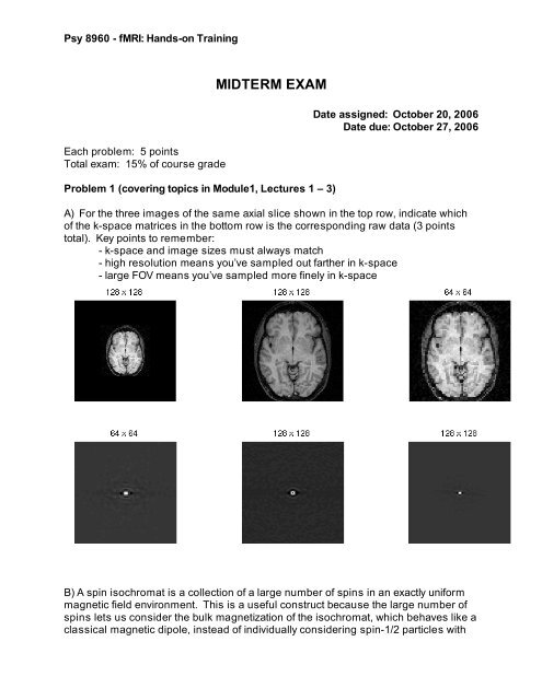
Psy 8960 - fMRI: Hands-on Training<br />
Each problem: 5 points<br />
Total exam: 15% of course grade<br />
<strong>MIDTERM</strong> <strong>EXAM</strong><br />
Problem 1 (covering topics in Module1, Lectures 1 – 3)<br />
Date assigned: October 20, 2006<br />
Date due: October 27, 2006<br />
A) For the three images of the same axial slice shown in the top row, indicate which<br />
of the k-space matrices in the bottom row is the corresponding raw data (3 points<br />
total). Key points to remember:<br />
- k-space and image sizes must always match<br />
- high resolution means you’ve sampled out farther in k-space<br />
- large FOV means you’ve sampled more finely in k-space<br />
B) A spin isochromat is a collection of a large number of spins in an exactly uniform<br />
magnetic field environment. This is a useful construct because the large number of<br />
spins lets us consider the bulk magnetization of the isochromat, which behaves like a<br />
classical magnetic dipole, instead of individually considering spin-1/2 particles with
Psy 8960 - fMRI: Hands-on Training<br />
quantized states. At equilibrium, in an external magnetic field oriented along the<br />
positive z- axis, what is the orientation of the net magnetic moment of a spin<br />
isochromat (1/2 point)?<br />
After application of a (perfect) inversion pulse, what is the orientation of the net<br />
magnetic moment of the isochromat (1/2 point)?<br />
C) For the illustrated (perfect) 45 degree RF pulse applied in conjunction with a linear<br />
magnetic field gradient, draw (on the blanks provided) the orientation of the bulk<br />
magnetization vector associated with the spin isochromats at the indicated positions<br />
along the x axis (1 point total).<br />
Frequency (Hz)<br />
X gradient: 11.7 mT/m = 500 Hz/mm<br />
RF pulse:<br />
Bandwidth: 2 kHz<br />
Flip angle: 45 deg.<br />
Indicated local M || after pulse by<br />
drawing an arrow on each line.<br />
Static field (B 0 ) points up:<br />
Position (cm)<br />
__ __ __ __ __ __ __ __ __ __<br />
For extra credit:<br />
How thick is the excited slice?<br />
What is the orientation of the slice?
Psy 8960 - fMRI: Hands-on Training<br />
Problem 2 (covering Module 2, Lectures 4 & 5)<br />
A) Label, on the drawing below, the T2 and T2* envelopes describing the magnitude<br />
of the net transverse magnetization generated by a collection of spin isochromats in<br />
an inhomogeneous magnetic field environment during a spin echo experiment (1<br />
point).<br />
Excitation<br />
pulse<br />
Refocusing<br />
pulse<br />
Echo<br />
Read-out<br />
M T<br />
S<br />
B) In the inversion-recovery experiment illustrated below, what would be the<br />
appropriate time to apply an excitation pulse and read-out an image if you wanted to<br />
null the signal from the gray matter? (1 point)<br />
White matter<br />
Gray matter<br />
CSF
Psy 8960 - fMRI: Hands-on Training<br />
C) In the data shown above, which has the longest T 1 : gray matter, white matter or<br />
CSF? (1 point)<br />
D) In the acquisition described in (b), what would be the relative image intensities of<br />
white matter and CSF in a magnitude image? (1 point)<br />
E) In any experiment, if the repetition time is very short, should the flip angle of the<br />
repeated RF pulse be large or small? (1 point)<br />
Problem 3 (covering Module 3, Lectures 6 & 7)<br />
A) Label the slice-select, read-out, and phase-encode gradient axes for the two pulse<br />
sequences illustrated on the next page. Describe each pulse sequence as FLASH or<br />
EPI, Spin Echo or Gradient Echo, and Single-shot or multi-Shot. (2 points)<br />
B) Draw on the grids provided below the k-space trajectories for each of the pulse<br />
sequences shown on the following page. Label the phase-encode direction. (2<br />
points) (It might help to number the gradients and refocusing pulse to match k-space<br />
excursions; no need to be precise once you get read-out lines.)<br />
Sequence 3.A.1<br />
Sequence 3.A.2
Psy 8960 - fMRI: Hands-on Training<br />
Pulse sequence 3.A.1: _________________________________________<br />
N = 4<br />
RF<br />
G ______<br />
G ______<br />
G ______<br />
DAC<br />
Pulse sequence 3.A.2: _________________________________________<br />
RF<br />
G ______<br />
G ______<br />
G ______<br />
DAC
Psy 8960 - fMRI: Hands-on Training<br />
C) For the EPI images shown on the following pages, indicate which was acquired<br />
with pulse sequence 3.A.1, and which was acquired with pulse sequence 3.A.2. (1<br />
point) Assume both had the same echo time during the acquisition.