download report - Sapienza
download report - Sapienza
download report - Sapienza

You also want an ePaper? Increase the reach of your titles
YUMPU automatically turns print PDFs into web optimized ePapers that Google loves.
Scientific Report 2007-2009<br />
Condensed matter physics and biophysics<br />
C40. Molecular diffusion and Molecular imaging studies by means of<br />
NMR techniques in materials, tissues, animal models and humans<br />
Diffusion Tensor (DTI) and Diffusion-weighted (DWI)<br />
imaging NMR techniques are the only non-invasive tool<br />
available today to investigate molecular diffusion processes<br />
in vivo. DTI and DWI provide information on<br />
biophysical properties of tissues which influence the diffusion<br />
of water molecules [1]. Molecular imaging is the<br />
non-invasive visualization in space and time of cellular<br />
processes at molecular or genetic level of function.<br />
A key component in molecular imaging is the imaging<br />
probe which homes in on the specific target of interest<br />
in the body providing pharmacokinetic and tracking information.<br />
Using NMR spectroscopic and/or scanning<br />
methods (magnetic resonance spectroscopy, MRS and<br />
imaging, MRI), the probe is labeled with 19F atoms<br />
and it is visualized by 19F-MRS and /or 19F-MRI.<br />
Specifically, target molecules are marked by substitution<br />
of hydrogen atoms with one or more 19F atoms<br />
(usually -CF3 group). Due to the lack of endogenous<br />
background signal in vivo and the high MR sensitivity<br />
of the 19F atoms (83Within the framework of Molecular<br />
Imaging investigations we developed in our laboratory<br />
an in vivo 19F MR Imaging and Spectroscopy<br />
protocol for the optimization of BNCT (Boron Neutron<br />
Capture therapy). BNCT is an experimental binary radiation<br />
therapy based on the cytotoxic effects of high<br />
LET particles released from the 10B(n,alpha)7Li reaction<br />
that occurs when 10B captures a thermal neutron.<br />
For BNCT effectiveness a large amount of 10B atoms (at<br />
least 109 atoms of 10B per targeted cell) should be accumulated<br />
within tumour cells in order to obtain a maximum<br />
tumour-to-brain (T:Br) 10B concentration ratio.<br />
Currently, the therapy is mainly used to treat malignant<br />
brain glioma for which the conventional therapies<br />
do not provide any substantial benefit. The main limitations<br />
for BNCT effectiveness are: 1) the lack of efficient<br />
imaging methods to monitor the bio-distribution of 10Blabeled<br />
drugs in order to estimate the efficiency of the<br />
carrier and the optimal timing of neutron irradiation.<br />
This ideal time is when tumour-to-brain (T:Br) 10B concentration<br />
ratio achieves the maximum value at the lowest<br />
blood concentration; 2) the insufficient intake of 10B<br />
nuclei in the tumour cells. The aim of our study was<br />
to evaluate the boron bio-distribution and pharmacokinetics<br />
of 4-borono-2-fluorophenylalanine (19F-BPA) using<br />
19F-MRI and 19F-MRS in animal model (C6-glioma<br />
rat brain). Moreover, the effect of L-DOPA as potential<br />
enhancer of BPA tumour intake was evaluated. 1H and<br />
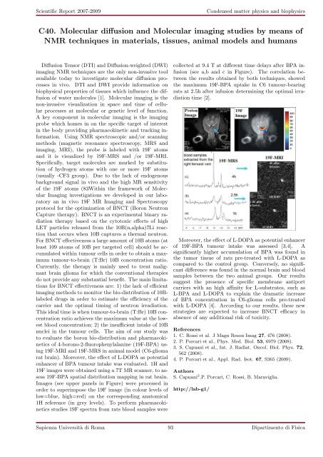
19F images were obtained using a 7T MR scanner, to assess<br />
19F-BPA spatial distribution mapping in rat brain.<br />
Images (see upper panels in Figure) were processed in<br />
order to superimpose the 19F image (in colour levels of<br />
low=blue, high=red) on the corresponding anatomical<br />
1H reference (in grey levels). To perform pharmacokinetics<br />
studies 19F spectra from rats blood samples were<br />
collected at 9.4 T at different time delays after BPA infusion<br />
(see a,b and c in Figure). The correlation between<br />
the results obtained by both techniques, showed<br />
the maximum 19F-BPA uptake in C6 tumour-bearing<br />
rats at 2.5h after infusion determining the optimal irradiation<br />
time [2].<br />
Moreover, the effect of L-DOPA as potential enhancer<br />
of 19F-BPA tumour intake was assessed [3,4]. A<br />
significantly higher accumulation of BPA was found in<br />
the tumor tissue of rats pre-treated with L-DOPA as<br />
compared to the control group. Conversely, no significant<br />
difference was found in the normal brain and blood<br />
samples between the two animal groups. Our results<br />
suggest the presence of specific membrane antiport<br />
carriers with an high affinity for L-substrates, such as<br />
L-BPA and L-DOPA to explain the dramatic increase<br />
of BPA concentration in C6-glioma cells pre-treated<br />
with L-DOPA [4]. According to our results, these new<br />
strategies are expected to increase BNCT efficacy in<br />
absence of any additional risk of toxicity.<br />
References<br />
1. C. Rossi et al. J Magn Reson Imag 27, 476 (2008).<br />
2. P. Porcari et al., Phys. Med. Biol. 53, 6979 (2008).<br />
3. S. Capuani et al., Int. J. Radiat. Oncol. Biol. Phys. 72,<br />
562 (2008).<br />
4. P. Porcari et al., Appl. Rad. Isot. 67, S365 (2009).<br />
Authors<br />
S. Capuani 2 ,P. Porcari, C. Rossi, B. Maraviglia.<br />
http://lab-g1/<br />
<strong>Sapienza</strong> Università di Roma 93 Dipartimento di Fisica