The red algal genus Reticulocaulis from the Arabian
The red algal genus Reticulocaulis from the Arabian
The red algal genus Reticulocaulis from the Arabian

You also want an ePaper? Increase the reach of your titles
YUMPU automatically turns print PDFs into web optimized ePapers that Google loves.
Schils et al.: <strong>Reticulocaulis</strong> in <strong>the</strong> Indian Ocean 45<br />
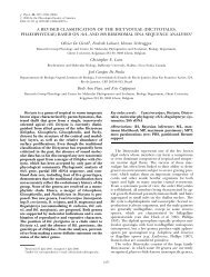
Figs 1–3. Collection sites of <strong>Reticulocaulis</strong> in <strong>the</strong> <strong>Arabian</strong> Sea. Scale<br />
bars 1000 km (Fig. 1); 20 km (Figs 2, 3).<br />
Fig. 1. <strong>The</strong> <strong>Arabian</strong> Peninsula showing Masirah Island and Socotra.<br />
Fig. 2. Sample site 9 (asterisk; 20.199N, 58.715E), near Ras Zarri,<br />
off Masirah Island, Oman.<br />
Fig. 3. Sample site ALG-40 (asterisk; 12.303N, 53.843E), west of<br />
Bidholih, off Socotra, Yemen.<br />
imens and microscope slides are deposited in GENT (Ghent<br />
University Herbarium, Krijgslaan 281/S8, 9000 Ghent, Belgium).<br />
Slides and formalin-preserved samples of Hawaiian R.<br />
mucosissimus I.A. Abbott were kindly supplied by I. A. Abbott<br />
of <strong>the</strong> Bernice Bishop Museum. Herbarium sheets of N.<br />
corymbosa J. Agardh and N. wiggii (Turner) Endlicher were<br />
borrowed <strong>from</strong> <strong>the</strong> National Herbarium of <strong>the</strong> Ne<strong>the</strong>rlands<br />
(L). Material for microscopical examination was stained with<br />
aniline blue, fast green or Lugol’s Iodine (for rhodoplasts).<br />
Material for nuclear and pit-connection studies was stained<br />
using Wittmann’s aceto-iron-haematoxylin–chloral hydrate<br />
(Wittmann 1965), following <strong>the</strong> procedures of Hommersand<br />
& F<strong>red</strong>ericq (1988). Anatomical and reproductive characteristics<br />
were observed <strong>from</strong> tissue squashes (whole-mounts in a<br />
50% corn syrup–water solution, containing a few drops of<br />
phenol) using light microscopy (Leitz Diaplan). Photographs<br />
were taken with a Wild MPS51 35 mm camera and on an<br />
Olympus DP50 digital camera.<br />
RESULTS<br />
<strong>Reticulocaulis</strong> mucosissimus I.A. Abbott 1985, p. 555<br />
SPECIMENS EXAMINED: Oman. Masirah Island (Figs 1, 2): sample site<br />
9 (20.199N, 58.715E), close to Ras Zarri. A rocky platform at 9<br />
m depth with scatte<strong>red</strong> rocky outcrops in an area of strong surge<br />
(Schils, 9 November 1999). MAS 138: female (Fig. 4) and male<br />
gametophytes. Hawaii. Mahukona, north-west coast of Hawaii.<br />
Plants growing on dead coral at a depth of 9 m (K. J. McDermid,<br />
26 May 1998). Formalin sample IA 23471 (female gametophyte)<br />
and slide KM 4481 (female gametophyte); Kawailoa, Oahu Island<br />
(W. H. Magruder & S. Carper, 10 May 1985). Slide IA 17225:<br />
female gametophyte.<br />
Thalli are bright <strong>red</strong>, mucilaginous and attached by a discoid<br />
holdfast (Fig. 4). Omani plants reach 13 cm in length and<br />
grow <strong>from</strong> dome-shaped apical cells that divide obliquely, <strong>the</strong><br />
immediate daughter cells being aligned in a nearly straight<br />
row (Fig. 5). <strong>The</strong> axial cells are slender and elongate, those<br />
lying 1 mm away <strong>from</strong> <strong>the</strong> apical cells having length–width<br />
ratios of 4 : 1. <strong>The</strong> first periaxial cell (<strong>the</strong> ‘superior’ periaxial)<br />
is cut off three axial cells <strong>from</strong> <strong>the</strong> apex, and superior<br />
periaxial cells on successive segments are produced in an irregular<br />
¼ spiral. A second periaxial cell (<strong>the</strong> ‘inferior’ periaxial)<br />
is always positioned proximal to <strong>the</strong> first. It is generally<br />
cut off in cells positioned 15–20 cells away <strong>from</strong> <strong>the</strong> apex<br />
(Fig. 5) and at a 90 angle to <strong>the</strong> first periaxial cell. At <strong>the</strong><br />
same time, several rhizoidal outgrowths develop <strong>from</strong> both<br />
periaxial cells; <strong>the</strong>se outgrowths branch. Besides differing in<br />
<strong>the</strong> timing of <strong>the</strong>ir initiation, <strong>the</strong> shapes of <strong>the</strong> two periaxial<br />
cells are also dissimilar: <strong>the</strong> superior periaxial cell becomes<br />
elongated and rectilinear, whereas <strong>the</strong> inferior one remains<br />
spherical (Fig. 5). <strong>The</strong> inferior lateral becomes <strong>the</strong> more developed<br />
of <strong>the</strong> two laterals and occasionally gives rise to indeterminate<br />
branches as it continues growing and initiates<br />
periaxial cells. Infrequently, an axial cell can initiate a third<br />
periaxial cell, which develops like <strong>the</strong> superior lateral. <strong>The</strong><br />
derivatives of <strong>the</strong> periaxial cells (<strong>from</strong> about <strong>the</strong> 15th axial<br />
cell) differentiate rapidly by branching and cell elongation<br />
into determinate filaments that constitute <strong>the</strong> cortex. <strong>The</strong> inner<br />
cortical cells are cylindrical (Fig. 6), whereas <strong>the</strong> outer cells<br />
remain ovoid to (sub)spherical.<br />
Two-celled propagules, reaching 16.5 m in diameter (Fig.<br />
7) and developing terminally on many of <strong>the</strong> cortical filaments,<br />
were observed on slide IA 17225 of a specimen <strong>from</strong><br />
Hawaii. One or two axial cells below <strong>the</strong> site where <strong>the</strong> second<br />
periaxial cell first forms, both periaxial cells initiate rhizoidal<br />
downgrowths. <strong>The</strong> periaxial cells and <strong>the</strong> rhizoidal<br />
downgrowths inflate into what were termed ‘jacket cells’ by<br />
Abbott (1985), viz. cells that mutually cross-connect by lateral<br />
secondary pit connections (Fig. 8) and constitute a sheath<br />
around <strong>the</strong> central-axial strand (Fig. 9). While maturing, <strong>the</strong><br />
pit connections of <strong>the</strong> jacket cells attenuate and become difficult<br />
to distinguish, which results in a seemingly parenchymatous<br />
covering. Before <strong>the</strong> covering is complete, <strong>the</strong> jacket<br />
cells initiate secondary cortical filaments that are ei<strong>the</strong>r fasciculate<br />
or unbranched, as well as secondary rhizoidal downgrowths.<br />
In older parts of <strong>the</strong> thallus, <strong>the</strong> jacket cells become<br />
densely cove<strong>red</strong> by <strong>the</strong>se secondary rhizoidal filaments, which<br />
rarely branch and form uniseriate rows that cross one ano<strong>the</strong>r,<br />
but actually constitute a single layer.<br />
<strong>The</strong> rhodoplasts are discoid but like erythrocytes in shape<br />
(2–4 m in diameter), having centres that are thinner than <strong>the</strong><br />
margins.<br />
Female gametophytes have carpogonial branches that are of<br />
accessory origin; <strong>the</strong>y were found throughout <strong>the</strong> thallus in<br />
various stages of development. Near <strong>the</strong> apex, carpogonial<br />
branches arise singly <strong>from</strong> ei<strong>the</strong>r of <strong>the</strong> periaxial cells. Fur<strong>the</strong>r<br />
down <strong>the</strong> thallus, <strong>the</strong>y also develop <strong>from</strong> o<strong>the</strong>r jacket cells<br />
(rhizoidal filament cells) and <strong>the</strong> lower cortical filament cells.<br />
Pairs of carpogonial branches on a single supporting cell are<br />
infrequently seen. <strong>The</strong> branches consist of 7–13 equally staining<br />
cells, which, following <strong>the</strong> terminology of Lindstrom<br />
(1984), can be designated by numbers starting with <strong>the</strong> carpogonium<br />
(#1). Eccentric positioning of <strong>the</strong> primary pit con-