The formation of the twin resting cysts in the dinoflagellate Dissodinium pseudolunula, a parasite of copepod eggs
The dinoflagellate Dissodinium pseudolunula is the most common and widespread ectoparasite of copepod eggs in neritic waters. When the host is absent, the species survives with a distinctive pair of twin resting cysts described as Pyrocystis margalefii. Based on live samples, the formation of the twin resting cysts is illustrated here for the first time. The gymnodinioid infective cells did not form overwintering cysts under unfavourable conditions. These are formed inside the secondary lunate sporangium
The dinoflagellate Dissodinium pseudolunula is the most common and widespread ectoparasite
of copepod eggs in neritic waters. When the host is absent, the species survives
with a distinctive pair of twin resting cysts described as Pyrocystis margalefii. Based
on live samples, the formation of the twin resting cysts is illustrated here for the first
time. The gymnodinioid infective cells did not form overwintering cysts under
unfavourable conditions. These are formed inside the secondary lunate sporangium

You also want an ePaper? Increase the reach of your titles
YUMPU automatically turns print PDFs into web optimized ePapers that Google loves.
F. GÓMEZ AND L. F. ARTIGAS j LIFE CYCLE OF DISSODINIUM, PARASITE OF COPEPOD EGGS<br />
<strong>the</strong> life cycle <strong>of</strong> Dissod<strong>in</strong>ium <strong>in</strong> which <strong>the</strong> <strong>rest<strong>in</strong>g</strong> <strong>cysts</strong> are<br />
formed rema<strong>in</strong>s unknown. This fact also represents a<br />
way to confirm <strong>the</strong> relationship between <strong>the</strong> <strong>tw<strong>in</strong></strong> <strong>rest<strong>in</strong>g</strong><br />
cyst and Dissod<strong>in</strong>ium. <strong>The</strong> most <strong>in</strong>tuitive explanation is<br />
that <strong>the</strong> <strong>in</strong>fective d<strong>in</strong>ospore forms a <strong>rest<strong>in</strong>g</strong> cyst under unfavourable<br />
conditions (when it is unable to f<strong>in</strong>d <strong>the</strong> host).<br />
In fact, <strong>the</strong> <strong>in</strong>fective d<strong>in</strong>ospore forms an outer membrane<br />
(Drebes, 1981). However, this corresponds to a temporary<br />
cyst which differs from <strong>the</strong> <strong>tw<strong>in</strong></strong> <strong>rest<strong>in</strong>g</strong> <strong>cysts</strong> <strong>of</strong><br />
Dissod<strong>in</strong>ium <strong>in</strong> size, shape and cell content. <strong>The</strong> present<br />
study identifies <strong>the</strong> miss<strong>in</strong>g l<strong>in</strong>k between <strong>the</strong> known life<br />
stages <strong>of</strong> Dissod<strong>in</strong>ium and its overw<strong>in</strong>ter<strong>in</strong>g <strong>rest<strong>in</strong>g</strong> cyst.<br />
Live phytoplankton samples were collected weekly<br />
with a 20 mm mesh net <strong>in</strong> 2010 and 2011 <strong>in</strong> coastal waters<br />
<strong>of</strong> <strong>the</strong> NE English Channel (<strong>of</strong>f Wimereux, France), which<br />
are characterized by recurrent blooms <strong>of</strong> diatoms and <strong>the</strong><br />
haptophyte Phaeocystis globosa. Daily sampl<strong>in</strong>g was also<br />
performed <strong>in</strong> a large pool (≏50 m diameter, ≏1 m<br />
depth) that formed dur<strong>in</strong>g low tide along <strong>the</strong> shore at<br />
Wimereux. In addition, <strong>in</strong>tertidal surface sediments at<br />
Wimereux were exam<strong>in</strong>ed follow<strong>in</strong>g <strong>the</strong> sampl<strong>in</strong>g procedure<br />
described by Gómez et al. (Gómez et al., 2011).<br />
Plankton and benthos live samples were settled <strong>in</strong><br />
Utermöhl chambers and exam<strong>in</strong>ed with an <strong>in</strong>verted<br />
microscope (Nikon Eclipse TE2000-S) equipped with a<br />
Nikon Digital Sight DS-2M camera.<br />
Infected <strong>eggs</strong> and globular and lunate sporangia <strong>of</strong><br />
D. <strong>pseudolunula</strong> were common <strong>in</strong> live samples from April<br />
to September, with more specimens <strong>in</strong> June (Fig. 1).<br />
<strong>The</strong> fast-swimm<strong>in</strong>g gymnod<strong>in</strong>ioid d<strong>in</strong>ospores were difficult<br />
to record, and <strong>the</strong> micrographs were taken when<br />
<strong>the</strong>y were enclosed <strong>in</strong>side a th<strong>in</strong> hyal<strong>in</strong>e outer membrane<br />
(Fig. 1AE–AG). Only 10 pairs <strong>of</strong> <strong>tw<strong>in</strong></strong> <strong>rest<strong>in</strong>g</strong> <strong>cysts</strong> were<br />
observed between July and September <strong>in</strong> 2010 and 2011<br />
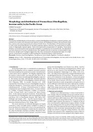
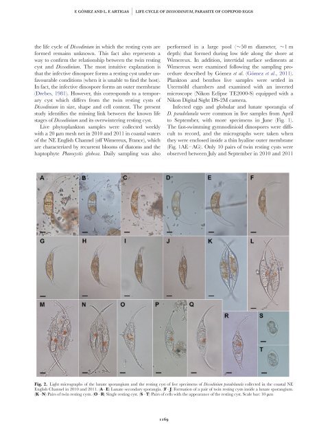
Fig. 2. Light micrographs <strong>of</strong> <strong>the</strong> lunate sporangium and <strong>the</strong> <strong>rest<strong>in</strong>g</strong> cyst <strong>of</strong> live specimens <strong>of</strong> Dissod<strong>in</strong>ium <strong>pseudolunula</strong> collected <strong>in</strong> <strong>the</strong> coastal NE<br />
English Channel <strong>in</strong> 2010 and 2011. (A–E) Lunate secondary sporangia. (F–J) Formation <strong>of</strong> a pair <strong>of</strong> <strong>tw<strong>in</strong></strong> <strong>rest<strong>in</strong>g</strong> <strong>cysts</strong> <strong>in</strong>side a lunate sporangium.<br />
(K–N) Pairs <strong>of</strong> <strong>tw<strong>in</strong></strong> <strong>rest<strong>in</strong>g</strong> <strong>cysts</strong>. (O–R) S<strong>in</strong>gle <strong>rest<strong>in</strong>g</strong> cyst. (S–T) Pairs <strong>of</strong> cells with <strong>the</strong> appearance <strong>of</strong> <strong>the</strong> <strong>rest<strong>in</strong>g</strong> cyst. Scale bar: 10 mm<br />
1169