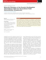
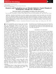
Solenicola setigera is the first characterized memberof the abundant and cosmopolitan uncultured marine stramenopile group MAST-3
Culture-independent molecular methods based on the amplification, cloning and sequencing of smallsubunit (SSU) rRNA genes are a powerful tool to study the diversity of prokaryotic and eukaryotic microorganisms for which morphological features are not conspicuous. In recent years, molecular data from environmental surveys have revealed several clades of protists lacking cultured and/or described members. Among them are various clades of marine stramenopiles (heterokonts), which are thought to play an essential ecological role as grazers, being abundant and distributed in oceans worldwide. In this work, we show that Solenicola setigera, a distinctive widespread colonial marine protist, is a member of the environmental clade MArine STramenopile 3 (MAST-3). Solenicola is generally considered as a parasite or an epiphyte of the diatom Leptocylindrus mediterraneus. So far, the ultrastructural, morphological and ecological data available were insufficient to elucidate its phylogenetic position, even at the division or class level. We determined SSU rRNA gene sequences of S. setigera specimens sampled from different locations and seasons in the type locality, the Gulf of Lions, France. They were closely related, though not identical, which, together with morphological differences under electron microscopy, suggest the occurrence of several species. Solenicola sequences were well nested within the MAST-3 clade in phylogenetic trees. Since Solenicola is the first identified member of this abundant marine clade, we propose the name Solenicolida for the MAST-3 phylogenetic group.
Culture-independent molecular methods based on
the amplification, cloning and sequencing of smallsubunit
(SSU) rRNA genes are a powerful tool to
study the diversity of prokaryotic and eukaryotic
microorganisms for which morphological features are
not conspicuous. In recent years, molecular data from
environmental surveys have revealed several clades
of protists lacking cultured and/or described
members. Among them are various clades of marine
stramenopiles (heterokonts), which are thought to
play an essential ecological role as grazers, being
abundant and distributed in oceans worldwide. In this
work, we show that Solenicola setigera, a distinctive
widespread colonial marine protist, is a member of
the environmental clade MArine STramenopile 3
(MAST-3). Solenicola is generally considered as a
parasite or an epiphyte of the diatom Leptocylindrus
mediterraneus. So far, the ultrastructural, morphological
and ecological data available were insufficient
to elucidate its phylogenetic position, even at the division
or class level. We determined SSU rRNA gene
sequences of S. setigera specimens sampled from
different locations and seasons in the type locality,
the Gulf of Lions, France. They were closely related,
though not identical, which, together with morphological
differences under electron microscopy,
suggest the occurrence of several species. Solenicola
sequences were well nested within the MAST-3
clade in phylogenetic trees. Since Solenicola is the
first identified member of this abundant marine clade,
we propose the name Solenicolida for the MAST-3
phylogenetic group.

Create successful ePaper yourself
Turn your PDF publications into a flip-book with our unique Google optimized e-Paper software.
196 F. Gómez, D. Moreira, K. Benzerara <strong>and</strong> P. López-García<br />
siliceous tube with an undulating movement (Fig. 1H–J,<br />
see Video S1 or http://www.youtube.com/watch?v=6<br />
COYUXjrBWI). Th<strong>is</strong> provoked <strong>the</strong> motility of <strong>the</strong> entire<br />
colony in <strong>the</strong> direction of one of <strong>the</strong> extremes of <strong>the</strong><br />
siliceous tube, but also enhanced <strong>the</strong> traffic of small particles<br />
towards <strong>the</strong> colony. Single individuals may detach<br />
from <strong>the</strong> colony (see Videos S1 <strong>and</strong> S2).<br />
The specimens of <strong>Solenicola</strong> that we observed were<br />
colourless. No trace of chlorophyll a autofluorescence<br />
was observed by epifluorescence microscopy when <strong>the</strong><br />
cells were excited by blue or green light. On rare occasions,<br />
one colony showed a partial yellow-green<strong>is</strong>h<br />
pigmentation (Fig. 1E, see Video S2 or http://<br />
www.youtube.com/watch?v=-B8m8YNlRas). However,<br />
<strong>the</strong> pigmentation did not correspond to chlorophyll a as<br />
tested by epifluorescence microscopy (Fig. 1F). The only<br />
signal of chlorophyll a corresponded to scattered spots of<br />
small circular spots of orange (phycoerythrin-derived pigments<br />
of cyanobacteria) or larger spots of red fluorescence<br />
(photosyn<strong>the</strong>tic picoeukaryotes) that might<br />
correspond to recently captured preys by <strong>Solenicola</strong>.<br />
During manipulation, <strong>the</strong> siliceous tubes often broke<br />
apart (Fig. 1E). <strong>Solenicola</strong> cells could suddenly detach<br />
from <strong>the</strong> siliceous tube. The detached cells did not swim,<br />
but remained <strong>group</strong>ed <strong>and</strong> motionless (Fig. 1G, see<br />
Video S3 or http://www.youtube.com/watch?v=q7<br />
mysOtm-eE).<br />
Scanning electron microscopy of <strong>Solenicola</strong><br />
Five colonies of <strong>Solenicola</strong> collected in December 2008<br />
from <strong>the</strong> port of Banyuls sur Mer, France, were fixed in<br />
glutaraldehyde <strong>and</strong> examined using scanning electron<br />
microscopy (Fig. 2). Two of <strong>the</strong> siliceous tubes colonized<br />
by <strong>Solenicola</strong> were fully covered with <strong>the</strong> nanoflagellate<br />
cells (Fig. 2A–J), while <strong>the</strong> o<strong>the</strong>r three colonies showed<br />
clusters of <strong>Solenicola</strong> restricted to localized areas of <strong>the</strong><br />
tubes (Fig. 2K–AA). In <strong>the</strong> former, <strong>the</strong> cells bore a v<strong>is</strong>ible<br />
flagellum, whereas in <strong>the</strong> second case, <strong>the</strong> cells that<br />
remained attached to <strong>the</strong> tubes appeared to have lost<br />
<strong>the</strong>ir flagella, as attested by remaining flagellar insertion<br />
regions (e.g. Fig. 2L).<br />
When v<strong>is</strong>ible, each cell showed a single smooth flagellum<br />
with a pointed end (Fig. 2C). The diameter was<br />
approximately 0.18 mm <strong>and</strong> <strong>the</strong> length was variable (up to<br />
24 mm long) (Fig. 2F). The flagellum emerged from a ringlike<br />
bas<strong>is</strong> (0.5 mm in diameter) that was apparently composed<br />
of 10 circularly arrayed globules (Fig. 2B, G <strong>and</strong> H).<br />
Although <strong>Solenicola</strong> cells were generally rounded or tear<br />
shaped (most cells shown in Fig. 2A <strong>and</strong> E), some cells,<br />
which were made often v<strong>is</strong>ible after <strong>the</strong> detachment of <strong>the</strong><br />
rest of cells covering <strong>the</strong>m, were ra<strong>the</strong>r pleomorphic,<br />
showing lobose pseudopodial <strong>and</strong>/or filopodial extensions<br />
that were intimately attached to <strong>the</strong>ir siliceous substrate<br />
(Fig. 2K, Q <strong>and</strong> W). Some of <strong>the</strong>se pseudopodial extensions<br />
often showed protruding bulges in <strong>the</strong> d<strong>is</strong>tal ends<br />
(Fig. 2N). The morphology <strong>and</strong> structural details of <strong>the</strong><br />
siliceous tubes colonized by <strong>Solenicola</strong> varied depending<br />
on <strong>the</strong> colonies (Fig. 2R–AA), which may suggest <strong>the</strong><br />
occurrence of different closely related <strong>Solenicola</strong> species<br />
or strains with different substrate specificity.<br />
Interestingly, one of <strong>the</strong> examined colonies showed particles<br />
attached to <strong>the</strong> flagella with a hexagonal contour of<br />
0.27 mm in diameter, tentatively large icosahedral viruses<br />
(Fig. 2D). In a second colony, <strong>the</strong> polyhedral particles<br />
were attached to <strong>the</strong> cell body of <strong>Solenicola</strong> (Fig. 2I <strong>and</strong><br />
J). Although rare, descriptions of large double-str<strong>and</strong>ed<br />
viruses infecting <strong>stramenopile</strong>s ex<strong>is</strong>t, for instance in <strong>the</strong><br />
pelagophyte Aureococcus (Gastrich et al., 1998) or <strong>the</strong><br />
bicosoecid Cafeteria (Massana et al., 2007), suggesting<br />
that viruses exert a control on <strong>the</strong>ir populations.<br />
Molecular phylogeny of <strong>Solenicola</strong> from<br />
individual filaments<br />
We obtained nearly complete SSU rDNA sequences from<br />
<strong>the</strong> three <strong>Solenicola</strong> colonies shown in Fig. 1A–D using<br />
PCR amplification with eukaryotic universal primers. All <strong>the</strong><br />
sequences obtained from <strong>the</strong> three SSU rDNA libraries<br />
(FG11, FG174 <strong>and</strong> FG712) were identical between <strong>the</strong>m in<br />
each case <strong>and</strong> corresponded to <strong>stramenopile</strong> sequences.<br />
The two closest BLAST hits to our <strong>stramenopile</strong> sequences<br />
were <strong>the</strong> environmental clones NPK2-2 <strong>and</strong> BL0000921-<br />
38, both sharing 91% identity with our sequences. We<br />
concluded that <strong>the</strong> <strong>stramenopile</strong> sequences did correspond<br />
to <strong>Solenicola</strong> <strong>and</strong> we included <strong>the</strong>m in a large<br />
alignment of eukaryotic sequences to determine its phylogenetic<br />
position. Preliminary phylogenetic analyses with<br />
SSU rDNA sequences from a variety of eukaryotic phyla<br />
revealed that <strong>Solenicola</strong> belonged to <strong>the</strong> <strong>stramenopile</strong>s<br />
(not shown). We <strong>the</strong>n carried out more detailed phylogenetic<br />
analyses with representatives of <strong>the</strong> different <strong>stramenopile</strong><br />
families. They showed that <strong>Solenicola</strong> branched<br />
with maximum support (100% bootstrap value – BV) within<br />
<strong>the</strong> environmental <strong>group</strong> <strong>MAST</strong>-3 (Fig. 3), defined by<br />
Massana <strong>and</strong> colleagues (2004). Our three <strong>Solenicola</strong><br />
sequences were closely related but not identical<br />
(maximum divergence was 3% between <strong>the</strong> sequence of<br />
<strong>the</strong> FG712 colony <strong>and</strong> <strong>the</strong> o<strong>the</strong>r two sequences). Th<strong>is</strong><br />
genetic diversity might indicate <strong>the</strong> occurrence of different<br />
species. The closest relative to <strong>Solenicola</strong> was <strong>the</strong> environmental<br />
clone NPK2-2, obtained from Artic waters at 2 m<br />
depth (Luo et al., 2009) <strong>and</strong> <strong>the</strong>n several o<strong>the</strong>r environmental<br />
sequences from diverse oceanic regions, including<br />
<strong>the</strong> Mediterranean Sea (BL0000921-38 <strong>and</strong> ME1-28), Sargasso<br />
Sea (SMC27C17) <strong>and</strong> <strong>the</strong> Framvaren Fjiord in<br />
Norway (NIF_1E11, NIF_3D5 <strong>and</strong> NIF_3F1). All of <strong>the</strong>m<br />
<strong>group</strong>ed with <strong>Solenicola</strong> with strong support (BV of 99%).<br />
© 2010 Society for Applied Microbiology <strong>and</strong> Blackwell Publ<strong>is</strong>hing Ltd, Environmental Microbiology, 13, 193–202