Life cycle and molecular phylogeny of the dinoflagellates Chytriodinium and Dissodinium, ectoparasites of copepod eggs
The dinoflagellates Chytriodinium affine, C. roseum and Dissodinium pseudolunula are ectoparasites of crustacean eggs. Here, we present new observations regarding their life cycle based on coastal plankton samples and incubations and analyze their molecular phylogeny using the small subunit ribosomal RNA gene (SSU rDNA) as a marker. In contrast to the typical stages already documented for its life cycle, we observed that D. pseudolunula dinospores may exceptionally differentiate inside a globular cyst. Despite its parasitic life style, the cysts and dinospores of D. pseudolunula contain chlorophyll a. We obtained the first SSU rDNA sequences for the genera Chytriodinium (the type C. roseum and C. affine) and Dissodinium (D. pseudolunula). Classical taxonomical schemes have ascribed these genera to the order Blastodiniales. However, our SSU rDNA-based phylogenetic analysis shows that these ectoparasites form a clade in the Gymnodinium sensu stricto group, unarmored dinokaryotic dinoflagellates of the order Gymnodiniales. They branch in a subgroup composed of warnowiids, polykrikoids, the type of Gymnodinium, G. fuscum and G. aureolum. Although Chytriodinium and Dissodinium appear to be relatives based on SSU rDNA phylogeny, feeding and host specificity, their life cycles are substantially different. Based on these data we consider that the type of life cycle is a poor criterion for classification at the family level. We suggest that the morphology of the infective cell is
The dinoflagellates Chytriodinium affine, C. roseum and Dissodinium pseudolunula are ectoparasites of crustacean
eggs. Here, we present new observations regarding their life cycle based on coastal plankton samples and incubations
and analyze their molecular phylogeny using the small subunit ribosomal RNA gene (SSU rDNA) as a marker. In
contrast to the typical stages already documented for its life cycle, we observed that D. pseudolunula dinospores may
exceptionally differentiate inside a globular cyst. Despite its parasitic life style, the cysts and dinospores of
D. pseudolunula contain chlorophyll a. We obtained the first SSU rDNA sequences for the genera Chytriodinium (the
type C. roseum and C. affine) and Dissodinium (D. pseudolunula). Classical taxonomical schemes have ascribed these
genera to the order Blastodiniales. However, our SSU rDNA-based phylogenetic analysis shows that these
ectoparasites form a clade in the Gymnodinium sensu stricto group, unarmored dinokaryotic dinoflagellates of the
order Gymnodiniales. They branch in a subgroup composed of warnowiids, polykrikoids, the type of Gymnodinium,
G. fuscum and G. aureolum. Although Chytriodinium and Dissodinium appear to be relatives based on SSU rDNA
phylogeny, feeding and host specificity, their life cycles are substantially different. Based on these data we consider that
the type of life cycle is a poor criterion for classification at the family level. We suggest that the morphology of the
infective cell is

Create successful ePaper yourself
Turn your PDF publications into a flip-book with our unique Google optimized e-Paper software.
ARTICLE IN PRESS<br />
European Journal <strong>of</strong> Protistology 45 (2009) 260–270<br />
European Journal <strong>of</strong><br />
PROTISTOLOGY<br />
www.elsevier.de/ejop<br />
<strong>Life</strong> <strong>cycle</strong> <strong>and</strong> <strong>molecular</strong> <strong>phylogeny</strong> <strong>of</strong> <strong>the</strong> din<strong>of</strong>lagellates <strong>Chytriodinium</strong><br />
<strong>and</strong> <strong>Dissodinium</strong>, <strong>ectoparasites</strong> <strong>of</strong> <strong>copepod</strong> <strong>eggs</strong><br />
Fern<strong>and</strong>o Gómez a, , David Moreira b , Purificación López-García b<br />
a Observatoire Océanologique de Banyuls sur Mer, Université Pierre et Marie Curie, CNRS-INSU UMR 7621, Avenue du Fontaulé,<br />
BP 44, 66651 Banyuls sur Mer, France<br />
b Unité d’Ecologie, Systématique et Evolution, CNRS UMR 8079, Université Paris-Sud, Bâtiment 360, 91405 Orsay Cedex, France<br />
Received 19 February 2009; received in revised form 5 May 2009; accepted 15 May 2009<br />
Abstract<br />
The din<strong>of</strong>lagellates <strong>Chytriodinium</strong> affine, C. roseum <strong>and</strong> <strong>Dissodinium</strong> pseudolunula are <strong>ectoparasites</strong> <strong>of</strong> crustacean<br />
<strong>eggs</strong>. Here, we present new observations regarding <strong>the</strong>ir life <strong>cycle</strong> based on coastal plankton samples <strong>and</strong> incubations<br />
<strong>and</strong> analyze <strong>the</strong>ir <strong>molecular</strong> <strong>phylogeny</strong> using <strong>the</strong> small subunit ribosomal RNA gene (SSU rDNA) as a marker. In<br />
contrast to <strong>the</strong> typical stages already documented for its life <strong>cycle</strong>, we observed that D. pseudolunula dinospores may<br />
exceptionally differentiate inside a globular cyst. Despite its parasitic life style, <strong>the</strong> cysts <strong>and</strong> dinospores <strong>of</strong><br />
D. pseudolunula contain chlorophyll a. We obtained <strong>the</strong> first SSU rDNA sequences for <strong>the</strong> genera <strong>Chytriodinium</strong> (<strong>the</strong><br />
type C. roseum <strong>and</strong> C. affine) <strong>and</strong><strong>Dissodinium</strong> (D. pseudolunula). Classical taxonomical schemes have ascribed <strong>the</strong>se<br />
genera to <strong>the</strong> order Blastodiniales. However, our SSU rDNA-based phylogenetic analysis shows that <strong>the</strong>se<br />
<strong>ectoparasites</strong> form a clade in <strong>the</strong> Gymnodinium sensu stricto group, unarmored dinokaryotic din<strong>of</strong>lagellates <strong>of</strong> <strong>the</strong><br />
order Gymnodiniales. They branch in a subgroup composed <strong>of</strong> warnowiids, polykrikoids, <strong>the</strong> type <strong>of</strong> Gymnodinium,<br />
G. fuscum <strong>and</strong> G. aureolum. Although <strong>Chytriodinium</strong> <strong>and</strong> <strong>Dissodinium</strong> appear to be relatives based on SSU rDNA<br />
<strong>phylogeny</strong>, feeding <strong>and</strong> host specificity, <strong>the</strong>ir life <strong>cycle</strong>s are substantially different. Based on <strong>the</strong>se data we consider that<br />
<strong>the</strong> type <strong>of</strong> life <strong>cycle</strong> is a poor criterion for classification at <strong>the</strong> family level. We suggest that <strong>the</strong> morphology <strong>of</strong> <strong>the</strong><br />
infective cell is probably <strong>the</strong> most reliable phenotypic characteristic to determine <strong>the</strong> systematic position <strong>of</strong> parasitic<br />
din<strong>of</strong>lagellates.<br />
r 2009 Elsevier GmbH. All rights reserved.<br />
Keywords: Alveolata; Blastodiniales; Gymnodinium; Parasitic dinophyceae; SSU rDNA<br />
Introduction<br />
Copepods are <strong>the</strong> most abundant metazoans in <strong>the</strong><br />
sea <strong>and</strong> represent a key trophic link in pelagic food<br />
webs (Mauchline 1998). Numerous parasites have been<br />
Corresponding author. Tel.: +33 4 68 88 73 25;<br />
fax: +33 468887398.<br />
E-mail address: fern<strong>and</strong>o.gomez@fitoplancton.com (F. Gómez).<br />
shown to influence <strong>the</strong> mortality <strong>and</strong> fecundity <strong>of</strong> <strong>the</strong><br />
<strong>copepod</strong> populations (Théodoridès 1989). The lipid-rich<br />
<strong>copepod</strong> <strong>eggs</strong> are <strong>the</strong> target <strong>of</strong> several specialized<br />
parasites. In particular, <strong>the</strong> din<strong>of</strong>lagellates <strong>Chytriodinium</strong><br />
Chatton <strong>and</strong> <strong>Dissodinium</strong> Klebs in Pascher<br />
( ¼ Diplodinium Klebs) have dinospores able to infest<br />
planktonic crustacean <strong>eggs</strong>, absorb <strong>the</strong> host content <strong>and</strong><br />
form one or two successive cysts that produce<br />
new dinospores. The cysts <strong>of</strong> <strong>Dissodinium</strong> pseudolunula<br />
0932-4739/$ - see front matter r 2009 Elsevier GmbH. All rights reserved.<br />
doi:10.1016/j.ejop.2009.05.004
ARTICLE IN PRESS<br />
F. Gómez et al. / European Journal <strong>of</strong> Protistology 45 (2009) 260–270 261<br />
Swift ex Elbrächter et Drebes, <strong>of</strong>ten reported as<br />
Gymnodinium lunula Schütt, <strong>Dissodinium</strong> lunula (Schütt)<br />
Pascher or Pyrocystis lunula (Schütt) Schütt, are known<br />
from earlier plankton studies (Claparède <strong>and</strong> Lachmann<br />
1858). <strong>Dissodinium</strong> pseudolunula has <strong>of</strong>ten been confused<br />
with <strong>the</strong> superficially similar free-living <strong>the</strong>cate<br />
din<strong>of</strong>lagellate Pyrocystis lunula, as both genera form<br />
primary <strong>and</strong> secondary cysts <strong>of</strong> somewhat similar size<br />
<strong>and</strong> shape during certain stages <strong>of</strong> <strong>the</strong>ir life <strong>cycle</strong>s<br />
(Elbrächter <strong>and</strong> Drebes 1978). The genus <strong>Dissodinium</strong><br />
contains two species: D. pseudocalani (Gönnert) Drebes<br />
( ¼ Sporodinium pseudocalani) <strong>and</strong>D. pseudolunula that<br />
were first considered as endoparasites. Initially it was<br />
understood that <strong>the</strong> dinospore was able to penetrate <strong>the</strong><br />
egg <strong>and</strong> consequently <strong>the</strong> primary cyst was confused with<br />
<strong>the</strong> egg membrane (Dogiel 1906; Gönnert 1936). However,<br />
subsequent work (Drebes 1969, 1978) demonstrated<br />
that both <strong>Dissodinium</strong> species were indeed <strong>ectoparasites</strong>.<br />
Dogiel (1906) investigated <strong>the</strong> life <strong>cycle</strong> <strong>of</strong><br />
D. pseudolunula (as Gymnodinium lunula) in <strong>the</strong> Mediterranean<br />
Sea, in addition to three o<strong>the</strong>r din<strong>of</strong>lagellates<br />
known to parasitize crustacean <strong>eggs</strong>. Since <strong>the</strong>ir<br />
dinospores resembled those <strong>of</strong> Gymnodinium, he classified<br />
<strong>the</strong>m into <strong>the</strong> genus Gymnodinium Stein as G. affine<br />
Dogiel, G. parasiticum Dogiel <strong>and</strong> G. roseum Dogiel.<br />
Chatton (1912, 1920) reinterpreted Dogiel’s observations,<br />
considering that <strong>the</strong>se species were <strong>ectoparasites</strong><br />
<strong>and</strong> multiplied by palinsporogenesis (unequal products<br />
<strong>of</strong> reproduction). Chatton transferred <strong>the</strong> three Dogiel’s<br />
new species into <strong>the</strong> genus <strong>Chytriodinium</strong> Chatton. Later,<br />
Cachon <strong>and</strong> Cachon (1968) demonstrated that reproduction<br />
took place by palintomic multiplication (repeated<br />
binary fission, without an intermediate stage <strong>of</strong> nutrition<br />
<strong>and</strong> growth, leading to <strong>the</strong> formation <strong>of</strong> identical<br />
products <strong>of</strong> reproduction). More recently, Schizochytriodinium<br />
calani Elbrächter <strong>and</strong> Syltodinium listii Drebes<br />
were added to <strong>the</strong> list <strong>of</strong> din<strong>of</strong>lagellate <strong>ectoparasites</strong> <strong>of</strong><br />
<strong>copepod</strong> <strong>eggs</strong> from <strong>the</strong> Arctic Ocean <strong>and</strong> North Sea,<br />
respectively (Elbrächter 1988; Drebes 1988).<br />
In earlier taxonomic schemes, <strong>Dissodinium</strong> pseudolunula<br />
(as Pyrocystis) <strong>and</strong> <strong>Chytriodinium</strong> were placed in<br />
<strong>the</strong> orders Dinococcales Pascher <strong>and</strong> Blastodiniales<br />
Chatton, respectively. O<strong>the</strong>r authors placed <strong>Dissodinium</strong><br />
toge<strong>the</strong>r with Pyrocystis, a free-living photosyn<strong>the</strong>tic<br />
<strong>the</strong>cate din<strong>of</strong>lagellate, in <strong>the</strong> order Pyrocystales Apstein<br />
(Chrétiennot-Dinet et al. 1993; Gómez 2005). However,<br />
most authors have placed <strong>Dissodinium</strong> <strong>and</strong> <strong>Chytriodinium</strong><br />
in <strong>the</strong> order Blastodiniales (Drebes 1969, 1978;<br />
Taylor 1987), more specifically within <strong>the</strong> family<br />
Chytriodiniaceae Cachon et Cachon, or under its own<br />
order, Chytriodiniales Loeblich III (Loeblich III 1982;<br />
Cachon et al. 1969; Cachon <strong>and</strong> Cachon 1987; Taylor<br />
1987). Fensome et al. (1993) placed <strong>Dissodinium</strong> <strong>and</strong><br />
Cachonella Rose et Cachon in <strong>the</strong> family Cachonellaceae<br />
P.C. Silva within <strong>the</strong> Blastodiniales <strong>and</strong> <strong>Chytriodinium</strong><br />
as incertae sedis in that same order.<br />
Consequently, based on morphological data, <strong>the</strong><br />
systematic position <strong>of</strong> <strong>Chytriodinium</strong> <strong>and</strong> <strong>Dissodinium</strong><br />
remains ambiguous. Recent <strong>molecular</strong> <strong>phylogeny</strong> studies<br />
have demonstrated that <strong>the</strong> Blastodiniales are<br />
polyphyletic, with several species branching among<br />
non-parasite dinokaryotic din<strong>of</strong>lagellates (Kühn <strong>and</strong><br />
Medlin 2005; Skovgaard et al. 2007). Phylogenetic<br />
analyses using partial large subunit ribosomal DNA<br />
(LSU rDNA) sequences, placed D. pseudolunula within<br />
<strong>the</strong> Gymnodinium sensu stricto group with <strong>the</strong> unarmored<br />
din<strong>of</strong>lagellates <strong>of</strong> <strong>the</strong> order Gymnodiniales<br />
Lemmermann (Kim et al. 2008).<br />
No records <strong>of</strong> <strong>the</strong> genus <strong>Chytriodinium</strong> have been<br />
reported within <strong>the</strong> last 40 years <strong>of</strong> published observations<br />
(Cachon <strong>and</strong> Cachon 1968). New observations <strong>of</strong><br />
<strong>the</strong> life <strong>cycle</strong> <strong>of</strong> <strong>Dissodinium</strong> pseudolunula have not been<br />
reported since <strong>the</strong> work published by Elbrächter <strong>and</strong><br />
Drebes (1978) in <strong>the</strong> North Sea. Here we illustrate, for<br />
<strong>the</strong> first time, photographic records <strong>of</strong> <strong>the</strong> life <strong>cycle</strong> <strong>of</strong><br />
<strong>the</strong>se organisms in <strong>the</strong> Mediterranean Sea. In addition,<br />
we present <strong>the</strong> first phylogenetic analyses based on SSU<br />
rDNA sequences <strong>of</strong> <strong>the</strong> genera <strong>Chytriodinium</strong> <strong>and</strong><br />
<strong>Dissodinium</strong> from single-cell specimens collected from<br />
<strong>the</strong> western Mediterranean, <strong>the</strong> type locality <strong>of</strong> both<br />
species <strong>of</strong> <strong>Chytriodinium</strong>. Finally, in <strong>the</strong> light <strong>of</strong><br />
<strong>molecular</strong> data <strong>of</strong> closely related species, we discuss<br />
<strong>the</strong> applicability <strong>of</strong> <strong>the</strong> type <strong>of</strong> life <strong>cycle</strong> (series <strong>of</strong> cysts),<br />
host, feeding, <strong>and</strong> general morphology <strong>of</strong> <strong>the</strong> trophont<br />
or infective dinospores for <strong>the</strong> classification <strong>of</strong> parasitic<br />
din<strong>of</strong>lagellates.<br />
Materials <strong>and</strong> methods<br />
Sampling <strong>and</strong> isolation<br />
From October 2007 to September 2008, seawater<br />
samples were collected from <strong>the</strong> pier at Station Marine<br />
d’Endoume, Marseille (43116 0 48 00 N, 5120 0 57 00 E, bottom<br />
depth 3 m). A strainer with netting apertures <strong>of</strong> 20, 40 or<br />
60-mm was used to collect <strong>the</strong> organisms. Between 10<br />
<strong>and</strong> 100 liters were filtered depending on <strong>the</strong> concentration<br />
<strong>of</strong> particulate matter. In addition, we also studied<br />
samples collected during several monitoring research<br />
cruises to <strong>the</strong> SOMLIT (Service d’Observation en Milieu<br />
LITtoral) station in <strong>the</strong> Bay <strong>of</strong> Marseille (43114 0 30 00 N,<br />
05117 0 30 00 E, bottom depth 60 m). Seawater samples were<br />
collected with a 12-l Niskin bottle at 40 <strong>and</strong> 55 m depth<br />
<strong>and</strong> filtered as described above. The plankton concentrate<br />
was scanned in settling chambers at 100 <br />
magnification with a Nikon Eclipse TE200 inverted<br />
microscope. Cells were photographed alive at 200 or<br />
400 magnification with a Nikon Coolpix E995 digital<br />
camera. In order to test <strong>the</strong> occurrence <strong>of</strong> chlorophyll a,<br />
live specimens were observed under blue-light with an
262<br />
ARTICLE IN PRESS<br />
F. Gómez et al. / European Journal <strong>of</strong> Protistology 45 (2009) 260–270<br />
inverted epifluorescence microscope (Nikon Eclipse<br />
TE2000). For single-cell PCR, <strong>the</strong> primary cyst <strong>of</strong><br />
D. pseudolunula or <strong>the</strong> trophont <strong>of</strong> C. affine (attached to<br />
<strong>the</strong> <strong>copepod</strong> egg) were isolated with a micropipette <strong>and</strong><br />
transferred to separate Utermöhl chambers containing<br />
filtered <strong>and</strong> sterilized seawater. After <strong>the</strong> formation <strong>of</strong><br />
cysts containing mature dinospores (24-48 h after), a fine<br />
capillary micropipette was used to transfer <strong>the</strong> samples<br />
to a second Utermöhl chamber <strong>and</strong> <strong>the</strong>y were washed<br />
several times in serial drops <strong>of</strong> 0.2-mm filtered <strong>and</strong><br />
sterilized seawater. Finally, <strong>the</strong> complete cyst with<br />
mature dinospores was picked up <strong>and</strong> deposited into a<br />
1.5 ml Eppendorf tube containing several drops <strong>of</strong> 100%<br />
ethanol. For <strong>Chytriodinium</strong> roseum, we observed only<br />
two chains <strong>of</strong> sporocytes that was immediately isolated<br />
for single-cell PCR. The samples were maintained<br />
at ambient temperature in darkness until <strong>molecular</strong><br />
analysis.<br />
PCR amplification <strong>of</strong> small subunit rRNA genes<br />
(SSU rDNAs) <strong>and</strong> sequencing<br />
Ethanol-fixed cysts were centrifuged for 5 minutes at<br />
3,000 rpm. Ethanol was removed by evaporation in a<br />
vacuum desiccator <strong>and</strong> <strong>the</strong> specimens re-suspended<br />
directly in 50 ml <strong>of</strong> Ex TaKaRa (TaKaRa, distributed<br />
by Lonza Cia., Levallois-Perret, France) PCR reaction<br />
mix containing 10 to 20 pmol <strong>of</strong> <strong>the</strong> eukaryotic-specific<br />
SSU rDNA primers EK-42F (5 0 -CTCAARGAY-<br />
TAAGCCATGCA-3 0 ) <strong>and</strong> EK-1520R (5 0 -CYGCAG<br />
GTTCACCTAC-3 0 ). PCR reactions were performed<br />
under <strong>the</strong> following conditions: 2 min denaturation at<br />
94 1C; 10 <strong>cycle</strong>s <strong>of</strong> ‘touch-down’ PCR (denaturation at<br />
94 1C for 15 s; a 30-s annealing step at decreasing<br />
temperature from 65 down to 55 1C 1 1C decrease with<br />
each <strong>cycle</strong>-, extension at 72 1C for 2 min); 20 additional<br />
<strong>cycle</strong>s with 55 1C <strong>of</strong> annealing temperature; <strong>and</strong> a final<br />
elongation step <strong>of</strong> 7 min at 72 1C. A nested PCR reaction<br />
was <strong>the</strong>n carried out using 2 to 5 ml <strong>of</strong> <strong>the</strong> first PCR<br />
reaction in a GoTaq (Promega, Lyon, France) polymerase<br />
reaction mix containing <strong>the</strong> eukaryotic-specific<br />
primers EK-82F (5 0 -GAAACTGCGAA;TGGCTC-3 0 )<br />
<strong>and</strong> EK-1498R (5 0 -CACCTACGGAAACCTTGT-<br />
TA-3 0 ) <strong>and</strong> similar PCR conditions as above except<br />
for an increase in <strong>the</strong> total number <strong>of</strong> <strong>cycle</strong>s from 30 to<br />
35. A third, semi-nested PCR was carried out under<br />
similar conditions using <strong>the</strong> din<strong>of</strong>lagellate specific<br />
primer DIN464F (5 0 -TAACAATACAGGGCATC-<br />
CAT-3 0 ) <strong>and</strong> 0.3 to 3 ml <strong>of</strong> <strong>the</strong> second PCR reaction as<br />
template. Amplicons <strong>of</strong> <strong>the</strong> expected size were <strong>the</strong>n<br />
sequenced bidirectionally using primers DIN464F <strong>and</strong><br />
EK-1498R (Cogenics, Meylan, France), yielding sequences<br />
<strong>of</strong> 1,200 bp. The sequences were deposited in<br />
GenBank with accession numbers FJ473378-FJ473380<br />
<strong>and</strong> FJ663049.<br />
Phylogenetic analyses<br />
The new sequences were aligned to a large multiple<br />
sequence alignment containing 1100 publicly available<br />
complete or nearly complete (41,300 bp) din<strong>of</strong>lagellate<br />
SSU rDNA sequences using <strong>the</strong> pr<strong>of</strong>ile alignment option<br />
<strong>of</strong> MUSCLE 3.7 (Edgar 2004). The resulting alignment<br />
was manually inspected using <strong>the</strong> program ED <strong>of</strong> <strong>the</strong><br />
MUST package (Philippe 1993). Ambiguously aligned<br />
regions <strong>and</strong> gaps were excluded in phylogenetic analyses.<br />
Preliminary phylogenetic trees with all sequences<br />
were constructed using <strong>the</strong> Neighbour Joining (NJ)<br />
method (Saitou <strong>and</strong> Nei 1987) implemented in <strong>the</strong><br />
MUST package (Philippe 1993). These trees allowed<br />
identifying <strong>the</strong> closest relatives <strong>of</strong> our sequences, which<br />
were selected toge<strong>the</strong>r with a sample <strong>of</strong> o<strong>the</strong>r din<strong>of</strong>lagellate<br />
species to carry out more computationallyintensive<br />
Maximum Likelihood (ML) <strong>and</strong> Bayesian<br />
Inference (BI) analyses with a data set <strong>of</strong> 1,202 sites.<br />
ML analyses were conducted with <strong>the</strong> program TREE-<br />
FINDER (Jobb et al. 2004) by applying a GTR+G+I<br />
model <strong>of</strong> nucleotide substitution, taking into account a<br />
proportion <strong>of</strong> invariable sites, <strong>and</strong> a G-shaped distribution<br />
<strong>of</strong> substitution rates with four rate categories. BI<br />
analyses were carried out with <strong>the</strong> program PHYLO-<br />
BAYES through <strong>the</strong> application <strong>of</strong> a GTR+CAT<br />
Bayesian mixture model (Lartillot <strong>and</strong> Philippe 2004).<br />
Results<br />
<strong>Dissodinium</strong> pseudolunula<br />
The primary (spherical) <strong>and</strong> secondary (lunate,<br />
crescent-shaped) cysts <strong>of</strong> D. pseudolunula were a<br />
common component <strong>of</strong> <strong>the</strong> spring phytoplankton<br />
assemblage in <strong>the</strong> coastal NW Mediterranean Sea. The<br />
first cysts appeared in early February, coinciding with<br />
<strong>the</strong> development <strong>of</strong> <strong>the</strong> spring diatom bloom <strong>and</strong> <strong>the</strong><br />
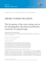
Figs 1–17. Light micrographs <strong>of</strong> different stages <strong>of</strong> <strong>the</strong> life <strong>cycle</strong> <strong>of</strong> <strong>Dissodinium</strong> pseudolunula collected <strong>of</strong>f Marseille, France, in<br />
spring 2008. 1. Primary cyst recently detached from <strong>the</strong> <strong>copepod</strong> egg. 2-7. Sequence <strong>of</strong> cell division inside <strong>of</strong> <strong>the</strong> primary cyst. The<br />
arrow in <strong>the</strong> Fig. 2 points to <strong>the</strong> rigid cellulosic wall. 9-10, 13. Binary division <strong>of</strong> <strong>the</strong> protoplasm to form <strong>the</strong> dinospores. The arrow<br />
in <strong>the</strong> Fig. 9 points to <strong>the</strong> tentative residual vacuole. The insets in <strong>the</strong> Figs 9, 10 showed <strong>the</strong> chloroplasts illuminated with blue light<br />
under epifluorescence microscopy. 11-12. Exceptional differentiation <strong>of</strong> <strong>the</strong> dinospores inside <strong>of</strong> a globular cyst. This cyst was used<br />
for PCR amplification. 14-16. Free dinospores, note <strong>the</strong> hyaline capsule. 17. Dinospore infecting a <strong>copepod</strong> egg. Scale bars: 50 mmin<br />
<strong>the</strong> Figs 1–8 <strong>and</strong> 10 mm in<strong>the</strong>Figs 9–17.
ARTICLE IN PRESS<br />
F. Gómez et al. / European Journal <strong>of</strong> Protistology 45 (2009) 260–270 263
264<br />
ARTICLE IN PRESS<br />
F. Gómez et al. / European Journal <strong>of</strong> Protistology 45 (2009) 260–270<br />
presence <strong>of</strong> large size opaque <strong>copepod</strong> <strong>eggs</strong>. The last<br />
cysts were observed in early June. The different stages <strong>of</strong><br />
<strong>the</strong> life <strong>cycle</strong> <strong>of</strong> <strong>Dissodinium</strong> pseudolunula are illustrated<br />
in <strong>the</strong> figures (Figs 1–17). We omitted <strong>the</strong> detailed<br />
description <strong>of</strong> our observations in <strong>the</strong> Mediterranean<br />
Sea because <strong>the</strong>y coincided to those already documented<br />
in <strong>the</strong> North Sea (Elbrächter <strong>and</strong> Drebes 1978).<br />
However, we exceptionally observed that <strong>the</strong> dinospores<br />
began to differentiate before <strong>the</strong> secondary cysts were<br />
released from <strong>the</strong> primary cyst. In fact, we observed two<br />
globular cysts containing dinospores in <strong>the</strong> natural<br />
samples (Figs 11, 12). In order to verify <strong>the</strong>ir identity, we<br />
carried out PCR analysis <strong>of</strong> each <strong>of</strong> <strong>the</strong>se globular cysts<br />
with dinospores. The SSU rDNA sequences were<br />
identical to that <strong>of</strong> <strong>the</strong> dinospores found in <strong>the</strong> lunate<br />
cyst <strong>of</strong> D. pseudolunula, showing that <strong>the</strong> dinospores<br />
may indeed occasionally differentiate before <strong>the</strong> rupture<br />
<strong>of</strong> <strong>the</strong> primary cyst wall.<br />
The fast-swimming dinospores <strong>of</strong> D. pseudolunula<br />
could not be easily distinguished from o<strong>the</strong>r surrounding<br />
gymnodinioid cells in <strong>the</strong> live plankton samples. We<br />
carried out short-term laboratory incubations with<br />
isolated primary cysts in order to investigate <strong>the</strong> life<br />
<strong>cycle</strong> <strong>and</strong> <strong>the</strong> morphology <strong>and</strong> behavior <strong>of</strong> <strong>the</strong> dinospores.<br />
A recently detached primary cyst required two<br />
days for <strong>the</strong> formation <strong>of</strong> <strong>the</strong> secondary cysts <strong>and</strong> <strong>the</strong><br />
dinospores. These incubations <strong>of</strong>ten resulted in aberrant<br />
secondary cysts with anomalous shapes <strong>and</strong> a lower<br />
number <strong>of</strong> dinospores when compared to natural<br />
samples. The released dinospores swam actively <strong>and</strong> after<br />
several minutes, <strong>the</strong>y became non-motile <strong>and</strong> encysted<br />
inside a hyaline membrane (Figs 14–16). No division <strong>of</strong><br />
<strong>the</strong> dinospores was observed. We added fresh <strong>copepod</strong><br />
<strong>eggs</strong> to test whe<strong>the</strong>r re-infections could occur. However,<br />
<strong>the</strong> dinospores did not infect <strong>the</strong>m, so that infections were<br />
only observed in natural samples (Fig. 17).<br />
As <strong>the</strong> secondary cysts <strong>and</strong> <strong>the</strong> dinospores showed a<br />
yellow-green pigmentation, we looked for <strong>the</strong> presence<br />
<strong>of</strong> chlorophyll a using epifluorescence microscopy.<br />
Instead <strong>of</strong> typical globular plastid accumulations, <strong>the</strong><br />
chlorophyll a showed a reticulate distribution in <strong>the</strong><br />
periphery <strong>of</strong> <strong>the</strong> cell (Figs 9, 10). The dinospores were<br />
placed in cultures under different light intensities in a<br />
nutrient-rich (f/2) medium that has been successfully<br />
used with o<strong>the</strong>r photosyn<strong>the</strong>tic unarmored din<strong>of</strong>lagellates.<br />
However, <strong>the</strong> dinospores did not proliferate under<br />
<strong>the</strong>se conditions.<br />
<strong>Chytriodinium</strong> affine<br />
<strong>Chytriodinium</strong> affine (Dogiel) Chatton was observed<br />
attached to small or medium-sized <strong>copepod</strong> <strong>eggs</strong> from<br />
June to September. The dinospore attached to <strong>the</strong> egg<br />
surface from <strong>the</strong> hyposome, it transformed into a<br />
spherical trophont stage <strong>and</strong> grew to a size <strong>of</strong> 50 to<br />
80 mm. Whereas <strong>the</strong> infective dinospore <strong>of</strong> D. pseudolunula<br />
detached as a primary cyst as soon as <strong>the</strong> egg<br />
contents were absorbed (less than 1 hour) <strong>and</strong> subsequently<br />
started to divide, <strong>the</strong> trophont <strong>of</strong> C. affine<br />
remained attached to <strong>the</strong> <strong>copepod</strong> egg until <strong>the</strong><br />
dinospores were mature. Multi-infection was a common<br />
feature in <strong>Chytriodinium</strong> (Figs 18, 19), although no more<br />
than two trophonts developed successfully from a single<br />
host (Figs 22, 26, 27). The dinospore was attached to <strong>the</strong><br />
host by means <strong>of</strong> a feeding tube, enlarged at its base. An<br />
ampulla with one large trophic vacuole formed at <strong>the</strong><br />
end <strong>of</strong> <strong>the</strong> peduncular disk that gradually absorbed <strong>the</strong><br />
egg cytoplasm (Figs 20–22, 24–25). These structures are<br />
here named following <strong>the</strong> terminology by Cachon <strong>and</strong><br />
Cachon (1968). The trophont maintained a spherical<br />
shape during this process (Figs 20–26). It continued<br />
feeding while <strong>the</strong> dinospores formed by palintomic<br />
sporogenesis. The chain <strong>of</strong> dinospores coiled itself inside<br />
<strong>of</strong> a fine hyaline membrane (Figs 28, 29). When all <strong>the</strong><br />
dinospores were formed, <strong>the</strong> cyst containing <strong>the</strong> chain <strong>of</strong><br />
dinospores detached from <strong>the</strong> empty egg, leaving <strong>the</strong><br />
trophont membrane (Fig. 30). After <strong>the</strong> rupture <strong>of</strong> <strong>the</strong><br />
membrane, <strong>the</strong> chain <strong>of</strong> dinospores gradually decomposed<br />
by releasing single cells that began to swim <strong>and</strong><br />
dispersed (Fig. 31). The colorless dinospores (9 mm long)<br />
showed a hemispheric episome. The dinospores divided<br />
synchronously again (Fig. 32). In contrast to<br />
D. pseudolunula, <strong>the</strong> dinospores <strong>of</strong> C. affine were able<br />
to infect fresh <strong>copepod</strong> <strong>eggs</strong> immediately after <strong>the</strong> last<br />
binary division. Each <strong>cycle</strong>, from <strong>the</strong> infection <strong>of</strong> <strong>the</strong><br />
host to <strong>the</strong> liberation <strong>of</strong> dinospores, was completed in<br />
24 hours <strong>and</strong> provided up to 200 dinospores from a<br />
trophont.<br />
<strong>Chytriodinium</strong> roseum<br />
One specimen ascribed to <strong>the</strong> genus <strong>Chytriodinium</strong><br />
type species, C. roseum, was found in samples from <strong>the</strong><br />
NW Mediterranean. It was identified from a sample<br />
collected in <strong>the</strong> Bay <strong>of</strong> Marseille on June 24th 2008 at<br />
55 m depth (Figs 33–35). Due to <strong>the</strong> paucity <strong>of</strong><br />
individuals, we decided to isolate it immediately for<br />
single-cell PCR instead <strong>of</strong> trying a temporal incubation<br />
that might have resulted in loss <strong>of</strong> <strong>the</strong> specimen. At <strong>the</strong><br />
observed stage <strong>of</strong> development, <strong>the</strong> chain was not fully<br />
developed <strong>and</strong> about half <strong>of</strong> <strong>the</strong> host content was<br />
already consumed. The host membrane began to<br />
collapse in <strong>the</strong> area where <strong>the</strong> trophont was attached<br />
(Fig. 35). The growing parasite formed a chain with two<br />
ellipsoidal lobules with a round junction. The proximal<br />
lobule (70 mm long), attached to <strong>the</strong> egg, showed<br />
transversal constrictions or septa in <strong>the</strong> distal extreme.<br />
In <strong>the</strong> proximal extreme, <strong>the</strong> trophont showed a large<br />
structure that appeared to be a vacuole. The distal<br />
lobule showed three transversal septa that divided it into
ARTICLE IN PRESS<br />
F. Gómez et al. / European Journal <strong>of</strong> Protistology 45 (2009) 260–270 265<br />
Figs 18–35. Light micrographs <strong>of</strong> <strong>the</strong> life <strong>cycle</strong> <strong>of</strong> <strong>Chytriodinium</strong> affine (Figs 18–32) <strong>and</strong> two groups <strong>of</strong> sporocytes <strong>of</strong> C. roseum<br />
(Figs 33–35) collected <strong>of</strong>f Marseille, France, in summer 2008; 18-19 Copepod <strong>eggs</strong> multi-infected; 20-25 Development <strong>of</strong> <strong>the</strong><br />
trophont; Peduncular disk (PD), Ampulla (Am). 25 Note <strong>the</strong> collapse <strong>of</strong> <strong>the</strong> <strong>copepod</strong> egg membrane; 26-27 Host infected by two<br />
trophonts; 27 Note <strong>the</strong> different degree <strong>of</strong> differentiation between <strong>the</strong> two parasites; 28 Chain <strong>of</strong> sporocytes detached from <strong>the</strong> host;<br />
29-32 The sporocytes detached from <strong>the</strong> host, leaving behind <strong>the</strong> primary cyst; 31 The chain fragmented <strong>and</strong> <strong>the</strong> dinospore began to<br />
disperse; 32 Dinospores under binary division; 33-35 Chain <strong>of</strong> sporocytes <strong>of</strong> <strong>Chytriodinium</strong> roseum used for PCR analysis; 36-38.<br />
O<strong>the</strong>r individuals <strong>of</strong> C. roseum dissociated into several chains. Scale bar <strong>of</strong> 50 mm inFigs 18–30 <strong>and</strong> <strong>of</strong> 10 mm inFigs 31–38.
266<br />
ARTICLE IN PRESS<br />
F. Gómez et al. / European Journal <strong>of</strong> Protistology 45 (2009) 260–270<br />
four sections (Fig. 34). We ascribed this chain <strong>of</strong><br />
dinospores to <strong>the</strong> species C. roseum in accordance with<br />
<strong>the</strong> illustration by Cachon <strong>and</strong> Cachon (1968, plate II,<br />
i–j). InC. affine, <strong>the</strong> numerous dinospores developed in<br />
a coiled chain inside <strong>of</strong> a hyaline spherical membrane<br />
that was absent in <strong>the</strong> chain <strong>of</strong> C. roseum.<br />
Ano<strong>the</strong>r specimen <strong>of</strong> C. roseum was retrieved from a<br />
surface sample collected in <strong>the</strong> Bay <strong>of</strong> Marseille on<br />
September 2nd 2008 (Figs 36–38). The sporocytes<br />
formed six chains. The closer chains to <strong>the</strong> empty egg<br />
were formed <strong>of</strong> eight sporocytes. One chain was formed<br />
<strong>of</strong> 12 sporocytes <strong>and</strong> o<strong>the</strong>r chain showed some<br />
sporocytes (11-14 mm in diameter) were dissociated <strong>and</strong><br />
under binary division (Fig. 38).<br />
Molecular <strong>phylogeny</strong><br />
Preliminary phylogenetic analyses <strong>of</strong> D. pseudolunula,<br />
C. affine <strong>and</strong> C. roseum sequences were included in a<br />
large din<strong>of</strong>lagellate SSU rDNA sequence alignment<br />
containing more than 1100 sequences. The preliminary<br />
analysis indicated that <strong>the</strong>se species branched close to<br />
representatives <strong>of</strong> <strong>the</strong> Gymnodiniales (data not shown).<br />
The <strong>phylogeny</strong> was fur<strong>the</strong>r investigated by applying<br />
Maximum Likelihood (ML) <strong>and</strong> Bayesian Inference (BI)<br />
methods upon a more restricted taxonomic sampling,<br />
including 67 taxa representing different Gymnodiniales<br />
<strong>and</strong> <strong>the</strong> main din<strong>of</strong>lagellate orders (Peridiniales, Dinophysiales,<br />
Prorocentrales, Gonyaulacales, Suessiales),<br />
with Syndiniales as outgroup taxa. The two species <strong>of</strong><br />
<strong>Chytriodinium</strong> were closely related (97% sequence<br />
identity), <strong>and</strong> formed a moderately supported group<br />
toge<strong>the</strong>r with <strong>the</strong> sequence <strong>of</strong> <strong>Dissodinium</strong> pseudolunula<br />
(ML bootstrap proportion, BP, <strong>of</strong> 76%, <strong>and</strong> BI posterior<br />
probability <strong>of</strong> 0.82). This low support could partly be<br />
due to <strong>the</strong> different evolutionary rates <strong>of</strong> <strong>the</strong> two<br />
<strong>Chytriodinium</strong> sequences. In fact, C. affine showed a<br />
branch twice longer than C. roseum, which could induce<br />
tree reconstruction problems, in particular long branch<br />
attraction artifacts. In agreement with this idea, <strong>the</strong><br />
removal <strong>of</strong> <strong>the</strong> C. affine sequence resulted in a tree where<br />
D. pseudolunula <strong>and</strong> C. roseum formed a group with<br />
better support (BP <strong>of</strong> 88% <strong>and</strong> PP <strong>of</strong> 0.96, data not<br />
shown).<br />
The clade formed by <strong>Chytriodinium</strong> <strong>and</strong> <strong>Dissodinium</strong><br />
branched with <strong>the</strong> Gymnodinium sensu stricto group<br />
(Fig. 39). This group appeared split in two major<br />
subgroups. The first one was composed <strong>of</strong> Gymnodinium<br />
species available from phototrophically growing cultures,<br />
colonial species related to Gymnodinium catenatum<br />
Graham (G. microreticulatum Bolch, Negri<br />
et Hallegraeff, G. nolleri Ellegaard et Moestrup,<br />
G. impudicum (Fraga et Bravo) G. Hansen et Moestrup)<br />
<strong>and</strong> <strong>the</strong> unicellular G. dorsalisulcum Murray, de Salas<br />
et Hallegraeff. Lepidodinium viride Watanabe,<br />
Suda, Inouye, Sawaguchi et Chihara/Lepidodinium<br />
chlorophorum (Elbrächter et Schnepf) G. Hansen, Botes<br />
et de Salas also branched in this subgroup, although<br />
with a low bootstrap support (Fig. 39). The o<strong>the</strong>r<br />
subgroup included both apochlorotic <strong>and</strong> chloroplastcontaining<br />
genera with a high diversity <strong>of</strong> ultrastructural<br />
features <strong>and</strong> trophic behaviors. Among <strong>the</strong> species<br />
forming this cluster, only <strong>the</strong> freshwater type <strong>of</strong><br />
Gymnodinium, G. fuscum Stein <strong>and</strong> G. aureolum<br />
(Hulburt) G. Hansen have been maintained growing<br />
phototrophically in stable cultures. Gymnodinium fuscum<br />
branched at a basal position in <strong>the</strong> clade <strong>of</strong> pseudocolonial<br />
species <strong>of</strong> Polykrikos Bütschli. The phagotrophic<br />
warnowiids represented by <strong>the</strong> genera Erythropsidinium<br />
Hertwig <strong>and</strong> Warnowia Lindemann, <strong>and</strong> <strong>the</strong><br />
type <strong>of</strong> Pheopolykrikos Chatton, <strong>the</strong> chloroplast-containing<br />
Pheopolykrikos beauchampii Chatton, formed a<br />
weakly supported clade. These species branched relatively<br />
close to G. fuscum/Polykrikos, although with a low<br />
bootstrap support (54%). At a basal position appeared<br />
a clade formed by Gymnodinium aureolum <strong>and</strong> ano<strong>the</strong>r<br />
unidentified Gymnodinium species. The <strong>Chytriodinium</strong><br />
clade appeared in <strong>the</strong> most basal position <strong>of</strong> this<br />
subgroup <strong>of</strong> <strong>the</strong> Gymnodinium s.s. group. The inclusion<br />
<strong>of</strong> <strong>the</strong>se new sequences reinforced <strong>the</strong> separation <strong>of</strong> <strong>the</strong><br />
Gymnodinium type <strong>and</strong> o<strong>the</strong>r congeneric species related<br />
to G. catenatum (Fig. 39).<br />
Discussion<br />
<strong>Dissodinium</strong> pseudolunula is widely distributed in<br />
marine neritic habitats. In contrast, <strong>the</strong> records <strong>of</strong><br />
<strong>Chytriodinium</strong> are scarce, mainly restricted to <strong>the</strong><br />
western Mediterranean Sea (Dogiel 1906; Cachon <strong>and</strong><br />
Cachon 1968) <strong>and</strong> <strong>the</strong> subtropical Atlantic Ocean<br />
(Elbrächter 1988). Drebes <strong>and</strong> Elbrächter carried out<br />
intensive studies on <strong>the</strong> parasitic din<strong>of</strong>lagellates in <strong>the</strong><br />
North Sea, but no record <strong>of</strong> <strong>Chytriodinium</strong> was<br />
reported. <strong>Chytriodinium</strong> appears to have a clear warmwater<br />
distribution.<br />
Our <strong>molecular</strong> <strong>phylogeny</strong> study suggests that <strong>Chytriodinium</strong><br />
<strong>and</strong> <strong>Dissodinium</strong> derived from a common<br />
ancestor. The type <strong>of</strong> feeding <strong>and</strong> host appear to be<br />
similar for <strong>the</strong>se ectoparasitic din<strong>of</strong>lagellates but,<br />
apparently, <strong>the</strong>y do not compete for <strong>the</strong> same resources<br />
since <strong>the</strong>ir peaks in abundance are temporally decoupled<br />
in <strong>the</strong> western Mediterranean Sea. <strong>Chytriodinium</strong> affine<br />
is able to rapidly respond to <strong>the</strong> increase in host<br />
availability during <strong>the</strong> summer with <strong>the</strong> formation <strong>of</strong> <strong>the</strong><br />
infective dinospores occurring in 24 hours. In contrast,<br />
D. pseudolunula requires at least two days from infection<br />
to <strong>the</strong> liberation <strong>of</strong> <strong>the</strong> new dinospores <strong>and</strong> probably<br />
some additional time for <strong>the</strong> dinospores to be infective<br />
since, according to our observations, recently released
ARTICLE IN PRESS<br />
F. Gómez et al. / European Journal <strong>of</strong> Protistology 45 (2009) 260–270 267<br />
Fig. 39. Maximum likelihood phylogenetic tree <strong>of</strong> din<strong>of</strong>lagellate SSU rDNA sequences, based on 1,202 aligned positions. Names in<br />
bold represent sequences obtained in this study. Numbers at <strong>the</strong> nodes are bootstrap proportions (values under 50% are omitted).<br />
Nodes supported by posterior probability values 40.90 in Bayesian Inference analyses are indicated by black circles. Several<br />
branches leading to fast-evolving species have been shortened to one third (indicated by 1/3). Accession numbers are provided<br />
between brackets. The scale bar represents <strong>the</strong> number <strong>of</strong> substitutions for a unit branch length.
268<br />
ARTICLE IN PRESS<br />
F. Gómez et al. / European Journal <strong>of</strong> Protistology 45 (2009) 260–270<br />
dinospores appeared unable to infect new hosts. This<br />
agrees with <strong>the</strong> results obtained by Drebes (1984) that<br />
suggested that <strong>the</strong> dinospores <strong>of</strong> D. pseudolunula may<br />
need a post-maturation time before <strong>the</strong>y become able to<br />
infect a host. Drebes (1984) also found that <strong>the</strong>se<br />
dinospores could survive for up to four weeks. In<br />
contrast, our incubation experiments suggest that <strong>the</strong><br />
C. affine dinospores disappeared in a few hours.<br />
The dinospores <strong>of</strong> C. affine were colorless <strong>and</strong> we did<br />
not observe any cyst formation, while <strong>the</strong> dinospores <strong>of</strong><br />
D. pseudolunula contained chlorophyll a <strong>and</strong> were<br />
encysted. Stoecker (1999) reported that phototrophy in<br />
D. pseudolunula might be a mechanism to increase<br />
survival during dispersal. However, it is uncertain<br />
whe<strong>the</strong>r <strong>the</strong> chlorophyll a is photosyn<strong>the</strong>tically functional<br />
<strong>and</strong> able to maintain <strong>the</strong> dinospores until a<br />
suitable host is available. Chloroplasts are well developed<br />
<strong>and</strong> appear functional in o<strong>the</strong>r parasitic din<strong>of</strong>lagellates<br />
such as Protoodinium Hovasse,<br />
Piscinoodinium Lom <strong>and</strong> Crepidoodinium Lom et Lawler<br />
<strong>and</strong> some species <strong>of</strong> Blastodinium Chatton (Cachon <strong>and</strong><br />
Cachon 1987). The o<strong>the</strong>r species <strong>of</strong> <strong>Dissodinium</strong>,<br />
D. pseudocalani, lack chloroplasts (Drebes 1969). Since<br />
din<strong>of</strong>lagellates show a strong tendency to lose <strong>and</strong><br />
replace plastids (Saldarriaga et al. 2001), <strong>the</strong> fact that a<br />
parasitic din<strong>of</strong>lagellate continues to syn<strong>the</strong>size chlorophyll<br />
a suggests a function related to <strong>the</strong> survival <strong>of</strong> <strong>the</strong><br />
dinospores. The ultrastructure <strong>of</strong> <strong>the</strong> putative chloroplasts<br />
<strong>of</strong> D. pseudolunula needs to be investigated.<br />
The warnowiids <strong>and</strong> heterotrophic polykrikoids<br />
branched in <strong>the</strong> SSU rDNA phylogenies within <strong>the</strong><br />
Gymnodinium s.s. group, whose members are endowed<br />
with a high diversity <strong>of</strong> chloroplasts (Daugbjerg et al.<br />
2000; Hansen et al. 2007; Hoppenrath <strong>and</strong> Le<strong>and</strong>er<br />
2007b). The <strong>Chytriodinium</strong> clade branched in <strong>the</strong><br />
subgroup <strong>of</strong> type G. fuscum. In <strong>the</strong> LSU rDNA<br />
phylogenies, G. fuscum appeared as sister <strong>of</strong> <strong>the</strong><br />
heterotrophic Gymnodinium venator Flø Jørgensen<br />
et Murray, previously known under <strong>the</strong> genus Amphidinium<br />
as A. pellucidum C. Herdman (Flø Jørgensen et<br />
al. 2004). The species <strong>of</strong> Lepidodinium <strong>and</strong> Gymnodinium<br />
aureolum are able to grow phototrophically in cultures.<br />
These species have a peduncle, whose function is<br />
speculated to be a feeding structure (Hansen 2001;<br />
Hansen et al. 2007). In our SSU rDNA tree, <strong>the</strong> clade<br />
Lepidodinium <strong>and</strong> G. aureolum/Gymnodinium sp. appeared<br />
in different subgroups <strong>of</strong> Gymnodinium s.s.<br />
group, although <strong>the</strong>se clades have a very low bootstrap<br />
support. Gymnodinium aureolum appeared relatively<br />
close to <strong>the</strong> <strong>Chytriodinium</strong> clade <strong>and</strong> it is uncertain<br />
whe<strong>the</strong>r its peduncle emerging from <strong>the</strong> sulcal region<br />
might be related to <strong>the</strong> peduncular disk issued from <strong>the</strong><br />
hyposome in <strong>the</strong> <strong>Chytriodinium</strong> clade.<br />
The SSU rDNA <strong>phylogeny</strong> would support <strong>the</strong><br />
reclassification <strong>of</strong> <strong>Chytriodinium</strong> roseum <strong>and</strong> C. affine<br />
under <strong>the</strong> genus Gymnodinium, as Dogiel (1906)<br />
originally described it. This is also <strong>the</strong> case for<br />
D. pseudolunula. The name Gymnodinium lunula has<br />
been largely used for life stages <strong>of</strong> both, D. pseudolunula<br />
<strong>and</strong> Pyrocystis lunula. To avoid confusion, a new<br />
combination, Gymnodinium pseudolunula, may be proposed.<br />
However, before proposing <strong>the</strong> transfer <strong>of</strong><br />
<strong>Chytriodinium</strong> <strong>and</strong> <strong>Dissodinium</strong> species into Gymnodinium,<br />
we must be sure that <strong>the</strong> species currently ascribed<br />
to Gymnodinium form a unique clade. The addition <strong>of</strong><br />
new sequences in <strong>the</strong> Gymnodinium s.s. group is<br />
revealing that <strong>the</strong> freshwater Gymnodinium type is<br />
separated from <strong>the</strong> marine representatives (G. catenatum<br />
<strong>and</strong> related species) (Hoppenrath <strong>and</strong> Le<strong>and</strong>er 2007a, b;<br />
Kim et al. 2008; Gómez et al. 2009). Hence, a fur<strong>the</strong>r<br />
split <strong>of</strong> <strong>the</strong> current Gymnodinium s.s. into separate<br />
genera cannot be discarded <strong>and</strong>, <strong>the</strong>refore, we prefer at<br />
present to conserve C. roseum, C. affine <strong>and</strong> D.<br />
pseudolunula under <strong>the</strong>ir current nomenclature. At any<br />
rate, since <strong>the</strong> SSU rDNA <strong>phylogeny</strong> showed that<br />
<strong>Chytriodinium</strong> <strong>and</strong> <strong>Dissodinium</strong> formed a clade, it would<br />
be possible to group all <strong>the</strong>se species under <strong>the</strong> genus<br />
<strong>Chytriodinium</strong>, which has <strong>the</strong> priority versus <strong>Dissodinium</strong>.<br />
The type <strong>of</strong> life <strong>cycle</strong> has been used traditionally for<br />
<strong>the</strong> classification <strong>of</strong> parasitic din<strong>of</strong>lagellates into families.<br />
Fensome et al. (1993, p. 175) placed <strong>Dissodinium</strong><br />
<strong>and</strong> Cachonella into <strong>the</strong> Cachonellaceae according to <strong>the</strong><br />
presence <strong>of</strong> at least two vegetative cyst stages. However,<br />
in this study we observed that <strong>the</strong> <strong>Dissodinium</strong><br />
dinospores can occasionally be formed inside a globular<br />
cyst (Figs 11, 12), indicating that <strong>Dissodinium</strong> has <strong>the</strong><br />
capacity to modify its life <strong>cycle</strong>. Therefore, <strong>the</strong> type <strong>of</strong><br />
life <strong>cycle</strong> is not an appropriate characteristic for <strong>the</strong><br />
classification <strong>of</strong> <strong>the</strong> parasitic din<strong>of</strong>lagellates. A similar<br />
argument may apply also to <strong>the</strong> mode <strong>of</strong> feeding as a<br />
general diagnostic feature. The genus Paulsenella<br />
Chatton was initially included within <strong>the</strong> Chytriodiniaceae<br />
(Cachon et al. 1969; Loeblich III 1982; Elbrächter<br />
1988), although <strong>the</strong> SSU rDNA <strong>phylogeny</strong> indicated<br />
later an affiliation among <strong>the</strong> <strong>the</strong>cate din<strong>of</strong>lagellates <strong>of</strong><br />
<strong>the</strong> order Peridiniales (Kühn <strong>and</strong> Medlin 2005). Paulsenella<br />
<strong>and</strong> its relatives, Pfiesteria <strong>and</strong> <strong>the</strong> cryptoperidiniopsoids,<br />
form a clade that is united by <strong>the</strong>ir mode <strong>of</strong><br />
feeding, myzocytotically by means <strong>of</strong> an extensible<br />
feeding tube. Consequently, Kühn <strong>and</strong> Medlin (2005)<br />
suggested <strong>the</strong> mode <strong>of</strong> feeding as a character useful for<br />
<strong>the</strong> classification <strong>of</strong> parasitic din<strong>of</strong>lagellates, but this<br />
assumption is not valid for <strong>Chytriodinium</strong> <strong>and</strong> <strong>Dissodinium</strong><br />
because, although <strong>the</strong>y share <strong>the</strong> type <strong>of</strong> feeding,<br />
<strong>the</strong>ir closest relatives include numerous photosyn<strong>the</strong>tic<br />
species.<br />
Although, in general, trophonts easily lose <strong>the</strong><br />
din<strong>of</strong>lagellate characters <strong>and</strong> <strong>the</strong> morphology <strong>of</strong> unrelated<br />
species may resemble by convergence (Cachon<br />
<strong>and</strong> Cachon 1987), in <strong>the</strong> case <strong>of</strong> <strong>Chytriodinium</strong> <strong>and</strong><br />
<strong>Dissodinium</strong>, <strong>the</strong> type <strong>of</strong> dinospore appeared informative
ARTICLE IN PRESS<br />
F. Gómez et al. / European Journal <strong>of</strong> Protistology 45 (2009) 260–270 269<br />
<strong>and</strong> supported <strong>the</strong>ir systematic affiliation. The members<br />
<strong>of</strong> <strong>the</strong> Gymnodinium s.s. group possess a loop-shaped<br />
apical groove running anticlockwise (Daugbjerg et al.<br />
2000). Due to <strong>the</strong> low resolution <strong>of</strong> <strong>the</strong> inverted light<br />
microscopy, we were unable to confirm <strong>the</strong> occurrence<br />
<strong>of</strong> this type <strong>of</strong> apical groove in C. affine <strong>and</strong> D.<br />
pseudolunula. Unfortunately, <strong>the</strong> swimming infective<br />
dinospores are delicate, have a short life <strong>and</strong>, as such,<br />
cannot be easily distinguished from o<strong>the</strong>r plankton cells.<br />
For example, <strong>the</strong> dinospores <strong>of</strong> Blastodinium have been<br />
recently demonstrated to be <strong>the</strong>cate, while <strong>the</strong>y were<br />
considered gymnodinioid in previous studies (Skovgaard<br />
et al. 2007). Paulsenella has been placed between<br />
<strong>the</strong> <strong>the</strong>cate din<strong>of</strong>lagellates, but despite <strong>the</strong> studies in<br />
cultures, <strong>the</strong>re is no evidence <strong>of</strong> <strong>the</strong>cal plates in any life<br />
stage (Kühn <strong>and</strong> Medlin 2005). It appears that <strong>the</strong><br />
diagnostic features present in <strong>the</strong> dinospores that may<br />
reveal <strong>the</strong>ir systematic position are only visible during<br />
discrete periods <strong>of</strong> <strong>the</strong>ir life <strong>cycle</strong>. Therefore, <strong>molecular</strong><br />
techniques are not only complementary to classical<br />
taxonomy, but <strong>the</strong>y appear to be <strong>the</strong> only alternative to<br />
resolve <strong>the</strong> <strong>phylogeny</strong> <strong>of</strong> parasitic din<strong>of</strong>lagellate species<br />
in cases where <strong>the</strong> diagnostic morphological characteristics<br />
cannot be observed.<br />
Acknowledgements<br />
This is a contribution to <strong>the</strong> project DIVERPLAN-<br />
MED supported by a post-doctoral grant to F.G. <strong>of</strong> <strong>the</strong><br />
Ministerio Español de Educación y Ciencia #2007-0213.<br />
P.L.G. <strong>and</strong> D.M. acknowledge financial support from<br />
<strong>the</strong> French CNRS <strong>and</strong> <strong>the</strong> ANR Biodiversity project<br />
‘Aquaparadox’. We thank I. Salter for assistance with<br />
<strong>the</strong> English edition.<br />
References<br />
Cachon, J., Cachon, M., 1968. Cytologie et <strong>cycle</strong> évolutif des<br />
<strong>Chytriodinium</strong>. Protistologica 4, 249–262.<br />
Cachon, J., Cachon, M., Bouquaheux, F., 1969. Myxodinium<br />
pipiens gen. nov., péridinien parasite d’Halosphaera. Phycologia<br />
8, 157–164.<br />
Cachon, J., Cachon, M., 1987. Parasitic din<strong>of</strong>lagellates. In:<br />
Taylor, F.J.R. (Ed.), The Biology <strong>of</strong> Din<strong>of</strong>lagellates.<br />
Blackwell, Oxford, pp. 571–610.<br />
Chatton, É., 1912. Diagnoses préliminaires de Péridiniens<br />
parasites nouveaux. Bull. Soc. Zool. Fr. 37, 85–93.<br />
Chatton, É., 1920. Les péridiniens parasites. Morphologie,<br />
reproduction, éthologie. Arch. Zool. Exp. Gén. 59, 1–475.<br />
Chrétiennot-Dinet, M.-J., Sournia, A., Ricard, M., Billard, C.,<br />
1993. A classification <strong>of</strong> <strong>the</strong> marine phytoplankton <strong>of</strong> <strong>the</strong><br />
world from class to genus. Phycologia 32, 159–179.<br />
Claparède, E., Lachmann, J., 1858. Études sur les Infusoires et<br />
les Rhizopodes. Mém. Inst. Nat. Geneva 5 (3), 1–260.<br />
Daugbjerg, N., Hansen, G., Larsen, J., Moestrup, Ø., 2000.<br />
Phylogeny <strong>of</strong> some <strong>of</strong> <strong>the</strong> major genera <strong>of</strong> din<strong>of</strong>lagellates<br />
based on ultra-structure <strong>and</strong> partial LSU rDNA sequence<br />
data, including <strong>the</strong> erection <strong>of</strong> three new genera <strong>of</strong><br />
unarmoured din<strong>of</strong>lagellates. Phycologia 39, 302–317.<br />
Dogiel, V., 1906. Beiträge zur Kenntnis der Peridineen. Mitt.<br />
Zool. Stn. Neapel. 18, 1–45.<br />
Drebes, G., 1969. <strong>Dissodinium</strong> pseudocalani sp. nov., ein<br />
parasitischer Din<strong>of</strong>lagellat auf Copepodeneiern. Helgol.<br />
wiss. Meeresunters. 19, 58–67.<br />
Drebes, G., 1978. <strong>Dissodinium</strong> pseudolunula (Dinophyta), a<br />
parasite on <strong>copepod</strong> <strong>eggs</strong>. Br. Phycol. J. 13, 319–327.<br />
Drebes, G., 1984. <strong>Life</strong> <strong>cycle</strong> <strong>and</strong> host specifity <strong>of</strong> marine<br />
parasitic dinophytes. Helgol. Meeresunters. 37, 603–622.<br />
Drebes, G., 1988. Syltodinium listii gen. et spec. nov., a marine<br />
ectoparasitic din<strong>of</strong>lagellate on egg <strong>of</strong> <strong>copepod</strong>s <strong>and</strong> rotifers.<br />
Helgol. Meeresunters. 42, 583–591.<br />
Edgar, R.C., 2004. MUSCLE: multiple sequence alignment<br />
with high accuracy <strong>and</strong> high throughput. Nucleic Acids<br />
Res. 32, 1792–1797.<br />
Elbrächter, M., 1988. <strong>Life</strong> <strong>cycle</strong> <strong>of</strong> Schizochytriodinium calani<br />
nov. gen. nov. spec., a din<strong>of</strong>lagellate parasitizing <strong>copepod</strong><br />
<strong>eggs</strong>. Helgol. Meeresunters. 42, 593–599.<br />
Elbrächter, M., Drebes, G., 1978. <strong>Life</strong> <strong>cycle</strong>s, <strong>phylogeny</strong> <strong>and</strong><br />
taxonomy <strong>of</strong> <strong>Dissodinium</strong> <strong>and</strong> Pyrocystis (Dinophyta).<br />
Helgol. Meeresunters. 31, 347–366.<br />
Fensome, R.A., Taylor, F.J.R., Norris, G., Sarjeant., W.A.S.,<br />
Wharton, D.I., Williams, G.L., 1993. A Classification <strong>of</strong><br />
Living <strong>and</strong> Fossil Din<strong>of</strong>lagellates. Sheridan Press, Hanover,<br />
Pennsylvania.<br />
Flø Jørgensen, M., Murray, S., Daugbjerg, N., 2004.<br />
Amphidinium revisited. I. Redefinition <strong>of</strong> Amphidinium<br />
(Dinophyceae) based on cladistic <strong>and</strong> <strong>molecular</strong> phylogenetic<br />
analyses. J. Phycol. 40, 351–365.<br />
Gómez, F., 2005. A list <strong>of</strong> din<strong>of</strong>lagellates in <strong>the</strong> world’s oceans.<br />
Acta Bot. Croat. 64, 129–212.<br />
Gómez, F., López-García, P., Moreira, D., 2009. Molecular<br />
<strong>phylogeny</strong> <strong>of</strong> <strong>the</strong> ocelloid-bearing din<strong>of</strong>lagellates Erythropsidinium<br />
<strong>and</strong> Warnowia (Warnowiaceae, Dinophyceae). J.<br />
Eukaryot. Microbiol. 56 (4), in press, doi:10.1111/<br />
j.1550-7408.2009.00420.x.<br />
Gönnert, R., 1936. Sporodinium pseudocalani n.g., n. sp., ein<br />
Parasit auf Copepodeneiern. Z. Parasitenkunde 9, 140–143.<br />
Hansen, G., 2001. Ultrastructure <strong>of</strong> Gymnodinium aureolum<br />
(Dinophyceae): toward a fur<strong>the</strong>r redefinition <strong>of</strong> Gymnodinium<br />
sensu stricto. J. Phycol. 37, 612–623.<br />
Hansen, G., Botes, L., De Salas, M., 2007. Ultrastructure <strong>and</strong><br />
large subunit rDNA sequences <strong>of</strong> Lepidodinium viride reveal<br />
a close relationship to Lepidodinium chlorophorum comb.<br />
nov. (Gymnodinium chlorophorum). Phycol. Res. 55, 25–41.<br />
Hoppenrath, M., Le<strong>and</strong>er, B.S., 2007a. Character evolution in<br />
polykrikoid din<strong>of</strong>lagellates. J. Phycol. 43, 366–377.<br />
Hoppenrath, M., Le<strong>and</strong>er, B.S., 2007b. Morphology <strong>and</strong><br />
<strong>phylogeny</strong> <strong>of</strong> <strong>the</strong> pseudocolonial din<strong>of</strong>lagellates Polykrikos<br />
lebourae <strong>and</strong> Polykrikos herdmanae n. sp. Protist 158,<br />
209–227.<br />
Jobb, G., von Haeseler, A., Strimmer, K., 2004. TREEFIN-<br />
DER: a powerful graphical analysis environment for<br />
<strong>molecular</strong> phylogenetics. BMC Evol. Biol. 4, 18.
270<br />
ARTICLE IN PRESS<br />
F. Gómez et al. / European Journal <strong>of</strong> Protistology 45 (2009) 260–270<br />
Kim, K.-Y., Iwataki, M., Kim, C.-H., 2008. Molecular<br />
phylogenetic affiliations <strong>of</strong> <strong>Dissodinium</strong> pseudolunula, Pheopolykrikos<br />
hartmannii, Polykrikos cf. schwartzii <strong>and</strong><br />
Polykrikos k<strong>of</strong>oidii to Gymnodinium sensu stricto species<br />
(Dinophyceae). Phycol. Res. 56, 89–92.<br />
Kühn, S.F., Medlin, L.K., 2005. The systematic position <strong>of</strong> <strong>the</strong><br />
parasitoid marine din<strong>of</strong>lagellate Paulsenella vonstoschii<br />
(Dinophyceae) inferred from nuclear-encoded small subunit<br />
ribosomal DNA. Protist 156, 393–398.<br />
Lartillot, N., Philippe, H., 2004. A Bayesian mixture model for<br />
across-site heterogeneities in <strong>the</strong> amino-acid replacement<br />
process. Mol. Biol. Evol. 21, 1095–1109.<br />
Loeblich III, A.R., 1982. Dinophyceae. In: Parker, S.P. (Ed.),<br />
Synopsis <strong>and</strong> Classification <strong>of</strong> Living Organisms. McGraw-<br />
Hill, New York, pp. 101–105.<br />
Mauchline, J., 1998. The biology <strong>of</strong> calanoid <strong>copepod</strong>s. Adv.<br />
Mar. Biol. 33, 1–710.<br />
Philippe, H., 1993. MUST, a computer package <strong>of</strong> management<br />
utilities for sequences <strong>and</strong> trees. Nucleic Acids Res.<br />
21, 5264–5272.<br />
Saldarriaga, J.F., Taylor, F.J.R., Keeling, P.J., Cavalier-<br />
Smith, T., 2001. Din<strong>of</strong>lagellate nuclear SSU rRNA<br />
<strong>phylogeny</strong> suggests multiple plastid losses <strong>and</strong> replacements.<br />
J. Mol. Evol. 53, 204–213.<br />
Saitou, N., Nei, M., 1987. The neighbor-joining method: a new<br />
method for reconstructing phylogenetic trees. Mol. Biol.<br />
Evol. 4, 406–425.<br />
Skovgaard, A., Massana, R., Saiz, E., 2007. Parasitic species <strong>of</strong><br />
<strong>the</strong> genus Blastodinium (Blastodiniphyceae) are peridinioid<br />
din<strong>of</strong>lagellates. J. Phycol. 43, 553–560.<br />
Stoecker, D.E., 1999. Mixotrophy among din<strong>of</strong>lagellates. J.<br />
Eukaryot. Microbiol. 24, 397–401.<br />
Taylor, F.J.R., 1987. Taxonomy <strong>and</strong> classification. In: Taylor,<br />
F.J.R. (Ed.), The Biology <strong>of</strong> Din<strong>of</strong>lagellates. Blackwell,<br />
Oxford, pp. 723–732.<br />
Théodoridès, J., 1989. Parasitology <strong>of</strong> marine zooplankton.<br />
Adv. Mar. Biol. 25, 117–177.