Horizontal Gaze Palsy
Horizontal Gaze Palsy
Horizontal Gaze Palsy

Create successful ePaper yourself
Turn your PDF publications into a flip-book with our unique Google optimized e-Paper software.
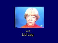
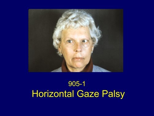
905-1<br />
<strong>Horizontal</strong> <strong>Gaze</strong> <strong>Palsy</strong>
Left esotropia; fascicular sixth nerve<br />
palsy, left horizontal gaze palsy
Full horizontal gaze to the right with<br />
gaze evoked nystagmus
Vergence movements induced the right<br />
eye to cross the midline
Full Downgaze
Impaired eye closure due to left facial<br />
palsy (Bell’s palsy)
Left lower motorneuron facial palsy<br />
(Bell’s palsy)
Figure 1 Axial NECT scan shows a focal hemorrhage in the<br />
posterior pons and fourth ventricle. Patient with known breast<br />
cancer.
Figure 2 Sagittal NECT scan showing the rostral-caudal<br />
extent of the pontine hemorrhage
Ocular Motility<br />
Unilateral horizontal gaze palsy to the left<br />
that impaired saccades and pursuit<br />
Esotropia of the left eye<br />
Fascicular sixth nerve palsy<br />
<strong>Horizontal</strong> gaze full to the right, gaze<br />
evoked nystagmus
Ocular Motility<br />
Normal convergence, right eye<br />
induced to cross the midline<br />
<strong>Horizontal</strong> oculocephalic reflex,<br />
absent (Doll’s head maneuver)<br />
Vertical eye movements normal
Signs in Leigh and Zee’s Case<br />
The patient was unable to move her eyes to the right<br />
past the midline using either saccadic or pursuit eye<br />
movements<br />
Head rotation to the left, however, drove the eyes past<br />
the midline, but the right eye abducted incompletely<br />
Vergence movements induced the left eye to cross the<br />
midline<br />
Vertical eye movements were normal<br />
<strong>Gaze</strong> evoked nystagmus was present on looking to the<br />
left, with slow phases toward the midline<br />
The patient developed a fascicular sixth nerve palsy
<strong>Horizontal</strong> <strong>Gaze</strong> <strong>Palsy</strong><br />
There are four theoretical possibilities to account<br />
for the ipsilateral horizontal gaze palsy due to a<br />
single unilateral lesion affecting<br />
1. The ipsilateral paramedial pontine reticular<br />
formation (PPRF) only<br />
2. The ipsilateral abducens nucleus (AN) alone<br />
3. Both the ipsilateral PPRF and the AN, or<br />
when two lesions are involved<br />
4. The motoneuron root fibers of the ipsilateral<br />
AN to the lateral rectus and the contralateral<br />
medial longitudinal fasciculus (MLF)
Figure 3 <strong>Horizontal</strong> section of the lower pons.<br />
1) Basis pontis syndrome. 2) Internuclear ophthalmoplegia 3) Abducens<br />
nucleus syndrome 4) Caudal PPRF syndrome 5) One-and-a-half syndrome 6)<br />
Paramedian midbrain syndrome
Figure 4 Sagittal section of brainstem
Clinical Findings with PPRF Lesion<br />
Loss of horizontal saccades towards the<br />
side of the lesion<br />
Contralateral gaze deviation, in acute<br />
phase<br />
<strong>Gaze</strong>-evoked nystagmus on looking<br />
contralateral to the lesion
Clinical Findings with PPRF Lesion<br />
Impaired smooth pursuit and vestibular<br />
eye movements may be preserved or<br />
impaired<br />
Bilateral lesions cause total horizontal<br />
gaze palsy and slowing of vertical<br />
saccades
Contralateral gaze deviation in an<br />
acute PPRF lesion
Clinical findings with lesion of the<br />
abducens nuclei<br />
Loss of all conjugate movements towards<br />
the side of the lesion – ipsilateral,<br />
horizontal gaze palsy<br />
Contralateral gaze deviation, in acute<br />
phase<br />
Vergence and vertical movements are<br />
spared
Clinical findings with lesion of the<br />
abducens nuclei<br />
In the intact hemifield of gaze, horizontal<br />
movements may be preserved, but<br />
ipsilaterally directed saccades are slow<br />
<strong>Horizontal</strong> gaze-evoked nystagmus on<br />
looking contralaterally<br />
Ipsilateral lower motoneuron facial palsy
Clinical signs of a lesion of the<br />
abducens nuclei
Clinical distinction PPRF: AN at the<br />
bedside<br />
PPRF lesions rostral to abducens<br />
paralysis of saccades and pursuit, but the<br />
eyes can be driven to the side of the<br />
ipsilateral gaze palsy with vestibular<br />
stimulation by the oculocephalic reflex<br />
and/or cold calorics
Clinical distinction PPRF: AN at the<br />
bedside<br />
PPRF lesions at the level of abducens<br />
are associated with ipsilateral gaze palsy<br />
and loss of reflex vestibular (and tonic<br />
neck) movements<br />
This presumes that there is a critical<br />
synapse within the caudal PPRF for the<br />
vestibulo-ocular pathways or that the<br />
functional integrity of the PPRF at that<br />
level is necessary for vestibulo-ocular eye<br />
movements
Figure 5 Ocular motor control system. Combination of previous illustrations<br />
indicates saccadic (s), pursuit (P), and vestibular (VIII), inputs to PPRF and its<br />
output to the oculomotor nucleus (III) and the abducens nucleus (VI).
Figure 6 Brainstem ocular motor control system
Figure 7 Brainstem ocular motor control system
The PRF contains three types of saccade – related<br />
neurons<br />
Burst neurons (BN)<br />
Excitatory BN (EBN) create saccadic eye velocity<br />
commands (the pulse)<br />
Inhibitory BN (IBN) permit reciprocal innervation<br />
to occur<br />
Tonic neurons (TN)<br />
Part of the neural integrator that integrates eye<br />
velocity commands and holds position for gaze<br />
Pause neurons (PN)<br />
Exert a normal inhibitory influence upon saccadic<br />
burst neurons during periods of fixation
Figure 8 The motor circuit for horizontal saccades in the brainstem
Conjugate horizontal deviation of the<br />
eyes in coma
http://www.library.med.utah.edu/NOVEL