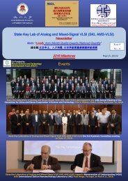
AMSV Newsletter 2014
AMSV Newsletter 2014
AMSV Newsletter 2014

Create successful ePaper yourself
Turn your PDF publications into a flip-book with our unique Google optimized e-Paper software.
In Royal Society of Chemistry - Analyst <strong>2014</strong><br />
NMR-DMF: A Modular Nuclear Magnetic Resonance–Digital<br />
Microfluidics System for Biological Assays<br />
Ka-Meng Lei, Pui-In Mak, Man-Kay Law, and Rui P. Martins<br />
From Multidisciplinary Research Area<br />
Motivation<br />
Architecture I<br />
We present a modular nuclear magnetic resonance–digital<br />
microfluidics (NMR-DMF) system<br />
as a portable diagnostic platform for miniaturized<br />
biological assays.<br />
With increasing numbers of combination between<br />
designed probes and a specific target,<br />
NMR becomes an accurate and rapid assay tool<br />
capable of detecting particular kinds of proteins,<br />
DNAs, bacteria and cells with a customized<br />
probe quantitatively.<br />
Traditional sample operation (e.g., manipulation<br />
and mixing) relied heavily on human efforts. We<br />
herein propose a modular NMR-DMF system to<br />
allow electronic automation of multi-step reaction-screening<br />
protocols.<br />
The overall schematics and operations of the NMR-DMF system. (a) The<br />
placement of the DMF chip, magnet, RF coil and PCB board in 3D view. Benefit<br />
from the plane-parallel magnetic field generated by the figure-8 shaped coil,<br />
the NMR system can be effectively integrated into the DMF system; (b) Schematics<br />
of the NMR Electronics. The transmitter which is formed by the digital<br />
logics such as flip flops is used to excite the hydrogen atom. On the receiver<br />
part, the capacitor together with the RF-coil (fabricated figure-8 shaped coil)<br />
forms a LC tank to provide passive gain enhancing of the system’s sensitivity.<br />
The signal is then amplified and down converted to f IF (intermediate frequency)<br />
and fed to the external filters and oscilloscope; (c) The filtered results from the<br />
PCB are captured by the oscilloscope for easier demonstration purpose.<br />
Waveforms are then analysed and the spin-spin relaxation time (T 2) is fitted by<br />
the algorithm written in MATLAB; (d) The photograph of the DMF chip and<br />
structure of the DMF platform. The droplets are squeezed between the top and<br />
bottom planes and surrounded by silicone oil; (e) The detection mechanism of<br />
the NMR-DMF system. The target-specific magnetic nanoparticles, which act<br />
as probes, are placed on the sensing site initially (in purple). The samples at<br />
other electrodes (in cyan) will be transported to the sensing site and mixed with<br />
the probes to perform NMR assays automatically by applying voltage on corresponding<br />
electrodes. Without the target, the probes stay monodispersed and<br />
will have a longer T 2. Otherwise, the target and probes will form clusters by<br />
forming bonds between each other and the T 2 will be decreased.<br />
Architecture II<br />
Result<br />
(a) Plot of unit magnetic field in y-direction of the 14-turn figure-8 shaped coil<br />
along z-axis. The magnetic field is stronger on the coil surface (1.8 mT) and<br />
starts to decrease above the coil. Inset shows the photograph of the 14-turn<br />
figure-8 shaped coil; (b) The magnetic flux lines of the simulated 14-turn<br />
figure-8 shaped coil. The magnetic fluxes are still pointing in the z-direction<br />
at the centres of each coil. However, between the two coils, the magnetic<br />
flux is pointing in the y-direction, generating plane-parallel magnetic flux. (c)<br />
Plane-parallel magnetic flux density map of the 14-turn figure-8 shaped coil<br />
at z = 0.6 mm (depth of the ITO glass). The sensing region, which is defined<br />
as the area have a plane-parallel magnetic flux density larger than 50% its<br />
peak value (1.43 mT) is located between the centers of two coils and has a<br />
shape of circle with diameter around 4.2 mm.<br />
(a) Illustration of droplets mixing. The droplets at electrode no. 1 (samples)<br />
and no. 8 (probe) were driven to electrode no.7 and mixed together. (b) The<br />
NMR assay results from the mixed droplets. Biotinylated magnetic nanoparticles<br />
acted as a probe. If the samples do not contain avidin, the nanoparticles<br />
will stay monodispersed and a longer T 2 will be obtained (181.5 ms). If<br />
avidin is with the samples, avidin and biotin will combine to form rigid bond<br />
and clusters will be presented. In consequence, T 2 will be decreased by the<br />
perturbation of the magnetic nanoparticle clusters (86.13 ms). This shows<br />
that the system is capable of detecting the existence of protein in the samples<br />
in a fully-automated way.