Assessment of Prognostic and Predictive Factors in Breast ... - Dako
Assessment of Prognostic and Predictive Factors in Breast ... - Dako Assessment of Prognostic and Predictive Factors in Breast ... - Dako
Assessment of Prognostic and Predictive Factors in Breast Cancer by Immunohistochemistry D. Craig Allred, MD Breast Center, Baylor College of Medicine, Houston Texas Most decisions for treatment of breast cancer are made on the basis of prognostic factors such as tumor, nodal, and metastasis staging variables. In addition to these traditional variables, predictive factors such as status of hormone receptors are playing increasingly important roles. Pathologists today are evaluating progesterone- and estrogen- receptors primarily by immunohistochemical methods which have largely replaced the earlier biochemical ligand-binding tests. Although many studies suggest that these immunohistochemical tests are at least as good in predicting patient outcomes as the older biochemical assays, there remain important methodological issues to resolve before immunohistochemistry achieves the clinical validation necessary to justify its routine use. Most important among these unresolved issues are standardization of test methodology and how to interpretation of results. 1 Some laboratories have gone to considerable efforts to validate their methods of testing and evaluating results, whereas other laboratories have not adequately addressed these issues and might not even be aware of this necessity. Unless laboratories are prepared to validate their own tests or use validated procedures developed by others, they run the risk of reporting meaningless and potentially harmful results. ER Estrogen receptor staining, 20x (PS4) + (IS2) = TS6 Estrogen Receptor For the past few years our laboratory has assessed estrogen-receptor status by immunohistochemistry on formalin-fixed, paraffin-embedded tissues. The staining signal was scored by estimating the proportion of positive tumor cells and their average staining intensity. The intra- and inter-observer reproducibility of this method was more than 90 percent in our laboratory. Figure 1 depicts the method of scoring both proportion and intensity and combining these values into a comprehensive total score (TS) that weighs both factors. Tumors with total scores (TS) of three or more were reported as “positive” on the basis of a cutpoint analysis of disease-free survival (DFS) in a study involving more than 1,900 patients. 2 This value separated patients into low- and high-risk subsets with approximately 15 percent difference in disease-free survival at five years. In the subset of almost 820 patients receiving no adjuvant therapy, the difference in DFS at five years was only approximately 10 percent indicating that estrogen-receptor was a weak prognostic indicator. However, as in the subset of nearly 800 patients receiving endocrine therapy (almost always tamoxifen), positive estrogen-receptor PR Progesterone receptor staining, 20x (PS3) + (IS1) = TS4 status was associated with a significant improvement in DFS (about 30 percent at five years), emphasizing the strong predictive power of assessing estrogenreceptor by this method. Progesterone Receptor The validation of progesterone-receptor by immunohistochemistry is still evolving. 3,4 In our laboratory we have performed a large clinical study involving more than 1,400 breast cancer patients. In this study immunostaining for progesterone-receptor was scored by the same method as illustrated for estrogenreceptor in Figure 1. Tumors with TS of three or more were defined as positive on the basis of a cutpoint analysis of DFS that optimally separated patients into low and high risk subsets. In the subset of 713 patients not receiving adjuvant therapy, the difference in DFS at five years was approximately five percent, suggesting a weak prognostic value. However, in the subset of 479 patients receiving adjuvant endocrine therapy (nearly always tamoxifen), positive progesteronereceptor status was associated with a relatively large improvement in DFS (about 20 percent at five years), emphasizing the strong predictive power of assessing progesterone-receptor by this method. | Connection 9 2005
<strong>Assessment</strong> <strong>of</strong> <strong>Prognostic</strong> <strong>and</strong> <strong>Predictive</strong><br />
<strong>Factors</strong> <strong>in</strong> <strong>Breast</strong> Cancer by Immunohistochemistry<br />
D. Craig Allred, MD<br />
<strong>Breast</strong> Center, Baylor College <strong>of</strong> Medic<strong>in</strong>e,<br />
Houston Texas<br />
Most decisions for treatment <strong>of</strong> breast<br />
cancer are made on the basis <strong>of</strong> prognostic<br />
factors such as tumor, nodal, <strong>and</strong><br />
metastasis stag<strong>in</strong>g variables. In addition<br />
to these traditional variables, predictive<br />
factors such as status <strong>of</strong> hormone<br />
receptors are play<strong>in</strong>g <strong>in</strong>creas<strong>in</strong>gly important<br />
roles. Pathologists today are evaluat<strong>in</strong>g<br />
progesterone- <strong>and</strong> estrogen- receptors<br />
primarily by immunohistochemical methods<br />
which have largely replaced the earlier<br />
biochemical lig<strong>and</strong>-b<strong>in</strong>d<strong>in</strong>g tests.<br />
Although many studies suggest that these<br />
immunohistochemical tests are at least as<br />
good <strong>in</strong> predict<strong>in</strong>g patient outcomes as<br />
the older biochemical assays, there rema<strong>in</strong><br />
important methodological issues to resolve<br />
before immunohistochemistry achieves the<br />
cl<strong>in</strong>ical validation necessary to justify its<br />
rout<strong>in</strong>e use.<br />
Most important among these unresolved<br />
issues are st<strong>and</strong>ardization <strong>of</strong> test methodology<br />
<strong>and</strong> how to <strong>in</strong>terpretation <strong>of</strong><br />
results. 1 Some laboratories have gone<br />
to considerable efforts to validate their<br />
methods <strong>of</strong> test<strong>in</strong>g <strong>and</strong> evaluat<strong>in</strong>g results,<br />
whereas other laboratories have not<br />
adequately addressed these issues <strong>and</strong><br />
might not even be aware <strong>of</strong> this necessity.<br />
Unless laboratories are prepared to validate<br />
their own tests or use validated procedures<br />
developed by others, they run the risk<br />
<strong>of</strong> report<strong>in</strong>g mean<strong>in</strong>gless <strong>and</strong> potentially<br />
harmful results.<br />
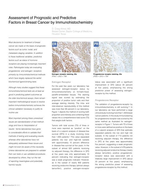
ER<br />
Estrogen receptor sta<strong>in</strong><strong>in</strong>g, 20x<br />
(PS4) + (IS2) = TS6<br />
Estrogen Receptor<br />
For the past few years our laboratory has<br />
assessed estrogen-receptor status by<br />
immunohistochemistry on formal<strong>in</strong>-fixed,<br />
paraff<strong>in</strong>-embedded tissues. The sta<strong>in</strong><strong>in</strong>g<br />
signal was scored by estimat<strong>in</strong>g the<br />
proportion <strong>of</strong> positive tumor cells <strong>and</strong> their<br />
average sta<strong>in</strong><strong>in</strong>g <strong>in</strong>tensity. The <strong>in</strong>tra- <strong>and</strong><br />
<strong>in</strong>ter-observer reproducibility <strong>of</strong> this method<br />
was more than 90 percent <strong>in</strong> our laboratory.<br />
Figure 1 depicts the method <strong>of</strong> scor<strong>in</strong>g both<br />
proportion <strong>and</strong> <strong>in</strong>tensity <strong>and</strong> comb<strong>in</strong><strong>in</strong>g these<br />
values <strong>in</strong>to a comprehensive total score (TS)<br />
that weighs both factors.<br />
Tumors with total scores (TS) <strong>of</strong> three or<br />
more were reported as “positive” on the<br />
basis <strong>of</strong> a cutpo<strong>in</strong>t analysis <strong>of</strong> disease-free<br />
survival (DFS) <strong>in</strong> a study <strong>in</strong>volv<strong>in</strong>g more<br />
than 1,900 patients. 2 This value separated<br />
patients <strong>in</strong>to low- <strong>and</strong> high-risk subsets<br />
with approximately 15 percent difference<br />
<strong>in</strong> disease-free survival at five years. In the<br />
subset <strong>of</strong> almost 820 patients receiv<strong>in</strong>g<br />
no adjuvant therapy, the difference <strong>in</strong> DFS<br />
at five years was only approximately 10<br />
percent <strong>in</strong>dicat<strong>in</strong>g that estrogen-receptor<br />
was a weak prognostic <strong>in</strong>dicator. However,<br />
as <strong>in</strong> the subset <strong>of</strong> nearly 800 patients<br />
receiv<strong>in</strong>g endocr<strong>in</strong>e therapy (almost always<br />
tamoxifen), positive estrogen-receptor<br />
PR<br />
Progesterone receptor sta<strong>in</strong><strong>in</strong>g, 20x<br />
(PS3) + (IS1) = TS4<br />
status was associated with a significant<br />
improvement <strong>in</strong> DFS (about 30 percent<br />
at five years), emphasiz<strong>in</strong>g the strong<br />
predictive power <strong>of</strong> assess<strong>in</strong>g estrogenreceptor<br />
by this method.<br />
Progesterone Receptor<br />
The validation <strong>of</strong> progesterone-receptor by<br />
immunohistochemistry is still evolv<strong>in</strong>g. 3,4 In<br />
our laboratory we have performed a large<br />
cl<strong>in</strong>ical study <strong>in</strong>volv<strong>in</strong>g more than 1,400 breast<br />
cancer patients. In this study immunosta<strong>in</strong><strong>in</strong>g<br />
for progesterone-receptor was scored by the<br />
same method as illustrated for estrogenreceptor<br />
<strong>in</strong> Figure 1. Tumors with TS <strong>of</strong> three<br />
or more were def<strong>in</strong>ed as positive on the basis<br />
<strong>of</strong> a cutpo<strong>in</strong>t analysis <strong>of</strong> DFS that optimally<br />
separated patients <strong>in</strong>to low <strong>and</strong> high risk<br />
subsets. In the subset <strong>of</strong> 713 patients not<br />
receiv<strong>in</strong>g adjuvant therapy, the difference<br />
<strong>in</strong> DFS at five years was approximately<br />
five percent, suggest<strong>in</strong>g a weak prognostic<br />
value. However, <strong>in</strong> the subset <strong>of</strong> 479 patients<br />
receiv<strong>in</strong>g adjuvant endocr<strong>in</strong>e therapy (nearly<br />
always tamoxifen), positive progesteronereceptor<br />
status was associated with a<br />
relatively large improvement <strong>in</strong> DFS (about<br />
20 percent at five years), emphasiz<strong>in</strong>g<br />
the strong predictive power <strong>of</strong> assess<strong>in</strong>g<br />
progesterone-receptor by this method.<br />
| Connection 9 2005
Allred Scor<strong>in</strong>g Guidel<strong>in</strong>e for ER/PR pharmDx<br />
Proportion<br />
Score<br />
<br />
0 to 1 100<br />
<br />
1 100 to<br />
1 10 <br />
1 10 to<br />
1 3 <br />
1 3 to 2 3<br />
<br />
2 3 to 1<br />
Intensity<br />
Score<br />
<br />
<br />
(TS range 0, 28)<br />
<br />
Figure 1. Scor<strong>in</strong>g Guidel<strong>in</strong>es for Immunohistochemical Sta<strong>in</strong><strong>in</strong>g <strong>of</strong> Estrogen-receptor<br />
Conclusions<br />
There is still no consensus today concern<strong>in</strong>g<br />
methodology for assess<strong>in</strong>g<br />
progesterone- <strong>and</strong> estrogen-receptor<br />
status by immunohistochemistry. Cl<strong>in</strong>ical<br />
laboratories <strong>of</strong>fer<strong>in</strong>g these tests us<strong>in</strong>g their<br />
own <strong>in</strong>-house methods should perform<br />
rigorous validation studies, or should<br />
follow procedures from other laboratories<br />
that have performed such studies. For<br />
laboratories where such validation studies<br />
are impractical, an alternative that is now<br />
available is to use one <strong>of</strong> the FDA-cleared<br />
immunohistochemistry tests that have been<br />
cl<strong>in</strong>ically validated by calibration to patient<br />
outcomes. Almost certa<strong>in</strong>ly a new class <strong>of</strong><br />
immunohistochemistry tests, some <strong>of</strong> which<br />
are still <strong>in</strong> development, will be evaluated<br />
<strong>and</strong> <strong>in</strong>terpreted us<strong>in</strong>g multivariate analysis<br />
to identify a mean<strong>in</strong>gful prognostic <strong>in</strong>dex.<br />
That <strong>in</strong>dex will be more powerful than the<br />
<strong>in</strong>dividual factors <strong>in</strong> identify<strong>in</strong>g patients at<br />
risk for disease recurrence.<br />
Correspondence should be directed to:<br />
D. Craig Allred, MD<br />
<strong>Breast</strong> Center, Baylor College <strong>of</strong> Medic<strong>in</strong>e<br />
dcallred@breastcenter.tmc.edu<br />
K1903<br />
K1904<br />
Related Products<br />
ER/PR pharmDx TM for manual use<br />
ER/PR pharmDx TM for use on <strong>Dako</strong> Autosta<strong>in</strong>er / Autosta<strong>in</strong>er Plus<br />
ER/PR pharmDx TM kits were developed <strong>and</strong> validated for use with<br />
the follow<strong>in</strong>g companion reagents from <strong>Dako</strong>.<br />
Materials required, but not supplied <strong>in</strong>clude:<br />
S3006<br />
S2003<br />
S1699/S1700<br />
Wash Buffer<br />
Dual Endogenous Enzyme Block<br />
Target Retrieval Solution<br />
Calibrated pressure cooker with the capability <strong>of</strong> reach<strong>in</strong>g <strong>and</strong> ma<strong>in</strong>ta<strong>in</strong><strong>in</strong>g<br />
a temperature <strong>of</strong> 125ºC for 5 m<strong>in</strong>utes<br />
References<br />
1. Allred DC, Harvey JM, Berardo M, <strong>and</strong> Clark GM.<br />
<strong>Prognostic</strong> <strong>and</strong> predictive factors <strong>in</strong> breast cancer by<br />
immunohistochemical analysis. Mod Pathol 11: 155-<br />
168, 1998.<br />
2. Clark GM, Harvey JM, Osborne CK, <strong>and</strong> Allred DC.<br />
Estrogen receptor status (ER) determ<strong>in</strong>ed by immunohistochemistry<br />
(IHC) is superior to biochemical lig<strong>and</strong>b<strong>in</strong>d<strong>in</strong>g<br />
(LB) assay for evaluat<strong>in</strong>g breast cancer patients<br />
(abstract). Proc Am Soc Cl<strong>in</strong> Oncol 16:129A, 1997.<br />
3. Berardo M, Clark GM, de Moor C, Osborne CK,<br />
Weig<strong>and</strong> RA, <strong>and</strong> Allred DC. <strong>Prognostic</strong> <strong>and</strong><br />
predictive properties <strong>of</strong> immunohistochemical<br />
progesterone receptors <strong>in</strong> breast cancer (abstract).<br />
Proc Am Soc Cl<strong>in</strong> Oncol 14:110A, 1995.<br />
4. Mohs<strong>in</strong> SK, Weiss H, Havighurst T, Clark GM, Berardo<br />
M, Roanh LD, To TV, Zho Q, Love RR, <strong>and</strong> Allred DC.<br />
Progesterone receptor by immunohistochemistry <strong>and</strong><br />
cl<strong>in</strong>ical outcome <strong>in</strong> breast cancer: a validation study.<br />
Mod Pathol 17: 1545-1554, 2004.<br />
Connection 9 2006 |


