

international institute of molecular and cell biology - IIMCB
international institute of molecular and cell biology - IIMCB
international institute of molecular and cell biology - IIMCB

You also want an ePaper? Increase the reach of your titles
YUMPU automatically turns print PDFs into web optimized ePapers that Google loves.
INTERNATIONAL INSTITUTE<br />
OF MOLECULAR AND CELL BIOLOGY<br />
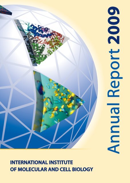
Annual Report 2009
Director<br />
Jacek Kuźnicki<br />
Deputy Scientific Director<br />
Michał Witt<br />
Financial Manager<br />
Hanna Iwaniukowicz<br />
Chairman <strong>of</strong> the International Advisory Board<br />
Angelo Azzi<br />
Deputy Chairman <strong>of</strong> the International Advisory Board<br />
Leszek Kaczmarek<br />
International Institute <strong>of</strong> Molecular <strong>and</strong> Cell Biology<br />
4 Ks. Trojdena Street<br />
02-109 Warsaw, Pol<strong>and</strong><br />
Tel. (48 22) 59 70 700; Fax (48 22) 59 70 715<br />
e-mail: secretariat@iimcb.gov.pl<br />
internet: www.iimcb.gov.pl<br />
This Report 2009 was edited by:<br />
Agnieszka Wagner-Ziemka <strong>and</strong> Michał Witt<br />
ISBN 978-83-917905-2-6
Contents<br />
10 th Anniversary <strong>of</strong> <strong>IIMCB</strong> 2<br />
Important Dates in the Institute’s History 3<br />
Directors <strong>and</strong> Administration 4<br />
Structure <strong>of</strong> the International Institute <strong>of</strong> Molecular <strong>and</strong> Cell Biology 5<br />
International Advisory Board <strong>of</strong> the International Institute <strong>of</strong> Molecular <strong>and</strong> Cell Biology 6<br />
Directors’ Note 7<br />
Description <strong>of</strong> the Institute’s Activities 8<br />
Scientific Meetings <strong>and</strong> Lectures 9<br />
Doctoral Degrees in 2009 11<br />
Grants 12<br />
Other Consortial Projects Coordinated at <strong>IIMCB</strong> 15<br />
Cooperation with Other Institutions 16<br />
Lab Leaders Competitions 17<br />
Proteins in Health <strong>and</strong> Disease HEALTH-PROT 18<br />
Diversity <strong>of</strong> Funding <strong>IIMCB</strong>’2009 21<br />
Department <strong>of</strong> Molecular Biology 22<br />
Laboratory <strong>of</strong> Bioinformatics <strong>and</strong> Protein Engineering 28<br />
Laboratory <strong>of</strong> Structural Biology MPG/PAN 34<br />
Laboratory <strong>of</strong> Neurodegeneration 40<br />
Laboratory <strong>of</strong> Biomodelling 48<br />
Laboratory <strong>of</strong> Cell Biology 54<br />
Laboratory <strong>of</strong> Molecular <strong>and</strong> Cellular Neuro<strong>biology</strong> 60<br />
Laboratory <strong>of</strong> Cell Cortex Mechanics MPG/PAN 68<br />
Laboratory <strong>of</strong> Protein Structure 74<br />
Laboratory <strong>of</strong> Mitochondrial Biogenesis 78<br />
Educational Activities 82<br />
Centre for Innovative Bioscience Education (CIBE) 83<br />
Staff at <strong>IIMCB</strong> 85
10 th Anniversary <strong>of</strong> <strong>IIMCB</strong><br />
From left to right: Ganna Jelskaja, Jacek Kuźnicki i Natalia Shulga during the Polish-Ukrainian poster session; Malgorzata Mossakowska receiving<br />
honorary diploma as the first employee <strong>of</strong> <strong>IIMCB</strong> (above); anniversary cake (below); anniversary balloons held by our administration staff.<br />
The 10 th anniversary <strong>of</strong> the <strong>IIMCB</strong> was not merely a festive<br />
event. In May 2009 we celebrated the end <strong>of</strong> the first decade<br />
<strong>of</strong> the <strong>IIMCB</strong> in a fashion which was as scientific as it was<br />
entertaining, with the anniversary dinner being very much just<br />
an addition to the proceedings. On this occasion the <strong>IIMCB</strong><br />
organized an <strong>international</strong> research symposium – a kick-<strong>of</strong>f<br />
meeting <strong>of</strong> the HeAlTH-PROT project presenting the project<br />
partners <strong>and</strong> potential partners <strong>of</strong> other european projects,<br />
with lectures delivered by: Wolfgang Zachariae, Vincenza<br />
Andrisano, Saulius Klimasauskas, Ted Hupp, Anita Becker,<br />
Samantha Spangler <strong>and</strong> Oleg Krishtal. It was preceded by a<br />
Polish-Ukrainian research collaboration meeting, where the<br />
<strong>IIMCB</strong> hosted a group <strong>of</strong> 23 Ukrainian scientists. The event<br />
was held in a form <strong>of</strong> a poster session, with 18 posters from<br />
the Institute <strong>of</strong> Molecular Biology <strong>and</strong> Genetics in Kiev <strong>and</strong><br />
18 posters from the <strong>IIMCB</strong>. The Ukrainian group was led by<br />
Pr<strong>of</strong>. Ganna Jelskaja, Director <strong>of</strong> the Institute <strong>of</strong> Molecular<br />
Biology <strong>and</strong> Genetics in Kiev <strong>and</strong> Dr. Natalia Shulga, CeO at<br />
the Ukrainian Scientific Club. The anniversary party followed,<br />
complete with a cake shaped like the Institute’s logo.<br />
It all dates back to 1994, when the newly established<br />
Committee for Scientific Research (KBN) <strong>and</strong> the Presidium<br />
<strong>of</strong> the Polish Academy <strong>of</strong> Sciences approved a UNeSCO<br />
initiative resulting in an <strong>international</strong> agreement signed in<br />
May 1995 by Pr<strong>of</strong>. F. Mayor, UNeSCO’s Director General, <strong>and</strong><br />
Pr<strong>of</strong>. A. Łuczak, Pol<strong>and</strong>’s Deputy Prime Minister <strong>and</strong> Head <strong>of</strong><br />
KBN. The agreement established the International Institute<br />
<strong>of</strong> Molecular <strong>and</strong> Cell Biology (<strong>IIMCB</strong>) in Warsaw. This formally<br />
fulfilled previous plans <strong>of</strong>: Pr<strong>of</strong>. Angelo Azzi, Pr<strong>of</strong>. Maciej<br />
Nałęcz, Pr<strong>of</strong>. Jacek Kuźnicki <strong>and</strong> Pr<strong>of</strong>. leszek Kaczmarek, later<br />
to be joined by Pr<strong>of</strong>. Ryszard Przewłocki <strong>and</strong> Pr<strong>of</strong>. Michał Witt.<br />
At the same time, the Polish Parliament (Sejm) <strong>and</strong> Pol<strong>and</strong>’s<br />
President ratified the May 1995 agreement. The correct<br />
legal foundation for the operations <strong>of</strong> the <strong>IIMCB</strong> was finally<br />
created by the Parliamentary bill <strong>of</strong> 26 June 1997 – until then<br />
the Polish legal system had been unable to accommodate<br />
scientific institutions <strong>of</strong> <strong>international</strong> st<strong>and</strong>ing. Pr<strong>of</strong>. Angelo<br />
Azzi was formally appointed as Head <strong>of</strong> the Institute, with<br />
deputies Pr<strong>of</strong>. Jacek Kuźnicki (as Acting Director) <strong>and</strong><br />
2 Annual Report 2009<br />
Pr<strong>of</strong>. Michał Witt, <strong>and</strong> the first employee was Dr. Małgorzata<br />
Mossakowska.<br />
Finally, on 1 January 1999, the International Institute for<br />
Molecular <strong>and</strong> Cell Biology began its independent existence.<br />
In the same year, the first pr<strong>of</strong>essorial positions were filled <strong>and</strong><br />
two research laboratories were launched: the laboratory <strong>of</strong><br />
Molecular Immunology, headed by Dr. Jarosław Dastych, <strong>and</strong><br />
the Department <strong>of</strong> Molecular Biology, under the leadership<br />
<strong>of</strong> Pr<strong>of</strong>. Maciej Żylicz. Since 2002, Pr<strong>of</strong>. J. Kuźnicki has been<br />
the Director <strong>of</strong> the Institute, <strong>and</strong> Pr<strong>of</strong>. M. Witt the Deputy<br />
Scientific Director.<br />
The principles <strong>of</strong> organization <strong>of</strong> the Institute differ from<br />
other research <strong>institute</strong>s in the country: an important body<br />
<strong>of</strong> the Institute is the International Advisory Board (IAB) which<br />
acts as the Scientific Council; the leader <strong>of</strong> each research team<br />
<strong>of</strong> the Institute is selected in an <strong>international</strong> competition;<br />
each group leader’s initial employment is limited to a fiveyear<br />
contract; <strong>and</strong> three years after the start <strong>of</strong> research work<br />
the progress <strong>of</strong> research is assessed by the IAB. Based on the<br />
IAB’s recommendation, a pr<strong>of</strong>essor’s contract may be either<br />
terminated or extended.<br />
The <strong>IIMCB</strong> is directly subordinate to the President <strong>of</strong> the<br />
Polish Academy <strong>of</strong> Sciences (PAN), who supervises the<br />
organization <strong>and</strong> activities <strong>of</strong> the Institute. The President <strong>of</strong> PAN<br />
nominates the members <strong>of</strong> the IAB <strong>and</strong> the Institute’s Directors.<br />
An important link between the Institute <strong>and</strong> the President <strong>of</strong><br />
PAN is the 2 nd Department <strong>of</strong> Biological Sciences <strong>of</strong> PAN, to<br />
which the Institute belongs along with sixteen PAN <strong>institute</strong>s.<br />
The <strong>IIMCB</strong> is financed in part from the national budget<br />
(statutory subvention; budgetary subvention via PAN)<br />
<strong>and</strong> in part from other sources (Ministry <strong>of</strong> Science <strong>and</strong><br />
Higher education, Foundation for Polish Science, UNeSCO,<br />
Framework Programs <strong>of</strong> eU, Max Planck Society, Howard<br />
Hughes Medical Institute, european Molecular Biology<br />
Organisation, National Institutes <strong>of</strong> Health, Wellcome Trust,<br />
etc.). About 70% <strong>of</strong> funds arrive as competitive grant awards<br />
received by the group leaders. The <strong>IIMCB</strong> is located in a<br />
building loaned by the Polish Academy <strong>of</strong> Sciences.<br />
The prospects for the second decade <strong>of</strong> the International<br />
Institute <strong>of</strong> Molecular <strong>and</strong> Cell Biology look promising.
Important Dates<br />
in the Institute’s History<br />
Sept. 1991 The proposal to create the Institute was published in the UNeSCO Bulletin <strong>of</strong> MCBN<br />
June 1994 State Committee for Scientific Research (KBN) accepts the activities aimed at establishing the Institute<br />
Oct. 1994 Presidium <strong>of</strong> Polish Academy <strong>of</strong> Sciences (PAN) votes to support the Institute<br />
May 1995 An agreement between Pol<strong>and</strong> <strong>and</strong> UNeSCO to establish the Institute<br />
June 1996 The Molecular <strong>and</strong> Cell Biology Department is created by PAN with Pr<strong>of</strong>. M.J. Nalecz as the Head<br />
June 1997 Polish Parliament passes a bill to found the Institute<br />
May 1998 Pr<strong>of</strong>. A. Azzi is nominated as the Director <strong>of</strong> <strong>IIMCB</strong><br />
Jan. 1999 The Institute commences its independent activities; Pr<strong>of</strong>. J. Kuźnicki appointed as Acting Director<br />
July 1999 Dr. J. Dastych is appointed as a first lab leader at <strong>IIMCB</strong><br />
Oct. 1999 Pr<strong>of</strong>. M. Żylicz is appointed as Chair <strong>of</strong> the Department <strong>of</strong> Molecular Biology<br />
April 2000 An agreement between the Max Planck Society (MPG) <strong>and</strong> the Polish Academy <strong>of</strong> Sciences (PAN) to launch<br />
a Joint MPG-PAN Junior Research Group<br />
Jan. 2001 The MPG-PAN Junior Research Group commences its activities with Dr. M. Bochtler as a lab leader<br />
June 2001 Pr<strong>of</strong>. J. Kuźnicki is elected by the International Advisory Board as Director <strong>of</strong> the Institute, begins to complete<br />
the laboratory <strong>of</strong> Neurodegeneration. After consultation with UNeSCO, the <strong>of</strong>ficial nomination was signed<br />
by the President <strong>of</strong> PAN on February 1, 2002<br />
Nov. 2002 New members <strong>of</strong> the International Advisory Board nominated for 2002-2006 term<br />
Jan. 2003 Status <strong>of</strong> the Centre <strong>of</strong> ex<strong>cell</strong>ence in Molecular Bio-Medicine is granted by the european Commission within<br />
5 th Framework Programme<br />
June 2005 Pr<strong>of</strong>essor J. Kuźnicki re-elected as Director <strong>of</strong> the Institute (term 2006-2010)<br />
May 2006 New members <strong>of</strong> the International Advisory Board nominated for 2006-2010 term<br />
Feb. 2006 Twin MPG-PAN laboratory established at the Max Planck Institute <strong>of</strong> Molecular Cell Biology <strong>and</strong> Genetics<br />
in Dresden with Dr. ewa Paluch as a lab leader.<br />
May 2009 Pr<strong>of</strong>essor J. Kuźnicki re-elected as Director <strong>of</strong> the Institute (term 2010-2014)<br />
Jan. 2010 New members <strong>of</strong> the International Advisory Board nominated for 2010-2014 term.<br />
Annual Report 2009 3
Directors <strong>and</strong> Administration<br />
Jacek Kuźnicki<br />
Director<br />
Michał Witt<br />
Deputy Scientific Director<br />
Hanna Iwaniukowicz<br />
Financial Manager<br />
4 Annual Report 2009<br />
Agnieszka Ziemka<br />
Director’s Representative for<br />
Research Management<br />
Zbigniew Przygoda<br />
Director’s Advisor (until Dec.2009)<br />
Beata Tkacz<br />
Human Resources Specialist<br />
Urszula Białek-<br />
-Wyrzykowska<br />
International Cooperation<br />
Manager<br />
Monika Nowicka<br />
Payroll Specialist<br />
Marcin Biedacha<br />
IT Manager (until Jan. 2010)<br />
Dorota Libiszowska<br />
Foreign Grants Specialist<br />
Agnieszka Karbowska<br />
Director’s Representative for<br />
Administrative Matters<br />
Dominika Dubicka-Boroch<br />
Director’s Assistant<br />
Krystyna Domańska<br />
Human Resources Specialist<br />
Renata Knyziak<br />
Accounting Specialist<br />
Jakub Skaruz<br />
IT Specialist<br />
Magdalena Powierża<br />
International Cooperation<br />
Specialist<br />
Roman Szczepanowski<br />
Director’s Representative for<br />
Information Technology & Research<br />
equipment<br />
Aleks<strong>and</strong>ra Olejniczak<br />
Secretary (until March 2010)<br />
Anna Brzezińska<br />
Tenders Specialist<br />
Marcin Ogonowski<br />
International Cooperation<br />
Specialist<br />
Mariola Arkuszewska<br />
Accounting Specialist<br />
Robert Banasiak<br />
Maintenance Specialist
International Advisory Board<br />
Structure <strong>of</strong> the International<br />
Institute <strong>of</strong> Molecular<br />
<strong>and</strong> Cell Biology<br />
Director<br />
Deputy Scientific<br />
Director<br />
Department <strong>of</strong> Molecular<br />
Biology<br />
Director’s<br />
Representative for<br />
Administrative Matters<br />
Director’s<br />
Representative for<br />
Information Technology<br />
& Research Equipment<br />
Financial Manager<br />
Director’s<br />
Representative for<br />
Research Management<br />
Laboratory <strong>of</strong> Neurodegeneration<br />
Director’s Assistant<br />
Secretariat<br />
Information<br />
Technology Unit<br />
Financial Unit<br />
Planning & Reporting<br />
Unit<br />
Laboratory <strong>of</strong> Bioinformatics<br />
<strong>and</strong> Protein Engineering<br />
Human Resources<br />
Unit<br />
Tenders <strong>and</strong><br />
Purchase Unit<br />
Research Equipment<br />
Laboratory & Lab<br />
Support<br />
Domestic Grants Unit<br />
Laboratory <strong>of</strong> Structural Biology<br />
MPG/PAN<br />
Technology Transfer<br />
Unit<br />
Laboratory <strong>of</strong> Biomodelling<br />
Technical Service<br />
Microscopy<br />
Laboratory<br />
Isotope Laboratory<br />
Public Relations Unit<br />
Centre for Innovative<br />
Bioscience Education<br />
Laboratory <strong>of</strong> Cell Biology<br />
Archive<br />
Laboratory <strong>of</strong> Molecular <strong>and</strong><br />
Cellular Neuro<strong>biology</strong><br />
Work Safety Unit<br />
International<br />
Cooperation Unit<br />
Laboratory <strong>of</strong> Protein Structure<br />
Ciliary Structure <strong>and</strong><br />
Function Project<br />
Former groups<br />
Laboratory <strong>of</strong> Cell Cortex Mechanics<br />
MPG/PAN (Dresden)<br />
Laboratory <strong>of</strong> Molecular Immunology (1999-2004)<br />
PolSenior Project<br />
Laboratory <strong>of</strong> Bioinformatics (1999-2002)<br />
Laboratory <strong>of</strong> Molecular Neurology (2000-2003)<br />
Laboratory <strong>of</strong> Mitochondrial<br />
Biogenesis<br />
Annual Report 2009 5
International Advisory Board<br />
<strong>of</strong> the International Institute<br />
<strong>of</strong> Molecular <strong>and</strong> Cell Biology<br />
2006-2010 term<br />
Chairman: Angelo Azzi<br />
Deputy Chairman: leszek Kaczmarek<br />
Members:<br />
Angelo Azzi. Pr<strong>of</strong>essor, Vascular Biology laboratory, Tufts University, Boston, MA, USA<br />
Francisco E. Baralle. Director-General <strong>of</strong> International Centre for Genetic engineering <strong>and</strong> Biotechnology, Trieste, Italy<br />
Alexey A. Bogdanov. Head <strong>of</strong> Department <strong>of</strong> Chemistry <strong>and</strong> Biochemistry <strong>of</strong> Nucleoproteins, Department <strong>of</strong> Chemistry,<br />
Moscow State University, Moscow, Russia<br />
Nicolaus Blin. Pr<strong>of</strong>essor <strong>of</strong> Molecular Genetics, Institute <strong>of</strong> Human Genetics, University <strong>of</strong> Tübingen, Tübingen, Germany;<br />
Foreign member <strong>of</strong> Polish Academy <strong>of</strong> Sciences<br />
Ineke Braakman. Pr<strong>of</strong>essor, Department <strong>of</strong> Cellular Protein Chemistry, Utrecht University, Utrecht, The Netherl<strong>and</strong>s<br />
Ivan Dikič. Pr<strong>of</strong>essor <strong>of</strong> Biochemistry, Institute <strong>of</strong> Biochemistry II, Goethe University Medical School, Frankfurt am Main,<br />
Germany<br />
Jerzy Duszyński. Undersecretary <strong>of</strong> State, Ministry <strong>of</strong> Science <strong>and</strong> Higher education, formerly Director, Nencki Institute <strong>of</strong><br />
experimental Biology, Polish Academy <strong>of</strong> Sciences, Warsaw, Pol<strong>and</strong><br />
Robert P. Erickson. Pr<strong>of</strong>essor, Department <strong>of</strong> Pediatrics, Section <strong>of</strong> Medical <strong>and</strong> Molecular Genetics, University <strong>of</strong> Arizona<br />
Health Sciences Center, Tucson, AZ, USA<br />
Klaus Hahlbrock. Pr<strong>of</strong>essor emeritus, Max-Planck Institute for Plant Breeding Research, Köln, Germany; laureate <strong>of</strong><br />
Alex<strong>and</strong>er von Humboldt Honorary Research Fellowship <strong>of</strong> Foundation for Polish Science<br />
Robert Huber. Head, Department <strong>of</strong> Structure Research, Max Planck Institute <strong>of</strong> Biochemistry, Martinsried, Germany<br />
Wiel<strong>and</strong> Huttner. Scientific Member <strong>and</strong> Director, Max Planck Institute <strong>of</strong> Molecular Cell Biology <strong>and</strong> Genetics, Dresden,<br />
Germany<br />
Leszek Kaczmarek. Pr<strong>of</strong>essor, Nencki Institute <strong>of</strong> experimental Biology, Polish Academy <strong>of</strong> Sciences, Warsaw, Pol<strong>and</strong><br />
Oleg A. Krishtal. Deputy Director <strong>of</strong> the Bogomoletz Institute <strong>of</strong> Physiology, Head <strong>of</strong> the Department <strong>of</strong> Cellular<br />
Membranology, Bogomoletz Institute <strong>of</strong> Physiology, Kiev, Ukraine<br />
Jacques Mallet. Pr<strong>of</strong>essor, laboratoire de Genetique Moleculaire de la Neurotransmission et des Processus<br />
Neurodegeneratifs, CNRS UMR 9923, Hopital de la Pitie-Salpetriere, Paris, France<br />
Maciej J. Nałęcz. Director, Division <strong>of</strong> Basic <strong>and</strong> engineering Sciences, UNeSCO, Paris, France<br />
Ryszard Przewłocki. Pr<strong>of</strong>essor, Institute <strong>of</strong> Pharmacology, Polish Academy <strong>of</strong> Sciences, Cracow, Pol<strong>and</strong><br />
J. Gregor Sutcliffe. Pr<strong>of</strong>essor, Department <strong>of</strong> Molecular Biology, The Scripps Research Institute, la Jolla, CA, USA<br />
Anna Tramontano. Pr<strong>of</strong>essor <strong>of</strong> Biochemistry, I Medical Faculty, University <strong>of</strong> Rome “la Sapienza”, Rome, Italy<br />
6 Annual Report 2009<br />
Participants <strong>of</strong> the meeting <strong>of</strong> the International Advisory Board, May 2009<br />
From left (first row): K. Hahlbrock, A. Tramontano, J. Kuźnicki, A. Azzi; (second row)<br />
M. Witt, R. Przewłocki, I. Braakman, J.G. Sutcliffe; (third row) I. Dikič, R.P. erickson, N. Blin,<br />
W. Huttner, M.J. Nałęcz, O.A. Krishtal.
Directors’ Note<br />
2009 was the year <strong>of</strong> the tenth<br />
anniversary <strong>of</strong> the International<br />
Institute <strong>of</strong> Molecular <strong>and</strong> Cell<br />
Biology (<strong>IIMCB</strong>). The celebrations<br />
featured a session <strong>of</strong> the<br />
International Advisory Board, the<br />
<strong>of</strong>ficial launch <strong>of</strong> the HeAlTH-<br />
PROT project (financed under<br />
the 7 th Framework Programme<br />
<strong>of</strong> the european Union) <strong>and</strong><br />
lectures by the foreign partners<br />
in this project, a Polish-Ukrainian<br />
poster session <strong>of</strong> 36 posters, <strong>and</strong><br />
the publication <strong>of</strong> the jubilee<br />
10 th edition <strong>of</strong> the report on the<br />
Institute’s operations – the 2008 Annual Report.<br />
Our tenth anniversary was, therefore, celebrated without a<br />
triumph <strong>of</strong> form over substance <strong>and</strong> with a strong focus on<br />
scientific content. We wished to emphasize our satisfaction<br />
with the success <strong>of</strong> <strong>IIMCB</strong>’s initial years, while making no secret<br />
<strong>of</strong> our conviction that there is always room for improvement:<br />
<strong>and</strong> this is what we have placed at the foundation <strong>of</strong> our<br />
program for another decade. We take exceptional pride<br />
in the fact that the employees <strong>of</strong> our Institute have been<br />
awarded some <strong>of</strong> the most prestigious <strong>and</strong> financially<br />
gratifying research grants, such as the Wellcome Trust Senior<br />
Research Fellowships, eMBO Installation Grants, Howard<br />
Hughes Medical Institute International Research Scholar<br />
Awards, grants from the National Institutes <strong>of</strong> Health, the<br />
Max Planck Institute Partner Group <strong>and</strong> Polish-Norwegian<br />
Research Funds, as well as grants received under the 5 th ,<br />
6 th <strong>and</strong> 7 th Framework Programmes or those awarded by<br />
the Foundation for Polish Science. Among our staff we<br />
have two newly appointed full pr<strong>of</strong>essors who began their<br />
independent scientific careers at our Institute <strong>and</strong> who<br />
are both below 40 years <strong>of</strong> age. Our papers are published<br />
in the leading foreign journals in increasing numbers, <strong>and</strong><br />
our alumni can readily find work in ex<strong>cell</strong>ent scientific or<br />
biomedical laboratories all around the world.<br />
The International Institute <strong>of</strong> Molecular <strong>and</strong> Cell Biology<br />
is entering into the second decade <strong>of</strong> its operations. There<br />
have been some specific consequences <strong>of</strong> the increasing<br />
length <strong>of</strong> the <strong>IIMCB</strong>’s history: a growing complexity <strong>of</strong><br />
the Institute’s structure, increasing numbers <strong>of</strong> staff, an<br />
expansion in the number <strong>of</strong> grants to be processed <strong>and</strong><br />
managed, <strong>and</strong> increasingly large administrative burdens.<br />
As a result, we have partially re-organized the Institute<br />
<strong>and</strong> created a number <strong>of</strong> new positions so that the new<br />
organizational structure can respond to our growing needs.<br />
The Scientific Office <strong>and</strong> the Technology Transfer Unit have<br />
been set up, <strong>and</strong> the supervision <strong>of</strong> the Institute’s computer<br />
network has been outsourced to an experienced IT firm.<br />
The economic crisis has not been without impact on our<br />
operations – government funding has become increasingly<br />
scarce <strong>and</strong>, to keep our budget balanced <strong>and</strong> ensure<br />
opportunities for growth, we rely on Polish <strong>and</strong> <strong>international</strong><br />
grants. There are some problems, however, which<br />
cannot be solved with the help <strong>of</strong> grants: modernization,<br />
renovations, major investment in equipment or further<br />
growth into exp<strong>and</strong>ing laboratory space.<br />
Our Institute is slowly approaching the capacity limits <strong>of</strong><br />
the facilities it has been occupying until now. In addition to<br />
the ten research groups (one <strong>of</strong> which operates in Dresden),<br />
we can place only two new laboratories in our building, <strong>and</strong><br />
this will occur quite soon. In this context, the question about<br />
continued development <strong>of</strong> the Institute is taking on a new<br />
meaning in organizational terms. There are several different<br />
scenarios under review, however each <strong>of</strong> them implies a<br />
need for significant investment in infrastructure, whether<br />
this be a completely new building, an extension <strong>of</strong> the old<br />
facility or, finally, use <strong>of</strong> other laboratory facilities available<br />
at or near the Ochota Campus. A sound <strong>and</strong> sensible size<br />
limit, which still allows a correct level <strong>of</strong> administrative <strong>and</strong><br />
scientific manageability to be maintained, is an <strong>institute</strong><br />
which does not exp<strong>and</strong> beyond 20 research groups. This is<br />
a target size which can, <strong>and</strong> certainly should, be envisaged<br />
in the long-term. Time will show whether any <strong>of</strong> the options<br />
considered now as a possibility will even have a chance<br />
<strong>of</strong> becoming reality - <strong>and</strong> the discussion over the future<br />
shape <strong>of</strong> the Institute continues. There are many problems<br />
to be solved but there are also increasing numbers <strong>of</strong> ideas<br />
how to do so. An important thing is that all the interested<br />
parties can take part in the discussion <strong>and</strong> that each voice is<br />
received with attention.<br />
Thanks to the structural funds obtained through<br />
a shared effort, the Ochota Campus has increasingly<br />
been gaining the status <strong>of</strong> a big, unified yet diversified,<br />
scientific organism, <strong>and</strong> it has began to operate as a whole<br />
by organizing initiatives which bring together diverse<br />
institutions. One such initiative is the Biocentrum-Ochota<br />
Consortium, which consists <strong>of</strong> five Polish Academy <strong>of</strong><br />
Science (PAN) <strong>institute</strong>s <strong>and</strong> the <strong>IIMCB</strong>, another is the<br />
Centre for Advanced Technology <strong>and</strong> Pre-Clinical Trials<br />
(CePT), which includes Biocentrum-Ochota <strong>and</strong> the three<br />
largest academic institutions in Warsaw (Warsaw University,<br />
Medical University <strong>of</strong> Warsaw <strong>and</strong> Warsaw University <strong>of</strong><br />
Technology). The shared activities include, for example,<br />
lecture series for all doctoral students, shared projects <strong>and</strong><br />
scientific conferences, purchases <strong>of</strong> cutting-edge research<br />
equipment, computer infrastructure for shared purpose<br />
<strong>and</strong> use, <strong>and</strong> new buildings. In our vision we envisage joint<br />
development <strong>and</strong> management <strong>of</strong> the Ochota complex –<br />
after all, this is the largest campus hosting biomedical <strong>and</strong><br />
biotechnology institutions in this part <strong>of</strong> europe.<br />
Annual Report 2009 7
Description <strong>of</strong> the Institute’s<br />
Activities<br />
Relation <strong>of</strong> <strong>IIMCB</strong> to PAN<br />
The International Institute <strong>of</strong> Molecular <strong>and</strong> Cell Biology is<br />
directly subordinate to the President <strong>of</strong> the Polish Academy<br />
<strong>of</strong> Sciences, who supervises the organization <strong>and</strong> activities<br />
<strong>of</strong> the Institute. The President <strong>of</strong> PAN nominates members <strong>of</strong><br />
IAB <strong>and</strong> the Institute’s Directors. An important link between<br />
the Institute <strong>and</strong> the President <strong>of</strong> PAN is the 2 nd Department<br />
<strong>of</strong> Biological Sciences <strong>of</strong> PAN, to which the Institute belongs<br />
together with sixteen <strong>institute</strong>s <strong>of</strong> PAN.<br />
The Organization <strong>of</strong> Research at <strong>IIMCB</strong><br />
Ten research groups comprise the structure <strong>of</strong> <strong>IIMCB</strong>:<br />
Department <strong>of</strong> Molecular Biology (Żylicz), laboratory <strong>of</strong><br />
Bioinformatics <strong>and</strong> Protein engineering (Bujnicki), laboratory<br />
<strong>of</strong> Structural Biology MPG/PAN (Bochtler), laboratory <strong>of</strong><br />
Neurodegeneration (Kuźnicki), laboratory <strong>of</strong> Biomodelling<br />
(Filipek), laboratory <strong>of</strong> Cell Biology (Miączyńska), laboratory<br />
<strong>of</strong> Molecular <strong>and</strong> Cellular Neuro<strong>biology</strong> (Jaworski),<br />
laboratory <strong>of</strong> Cell Cortex Mechanics MPG/PAN in Dresden<br />
(Paluch), laboratory <strong>of</strong> Protein Structure (Nowotny) <strong>and</strong><br />
laboratory <strong>of</strong> Mitochondrial Biogenesis (Chacińska).<br />
The research carried out at <strong>IIMCB</strong> is mainly focused on<br />
fundamental biomedical problems. The major research<br />
topics include the following:<br />
1. Role <strong>of</strong> <strong>molecular</strong> chaperones in <strong>cell</strong> transformation,<br />
including analysis <strong>of</strong> interactions between human p53<br />
<strong>and</strong> <strong>molecular</strong> chaperones <strong>and</strong> oncogenic activity <strong>of</strong><br />
MDM2 <strong>and</strong> mutated forms <strong>of</strong> p53 (Żylicz’s group).<br />
2. Development <strong>and</strong> application <strong>of</strong> computer s<strong>of</strong>tware for<br />
structural bioinformatics <strong>of</strong> proteins <strong>and</strong> nucleic acids<br />
<strong>and</strong> theoretical <strong>and</strong> experimental studies on enzymes<br />
acting on nucleic acids (protein structure prediction,<br />
evolutionary analyses, functional characterization,<br />
protein engineering) (Bujnicki’s group).<br />
3. Crystallographic structure determination <strong>of</strong> biological<br />
macromolecules (Bochtler’s group).<br />
4. Studies on the <strong>molecular</strong> basis <strong>of</strong> neurodegenerative<br />
disease (identification <strong>of</strong> mutations in Alzheimer’s<br />
disease-related genes <strong>of</strong> Polish patients <strong>and</strong> functional<br />
analysis <strong>of</strong> these mutations, the search for biomarkers,<br />
<strong>and</strong> potential therapeutic targets <strong>of</strong> Alzheimer’s disease)<br />
<strong>and</strong> studies <strong>of</strong> proteins implicated in the mechanisms <strong>of</strong><br />
learning <strong>and</strong> memory <strong>and</strong> pathogenesis <strong>of</strong> Alzheimer’s<br />
disease (Ca2+ sensors belonging to calmyrin family,<br />
b-catenin, cyclin-dependent kinase 5) (Kuźnicki’s group).<br />
5. Molecular modeling <strong>of</strong> structure <strong>and</strong> function (<strong>molecular</strong><br />
switches) <strong>of</strong> proteins <strong>and</strong> their oligomerization <strong>and</strong><br />
complexes, focusing on rhodopsin <strong>and</strong> other G proteincoupled<br />
receptors; <strong>molecular</strong> role <strong>of</strong> mutations <strong>of</strong><br />
presenilins in neurodegenerative diseases (Filipek’s group).<br />
8 Annual Report 2009<br />
6. Interdependence between intra<strong>cell</strong>ular endocytic<br />
transport <strong>and</strong> nuclear signal transduction (Miączynska’s<br />
group)<br />
7. Molecular processes, including gene transcription,<br />
kinase-dependent <strong>cell</strong> signaling, cytoskeleton dynamics,<br />
intra<strong>cell</strong>ular trafficking underlying neuronal development<br />
<strong>and</strong> plasticity, <strong>and</strong> central nervous system pathologies<br />
(tuberous sclerosis, epilepsy, neurodegenerative<br />
disorders) (Jaworski’s group).<br />
8. Mechanics <strong>of</strong> the actomyosin cortex; study <strong>of</strong> cortical<br />
contractility <strong>and</strong> the role <strong>of</strong> cortical mechanics during<br />
cytokinesis <strong>and</strong> migration (Paluch’s group).<br />
9. Structural <strong>and</strong> biochemical studies <strong>of</strong> nucleic acid<br />
enzymes (Nowotny’s group).<br />
10. Biogenesis <strong>of</strong> mitochondrial proteins (Chacińska’s group).<br />
Awards, Honors <strong>and</strong> Titles<br />
• Janusz M. Bujnicki, Pr<strong>of</strong>essorial title by the President <strong>of</strong><br />
Pol<strong>and</strong><br />
• Matthias Bochtler, Pr<strong>of</strong>essorial title by the President <strong>of</strong><br />
Pol<strong>and</strong><br />
• Janusz M. Bujnicki, Fellowship for Outst<strong>and</strong>ing Young<br />
Scientists <strong>of</strong> the Ministry <strong>of</strong> Science <strong>and</strong> Higher education<br />
• Janusz M. Bujnicki, 1-class award <strong>of</strong> the Ministry <strong>of</strong> Science<br />
<strong>and</strong> Higher education for research achievements <strong>and</strong> a<br />
book „Prediction <strong>of</strong> Protein Structures, Functions, <strong>and</strong><br />
Interactions” published in 2008<br />
• Jacek Jaworski’s Neuron 2009 Jan 15 paper as Highlight <strong>of</strong><br />
the Year on the FeNS website<br />
• Agnieszka Obarska-Kosińska <strong>and</strong> elżbieta Purta, START<br />
Fellowship for young scientists for 2009 by Foundation<br />
for Polish Science (FNP). Advisor <strong>of</strong> both theses: Janusz M.<br />
Bujnicki<br />
• Magdalena Błażejczyk <strong>and</strong> Adam Sobczak, awards for their<br />
PhD theses distinguished by the Scientific Council <strong>of</strong> the<br />
Nencki Institute. Advisor <strong>of</strong> both theses: Urszula Wojda<br />
• emilia Białopiotrowicz, the Mozołowski Award presented<br />
by the Polish Biochemical Society for outst<strong>and</strong>ing young<br />
biochemists in 2009 for her project <strong>and</strong> presentation<br />
entitled: „Disturbances <strong>of</strong> the <strong>cell</strong> cycle in lymphocytes<br />
from Alzheimer’s dementia patients”. Thesis advisor<br />
Urszula Wojda<br />
• Bożena Kuźniewska, the main award at the 5th Conference<br />
<strong>of</strong> International Natural Sciences Students in Wilno for the<br />
presentation <strong>of</strong> her MSc research on apoptosis <strong>and</strong> the<br />
<strong>cell</strong> cycle <strong>of</strong> lymphocytes in Alzheimer’s disease patients.<br />
Thesis advisor Urszula Wojda
• Łukasz Świech, Young Investigator Award, IX International<br />
Polish Neuroscience Meeting/Ist Regional FeNS Meeting,<br />
Warsaw, Pol<strong>and</strong><br />
Publishing NEWSKO<br />
Since 2000 e-bulletin NeWSKO provides the Ochota<br />
Campus community with current information on seminars,<br />
symposia, conferences, job opportunities <strong>and</strong> other<br />
essential events. NeWSKO, which has been published at<br />
the Institute for the last nine years, integrates scientists,<br />
students <strong>and</strong> medical doctors at the Ochota Campus <strong>and</strong><br />
plays a significant role as the communication platform for all<br />
Centres <strong>of</strong> ex<strong>cell</strong>ence at the Ochota Campus. Currently this<br />
information is available at www.iimcb.gov.pl/seminars.php.<br />
Computer Network<br />
During 2009 we managed to successfully finish<br />
modernization <strong>of</strong> our network infrastructure by constructing<br />
additional internet access points. This allows users to log on<br />
to the net without the need to actually plug in.<br />
The Institute’s servers <strong>and</strong> computer cluster (Property <strong>of</strong><br />
the laboratory <strong>of</strong> Bioinformatics <strong>and</strong> Protein engineering <strong>and</strong><br />
the laboratory <strong>of</strong> Biomodelling) were moved to a special<br />
basement location. This enabled us to integrate the most<br />
valuable technology assets <strong>of</strong> the Institute in one place,<br />
allowing us to make better use <strong>of</strong> them. To ensure an<br />
optimum working environment <strong>and</strong> keep the equipment in<br />
good condition, we installed almost all <strong>of</strong> the computers into<br />
rack cabinets <strong>and</strong> provided our equipment with a double<br />
AC unit cooling system. The server room is also protected<br />
against unauthorized access.<br />
In 2009, a s<strong>of</strong>tware audit was launched by an external<br />
validation firm, to establish which s<strong>of</strong>tware was used <strong>and</strong><br />
whether all the s<strong>of</strong>tware was licensed. S<strong>of</strong>tware without<br />
valid licenses was removed from computers.<br />
To improve the quality <strong>of</strong> network services our IT<br />
department has been outsourced. We are hoping that<br />
thanks to such measures our efficiency will be higher <strong>and</strong><br />
our operating costs lower than before. We chose the best<br />
<strong>of</strong>fer out <strong>of</strong> three possibilities on the market <strong>and</strong>, since<br />
the successful bidder - New Business Technologies - is a<br />
company with extensive experience in computer systems<br />
integration, it will hopefully meet our expectations. NBT’s<br />
current task is to provide a pr<strong>of</strong>essional IT growth strategy<br />
appropriate to our needs.<br />
Scientific Meetings <strong>and</strong> Lectures<br />
• Polish-Ukrainian research collaboration meeting with<br />
poster session (posters from the Institute <strong>of</strong> Molecular<br />
Biology <strong>and</strong> Genetics in Kiev <strong>and</strong> <strong>IIMCB</strong>). 14.05.2009,<br />
Warsaw, Pol<strong>and</strong><br />
• Kick-<strong>of</strong>f meeting <strong>of</strong> the HeAlTH-PROT project, 7 th<br />
Framework Programme european Commission,<br />
15.05.2009, Warsaw, Pol<strong>and</strong><br />
• International Research Symposium “10 th Anniversary <strong>of</strong><br />
the International Institute <strong>of</strong> Molecular <strong>and</strong> Cell Biology”,<br />
15.05.2009, Warsaw, Pol<strong>and</strong><br />
• International Conference “Challenges <strong>of</strong> <strong>molecular</strong><br />
genetic testing in Pol<strong>and</strong> – the proposal for regulations”,<br />
3.06.2009, Warsaw, Pol<strong>and</strong>, coorganized by <strong>IIMCB</strong><br />
• <strong>IIMCB</strong> Annual Report Session, 5.06.2009, Warsaw, Pol<strong>and</strong><br />
Seminars <strong>of</strong> invited speakers<br />
• Special Lecture Series: Frontiers <strong>of</strong> Polish Biosciences*<br />
Jerzy Mozrzymas (Department <strong>of</strong> Biophysics, Medical<br />
Academy, Wrocław; Recipient <strong>of</strong> the Wellcome Trust<br />
International Senior Fellowship) “Modulation <strong>and</strong> plasticity<br />
<strong>of</strong> synaptic transmission in the CNS”, 22.01.2009<br />
Artur Osyczka (Faculty <strong>of</strong> Biochemistry, Biophysics <strong>and</strong><br />
Biotechnology, Jagiellonian University, Kraków; Recipient<br />
<strong>of</strong> the Wellcome Trust International Senior Fellowship)<br />
“Productive <strong>and</strong> unproductive electron transfers in c<strong>of</strong>actor<br />
chains <strong>of</strong> cytochrome bc1”, 19.02.2009<br />
Maria Anna Ciemerych-Litwinienko (Department <strong>of</strong><br />
Cytology, University <strong>of</strong> Warsaw; Recipient <strong>of</strong> fellowships<br />
from l’Oreal Pol<strong>and</strong> <strong>and</strong> “Polityka”) “Stem <strong>cell</strong>s <strong>and</strong> muscle<br />
regeneration”, 7.05.2009<br />
Agnieszka Dobrzyń (The Nencki Institute <strong>of</strong> experimental<br />
Biology, Warsaw; Recipient <strong>of</strong> the eMBO Installation Grant)<br />
“Stearoyl-CoA desaturase – a novel control point <strong>of</strong> lipid<br />
metabolism <strong>and</strong> insulin sensitivity”, 28.05.2009<br />
Stanisław Karpiński (Department <strong>of</strong> Plant Genetics,<br />
Breeding <strong>and</strong> Biotechnology, Warsaw University <strong>of</strong><br />
life Sciences; Recipient <strong>of</strong> the WelCOMe Award <strong>of</strong> the<br />
Foundation for Polish Science) “Can plants think? evidences<br />
for light wave-length specific signaling <strong>and</strong> <strong>cell</strong>ular light<br />
memory in Arabidopsis”, 29.10.2009<br />
• Research Symposium “10th Anniversary <strong>of</strong> the<br />
International Institute <strong>of</strong> Molecular <strong>and</strong> Cell Biology”,<br />
15.05.2009<br />
Wolfgang Zachariae (Max Planck Institute <strong>of</strong> Molecular Cell<br />
Biology <strong>and</strong> Genetics, Dresden, Germany) “Of daughters <strong>and</strong><br />
sisters: control <strong>of</strong> chromosome segregation in meiosis”<br />
Vincenza Andrisano (Department <strong>of</strong> Pharmaceutical<br />
Sciences, University <strong>of</strong> Bologna, Italy) “Multitarget directed<br />
lig<strong>and</strong>s (MTDl) characterization strategy in the context <strong>of</strong><br />
Alzheimer’s disease”<br />
Saulius Klimasauskas (laboratory <strong>of</strong> Biological DNA<br />
Modi cation, Institute <strong>of</strong> Biotechnology, Vilnius, lithuania)<br />
*A seminar series entitled „Frontiers <strong>of</strong> Polish Bioscience” was coordinated by Dr. Marta Miączyńska <strong>and</strong> Dr. Jacek Jaworski. These seminars<br />
provided an opportunity to listen to <strong>and</strong> meet the top Polish scientists who received prestigious awards or grants in a broad field <strong>of</strong> bioscience.<br />
Annual Report 2009 9
“DNA methyltransferases: structural studies <strong>and</strong> redesign<br />
for novel functions”<br />
Ted Hupp (Cancer Research <strong>of</strong> UK Cell Signalling Unit,<br />
edinburgh Cancer Research Centre, University <strong>of</strong> edinburgh,<br />
UK) “Phosphorylation switch in MDM2 control <strong>of</strong> the p53<br />
tumour suppressor”<br />
Anita Becker (Department <strong>of</strong> Pediatrics <strong>and</strong> Adolescent<br />
Medicine, University <strong>of</strong> Freiburg, Germany) “Novel gene<br />
defects in primary ciliary dyskinesia”<br />
Samantha Spangler (Dept. <strong>of</strong> Neuroscience, erasmus<br />
Medical Center, Rotterdam, The Netherl<strong>and</strong>s) “Regulation<br />
<strong>of</strong> presynaptic composition <strong>and</strong> function by distinct liprinalpha<br />
family proteins”<br />
Oleg A. Krishtal (Department <strong>of</strong> Cellular Membranology,<br />
Bogomoletz Institute <strong>of</strong> Physiology, Kyiv, Ukraine)<br />
“Purinergic receptors in sensory neurons”<br />
• Regular <strong>IIMCB</strong> seminars<br />
Teresa Żołądek (Institute <strong>of</strong> Biochemistry <strong>and</strong> Biophysics,<br />
Warsaw, Pol<strong>and</strong>) “The influence <strong>of</strong> ubiquitination on<br />
intra<strong>cell</strong>ular trafficking <strong>and</strong> actin cytoskeleton organization<br />
in yeast S. cerevisiae”, 8.01.2009<br />
Axel Methner (Klinik für Neurologie, Universitätsklinikum<br />
Düsseldorf, Germany) “STIM1 in the brain: A multielectrode<br />
array approach”, 26.01.2009<br />
Agnieszka Chacińska (Institute for Biochemistry <strong>and</strong><br />
Molecular Biology, Freiburg, Germany) “Biogenesis <strong>and</strong><br />
turnover <strong>of</strong> mitochondrial intermembrane space proteins”,<br />
28.01.2009<br />
Adrianna Łoniewska-Lwowska (Department <strong>of</strong> Protein<br />
Biosynthesis, IBB PAN, Warsaw, Pol<strong>and</strong>) “How do the (+)<br />
str<strong>and</strong>ed RNA viruses express their genes – mechanism <strong>of</strong><br />
the subgenomic RNA synthesis”, 29.01.2009<br />
Fabiana Renzi (Department <strong>of</strong> Structural <strong>and</strong> Chemical<br />
Biology, Mount Sinai School <strong>of</strong> Medicine <strong>and</strong> New York<br />
Structural Biology Centre Cryo-electron Microscopy Unit,<br />
USA) “Structural studies <strong>of</strong> gamma-secretase by electron<br />
Microscopy: An intra-membrane protease involved in<br />
Alzheimer’s disease”, 2.04.2009<br />
Tomasz Węgierski (University Hospital <strong>of</strong> Freiburg,<br />
Germany) “TRP calcium channels: Cellular functions <strong>and</strong><br />
regulatory mechanisms”, 23.04. 2009<br />
Fred Van Leuven (experimental Genetics Group –<br />
leGTeGG, Department <strong>of</strong> Human Genetics, Katholieke<br />
Universiteit leuven, Belgium) “GSK3 links amyloid tau<br />
pathology in Alzheimer’s disease”, 28.04.2009<br />
Zuzanna Bukowy (Institute <strong>of</strong> Biochemistry <strong>and</strong> Biophysics,<br />
Warsaw, Pol<strong>and</strong>) “Role <strong>of</strong> oxidative stress in the regulation <strong>of</strong><br />
the enzymatic activities <strong>of</strong> WRN protein”, 25.06.2009<br />
Jędrzej Szymański (German Cancer Research Center,<br />
Heidelberg, Germany) “Dynamic sub<strong>cell</strong>ular partitioning<br />
<strong>of</strong> the nucleolar transcription factor TIF-IA under ribotoxic<br />
stress”, 16.07.2009<br />
Maria Gorna (Department <strong>of</strong> Biochemistry, Ben luisi group,<br />
University <strong>of</strong> Cambridge, UK) “Functional <strong>and</strong> structural<br />
studies <strong>of</strong> the escherichia coli RNA degradosome”, 31.08.2009<br />
10 Annual Report 2009<br />
Tassos Perrakis (National Cancer Institute, Amsterdam, The<br />
Netherl<strong>and</strong>s) “Structure-function studies <strong>of</strong> the Geminin-<br />
Cdt1 complex in DNA replication licensing”, 2.09.2009<br />
Dariusz Ekonomiuk (Computational Structural Biology<br />
Group, Department <strong>of</strong> Biochemistry, University <strong>of</strong> Zurich,<br />
Switzerl<strong>and</strong>) “Several keyboards against world-wide spread<br />
viral disease”, 15.09.2009<br />
Andrew Byrd (Center for Cancer Research, National Cancer<br />
Institute, Frederick, USA) “Allosteric effects on ubiqitin<br />
ligase activity by a novel e2 binding region: Integration <strong>of</strong><br />
NMR, crystallography, <strong>and</strong> <strong>molecular</strong> <strong>biology</strong>”, 16.09.2009<br />
S. Michał Jaźwiński (Co-Director, Center on Aging,<br />
Department <strong>of</strong> Biochemistry & Molecular Biology, New<br />
Orleans, USA) “Taking aging personally – individual paths to<br />
exceptional longevity”, 8.10.2009<br />
Oliver Griesbeck (Max Planck Institute <strong>of</strong> Neuro<strong>biology</strong>,<br />
Martinsried, Germany) “Probing the brain with fluorescent<br />
protein”, 16.10.2009<br />
Gyula Batta (Department <strong>of</strong> Biochemistry Centre <strong>of</strong><br />
Arts, Humanities <strong>and</strong> Sciences, University <strong>of</strong> Debrecen,<br />
Hungary) “NMR structural <strong>biology</strong> research in Debrecen (an<br />
overview)”, 19.11.2009<br />
<strong>IIMCB</strong> researchers’ seminars<br />
Paweł Krawczyk (laboratory <strong>of</strong> Molecular <strong>and</strong> Cellular<br />
Neuro<strong>biology</strong>) “Need For Speed. Aha1 Challenge”,<br />
15.01.2009<br />
Ewa Wywiał (laboratory <strong>of</strong> Bioinformatics <strong>and</strong> Protein<br />
engineering) “evolution <strong>of</strong> diversity in the membrane<br />
targeting behavior <strong>of</strong> Pleckstrin homology domains <strong>of</strong> the<br />
phospholipase C family <strong>of</strong> proteins”, 22.01.2009<br />
Sabah El Alaoui (laboratory <strong>of</strong> Structural<br />
Biology) “Photosystem II <strong>of</strong> the cyanobacterium<br />
Thermosynechoccocus elongates: Constructions <strong>of</strong><br />
mutants containing His-tagged (CP47;Cytb559) <strong>and</strong> point<br />
mutations”, 5.02.2009<br />
Wojciech Puławski (laboratory <strong>of</strong> Biomodelling)<br />
“Modeling <strong>of</strong> dynamic behaviour <strong>of</strong> biological systems<br />
using the coarse-grain methods”, 26.02.2009<br />
Łukasz Bojarski (laboratory <strong>of</strong> Neurodegeneration)<br />
“Alzheimer’s disease as a lifelong ‘calciumopathy”, 5.03.2009<br />
Łukasz Świech (laboratory <strong>of</strong> Molecular <strong>and</strong> Cellular<br />
Neuro<strong>biology</strong>) “TIPs for dendritogenesis: ClIP-170 in<br />
dendritic arbor development”, 12.03.2009<br />
Łukasz Sadowski (laboratory <strong>of</strong> Cell Biology) “On the<br />
(endo)track <strong>of</strong> PDGF”, 26.03.2009<br />
Anna Żurawska (Department <strong>of</strong> Molecular Biology) “Hsp90<br />
inhibitor – resistant mutants”, 16.04.2009<br />
Wojciech Potrzebowski (laboratory <strong>of</strong> Bioinformatics <strong>and</strong><br />
Protein engineering) “Protein structure modeling based on<br />
various experimental data”, 30.04.2009<br />
Marta Wiśniewska (laboratory <strong>of</strong> Neurodegeneration) “A<br />
new role for b-catenin in the brain”, 21.05.2009<br />
Marcin Klejman (Department <strong>of</strong> Molecular Biology) “HSP90<br />
– microtubules <strong>and</strong> mitosis”, 18.06.2009
Jan Kosiński (laboratory <strong>of</strong> Bioinformatics <strong>and</strong> Protein<br />
engineering) “Dr. Kosa Farewell Seminar”, 22.10.2009<br />
Maciej Żylicz (Department <strong>of</strong> Molecular Biology) “Tumor<br />
suppressors – good <strong>and</strong> bad guys”, 5.11.2009<br />
Umesh Ghoshdastider (laboratory <strong>of</strong> Biomodeling) “Study<br />
<strong>of</strong> bacterial cytoskeletal proteins”, 12.11.2009<br />
Marek Wojciechowski (laboratory <strong>of</strong> Structural Biology)<br />
“Crystal structure <strong>of</strong> ThaI restriction endonuclease”, 26.11.2009<br />
Paweł Wiśniewski (Department <strong>of</strong> Molecular Biology) “Role<br />
<strong>of</strong> Fasl in glioma pathogenesis”, 3.12.2009<br />
Emilia Białopiotrowicz (laboratory <strong>of</strong> Neurodegeneration)<br />
“Genomic <strong>and</strong> proteomic instability in lymphocytes from<br />
Alzheimer’s disease patients”, 17.12.2009<br />
<strong>IIMCB</strong> Annual Report Session, 5.06.2009<br />
Maté Biro (laboratory <strong>of</strong> Cell Cortex Mechanics MPG/PAN in<br />
Dresden) “Mechanics <strong>and</strong> regulation <strong>of</strong> the <strong>cell</strong> cortex”<br />
Karolina Górecka (laboratory <strong>of</strong> Protein Structure)<br />
“Structural studies <strong>of</strong> RuvC resolvase”<br />
Małgorzata Perycz (laboratory <strong>of</strong> Molecular <strong>and</strong> Cellular<br />
Neuro<strong>biology</strong>) “Zipping up dendritogenesis: a role for RNAbinding<br />
proteins”<br />
Sajid Rashid (laboratory <strong>of</strong> Cell Biology) “APPl proteins: the<br />
novel activators <strong>of</strong> canonical Wnt/TCF signaling”<br />
Aleks<strong>and</strong>er Dębiński (laboratory <strong>of</strong> Biomodelling) “Mechanical<br />
unfolding <strong>of</strong> proteins using <strong>molecular</strong> dynamics simulations”<br />
Michal Gajda, PhD thesis: „Automated protein structure<br />
prediction by the FRankenstein approach”. Thesis advisor:<br />
J.M. Bujnicki; Warsaw, 12.05.2009, Institute <strong>of</strong> Biochemistry<br />
<strong>and</strong> Biophysics PAN<br />
Marcin Pawłowski, PhD thesis: „Novel approaches to predict<br />
<strong>and</strong> refine theoretical models <strong>of</strong> protein structure, to predict<br />
their quality <strong>and</strong> to utilize them in Molecular Replacement”.<br />
Thesis advisor: J.M. Bujnicki; Warsaw, 9.06.2009, Institute <strong>of</strong><br />
Biochemistry <strong>and</strong> Biophysics PAN<br />
Joanna Sasin-Kurowska, PhD thesis: „Development <strong>of</strong><br />
bioinformatics tools for the classification <strong>of</strong> proteins based<br />
on their tertiary structure: their application to selected<br />
family <strong>of</strong> proteins - human tyrosine phosphatases”. Thesis<br />
advisor: J.M. Bujnicki; Warsaw, 14.09.2009, Nencki Institute <strong>of</strong><br />
experimental Biology PAN<br />
Jan Kosiński, PhD thesis: „Dissecting <strong>molecular</strong> basis <strong>of</strong><br />
dysfunction <strong>of</strong> protein complexes involved in DNA mismatch<br />
repair by characterization <strong>of</strong> their structure <strong>and</strong> mutual<br />
interactions, <strong>and</strong> their mechanism <strong>of</strong> action”. Thesis advisor:<br />
Emilia Białopiotrowicz (laboratory <strong>of</strong> Neurodegeneration)<br />
“Changes in the <strong>cell</strong> cycle regulation <strong>of</strong> human lymphocytes<br />
differentiate sporadic from familial Alzheimer’s disease”<br />
Monika Sokołowska (laboratory <strong>of</strong> Structural Biology<br />
MPG/PAN) “Crystal structure <strong>of</strong> the ββα-Me type II restriction<br />
endonuclease Hpy99I with target DNA”<br />
Kristian Rother (laboratory <strong>of</strong> Bioinformatics <strong>and</strong> Protein<br />
engineering) “The Genesilico RNA structure prediction<br />
framework”<br />
Jakub Urbański (Department <strong>of</strong> Molecular Biology)<br />
“Modulation <strong>of</strong> Hsp90 activity in normal <strong>and</strong> cancer <strong>cell</strong>s”<br />
Matylda Macias (laboratory <strong>of</strong> Molecular <strong>and</strong> Cellular<br />
Neuro<strong>biology</strong>) “mTOR kinase activity <strong>and</strong> aberrant<br />
neurotransmission”<br />
Julia Roensch (laboratory <strong>of</strong> Cell Cortex Mechanics MPG/<br />
PAN in Dresden) “Role <strong>of</strong> cortical tension in bleb growth”<br />
Marta Olchowik (laboratory <strong>of</strong> Cell Biology) “A novel<br />
compartment APPlied in endocytosis”<br />
Marcin Pawłowski (laboratory <strong>of</strong> Bioinformatics <strong>and</strong><br />
Protein engineering) “Novel approaches to predict <strong>and</strong><br />
refine theoretical models <strong>of</strong> protein structure, to predict their<br />
quality <strong>and</strong> to utilize them in crystallography”<br />
Marek Wojciechowski (laboratory <strong>of</strong> Structural Biology<br />
MPG/PAN) “The structure <strong>of</strong> ThaI restriction endonuclease”<br />
Wojciech Michowski (laboratory <strong>of</strong> Neurodegeneration)<br />
“Role <strong>of</strong> zinc-binding protein CHP-1 in <strong>cell</strong> response to stress<br />
conditions”.<br />
Doctoral Degrees in 2009<br />
J.M. Bujnicki; Warsaw, 22.09.2009, Institute <strong>of</strong> Biochemistry<br />
<strong>and</strong> Biophysics PAN<br />
Magdalena Lipka, PhD thesis: „Streptogramin B lyase Vgb -<br />
structure <strong>and</strong> mechanism”. Thesis advisor: M. Bochtler, Warsaw,<br />
29.09.2009, Institute <strong>of</strong> Biochemistry <strong>and</strong> Biophysics PAN<br />
Roman Szczepanowski, PhD thesis: „DNA base flipping by<br />
restriction endonucleases”. Thesis advisor: M. Bochtler, Warsaw,<br />
29.09.2009, Institute <strong>of</strong> Biochemistry <strong>and</strong> Biophysics PAN<br />
Magdalena Kaus-Drobek, PhD thesis: „Restriction<br />
endonuclease MvaI”. Warsaw, 5.10.2009, Thesis advisor:<br />
M. Bochtler, Institute <strong>of</strong> Biochemistry <strong>and</strong> Biophysics PAN<br />
Wojciech Michowski, PhD thesis: „Role <strong>of</strong> the CHORD<br />
domain containing protein 1 (CHP-1) in <strong>cell</strong>ular stress<br />
response”. Thesis advisor: J. Kuźnicki, Warsaw, 3.11.2009,<br />
Nencki Institute <strong>of</strong> experimental Biology PAN<br />
Monika Sokołowska, PhD thesis: „Structural <strong>and</strong><br />
biochemical studies <strong>of</strong> restriction endonucleases BcnI <strong>and</strong><br />
Hpy99I”. Thesis advisor: M. Bochtler, Warsaw, 10.11.2009,<br />
Institute <strong>of</strong> Biochemistry <strong>and</strong> Biophysics PAN.<br />
Annual Report 2009 11
Grants<br />
7 th Framework Programme<br />
• HeAlTH-PROT “Proteins in Health <strong>and</strong> Disease” (229676);<br />
954,100 eUR; matching funds 4,099,289 PlN; 2009-2012;<br />
J. Kuźnicki<br />
• NeURO.GSK3 “GSK-3 in neuronal plasticity <strong>and</strong><br />
neurodegeneration: basic mechanisms <strong>and</strong> pre-clinical<br />
assessment” (223276); 280,840 eUR; matching funds<br />
363,315 PlN 2008-2011; J. Jaworski<br />
• SBMPs “Structural Biology <strong>of</strong> Membrane Proteins” (211800);<br />
263,284 eUR; 2008-2012; S. Filipek<br />
6 th Framework Programme<br />
• eURASNeT “european alternative splicing network<br />
<strong>of</strong> ex<strong>cell</strong>ence” (lSHG-CT-2005-518238); 120,000<br />
eUR, matching funds 612,792 PlN; 2006-2010; <strong>IIMCB</strong><br />
participation 2008-2010; J.M. Bujnicki<br />
• MemProt “Structural studies <strong>of</strong> membrane proteases”<br />
(MTKD-CT-2006-042486); 626,800 eUR, matching funds<br />
1,453,851 PlN; 2006-2010; M. Bochtler<br />
• endoTrack “Tracking the endocytic routes <strong>of</strong><br />
polypeptidegrowth factor receptor complexes <strong>and</strong> their<br />
modulatory role on signalling” (lSHG-CT-2006-019050);<br />
428,400 eUR; matching funds 1,011,709 PlN; 2006-2010;<br />
M. Miączyńska<br />
• DNA enzymes “A multidisciplinary approach to the study<br />
<strong>of</strong> DNA enzymes down to the single molecule level”<br />
(MRTN-CT-2005-019566); 254,452 eUR, matching funds<br />
606,181 PlN; 2005-2009; J.M. Bujnicki<br />
• PROMeMORIA “From <strong>cell</strong>-<strong>cell</strong> recognition to memory<br />
formation. New strategies for the treatment <strong>of</strong><br />
dysfunctional plasticity, learning <strong>and</strong> memory” (lSHM-<br />
CT-2005-512012); 478,000 eUR, matching funds 1,203,600<br />
PlN; 2005-2009; J. Kuźnicki<br />
• eUROGeNTeST “Genetic testing in europe – Network<br />
for test development harmonization, validation <strong>and</strong><br />
st<strong>and</strong>ardization <strong>of</strong> services” (lSHB-CT-2004-512148); 30,000<br />
eUR, matching funds 60,002 PlN; 2005-2009; M. Witt<br />
Other International Funds<br />
• eMBO Installation Grant “Protein biogenesis <strong>and</strong> redox<br />
homeostasis in mitochondria” (1966); 150,000 eUR; 2010-<br />
2012, A. Chacińska<br />
• Polish Norwegian Research Fund “Screening for novel<br />
functions <strong>of</strong> endocytic <strong>and</strong> autophagic proteins in the<br />
regulation <strong>of</strong> gene expression, <strong>cell</strong> growth <strong>and</strong> carcinogenesis”<br />
(PNRF-27-AI-1/07); 672,572 eUR; 2010-2011; I. Pilecka<br />
• Polish Norwegian Research Fund “Aberrant synaptic<br />
plasticity in epilepsy” (PNRF-96-AI-1/07); 362,200 eUR;<br />
2008-2010; J. Jaworski<br />
12 Annual Report 2009<br />
• eMBO Installation Grant “Structural <strong>and</strong> biochemical<br />
studies <strong>of</strong> UvrA DNA repair protein” (1476); 250,000 eUR;<br />
2007-2012; M. Nowotny<br />
• Wellcome Trust International Senior Research Fellowship<br />
“Structural <strong>and</strong> functional studies <strong>of</strong> two members <strong>of</strong><br />
integrase superfamily – type 2 RNase H <strong>and</strong> Ruvc resolvase<br />
– from substrate recognition to catalysis” (081760);<br />
4,106,806 PlN; 2007-2012; M. Nowotny<br />
• NIH Grant “High-accuracy protein models derived from<br />
lower resolution data” subcontract (430-46-22 B) within<br />
a collaborative grant coordinated by A. Kloczkowski,<br />
Iowa State University, USA; 60,000 USD; 2007-2010;<br />
J.M. Bujnicki<br />
• Howard Hughes Medical Institute, International Research<br />
Scholars “Signaling from endosomes to the nucleus:<br />
The role <strong>of</strong> APPl proteins <strong>and</strong> their interacting partners”;<br />
500,000 USD; 2006-2010; M. Miączyńska<br />
• The MPI-CBG/<strong>IIMCB</strong> Partner Group at the <strong>IIMCB</strong><br />
“Biochemical <strong>and</strong> microscopial characterization <strong>of</strong><br />
APPl-positive endosomes; 109,000 eUR; 2006-2010;<br />
M. Miączyńska<br />
• Wellcome Trust International Senior Research Fellowship<br />
“Communication between intra<strong>cell</strong>ular organelles in<br />
trafficking <strong>and</strong> signalling: The role <strong>of</strong> APPl proteins”<br />
(076469); 4,315,706 PlN; 2005-2010; M. Miączyńska<br />
• NIH Grant “Kinetoplastid Sl RNA biogenesis”, subcontract<br />
(2301 G eN541) within a collaborative grant coordinated by<br />
D.A. Campbell, University <strong>of</strong> California, USA; 100,440 USD;<br />
2004-2009; J.M. Bujnicki<br />
• Utrecht University fellowships for five PhD students<br />
(M. Witt’s lab, <strong>IIMCB</strong> <strong>and</strong> Institute <strong>of</strong> Human Genetics PAN,<br />
Poznań; M. Żylicz’s lab, <strong>IIMCB</strong>; A. lipkowski’s lab, Center<br />
for experimental <strong>and</strong> Clinical Medicine, PAN, Warsaw;<br />
l. Kaczmarek’s lab, Nencki Institute PAN, Warsaw); 10,000<br />
eUR annually from 2004 to 2009<br />
• The Max Planck Society (MPG) – the Polish Academy <strong>of</strong><br />
Sciences (PAN) MPI-CBG Junior Research Group Program –<br />
laboratory <strong>of</strong> Structural Biology MPG/PAN; 1,500,000 eUR,<br />
2001-2010; M. Bochtler<br />
Structural Funds<br />
• POIG 1.1.2 Programme TeAM “Modeling <strong>of</strong> RNA <strong>and</strong><br />
protein-RNA complexes: from sequence to structure to<br />
function”; 2,200,000; 2010-2014; J.M. Bujnicki<br />
• POIG 1.1.2 Programme MPD “PhD Programme in Molecular<br />
Biology: Studies <strong>of</strong> nucleic acids <strong>and</strong> proteins - from basic<br />
to applied research”; (MPD/2009-3/2); 2,265,421 PlN; 2010-<br />
2015; M. Witt<br />
• POKl 8.2.1 “Support for bio tech med. scientists in<br />
technology transfer”; (UDA-POKl.08.02.01-14-041/09-00),<br />
2,586,221 PlN; 2010-2013; M. Powierża
• POIG 1.1.2 Programme WelCOMe “Biogenesis <strong>and</strong><br />
turnover <strong>of</strong> mitochondria intermembrane space<br />
proteins” (WelCOMe/2009/1); 5,940,670 PlN; 2009-2014;<br />
A. Chacińska<br />
• POIG 2.2.3 “Biocentrum Ochota – IT infrastructure for<br />
development <strong>of</strong> strategic directions <strong>of</strong> the <strong>biology</strong> <strong>and</strong><br />
medicine”, (POIG.02.03.00-00-003/09); 4,834,300 PlN;<br />
2009-2013; J.M. Bujnicki <strong>and</strong> S. Filipek<br />
• POIG 2.2.2 “Centre <strong>of</strong> Pre-clinical Research <strong>and</strong> Technology<br />
(CePT)” (POIG.02.02.00-14-024/08-00); 14,625,545 PlN;<br />
2008–2013; J. Kuźnicki<br />
Ministerial Research Grants<br />
• “Mechanism <strong>of</strong> oncogenic activities <strong>of</strong> mutated TP53” (NN<br />
302621838); 600,000 PlN; 2010-2013; A. Żylicz<br />
• “Biochemical <strong>and</strong> structural studies <strong>of</strong> lentiviral reverse<br />
transcriptases” (NN301439738); 599,800 PlN; 2010-2013;<br />
M. Nowotny<br />
• “experimental characterization <strong>of</strong> hMTcap1 <strong>and</strong> hMTcap2<br />
- last missing enzymes taking part in biosynthesis <strong>of</strong> the<br />
cap structure on human mRNA” (NN301425338); 500,000<br />
PlN; 2010-2013; J.M. Bujnicki<br />
• “Identifiction <strong>of</strong> the genetic program activated by lef1/bcatenin<br />
complex in mature neurons” 372,000 PlN, 2010-<br />
2013, M. Wiśniewska<br />
• “Structural studies <strong>of</strong> ββα-Me restriction endonucleases”<br />
(NN3014250378); 400,000 PlN; 2010-2012; H. Czapińska<br />
• “Towards a new drug against influenza: Identification <strong>and</strong><br />
characterization <strong>of</strong> compounds which abolish the activity<br />
<strong>of</strong> the influenza virus mRNA polymerase by the inhibition<br />
<strong>of</strong> virus endonuclease” (NN401 585738), 150,000 PlN; 2010-<br />
2011; K. Kamińska<br />
• “The role <strong>of</strong> mitochondria in biogenesis <strong>and</strong> pathogenesis<br />
<strong>of</strong> superoxide dismutase Sod1” (NN301298337); 476,000<br />
PlN; 2009-2012; A. Chacińska<br />
• “Identification <strong>and</strong> characteristics <strong>of</strong> endocytic<br />
proteins involved in regulation <strong>of</strong> gene transcription”<br />
(NN301296437); 340,740 PlN; 2009-2012; I. Pilecka<br />
• International Project Grant (MPG Program) “The role <strong>of</strong> <strong>cell</strong><br />
cortex mechanics in <strong>cell</strong> motility” (454/N-MPG/2009/0),<br />
4,692,929 PlN; 2009-2012; e. Paluch<br />
• Polish-German Special Grant „Development <strong>and</strong><br />
implementation <strong>of</strong> methods for improving protein’s<br />
crystals quality by engineering <strong>of</strong> protein-protein<br />
contacts”; 940,000 PlN; 2008-2011; J.M. Bujnicki<br />
• “Modulation <strong>of</strong> activity <strong>of</strong> transcription factors involved in<br />
tumorigenesis, by MDM2 <strong>and</strong> other e3 ubiquitin ligases”<br />
(NN301 032534); 750,000 PlN; 2008-2011; M. Żylicz<br />
• “Structural <strong>and</strong> biochemical studies <strong>of</strong> restriction enzymes<br />
specific for pseudopalindromic sequences” (NN301<br />
029534); 344,400 PlN; 2008-2011; M. Bochtler<br />
• “Functional characterization <strong>of</strong> exonuclease G - the role in<br />
the apoptosis <strong>and</strong> diabetes” (NN401 061535); 290,400 PlN;<br />
2008-2011; I. Cymerman<br />
• Polish-German Special Grant “Relationship between<br />
dysregulated calcium homeostasis <strong>and</strong> synaptic<br />
pathology in Alzheimer’s disease as a target for therapy”<br />
(P-N/001/2006); 1,050,000 PlN; 2007-2010; J. Kuźnicki<br />
• Polish-Spanish Special Grant “Computer prediction<br />
<strong>and</strong> simulation <strong>of</strong> RNA tertiary structure formation”<br />
(HISZPANIA/152/2006); 553,600 PlN; 2007-2010;<br />
J.M. Bujnicki<br />
• “experimental characterization <strong>of</strong> the complete set <strong>of</strong><br />
RNA methyltransferases in the model organisms <strong>and</strong><br />
identification <strong>of</strong> their counterparts in sequenced genomes”<br />
(NN301239633); 460,000 PlN; 2007-2010; J.M. Bujnicki<br />
• “Investigation <strong>of</strong> the mechanisms regulating expression<br />
<strong>of</strong> calmyrin2, a novel eF-h<strong>and</strong> Ca 2+ -binding protein,<br />
<strong>and</strong> elucidation <strong>of</strong> its role in Ca 2+ -signal transduction in<br />
physiology <strong>and</strong> in death <strong>of</strong> neurons” (N30110932/3854);<br />
303,000 PlN; 2007-2010; U. Wojda<br />
• “Role <strong>of</strong> dendritic mRNA transport <strong>and</strong> local protein<br />
synthesis in development <strong>of</strong> dendritic arbor <strong>of</strong> neurons”<br />
(N N301 314733); 300,000 PlN; 2007-2010; J. Jaworski<br />
• “Investigations <strong>of</strong> activation <strong>of</strong> GPCRs by theoretical<br />
methods” (N N301 203833); 205,000 PlN; 2007-2010; S. Filipek<br />
• “Proteins S-nitrosylation <strong>and</strong> Cdk5 kinase-dependent<br />
phosphorylation. Proteomic studies <strong>of</strong> synaptosomal<br />
fractions from transgenic mice - Alzheimer’s disease<br />
models” (NN301254333); 70,000 PlN; 2007-2010;<br />
A. Szybinska (subcontractor). Coordinator: M. Dadlez, IBB<br />
• “Role <strong>of</strong> mTOR-regulated proteins in development <strong>of</strong><br />
dendritic tree <strong>of</strong> hippocampal <strong>and</strong> cortical neurons”<br />
(2 P04A 015 30); 220,800 PlN; 2006-2009; J. Jaworski<br />
• Polish-German Special Grant “The role <strong>of</strong> <strong>cell</strong> cortex<br />
contractility in the establishment <strong>and</strong> positioning <strong>of</strong><br />
the cleavage furrow”, (JRGP/37/2005) The Max Planck<br />
Society (MPG) – the Polish Academy <strong>of</strong> Sciences (PAN)<br />
MPICBG Junior Research Program – laboratory <strong>of</strong> Cortex<br />
Movement <strong>and</strong> Cell Division MGP/PAN; 3,024,200 PlN;<br />
2006-2009; e. Paluch<br />
Ministerial Habilitation Grants<br />
• “Variation <strong>of</strong> restriction enzymes sequence specificities<br />
by combination <strong>of</strong> different methods <strong>of</strong> bioinformatics<br />
<strong>and</strong> protein engineering” (N30110031/3043); 278,000 PlN;<br />
2006-2009; K. Skowronek<br />
Ministerial Doctoral Grants<br />
• “Automated creation <strong>and</strong> implementation <strong>of</strong> data flow<br />
schemes between bioinformatics tools” (NN301297337);<br />
49,680 PlN; 2009-2011; J.M. Bujnicki/J. Orłowski<br />
• “Structural <strong>and</strong> biochemical characterization <strong>of</strong> two<br />
restriction enzymes: BcnI recognizing asymmetric DNA<br />
<strong>and</strong> AvaII cutting RNA-DNA hybrids” (NN301028934);<br />
30,600 PlN; 2008-2009; M. Bochtler/M. Sokołowska<br />
• “MvaI restriction-modification system: different ways to<br />
recognize the same DNA sequence” (N N301 030634);<br />
31,080 PlN; 2008-2009; M. Bochtler/M. Kaus-Drobek<br />
Annual Report 2009 13
• “Structural, evolutionary <strong>and</strong> functional classification <strong>of</strong><br />
methyltransferases” (N30110532/3599); 49,000 PlN; 2007-<br />
2009; J. M Bujnicki/K. Tkaczuk<br />
• “Modification <strong>of</strong> the substrate specificity <strong>of</strong> Bsp6I<br />
restriction endonuclease with novel methods <strong>of</strong> directed<br />
evolution” (N30204532/3598); 50,020 PlN; 2007-2009;<br />
J.M. Bujnicki/S. Pawlak<br />
• “A novel method for assessment <strong>of</strong> global credibility<br />
<strong>and</strong> local correctness <strong>of</strong> protein structure models”<br />
(N30110632/3600); 36,600 PlN; 2007-2009; J.M. Bujnicki/<br />
M. Pawłowski<br />
• “Studies <strong>of</strong> agonist <strong>and</strong> antagonist binding modes in<br />
opioid receptors” (NN401140133); 50,000 PlN; 2007-2009;<br />
S. Filipek/M. Koliński<br />
Ministerial Commissioned Grants<br />
• “Ageing <strong>of</strong> the Polish population – medical, psychological,<br />
sociological <strong>and</strong> economic aspects” (PBZ-MeiN-9/2/2006);<br />
12,178,420 PlN; 2007-2010; Director: P. Błędowski,<br />
coordinator M. Mossakowska<br />
• “Novel computer programs for homology modelling<br />
<strong>and</strong> fold recognition <strong>of</strong> RNA” (PBZ/MNiSW/07/2006/04<br />
POlPOSTDOC III); 240,000 PlN; 2007-2010; M. Boniecki<br />
• “Structural studies <strong>of</strong> restriction endonucleases generating<br />
unusual cleavage patterns” (PBZ/MeiN/01/2006/24<br />
POlPOSTDOC II); 160,000 PlN; 2007-2009; H. Czapińska<br />
• “Advanced <strong>molecular</strong> methods in haematology.<br />
Development <strong>and</strong> implementation <strong>of</strong> st<strong>and</strong>arized<br />
research procedures for minimal residual disease,<br />
posttransplantation chimerism <strong>and</strong> marker translocations”<br />
(PBZ-KBN-120/P05/2004); 3,027,500 PlN; 2005-2009;<br />
13 groups in Pol<strong>and</strong>; Director: M. Witt<br />
Ministerial Commissioned Grants coordinated<br />
by other institutions<br />
• Three tasks within a commissioned grant (PBZ-<br />
MNiI-2/1/2005) “Application <strong>of</strong> contemporary functional<br />
genomics <strong>and</strong> bioinformatics to characterize <strong>and</strong> develop<br />
models <strong>of</strong> biological processes <strong>of</strong> medical <strong>and</strong> agricultural<br />
interest”: 1) Modeling <strong>of</strong> protein structures <strong>and</strong> their<br />
complexes, 2) A database <strong>of</strong> systems for DNA repair <strong>and</strong><br />
degradation, 3) experimental analyses <strong>of</strong> DNA repair<br />
proteins; 340,000PlN; 2006-2009; J.M. Bujnicki<br />
• “From patterns <strong>of</strong> gene expression <strong>and</strong> regulatory<br />
motifs towards prediction <strong>and</strong> modeling <strong>of</strong> global<br />
gene expression in brain physiology <strong>and</strong> pathology”,<br />
14 Annual Report 2009<br />
Director: B. Kamińska-Kaczmarek from Nencki Institute <strong>of</strong><br />
experimental Biology; within the commissioned grant:<br />
„Application <strong>of</strong> functional genomics <strong>and</strong> bioinformatics<br />
for characterization <strong>and</strong> modeling <strong>of</strong> biological processes<br />
<strong>of</strong> critical importance for medicine <strong>and</strong> agriculture”<br />
(3/0-PBZMNiI-2/1/2005); 375,000 PlN; 2006-2009; J. Jaworski<br />
Ministerial Research-<strong>and</strong>-Development Grant<br />
• “New tools for analysis <strong>and</strong> manipulations <strong>of</strong> nucleic acids:<br />
restriction enzymes acting on RNA <strong>and</strong> DNA-RNA hybrids”<br />
(R1200202); 1,000,000 PlN; 2007-2010; JM. Bujnicki<br />
Other Research Grants<br />
• eRA-NeT NeURON, ImageNinND “Imaging Neurogenesis<br />
in Neurodegenerative Disease: In vivo imaging <strong>of</strong><br />
dopaminergic adult-born neurons in the olfactory bulb <strong>of</strong><br />
animal models <strong>of</strong> Parkinson’s disease” (1/2010); 1,085,875<br />
PlN; 2010-2013; J. Jaworski<br />
• Project for International Scientific Cooperation (PICS) „How<br />
RNA methyltransferases may control mRNA expression”<br />
2010-2012; B. lapeyre/J.M. Bujnicki<br />
• Scientific Network “Visualization <strong>of</strong> biomedical phenomena”<br />
– BIOWIZJA coordinated by the Institute <strong>of</strong> Fundamental<br />
Technological Research (63/e-89/BWSN-0142/2008);<br />
200,000 PlN; 2008-2009; J. Bujnicki <strong>and</strong> M. Nowotny<br />
• Grant from Foundation for Polish Science (Homing<br />
Programme) “Post-translational modifications <strong>and</strong><br />
nuclear functions <strong>of</strong> endosomal APPl proteins” (HOM/<br />
ed2007/126); 80,000 PlN; 2007-2009; I. Pilecka<br />
• Scientific Network organized by Institute <strong>of</strong> Pharmacology<br />
PAN – “looking for systemic targets <strong>of</strong> potential<br />
neurotrophic drugs” (26/e-40/BWSN-0023/2008); 54,680<br />
PlN; 2006-2009; J. Kuźnicki/M. Wiśniewska<br />
Publications resulting from grants (not affiliated<br />
to <strong>IIMCB</strong> research groups)<br />
• Polosak J, Roszkowska-Gancarz M, Kurylowicz A,<br />
Owczarz M, Dobosz P, Mossakowska M, Szybinska A,<br />
Puzianowska-Kuznicka M. Decreased expression <strong>and</strong> the<br />
lys751Gln polymorphism <strong>of</strong> the XPD gene are associated<br />
with extreme longevity. Biogerontology. 2009 Aug 26.<br />
[epub ahead <strong>of</strong> print]<br />
• Zietkiewicz e, Wojda A, Witt M. Cytogenetic perspective<br />
<strong>of</strong> ageing <strong>and</strong> longevity in men <strong>and</strong> women. J Appl<br />
Genet. 2009; 50(3):261-73
Other Consortial Projects<br />
Coordinated at <strong>IIMCB</strong><br />
Molecular haematology project<br />
2009 was the year <strong>of</strong> completion <strong>of</strong> the project entitled<br />
Advanced <strong>molecular</strong> methods in hematology. Development<br />
<strong>and</strong> implementation <strong>of</strong> st<strong>and</strong>ards <strong>of</strong> minimal residual disease,<br />
post-transplant chimerism <strong>and</strong> marker translocations analysis,<br />
commissioned at the government minister level. The<br />
project, with an overall budget <strong>of</strong> about 3 mln PlN, linked<br />
elements <strong>of</strong> pediatric <strong>and</strong> adult <strong>molecular</strong> hematology<br />
related to basic research <strong>and</strong> practical applications. The<br />
project consortium was composed <strong>of</strong> the major Polish<br />
centers <strong>of</strong> <strong>molecular</strong> hematology in Poznań, Warsaw,<br />
Kraków, lublin <strong>and</strong> Zabrze, which created the core <strong>of</strong> a<br />
reference laboratory network active in this area in Pol<strong>and</strong>.<br />
Hematological disorders selected for the study by the<br />
consortium were: pediatric acute lymphoblastic leukemia<br />
(All), pediatric <strong>and</strong> adult acute myeloid leukemia (AMl),<br />
chronic myeloid leukemia (CMl) <strong>and</strong> non-Hodgkin<br />
lymphomas (NHl), chosen as model disorders. Posttransplant<br />
chimerism <strong>and</strong> minimal residual disease (MRD)<br />
were analyzed in connection to the disorders listed above.<br />
Specific computer databases for All <strong>and</strong> AMl were<br />
created, linking the available clinical <strong>and</strong> <strong>molecular</strong> data<br />
<strong>of</strong> patients recruited to the program. During the course <strong>of</strong><br />
this project a multicenter collection <strong>of</strong> relevant biological<br />
material was created. The program was initiated at <strong>IIMCB</strong><br />
<strong>and</strong> was conducted by Pr<strong>of</strong>. Michał Witt <strong>and</strong> coordinated<br />
by Dr. Małgorzata Mossakowska. The major outcome <strong>of</strong><br />
the project, in addition to the organizational results <strong>and</strong><br />
research findings, was a monographic book “Molecular<br />
hematology”, edited by M. Witt, T. Szczepański <strong>and</strong> M.<br />
Dawidowska, which was published in 2009 by OWN<br />
Poznań <strong>and</strong> widely acclaimed by Polish hematologists,<br />
biotechnologists <strong>and</strong> laboratory diagnosticians.<br />
PolSenior project<br />
The <strong>IIMCB</strong> was one <strong>of</strong> the major initiators <strong>of</strong> multidisciplinary<br />
projects on ageing <strong>and</strong> is currently a coordinator <strong>of</strong> the<br />
ministerial commissioned project PolSenior entitled<br />
“Medical, psychological <strong>and</strong> economical aspects <strong>of</strong> ageing in<br />
Pol<strong>and</strong>”. The country’s largest project in this area <strong>of</strong> research,<br />
with a budget <strong>of</strong> over 12 million PlN, aims to conduct an<br />
interdisciplinary study <strong>of</strong> various ageing-related problems<br />
encompassing diverse research disciplines concerning the<br />
ageing process in the Polish society. Specialists involved<br />
range from sociologists, psychologists, economists <strong>and</strong><br />
demographers to geriatricians, cardiologists, nephrologists,<br />
neurologists, epidemiologists <strong>and</strong> <strong>molecular</strong> biologists from<br />
research centers <strong>of</strong> Białystok, Bydgoszcz, Gdańsk, lublin, lódź,<br />
Katowice, Kraków, Poznań, Szczecin, Wrocław, <strong>and</strong> Warszawa.<br />
About 40 research groups are engaged in the project. Till the<br />
end <strong>of</strong> 2009 about 3,500 (out <strong>of</strong> 6,000 planned) respondents<br />
have been visited by nurses, the questionnaires have been<br />
filed out <strong>and</strong> morphological <strong>and</strong> biochemical analysis<br />
performed. Institute also provides the whole consortium with<br />
samples <strong>of</strong> biological material <strong>and</strong> with database constructed<br />
specifically for the project. Biological material from 2 900<br />
respondents is deposited at the <strong>IIMCB</strong>. The genomic DNA has<br />
been isolated from 600 samples.<br />
The project is being conducted by the International<br />
Institute <strong>of</strong> Molecular <strong>and</strong> Cell Biology in Warsaw with Pr<strong>of</strong>.<br />
Piotr Błędowski (President <strong>of</strong> the Polish Gerontological<br />
Society) as a head <strong>of</strong> the project <strong>and</strong> Dr. Małgorzata<br />
Mossakowska as a coordinator.<br />
Annual Report 2009 15
Cooperation with Other<br />
Institutions<br />
Ochota Biocenter<br />
In 2008 the Ochota Biocenter consortium was created,<br />
consisting <strong>of</strong> six <strong>institute</strong>s belonging to or connected<br />
with the Polish Academy <strong>of</strong> Sciences: International<br />
Institute <strong>of</strong> Molecular <strong>and</strong> Cell Biology, Nencki Institute<br />
<strong>of</strong> experimental Biology, Mossakowski Medical Research<br />
Center, Institute <strong>of</strong> Biochemistry <strong>and</strong> Biophysics, Nałęcz<br />
Institute <strong>of</strong> Biocybernetics <strong>and</strong> Biomedical engineering,<br />
Institute <strong>of</strong> Fundamental Technological Research. The aim<br />
for this multilateral agreement was a concentration <strong>of</strong><br />
research potential <strong>of</strong> all consortiants enabling applications<br />
for <strong>and</strong> realizations <strong>of</strong> large-scale projects in the field <strong>of</strong><br />
biomedicine <strong>and</strong> bioengineering. The Ochota Biocenter<br />
is governed by the Board <strong>of</strong> Directors with the President<br />
rotating among all six directors every 6 months. The<br />
financial basis <strong>of</strong> activities <strong>of</strong> Ochota Biocenter is currently<br />
formed by a number <strong>of</strong> grants (e.g. for technology<br />
transfer, for bioinformatic infrastructure, for biomedical<br />
visualization).<br />
Centre <strong>of</strong> Pre-clinical Research <strong>and</strong> Technology<br />
(CePT)<br />
Together with other research <strong>institute</strong>s, <strong>IIMCB</strong> implements<br />
a Project „Centre <strong>of</strong> Pre-clinical Research <strong>and</strong> Technology”<br />
(CePT), supported by Structural Funds, Operational<br />
Programme Innovative economy (OP Ie), Priority 2 R&D<br />
Infrastructure, Measure 2.2, Support for development <strong>of</strong><br />
research infrastructure <strong>of</strong> scientific entities. The aim <strong>of</strong><br />
the project is to create the biggest biomedical research<br />
centre in Pol<strong>and</strong> within the Ochota Campus. Ten research<br />
institutions engaged in CePT (six above mentioned Ochota<br />
Biocenter <strong>institute</strong>s together with the Institute <strong>of</strong> High<br />
Pressure Physics, Warsaw University <strong>of</strong> Technology , Warsaw<br />
University <strong>and</strong> Medical University <strong>of</strong> Warsaw) will carry out<br />
basic <strong>and</strong> pre-clinical research in fields such as structural<br />
<strong>and</strong> functional analysis <strong>of</strong> proteins, physicochemistry,<br />
nanotechnology <strong>of</strong> biomaterials, <strong>molecular</strong> biotechnology,<br />
pathophysiology <strong>and</strong> physiology, oncology, genomics,<br />
neuro<strong>biology</strong> <strong>and</strong> neurological diseases. Research activities<br />
<strong>of</strong> the Ochota Research Campus will be supported by<br />
a large-scale data processing <strong>and</strong> computation centre<br />
with acknowledged <strong>international</strong> achievements. CePT<br />
infrastructure will consist <strong>of</strong> 10 interrelated core facilities<br />
integrating the main research activities <strong>of</strong> the Ochota<br />
Research Campus. Achieving CePT objectives requires<br />
construction <strong>of</strong> two buildings <strong>and</strong> utilizing other buildings<br />
financed from extramural sources. expenditures for<br />
purchasing equipment for 10 core facilities are estimated<br />
at 250 million PlN, bringing the total estimated project cost<br />
to almost 400 million PlN. The investment will be executed<br />
in stages between 2009 <strong>and</strong> 2013. Within CePT <strong>IIMCB</strong> will<br />
16 Annual Report 2009<br />
create the Centre <strong>of</strong> Function <strong>of</strong> Protein Structure Analysis.<br />
The project is coordinated by the Medical University <strong>of</strong><br />
Warsaw (WUM).<br />
Max Planck Society<br />
The laboratory <strong>of</strong> Cell Cortex Mechanics MPG/PAN headed<br />
by Dr. ewa Paluch, a twin lab <strong>of</strong> Matthias Bochtler’s MPG/PAN<br />
laboratory operating at <strong>IIMCB</strong> since 2001, continued its activities<br />
(started February 2006). The equipment <strong>and</strong> running costs for<br />
the lab, including personnel, are covered from the Polish funds,<br />
but the Max Planck Institute <strong>of</strong> Molecular Cell Biology <strong>and</strong><br />
Genetics in Dresden (MPI-CBG), being a host for this laboratory,<br />
is responsible for local operational costs, maintenance, <strong>and</strong><br />
administrative support. Dr. ewa Paluch’s group focuses on<br />
biochemical <strong>and</strong> physical mechanisms <strong>of</strong> <strong>cell</strong> shape <strong>and</strong><br />
deformations. The research is concentrated on movements<br />
<strong>of</strong> the actomyosin cortex, the involvement <strong>of</strong> spontaneous<br />
cortical ruptures <strong>and</strong> flows in <strong>cell</strong> division in particular.<br />
Dr. Marta Miączyńska, a leader <strong>of</strong> laboratory <strong>of</strong> Cell Biology<br />
at the International Institute <strong>of</strong> Molecular <strong>and</strong> Cell Biology<br />
(<strong>IIMCB</strong>) in Warsaw, is heading a Partner Group <strong>of</strong> the Max Planck<br />
Institute <strong>of</strong> Molecular Cell Biology <strong>and</strong> Genetics (MPI-CBG) in<br />
Dresden. The MPICBG/<strong>IIMCB</strong> Partner Group was established<br />
based on a 3-years contract between the two institutions in<br />
January 2006, further extended until December 2010. From<br />
the side <strong>of</strong> MPI-CBG, Pr<strong>of</strong>. Marino Zerial (Managing Director <strong>of</strong><br />
MPI-CBG) is the cooperation partner <strong>and</strong> a scientific mentor<br />
<strong>of</strong> the Partner Group. Dr. Miączyńska has been working in the<br />
group <strong>of</strong> Pr<strong>of</strong>. Zerial in Dresden as a senior postdoctoral fellow<br />
in years 2001-2005, before her return to Pol<strong>and</strong> in April 2005.<br />
The scientific project <strong>of</strong> the Partner Group dealing with the<br />
characterization <strong>of</strong> APPl-positive endosomes is a continuation<br />
<strong>of</strong> the work that Dr. Miączyńska carried out in the laboratory <strong>of</strong><br />
Pr<strong>of</strong>. Zerial in Dresden.<br />
Foreign scientists at <strong>IIMCB</strong><br />
• Frank King, MSc (USA) – PhD student in the Department<br />
<strong>of</strong> Molecular Biology, 1999-2001; graduated in Oct., 2001<br />
• Sanne Mikkelsen, MSc (Denmark) – involved in Polish<br />
Centenarians Program PolStu99, then in the laboratory <strong>of</strong><br />
Neurodegeneration, 1999-2001<br />
• Sophie Chiron (France) – senior technican at Cell <strong>and</strong> DNA<br />
Bank, Polish Centenarians Program PolStu99, 1999-2000<br />
• Matthias Bochtler, PhD (Germany) – Head <strong>of</strong> the<br />
laboratory <strong>of</strong> Structural Biology MPG/PAN Junior Research<br />
Group, 2000-present<br />
• Sergey Odintsov, MSc (Belarus) – SMM’s PhD student in<br />
the laboratory <strong>of</strong> Structural Biology MPG/PAN, 2001-2004<br />
• Ahmad Noor Jalili, MD (Iran) – PhD student in the<br />
laboratory <strong>of</strong> Molecular Neurology, 2002-2003
• Tiziana Cacciamani, PhD (Italy) – Post-doctoral fellow in<br />
the laboratory <strong>of</strong> Department <strong>of</strong> Molecular Biology, 2003<br />
(2 months)<br />
• Gang Zhao, PhD (China) – Post-doctoral fellow in the<br />
laboratory <strong>of</strong> Neurodegeneration, 2003-2004<br />
• Michael Kreutz, PhD (Germany) – Senior researcher in the<br />
laboratory <strong>of</strong> Neurodegeneration, 2004 (2 months)<br />
• Rashid Sajid, PhD (Pakistan) – Post-doctoral fellow in the<br />
laboratory <strong>of</strong> Cell Biology, 2006-2009<br />
• Kristian Rother, PhD (Germany/Finl<strong>and</strong>) – Post-doctoral<br />
fellow in the laboratory <strong>of</strong> Bioinformatics <strong>and</strong> Protein<br />
engineering, 2006-2009<br />
• Neli Kachamakova, PhD (Bulgaria) – Post-doctoral fellow<br />
in the laboratory <strong>of</strong> Neurodegeneration, 2006-2007<br />
• Laura Lopez Munoz, BSc (Spain) – MSc student in the<br />
laboratory <strong>of</strong> Bioinformatics <strong>and</strong> Protein engineering<br />
2006-2007 (one semester)<br />
• Tran Cat Dong, PhD (Vietnam) – Post-doctoral fellow in<br />
the laboratory <strong>of</strong> Neurodegeneration, 2007 (2 months)<br />
• Nguyen Trong Hung, MD (Vietnam) – PhD student in the<br />
laboratory <strong>of</strong> Neurodegeneration, 2007 (1 month)<br />
• Dario Piano, PhD (Italy) – expert involved in eU grant<br />
MeMPROT, the laboratory <strong>of</strong> Structure Biology, 2007-2009<br />
• Elisa Tomat, PhD (Italy) – visiting researcher (Dept. <strong>of</strong><br />
Chemistry, MIT) in the laboratory <strong>of</strong> Molecular <strong>and</strong><br />
Cellular Neuro<strong>biology</strong>, July 7-25, 2008<br />
• Sabah El Alaoui, PhD (Spain) – expert involved in eU<br />
grant – MeMPROT, the laboratory <strong>of</strong> Structure Biology,<br />
2008–2009<br />
• Umesh Ghoshdastider, MSc (India) – PhD student<br />
involved in eU grant SBMPs within 7th FP”Marie<br />
Curie Networks for Initial Training”, the laboratory <strong>of</strong><br />
Biomodelling, since Aug. 2009<br />
• Dragos Trinca, PhD (Romania) – experienced researcher<br />
involved in eU grant SBMPs within 7th FP”Marie<br />
Curie Networks for Initial Training”, the laboratory <strong>of</strong><br />
Biomodelling, 2009 (3 months)<br />
• Jean-Philippe Borges, PhD (France) – researcher involved<br />
in eU grant MeMPROT, the laboratory <strong>of</strong> Structure Biology,<br />
since Jan. 2010<br />
• Inmaculada Mora Espi, MSc student (Spain) – volunteer<br />
in the laboratory <strong>of</strong> Mitochondrial Biogenesis, since<br />
March 2010<br />
Lab Leaders Competitions<br />
The principles <strong>of</strong> organization <strong>of</strong> the Institute are distinct<br />
<strong>and</strong> differ from other research <strong>institute</strong>s in Pol<strong>and</strong>: an<br />
important body <strong>of</strong> the Institute is the International Advisory<br />
Board, acting as the Scientific Council; the leader <strong>of</strong> each<br />
research team <strong>of</strong> the Institute is selected in an <strong>international</strong><br />
competition; group leader’s initial employment is limited<br />
to five years; the progress <strong>of</strong> research is assessed by the<br />
International Advisory Board after the first 3 years <strong>and</strong> then<br />
every 2 years, <strong>and</strong> the pr<strong>of</strong>essor’s contract may be either<br />
terminated or extended. There are no permanent scientific<br />
positions at the Institute, however a successful lab leader<br />
after seven years can be promoted to a senior position <strong>and</strong><br />
fall into a rolling-tenure mechanism <strong>of</strong> employment.<br />
Competition Year Number <strong>of</strong><br />
c<strong>and</strong>idates<br />
According to the <strong>IIMCB</strong> bylaws, the lab leader position<br />
constitutes an equivalent <strong>of</strong> a pr<strong>of</strong>essorial position <strong>of</strong> other<br />
Polish research <strong>institute</strong>s. each competition is advertised in<br />
<strong>international</strong>ly visible sources, both hard copy <strong>and</strong> electronic.<br />
The applicants are initially screened formally at the<br />
Institute, <strong>and</strong> later, get evaluated by the Selection Committee,<br />
made up <strong>of</strong> several members <strong>of</strong> the International Advisory<br />
Board (IAB). Short-listed c<strong>and</strong>idates with the highest score<br />
receive invitations to give a presentation in a symposium<br />
run publicly with the participation <strong>of</strong> IAB members. The final<br />
recommendation is made by IAB <strong>and</strong> passed to the Director,<br />
who is supposed to come with the binding decision based<br />
on this recommendation.<br />
Winners employed at <strong>IIMCB</strong><br />
I 1998 6 Jarosław Dastych<br />
II 1999 3 Maciej Żylicz<br />
III 2000 6 Michał Hetman<br />
IV 1) 2000 7 Matthias Bochtler, Leszek Rychlewski<br />
V 2002 9 Janusz M. Bujnicki, Sławomir Filipek<br />
VI 2) 2002 9 –<br />
VII 2003 18 Marta Miączyńska<br />
VIII 3) 2004 26 –<br />
IX 2005 26 Jacek Jaworski<br />
X 1) 2005 17 Ewa Paluch<br />
XI 2006 25 Marcin Nowotny<br />
XII 3) 2007 16 –<br />
XIII 2008 14 Agnieszka Chacińska<br />
1) these competitions fulfilled the MPG/PAN agreement<br />
2) no result<br />
3) the winner did not accept the <strong>of</strong>fer<br />
Annual Report 2009 17
Proteins in Health <strong>and</strong> Disease<br />
HEALTH-PROT<br />
Coordination <strong>and</strong> support actions project financed<br />
by the 7 th Framework Programme <strong>of</strong> the european Union<br />
within the Research Potential scheme<br />
Objectives<br />
HeAlTH-PROT project is targeted at continuation <strong>and</strong><br />
expansion <strong>of</strong> activities initiated as the 5 th FP Centre <strong>of</strong> ex<strong>cell</strong>ence<br />
in Molecular Bio-Medicine (CeMBM). In the past as the Centre<br />
<strong>of</strong> ex<strong>cell</strong>ence we developed an advanced methodology <strong>of</strong><br />
analysis <strong>of</strong> complex protein structures with the use <strong>of</strong> <strong>cell</strong><br />
<strong>and</strong> <strong>molecular</strong> <strong>biology</strong> techniques, biochemical methods,<br />
crystallographic analysis <strong>and</strong> computer modelling. Our goal is<br />
to become a top protein studies Centre in the region by<br />
unlocking the potential <strong>of</strong> all our research groups. This will be<br />
achieved mainly by twinning each <strong>of</strong> the Institute’s groups<br />
with European groups leading in the field (first objective),<br />
through joint research activities, organization <strong>of</strong> workshops<br />
<strong>and</strong> conferences <strong>and</strong> participation in consortia within FP7. We<br />
plan to create the place for experienced researchers to<br />
conduct research at the highest level (second objective),<br />
<strong>and</strong> for junior researchers to obtain the best possible<br />
mentoring <strong>and</strong> a degree based on the top-flight theses. We<br />
also intend to reach an ultimate critical mass by completing<br />
the organisation <strong>of</strong> <strong>IIMCB</strong>’s structure (third objective). We<br />
aspire to be more innovative towards applications in<br />
medicine <strong>and</strong> biotechnology (fourth objective). In parallel,<br />
we will popularize science <strong>and</strong> raise social awareness <strong>of</strong><br />
the benefits <strong>of</strong> modern <strong>biology</strong> <strong>and</strong> biotechnology (fifth<br />
objective). We will leverage the activities <strong>of</strong> the existing<br />
pr<strong>of</strong>essional popularization science unit <strong>and</strong> use the available<br />
electronic media to disseminate knowledge on protein<br />
research. Ultimately, a reinforced S&T potential <strong>of</strong> our research<br />
groups will allow us to become more visible <strong>and</strong> attractive<br />
as a collaborating partner in the European Research<br />
Area, for both academia <strong>and</strong> industry.<br />
Twinning partners <strong>and</strong> their projects<br />
Matthias Bochtler, laboratory <strong>of</strong> Structural Biology,<br />
<strong>IIMCB</strong> <strong>and</strong> Ruedi Allemann, University <strong>of</strong> Cardiff, UK. The<br />
structure <strong>and</strong> function <strong>of</strong> proteases <strong>and</strong> endonucleases<br />
with relevance to human medicine.<br />
Janusz M. Bujnicki, laboratory <strong>of</strong> Bioinformatics <strong>and</strong> Protein<br />
engineering, <strong>IIMCB</strong> <strong>and</strong> Saulius Klimasauskas, laboratory<br />
<strong>of</strong> Biological DNA Modification, Institute <strong>of</strong> Biotechnology,<br />
Vilnius, lithuania. Enzymes acting on nucleic acids.<br />
Sławomir Filipek, Biomodelling laboratory, <strong>IIMCB</strong><br />
<strong>and</strong> Vicenza Andrisano, Department <strong>of</strong> Pharmaceutical<br />
18 Annual Report 2009<br />
Sciences, University <strong>of</strong> Bologna, Italy. Underst<strong>and</strong>ing <strong>of</strong><br />
beta-amyloid formation in Alzheimer’s Disease.<br />
Jacek Jaworski, laboratory <strong>of</strong> Molecular <strong>and</strong> Cellular<br />
Neuro<strong>biology</strong>, <strong>IIMCB</strong> <strong>and</strong> Casper Hoogenraad, erasmus MC,<br />
Rotterdam, The Netherl<strong>and</strong>s. mTOR dependent microtubule<br />
dynamics in shaping dendritic arbor in physiological <strong>and</strong><br />
pathological brain development.<br />
Jacek Kuźnicki, laboratory <strong>of</strong> Neurodegeneration, <strong>IIMCB</strong><br />
<strong>and</strong> Jochen Herms, ludwig-Maximilians-University <strong>of</strong><br />
Munich, Centre for Neuropathology, Germany. Relationship<br />
between deregulated calcium homeostasis <strong>and</strong> synaptic<br />
pathology in Alzheimer’s disease as a target for therapy.<br />
Marta Miączyńska, laboratory <strong>of</strong> Cell Biology, <strong>IIMCB</strong><br />
<strong>and</strong> Harald Stenmark, Institute for Cancer Research, The<br />
Norwegian Radium Hospital, Oslo, Norway. Endosomal<br />
proteins in regulation <strong>of</strong> <strong>cell</strong> signaling <strong>and</strong> proliferation.<br />
Marcin Nowotny, laboratory <strong>of</strong> Protein Structure,<br />
<strong>IIMCB</strong> <strong>and</strong> Rol<strong>and</strong> Marquet, Retroviruses <strong>and</strong> RNA viruses<br />
laboratory, RNA Architecture <strong>and</strong> Reactivity Unit, Université<br />
louis Pasteur, CNRS, Strasbourg, France. Structural <strong>and</strong><br />
biochemical studies <strong>of</strong> reverse transcriptases.<br />
Michał Witt, Ciliary Proteins Function Project, <strong>IIMCB</strong> <strong>and</strong><br />
Heimut Omran, Department <strong>of</strong> Pediatrics <strong>and</strong> Adolescent<br />
Medicine, University <strong>of</strong> Freiburg, Germany. Role <strong>of</strong> ciliary<br />
proteins in pathogenesis <strong>of</strong> cilia-related disorders.<br />
Maciej Żylicz, Department <strong>of</strong> Molecular Biology, <strong>IIMCB</strong><br />
<strong>and</strong> Ted Hupp, Cancer Research <strong>of</strong> UK Cell Signalling Unit,<br />
edinburgh Cancer Research Centre, University <strong>of</strong> edinburgh,<br />
UK. Molecular chaperones in <strong>cell</strong> transformation.<br />
Project progress (April 2009 – March 2010)<br />
Increasing scientific expertise through twinning<br />
Within this workpackage 6 experienced researchers<br />
from twining laboratories came to <strong>IIMCB</strong> to give lectures<br />
<strong>and</strong> to discuss cooperation within the project at it’s<br />
kick-<strong>of</strong>f meeting: Wolfgang Zachariae from the Max<br />
Planck Institute <strong>of</strong> Molecular Cell Biology <strong>and</strong> Genetics<br />
in Dresden, Germany; Saulius Klimasauskas from the<br />
laboratory <strong>of</strong> Biological DNA Modification at the Institute<br />
<strong>of</strong> Biotechnology in Vilnius, lithuania; Vincenza Andrisano<br />
from the Department <strong>of</strong> Pharmaceutical Sciences, University
<strong>of</strong> Bologna, Italy; Samantha Spangler from the Department<br />
<strong>of</strong> Neuroscience at the erasmus Medical Center in Rotterdam,<br />
The Netherl<strong>and</strong>s; Anita Becker from the Department <strong>of</strong><br />
Pediatrics <strong>and</strong> Adolescent Medicine, University <strong>of</strong> Freiburg,<br />
Germany; <strong>and</strong> Ted Hupp from the Cancer Research <strong>of</strong> UK<br />
Cell Signalling Unit, edinburgh Cancer Research Centre,<br />
University <strong>of</strong> edinburgh, United Kingdom.<br />
Other visits to <strong>IIMCB</strong><br />
Olivier Griesbeck, Max Planck Institute <strong>of</strong> Neuro<strong>biology</strong>,<br />
Martinsried, Germany<br />
Visits <strong>of</strong> <strong>IIMCB</strong> experienced researchers at the twinning<br />
institutions<br />
A. Żylicz <strong>and</strong> M. Żylicz visited Ted Hupp at the Cancer<br />
Research <strong>of</strong> UK Cell Signalling Unit, edinburgh Cancer<br />
Research Centre, University <strong>of</strong> edinburgh, UK<br />
Jacek Kuźnicki visited Jochen Herms in the Centre for<br />
Neuropathology at the ludwig-Maximilians-University <strong>of</strong><br />
Munich, Germany.<br />
Jacek Jaworski visited Casper Hoogenraad at the<br />
erasmus Medical Centre in Rotterdam, The Netherl<strong>and</strong>s.<br />
Beata Pyrzyńska <strong>and</strong> Ewelina Szymańska visited<br />
Marino Zerial’s laboratory at the Max Planck Institute <strong>of</strong><br />
Molecular Cell Biology <strong>and</strong> Genetics in Dresden, Germany<br />
Aleks<strong>and</strong>ra Szybińska visited Jochen Herms’s<br />
laboratory in the Centre for Neuropathology at the ludwig-<br />
Maximilians-University <strong>of</strong> Munich, Germany<br />
Ewa Ziętkiewicz visited Heimut Omran’s laboratory<br />
at the Department <strong>of</strong> Pediatrics <strong>and</strong> Adolescent Medicine,<br />
University <strong>of</strong> Freiburg, Germany<br />
Honorata Czapińska visited Ruedi Allemann’s<br />
laboratory at the University <strong>of</strong> Cardiff, Great Britain<br />
Exp<strong>and</strong>ing research capacity<br />
To increase research capacity at <strong>IIMCB</strong> 7 experienced<br />
scientists selected through an open <strong>international</strong><br />
competition were employed. Additionally, an equipment<br />
Specialist was employed to support scientists in specialized<br />
equipment usage.<br />
Honorata Czapińska, laboratory <strong>of</strong> Structural Biology,<br />
head Matthias Bochtler.<br />
In the protease field, a post-doc uses TAP-tagged strains<br />
to isolate 26S proteasome subunits or subcomplexes. In the<br />
endonuclease field, we focus on Spo11 <strong>and</strong> its interaction<br />
partners, which play a key role in meiotic recombination,<br />
<strong>and</strong> on the RAG1/2 recombination endonucleases, which are<br />
responsible for somatic recombination.<br />
Marcin Pawłowski, laboratory <strong>of</strong> Bioinformatics <strong>and</strong><br />
Protein engineering, head Janusz M. Bujnicki.<br />
A postdoctoral researcher is involved in the development<br />
<strong>of</strong> computer s<strong>of</strong>tware dedicated to prediction, modeling,<br />
<strong>and</strong> analysis <strong>of</strong> protein <strong>and</strong> nucleic acid structures. In<br />
particular, he develops computer programs for modeling<br />
<strong>of</strong> protein-RNA complexes <strong>and</strong> uses these programs to<br />
build models <strong>of</strong> RNPs implicated in medically important<br />
processes.<br />
Marta Wiśniewska <strong>and</strong> Tomasz Węgierski, laboratory<br />
<strong>of</strong> Neurodegeneration, head Jacek Kuźnicki.<br />
Newly hired post-docs are involved in the analysis <strong>of</strong><br />
changes in calcium homeostasis <strong>and</strong> synaptic function in <strong>cell</strong>s<br />
with mutations causing familial Alzheimer disease (FAD).<br />
Elżbieta Nowak, laboratory <strong>of</strong> Protein Structure, head<br />
Marcin Nowotny.<br />
The postdoctoral researcher works on purification <strong>and</strong><br />
crystallization <strong>of</strong> reverse transcriptase from S. cerevisiae lTR<br />
retrotransposon Ty3. We plan to determine crystal structures<br />
<strong>of</strong> Ty3 RT in complex with RNA/DNA hybrids. Unlike HIV <strong>and</strong><br />
HBV RT, Ty3 enzyme is monomeric <strong>and</strong> there is no structural<br />
information available about the mode <strong>of</strong> substrate binding<br />
for monomeric reverse transcriptases.<br />
Zuzanna Bukowy, Ciliary Proteins Function Project, head<br />
Michal Witt.<br />
The postdoctoral researcher performs in vitro culturing <strong>of</strong><br />
airway epithelial <strong>cell</strong>s <strong>and</strong> detail microscopic analysis <strong>of</strong> de-<br />
<strong>and</strong> reciliation <strong>of</strong> these <strong>cell</strong>s. Slow-motion video microscopic<br />
analysis <strong>of</strong> ciliary beat pattern <strong>of</strong> various PCD patients is<br />
being performed.<br />
Paweł Wiśniewski, Department <strong>of</strong> Molecular Biology,<br />
head Maciej Żylicz.<br />
A new postdoctoral researcher works on modulation <strong>of</strong><br />
activity <strong>of</strong> transcription factors involved in tumorigenesis,<br />
by MDM2 <strong>and</strong> other e3 ubiquitin ligases. Screening for<br />
such transcription factors which react with MDM2 but<br />
are not ubiquitylated <strong>and</strong> degraded by proteasome is<br />
being performed aimed at elucidation if MDM2 exerts its<br />
chaperone activity on some transcription factors.<br />
Roman Szczepanowski, equipment specialist assists<br />
newly employed postdoctoral researchers in scientific <strong>and</strong><br />
technical matters related to the usage <strong>of</strong> highly specialized<br />
research equipment.<br />
Organization <strong>of</strong> scientific events<br />
During the first project’s year organization <strong>of</strong> the following<br />
events has been initiated:<br />
• Workshop „Biology <strong>of</strong> cancer“ 12-13.06.2010, Warsaw, 30<br />
participants; organizers Alicja Żylicz <strong>and</strong> Maciej Żylicz<br />
• ECS meeting 6-9.09.2010; workshop „Calcium <strong>and</strong><br />
toolkit”, Warsaw, 300 participants; organizer Jacek<br />
Kuźnicki<br />
Annual Report 2009 19
• Workshop „Proteins: structures, folding, <strong>and</strong><br />
interactions’’, 27-30.08.2010, Warsaw, around 60<br />
participants; organizers Janusz M. Bujnicki, Matthias<br />
Bochtler <strong>and</strong> Marcin Nowotny<br />
• Workshop „Mechanisms <strong>of</strong> cytoskeleton dynamics<br />
<strong>and</strong> intra<strong>cell</strong>ular trafficking” 21-24.10.2010, Warsaw,<br />
around 60 participants; organizers Marta Miączyńska <strong>and</strong><br />
Jacek Jaworski<br />
Participation in <strong>international</strong> events<br />
• Jan Kosiński, ISMB/eCCB, Sweden<br />
Poster: Kosinski J, Bujnicki JM “Prediction <strong>of</strong> causative<br />
effects <strong>of</strong> disease related mutations at the <strong>molecular</strong> level<br />
by integrated bioinformatics analyses”<br />
• Marcin Pawłowski, ISMB/eCCB, Sweden<br />
Poster: Pawlowski M, Bujnicki JM “Importance <strong>of</strong> local<br />
model quality in <strong>molecular</strong> replacement method”<br />
• Janusz M. Bujnicki, ISMB/eCCB, Sweden<br />
Poster: Musielak M, Puton T, Rother K, Bujnicki JM<br />
“ModeRNA: a new s<strong>of</strong>tware tool for comparative modeling<br />
<strong>of</strong> RNA 3D structures”<br />
• Marcin Nowotny, european Crystallographic Meeting,<br />
Turkey<br />
Poster: Nowotny M, Gaidamakov S, Crouch RJ, Yang W<br />
“Structural Studies <strong>of</strong> RNases H <strong>and</strong> their Complexes with<br />
RNA/DNA Hybrids”<br />
• Matthias Bochtler, european Crystallographic Meeting,<br />
Turkey<br />
• Roman Szczepanowski, european Crystallographic<br />
Meeting, Turkey<br />
Poster: Szczepanowski R, Czapinska H, Carpenter M,<br />
Zaremba M, Tamulaitis G, Siksys V, Bhagwat A, Bochtler M<br />
“DNA flipping by restriction endonucleases”<br />
• Honorata Czapińska, european Crystallographic<br />
Meeting, Turkey<br />
Poster: Czapińska H, Sokolowska M, Bochtler M „ββα-Me<br />
restriction endonuclease Hpy99I in complex with target<br />
DNA”<br />
• Grzegorz Chojnowski, european Crystallographic<br />
Meeting, Istambul<br />
Poster: Chojnowski G, Bochtler M „DIBeR: protein, DNA, or<br />
both?”<br />
• Łukasz Świech, 22nd Biennial Meeting <strong>of</strong> the International<br />
Society for Neurochemistry (ISN), Korea<br />
Poster: Świech l, Blazejczyk M, Dortl<strong>and</strong>, Hoogenraad,<br />
Jaworski J “In search for the missing link: ClIP-170 <strong>and</strong><br />
mTOR kinase in dendritic arbor development”<br />
• Anna Toruń, eMBO endocytosis 2009, Greece<br />
Poster: Toruń A, Banach-Orłowska M, Rashid S, Pilecka I,<br />
Pyrzyńska B, Miączyńska M „APPl proteins as components<br />
<strong>and</strong> regulators <strong>of</strong> histone deacetylase-containing<br />
complexes”<br />
20 Annual Report 2009<br />
• Marta Miączyńska, eMBO Conference Series on<br />
“Membrane Dynamics in endocytosis“, Greece<br />
Oral presentation: “APPl proteins: endosomal adaptors as<br />
regulators <strong>of</strong> transcription”<br />
• Michał Witt, Ewa Ziętkiewicz, Keystone Symposium: (G1)<br />
“Cilia, Signaling <strong>and</strong> Human Disease”, USA<br />
Poster: Zietkiewicz e, Nitka B, Rutkiewicz e, Skrzypczak U,<br />
Voelkel K, Witt M “Genetic background <strong>of</strong> primary ciliary<br />
dyskinesia in Polish patients – search for mutations in<br />
c<strong>and</strong>idate genes”.<br />
Promotion<br />
Promotional actions <strong>of</strong> the HeAlTH-PROT started already<br />
at the beginning <strong>of</strong> the project <strong>and</strong> continued throughout<br />
the first year <strong>of</strong> project implementation. In May 2009 the<br />
project was initiated with a kick-<strong>of</strong>f meeting, held in Warsaw,<br />
which was attended by <strong>of</strong>ficials from the Ministry <strong>of</strong> Science<br />
<strong>and</strong> Higher education, National Contact Point, Polish<br />
Academy <strong>of</strong> Sciences, HeAlTH-PROT twinning partners <strong>and</strong><br />
<strong>IIMCB</strong> International Advisory Board members.<br />
In June the project website was launched which was<br />
subsequently exp<strong>and</strong>ed with the HeAlTH-PROT events <strong>and</strong><br />
activities. It can be viewed at: http://www.iimcb.gov.pl/hp for<br />
further details.<br />
The project was also promoted by its poster which<br />
was presented during various <strong>international</strong> <strong>and</strong> domestic<br />
events. The poster is displayed on the cover <strong>of</strong> the Annual<br />
Report.<br />
In October 2009 Joanna lilpop representing the <strong>IIMCB</strong>’s<br />
Centre for Innovative Bioscience education visited the<br />
National Science learning Centre, University <strong>of</strong> York, UK.<br />
The following actions were delivered: (i) oral presentation <strong>of</strong><br />
the HeAlTH-PROT – its aims, research groups <strong>and</strong> scientific<br />
objectives. (ii) HeAlTH-PROT poster exhibited at the poster<br />
session “Sharing science as a process <strong>of</strong> discovery”, (iii)<br />
leaflets left in the area open for visitors at the NSlC.<br />
In March 2010 <strong>IIMCB</strong> organized open days <strong>of</strong> the HeAlTH-<br />
PROT project. 32 secondary school students from whole<br />
Pol<strong>and</strong> listened to presentations delivered by postdoctoral<br />
researchers employed in the project <strong>and</strong> by the project’s<br />
managers. Then they visited the Institute.<br />
In March 2010 Urszula Białek-Wyrzykowska participated<br />
in the conference „Week <strong>of</strong> Innovative Regions in europe”<br />
(WIRe2010), Granada, Spain co-organized by The Spanish<br />
Presidency to the eU Council <strong>and</strong> by the european<br />
Commission. She presented there a case study on the<br />
project.<br />
Management<br />
The project’s progress was reviewed during 7 monthly<br />
meetings <strong>of</strong> Task leaders <strong>and</strong> Project Managers. In November<br />
2009 a meeting <strong>of</strong> Workpackage leaders, <strong>and</strong> Task leaders<br />
<strong>and</strong> Project Managers was organized to summarize the<br />
project achievements during the first six months <strong>of</strong> it’s<br />
implementation (the report available at: http://www.iimcb.<br />
gov.pl/hp/management.html).
Diversity <strong>of</strong> Funding <strong>IIMCB</strong>’2009<br />
Annual income (in PLN)<br />
20 000 000<br />
15 000 000<br />
10 000 000<br />
5 000 000<br />
0<br />
Sources <strong>of</strong> Funding amounts amounts<br />
in PLN in EUR (1)<br />
Statutory Subvention 4 248 326 1 034 109<br />
Budgetary Subvention 1 066 000 259 481<br />
Individual Domestic Grants 3 564 040 867 543<br />
*Consortial Domestic Grants 2 334 860 568 341<br />
Structural Funds 467 700 113 845<br />
**Supplementary Financial Support<br />
<strong>of</strong> Foreign Grants 1 144 746 278 649<br />
**Foreign Grants 5 231 986 1 273 547<br />
equipment Subvention 523 345 127 390<br />
Total 18 581 003 4 522 906<br />
(1) 1eUR – 4,1082 @ 31st Dec’ 2009<br />
* Funds for Partners <strong>of</strong> the PolSenior Project excluded<br />
** Funds for 2010 <strong>and</strong> 2011 <strong>of</strong> HeAlTH-PROT Project excluded<br />
Consortial Domestic<br />
Grants 12.6%<br />
equipment<br />
Subvention<br />
Structural Funds 2.5%<br />
equipment<br />
Subvention<br />
1999 2001 2003 2005 2007 2009<br />
Individual Domestic<br />
Grants 19.2%<br />
Pr<strong>of</strong>it & loss statement<br />
amounts in PLN<br />
A. net revenue on sales <strong>and</strong> equivalents 16 703 147<br />
B. operational activity costs: 17 510 100<br />
Depreciation (equipment) 1 255 739<br />
Research materials 3 770 976<br />
Utilities 412 030<br />
Services 2 782 295<br />
Fees <strong>and</strong> taxes 706 743<br />
Salaries <strong>and</strong> wages 5 684 805<br />
Social <strong>and</strong> health insurance 1 331 823<br />
Other operational expenses, in this: 1 565 689<br />
business trips 712 872<br />
property insurance 18 020<br />
fellowships 815 325<br />
others 19 472<br />
C. other operational income (subventions) 806 953<br />
D. other operational expenses: 10 358<br />
E. financial income (interest): 107 034<br />
F. financial expenses (other): 47 468<br />
Pr<strong>of</strong>it on business activity (A-B+C-D+e-F) 49 208<br />
Supplementary Financial<br />
Support <strong>of</strong> Foreign Grants<br />
6.2%<br />
Foreign Grants 28.2%<br />
Budgetary Subvention 5.7%<br />
equipment Subvention 2.8%<br />
Statutory Subvention 22.9%<br />
Annual Report 2009 21
GFP-ClIP170 microtubule plus-tip interacting protein (green) colocalizes with HA-lis1 (red) in Hela <strong>cell</strong>s. DNA was<br />
stained with DAPI (blue) (author: Marcin Klejman).
Department <strong>of</strong><br />
Molecular Biology<br />
Lab Leader:<br />
Maciej Żylicz, PhD, Pr<strong>of</strong>essor<br />
Vice Head:<br />
Alicja Żylicz, PhD, Pr<strong>of</strong>essor<br />
Research Associates:<br />
Marcin Klejman, PhD<br />
Dawid Walerych, PhD<br />
Paweł Wiśniewski, PhD<br />
Junior Researchers:<br />
Marta Małuszek, MSc<br />
Zuzanna Szymańska, MSc<br />
Zuzanna Tracz, MSc<br />
Jakub Urbański, MSc<br />
Milena Wiech, MSc<br />
Aleks<strong>and</strong>ra Dudek, MSc student<br />
Natalia Sikorska, MSc student<br />
Secretary:<br />
Grażyna Orleańska, MSc<br />
Technician:<br />
W<strong>and</strong>a Gocal
DEGREES<br />
• Pr<strong>of</strong>essor, 1992<br />
• DSc. Habil. in <strong>molecular</strong> <strong>biology</strong>, Institute <strong>of</strong> Biochemistry<br />
<strong>and</strong> Biophysics PAN, Warsaw, Pol<strong>and</strong>, 1986<br />
• PhD in biochemistry, Medical University <strong>of</strong> Gdańsk,<br />
Pol<strong>and</strong>, 1979<br />
• MSc in physics, University <strong>of</strong> Gdańsk, Pol<strong>and</strong>, 1977<br />
(student <strong>of</strong> physics <strong>and</strong> <strong>biology</strong>)<br />
POST-DOCTORAL TRAINING<br />
1982-1984 University <strong>of</strong> Utah, Department <strong>of</strong> Cellular,<br />
Viral <strong>and</strong> Molecular Biology, Salt lake City, UT,<br />
USA <strong>and</strong> Stanford University, Department <strong>of</strong><br />
Biochemistry, USA<br />
1979-1981 University <strong>of</strong> Gdańsk, Department <strong>of</strong><br />
Biochemistry, Gdańsk<br />
PROFESSIONAL EMPLOyMENT<br />
since 2005 President, executive Director <strong>of</strong> the Foundation<br />
For Polish Science (FNP)<br />
since 1999 Head <strong>of</strong> the Department <strong>of</strong> Molecular Biology,<br />
<strong>IIMCB</strong><br />
1994-1999 Head <strong>of</strong> the Department <strong>of</strong> Molecular <strong>and</strong><br />
Cellular Biology, Faculty <strong>of</strong> Biotechnology,<br />
University <strong>of</strong> Gdańsk<br />
1991-1994 Head <strong>of</strong> the Department <strong>of</strong> Molecular Biology,<br />
University <strong>of</strong> Gdańsk<br />
1993-1994 Visiting Pr<strong>of</strong>essor, University <strong>of</strong> Utah, Medical<br />
Center, Institute <strong>of</strong> Oncology, Salt lake City, UT, USA<br />
1990-1993 Vice President, University <strong>of</strong> Gdańsk<br />
1988-1991 Associate Pr<strong>of</strong>essor, Department <strong>of</strong> Molecular<br />
Biology, University <strong>of</strong> Gdańsk<br />
1981-1988 Assistant Pr<strong>of</strong>essor, Department <strong>of</strong> Biochemistry,<br />
University <strong>of</strong> Gdańsk<br />
OTHER PROFESSIONAL ACTIVITIES<br />
2000-2004 Chair <strong>of</strong> Biology, earth Sciences <strong>and</strong><br />
environmental Protection Commission <strong>of</strong> the<br />
State Committee for Scientific Research (Pol<strong>and</strong>)<br />
since 2008 -Panel Chair, Molecular <strong>and</strong> Structural Biology<br />
<strong>and</strong> Biochemistry (lS1), eRC, Brussels<br />
MEMBERSHIP IN SCIENTIFIC SOCIETIES,<br />
ORGANIZATIONS AND PANELS<br />
• Full Member <strong>of</strong> the Polish Academy <strong>of</strong> Sciences<br />
• Member <strong>of</strong> the Polish Academy <strong>of</strong> Arts <strong>and</strong> Sciences<br />
• Member <strong>of</strong> the Academia europaea<br />
• Member <strong>of</strong> the American Society <strong>of</strong> Biochemistry<br />
<strong>and</strong> Molecular Biology<br />
24 Annual Report 2009<br />
Maciej Żylicz<br />
PhD, Pr<strong>of</strong>essor<br />
• Member <strong>of</strong> eMBO<br />
• Member <strong>of</strong> the Advisory editorial Board <strong>of</strong> eMBO Journal,<br />
eMBO Reports (2004-2008) <strong>and</strong> IUBMB life<br />
• Member <strong>of</strong> eMBO Council (2004-2007)<br />
• Member <strong>of</strong> the Selection Committee for eMBO YIP<br />
(2001-2003)<br />
• Polish delegate to eMBC (2001-2004)<br />
• Member <strong>of</strong> the State Committee for Scientific Research<br />
(1997-2004)<br />
• Polish delegate to the life Science Committee <strong>of</strong> eSF<br />
(2003-2005)<br />
• Member <strong>of</strong> the Selection Committee for the special DFG<br />
programmes (2001-2005)<br />
HONORS, PRIZES, AWARDS<br />
1. Officer’s Cross <strong>of</strong> the Order <strong>of</strong> Polonia Restituta awarded<br />
by the President <strong>of</strong> the Republic <strong>of</strong> Pol<strong>and</strong> (2008)<br />
2. Doctor Honoris Causa <strong>of</strong> University <strong>of</strong> Wrocław, 2007<br />
3. Prime Minister Award for Scientific Achievements, 2002<br />
4. “l. Marchlewski” Award from the Biochemistry <strong>and</strong><br />
Biophysics Committee PAN, 2001<br />
5. Award from the Foundation for Polish Science (FNP) in<br />
biological/medical sciences, 1999<br />
6. Awards from the Polish Biochemical Society for the best<br />
biochemistry work performed in Polish laboratories,<br />
1996, 2007<br />
7. Award from the Ministry <strong>of</strong> education, 1994<br />
8. “Heweliusz” Prize for the Scientific Achievements,<br />
awarded by the President <strong>of</strong> Gdańsk, 1993<br />
9. Award from the Polish Academy <strong>of</strong> Sciences, 1990<br />
10. Individual Award from the Polish Academy <strong>of</strong> Sciences<br />
for Scientific Achievements, 1986<br />
DOCTORATES<br />
liberek K, Skowyra D, Osipiuk J, Banecki B, Wojtkowiak D,<br />
Jakóbkiewicz J, Puzewicz J, Barski P, King F, Bućko-Justyna M,<br />
Kudła G, Helwak A, lipiński l.<br />
ACADEMIC HABILITATIONS<br />
liberek K, Werel W, Marszałek J, Konieczny I, Wawrzynów A,<br />
Banecki B, Bieganowski P.<br />
PROFESSOR TITLES RECEIVED<br />
liberek K, Marszalek J, Konieczny I, Wawrzynów A.<br />
PUBLICATIONS<br />
Over 80 publications in primary scientific journals including:<br />
two papers published in Cell, six in eMBO J, six in PNAS<br />
<strong>and</strong> more than 25 in J Biol Chem. These papers were cited<br />
around 5 500 times. The Hirsch parameter around H=40.
Selected publications<br />
• Walerych D, Olszewski MB, Gutkowska M, Helwak A,<br />
Zylicz M, Zylicz A. Hsp70 <strong>molecular</strong> chaperones are<br />
required to support p53 tumor suppressor activity under<br />
stress conditions. Oncogene, 2009; 28:4284-94<br />
• Narayan V, eckert M, Zylicz A, Zylicz M, Ball Kl. Cooperative<br />
regulation <strong>of</strong> the interferon regulatory factor-1 tumor<br />
suppressor protein by core components <strong>of</strong> the <strong>molecular</strong><br />
chaperone machinery. J Biol Chem, 2009; 284:25889-99<br />
• Wawrzynow B, Pettersson S, Zylicz A, Bramham J,<br />
Worrall e, Hupp TR, Ball Kl. (2009) A function for the<br />
RING finger domain in the allosteric control <strong>of</strong> MDM2<br />
conformation <strong>and</strong> activity. J Biol Chem, 2009; 284:11517-30<br />
• Szymanska Z, Zylicz M. Mathematical modeling <strong>of</strong><br />
heat shock protein synthesis in response to temperature<br />
change. J Theor Biol, 2009; 259:562-569<br />
• Szymanska Z, Urbanski J, Marciniak-Czochra A.<br />
Mathematical modelling <strong>of</strong> the influence <strong>of</strong> heat shock<br />
proteins on cancer invasion <strong>of</strong> tissue. J Math Biol, 2009;<br />
58:819-44<br />
• Zurawska A, Urbanski J, Bieganowski P. Hsp90n -<br />
An accidental product <strong>of</strong> a fortuitous chromosomal<br />
translocation rather than a regular Hsp90 family member<br />
<strong>of</strong> human proteome. Biochim Biophys Acta, 2008;<br />
1784:1844-6<br />
• Stevens C, Pettersson S, Wawrzynow B, Wallace M,<br />
Ball K, Zylicz A, Hupp TR. ATP stimulates MDM2-mediated<br />
inhibition <strong>of</strong> the DNA-binding function <strong>of</strong> e2F1. FeBS J,<br />
2008; 275:4875-86<br />
• Wawrzynow B, Zylicz A, Wallace M, Hupp T, Zylicz M.<br />
MDM2 chaperones the p53 tumor suppressor. J Biol Chem,<br />
2007; 282:32603-12<br />
• Issat T, Nowis D, legat M, Makowski M, Klejman MP,<br />
Urbanski J, Skierski J, Koronkiewicz M, Stoklosa T,<br />
Brzezinska A, Bil J, Gietka J, Jakobisiak M, Golab J.<br />
Potentiated antitumor effects <strong>of</strong> the combination<br />
treatment with statins <strong>and</strong> pamidronate in vitro <strong>and</strong> in vivo.<br />
Int J Oncol, 2007; 30:1413-25<br />
• Spiechowicz M, Zylicz A, Bieganowski P, Kuznicki J,<br />
Filipek A. Hsp70 is a new target <strong>of</strong> Sgt1- an interaction<br />
modulated by S100A6A. Biochem Biophys Res Commun,<br />
2007; 357:1148-53<br />
• Kudla G, Lipinski L, Caffin F, Helwak A, Zylicz M. High<br />
guanine <strong>and</strong> cytosine content increases mRNA levels in<br />
mammalian <strong>cell</strong>s. PloS Biology, 2006; 4:0933-42<br />
• Walerych D, Kudla G, Gutkowska M, Wawrzynow B,<br />
Muller l, King FW, Helwak A, Boros J, Zylicz A, Zylicz M.<br />
Hsp90 chaperones wild-type p53 tumor suppressor protein.<br />
J Biol Chem, 2004; 279: 48836-45<br />
• Dworakowska D, Jassem e, Jassem J, Peters B,<br />
Dziadziuszko R, Zylicz M, Jakobkiewicz-Banecka J,<br />
Kobierska-Gulida G, Szymanowska A, Skokowski J, Roessner<br />
A, Schneider-Stock R. MDM2 gene amplification: a new<br />
independent factor <strong>of</strong> adverse prognosis in non-small <strong>cell</strong><br />
lung cancer (NSClC) lung Cancer, 2004; 43:285-295<br />
• Kudla G, Helwak A, Lipinski L. Gene conversion <strong>and</strong> GCcontent<br />
evolution in mammalian Hsp70. Mol Biol evol,<br />
2004; 21:1438-44<br />
• Zylicz M, King FW, Wawrzynow A. Hsp70 interactions<br />
with the p53 tumour suppressor protein. eMBO J, 2001;<br />
20:4634-8<br />
• King FW, Wawrzynow A, Hohfeld J, Zylicz M.<br />
Cochaperones Bag-1, Hop <strong>and</strong> Hsp40 regulate Hsc70 <strong>and</strong><br />
Hsp90 interactions with wild-type or mutant p53. eMBO J,<br />
2001; 20:6297-305.<br />
Summary <strong>of</strong> work<br />
The research conducted in the Department <strong>of</strong><br />
Molecular Biology is mainly focused on activities <strong>of</strong><br />
<strong>molecular</strong> chaperones in mammalian <strong>cell</strong>s, including <strong>cell</strong><br />
transformation (for review, see Zylicz et al., eMBO J, 2001).<br />
Using highly purified recombinant human proteins, we<br />
previously identified intermediate reactions leading to<br />
the assembly <strong>of</strong> <strong>molecular</strong> chaperone complexes with the<br />
wild-type or mutant p53 tumor suppressor protein (King et<br />
al., eMBO J, 2001). More recently, we demonstrated that the<br />
Hsp90 <strong>molecular</strong> chaperone is required for binding <strong>of</strong> wildtype<br />
p53 to the promoter sequences under a physiological<br />
temperature <strong>of</strong> 37°C <strong>and</strong> that this chaperoning activity is<br />
ATP-dependent (Walerych et al., J Biol Chem, 2004). We<br />
provided in vivo evidence that Hsp90 <strong>and</strong> Hsp70 chaperone<br />
machines are required for proper folding <strong>of</strong> wild-type p53,<br />
its specific binding to chromatin, <strong>and</strong> transcription <strong>of</strong> p53dependent<br />
genes (Walerych et al., Oncogene, 2009). p53 as<br />
an unstable protein in vitro likely requires stabilizing factors<br />
to act as a tumor suppressor in vivo. We have shown that<br />
in human <strong>cell</strong>s transfected with wild-type p53, Hsp90, <strong>and</strong><br />
Hsp70, <strong>molecular</strong> chaperones maintain the p53 native<br />
conformation under heat-shock conditions (42°C) <strong>and</strong> assist<br />
p53 refolding at 37°C during the recovery from heat-shock.<br />
We also demonstrated that the interaction <strong>of</strong> wild-type<br />
p53 with the WAF1 promoter in <strong>cell</strong>s is sensitive to Hsp70<br />
<strong>and</strong> Hsp90 inhibition at 37°C <strong>and</strong> further decreased upon<br />
heat-shock. The influence <strong>of</strong> chaperones on p53 binding to<br />
the WAF1 promoter sequence has been confirmed in vitro<br />
using highly purified proteins. Hsp90 stabilizes p53 binding<br />
to the promoter sequence at 37°C, whereas under heatshock<br />
conditions, the requirement for the Hsp70-Hsp40<br />
system <strong>and</strong> its cooperation with Hsp90 increases. The<br />
Hop co-chaperone additionally stimulates these reactions.<br />
Interestingly, the combined Hsp90 <strong>and</strong> Hsp70-Hsp40<br />
allows for a limited in vitro restoration <strong>of</strong> the DNA binding<br />
activity by the p53 oncogenic variant R249S <strong>and</strong> affects its<br />
conformation in <strong>cell</strong>s. Our results indicate for the first time<br />
that, especially under stress conditions, not only Hsp90 but<br />
also Hsp70 is required for the chaperoning <strong>of</strong> wild-type <strong>and</strong><br />
R249S p53.<br />
In collaboration with Pr<strong>of</strong>. Jacek Jassem, a clinician at the<br />
Medical University <strong>of</strong> Gdańsk, we previously demonstrated<br />
that MDM2 overexpression was a new independent<br />
factor <strong>of</strong> adverse prognosis in non-small <strong>cell</strong> lung cancer<br />
(Dworakowska et al., lung Cancer 2004). We recently<br />
discovered that MDM2, in addition to its e3-ubiquitin ligase<br />
activity, also possesses <strong>molecular</strong> chaperone activity. We<br />
demonstrated that a MDM2 mutant protein defective in<br />
Department <strong>of</strong> Molecular Biology 25
ATP binding (K454A) lacked chaperone activity both in vivo<br />
<strong>and</strong> in vitro. Wildtype MDM2 coexpressed with wildtype p53<br />
stimulated efficient p53 protein folding in vivo, <strong>and</strong> this effect<br />
was abrogated with an ATP-binding defective form <strong>of</strong> MDM2<br />
(Wawrzynow et al., J Biol Chem, 2007). In collaboration with<br />
the Pr<strong>of</strong>. Ted Hupp laboratory, we developed a system for<br />
the analysis <strong>of</strong> the <strong>molecular</strong> chaperone function <strong>of</strong> MDM2<br />
toward its target proteins (e.g., transcription factor e2F1;<br />
Stevens et al.,FeBS J, 2008).<br />
The MDM2 oncoprotein plays multiple regulatory roles<br />
in the control <strong>of</strong> p53-dependent gene expression. A picture<br />
<strong>of</strong> MDM2 is emerging in which structurally discrete but<br />
interdependent functional domains are linked through<br />
changes in conformation. The domain structure includes<br />
the following: (i) a hydrophobic pocket at the N-terminus<br />
<strong>of</strong> MDM2 that is involved in both its transrepressor <strong>and</strong> e3ubiqutin<br />
ligase functions, (ii) a central acid domain that<br />
recognizes a ubiquitination signal in the core DNA binding<br />
domain <strong>of</strong> p53, <strong>and</strong> (iii) a C-terminal C2H2C4 RING finger<br />
domain that is required for e2 enzyme-binding <strong>and</strong> ATPdependent<br />
<strong>molecular</strong> chaperone activity. In collaboration<br />
26 Annual Report 2009<br />
with the Pr<strong>of</strong>. Kathryn Ball laboratory (University <strong>of</strong><br />
edinburgh), we showed that the binding affinity <strong>of</strong> MDM2’s<br />
hydrophobic pocket can be regulated through the<br />
RING finger domain <strong>and</strong> that increases in pocket affinity<br />
are reflected by a gain in MDM2 transrepressor activity<br />
(Wawrzynow et al., J Biol Chem, 2009). Thus, mutations<br />
within the RING domain that affect zinc coordination,<br />
but not mutations that inhibit ATP binding, produce<br />
MDM2 proteins that have a higher affinity for the BOX-I<br />
transactivation domain <strong>of</strong> p53 <strong>and</strong> a reduced I 0.5 for p53<br />
transrepression. An allosteric model for regulation <strong>of</strong> the<br />
hydrophobic pocket is supported by differences in protein<br />
conformation <strong>and</strong> pocket accessibility between wild-type<br />
<strong>and</strong> RING domain mutant MDM2 proteins. Additionally, the<br />
data demonstrate that the complex relationship between<br />
different domains <strong>of</strong> MDM2 can impact the efficacy <strong>of</strong><br />
anticancer drugs directed toward its hydrophobic pocket<br />
(Wawrzynow et al., J Biol Chem, 2009).<br />
Interferon regulatory factor-1 (IRF-1), the founding member<br />
<strong>of</strong> the interferon regulatory factor family, is a transcription<br />
factor that regulates a diverse range <strong>of</strong> target genes during<br />
Fig. 1. GFP-ClIP170 microtubule plus-tip interacting protein (green) colocalizes with HA-lis1 (red) in Hela <strong>cell</strong>s. DNA was stained with DAPI (blue)<br />
(author: Marcin Klejman).
Fig. 2. GFP-ClIP170 microtubule plus-tip interacting protein (green) colocalizes with HA-lis1 (red) in Hela <strong>cell</strong>s. DNA was stained with DAPI (blue)<br />
(author: Marcin Klejman).<br />
the response to stimuli, such as pathogen infection,<br />
DNA damage, <strong>and</strong> hypoxia. Additionally, the loss <strong>of</strong> IRF-1<br />
can cooperate with c-Ha-ras in <strong>cell</strong>ular transformation; it<br />
becomes up-regulated in <strong>cell</strong>s that bear oncogenic lesions,<br />
<strong>and</strong> deletions <strong>of</strong> IRF-1 are associated with the development<br />
<strong>of</strong> gastric <strong>and</strong> esophageal tumors <strong>and</strong> some leukemias.<br />
In collaboration with the Pr<strong>of</strong>. Kathryn Ball laboratory, we<br />
provided evidence linking IRF-1 to the Hsp70 family <strong>and</strong><br />
Hsp90, the core components <strong>of</strong> the <strong>molecular</strong> chaperone<br />
machinery. Narayan et al. (J Biol Chem, 2009) demonstrated<br />
a requirement for the C-terminal multifunctional-1 (Mf1;<br />
amino acids 301-325) domain <strong>of</strong> IRF-1 in the recruitment <strong>of</strong><br />
Hsp70 proteins. Consequently, Hsp70 was shown to recruit<br />
Hsp90, which together impact turnover, localization, <strong>and</strong><br />
activity <strong>of</strong> IRF-1. The data highlight a novel IRF-1 interaction<br />
that contributes to its activation pathway, suggesting<br />
that the <strong>molecular</strong> chaperones are key components <strong>of</strong> a<br />
regulatory network that maintains IRF-1 tumor suppressor<br />
function.<br />
In the search for novel Hsp90-interacting proteins, we<br />
identified a family <strong>of</strong> Hsp90-interacting proteins—NudC<br />
(nuclear distribution protein C homolog). The NudC family<br />
shares a CS domain with other Hsp90-interacting proteins,<br />
including p23 <strong>and</strong> Sgt1. We showed that NudC proteins differ<br />
in their ability to bind Hsp90. NudC <strong>and</strong> NudCD3, but not<br />
NudCD1, interact with Hsp90 in an ATP-dependent manner.<br />
Hsp90 binding is not necessary for NudC stability; therefore,<br />
NudC appears to be a co-chaperone or an auxiliary protein. We<br />
are currently studying the mechanism <strong>of</strong> NudC interactions<br />
with Hsp90 using both purified proteins <strong>and</strong> <strong>cell</strong> culture-based<br />
assays. NudC is known to interact <strong>and</strong> modulate the function<br />
<strong>of</strong> mitotic Polo-like kinase 1. Moreover, it regulates trafficking<br />
<strong>of</strong> cargo on microtubules via the dynein/dynactin complex.<br />
Hsp90 also interacts with Plk1 <strong>and</strong> was implicated in <strong>cell</strong>-cycle<br />
regulation. To investigate the interplay between NudC, Plk1,<br />
<strong>and</strong> NudC, we designed several NudC mutants defective in<br />
Hsp90 binding. We speculate that the Hsp90-NudC interaction<br />
is necessary to properly control mitosis by Plk1.<br />
Department <strong>of</strong> Molecular Biology 27
Structural model <strong>of</strong> the N-terminal „catalytically active” Sec63 module in the spliceosomal RNA helicase Brr2 from<br />
S. cerevisiae. The protein is shown in the „cartoon” rendering, with different domains shown in distinct colors. The<br />
tentative position <strong>of</strong> RNA is shown in red. This model has been constructed by Jerzy Orłowski <strong>and</strong> Janusz M. Bujnicki<br />
based on the crystal structure <strong>of</strong> homologous „inactive” C-terminal Sec63 module <strong>of</strong> Brr2 solved in the group <strong>of</strong><br />
Markus Wahl (Freie Universität Berlin). This research has been published in Mol Cell. 2009 Aug 28;35(4):454-66,<br />
„Common design principles in the spliceosomal RNA helicase Brr2 <strong>and</strong> in the Hel308 DNA helicase”, Pena V, Jovin SM,<br />
Fabrizio P, Orlowski J, Bujnicki JM, lührmann R, Wahl MC.
laboratory <strong>of</strong><br />
Bioinformatics<br />
<strong>and</strong> Protein<br />
Engineering<br />
Lab Leader:<br />
Janusz M. Bujnicki, PhD, Pr<strong>of</strong>essor<br />
Post-doctoral Fellows:<br />
Krzyszt<strong>of</strong> J. Skowronek, PhD,<br />
Kristian Rother, PhD,<br />
Michał Boniecki, PhD,<br />
Marcin Pawłowski, PhD,<br />
ewa Wywiał, PhD<br />
Junior Researchers:<br />
Małgorzata Durawa, MSc; Agata Kamaszewska, MSc;<br />
Katarzyna H. Kamińska, MSc; Jan Kosiński, MSc;<br />
Iga Korneta, MSc; Łukasz Kozłowski, MSc; Agnieszka<br />
Obarska-Kosińska, MSc; Jerzy Orłowski, MSc; Dariusz<br />
Pianka, MSc; Michał J. Piętal, MSc; Katarzyna<br />
Poleszak, MSc; Wojciech Potrzebowski, MSc;<br />
elżbieta Purta, MSc; Wojciech Siwek, MSc;<br />
Karolina l. Tkaczuk, MSc; Irina Tuszyńska, MSc; Maria<br />
Werner, MSc, Paweł Łukasz, MSc, Konrad Tomala, MSc<br />
Undergraduate Students:<br />
Piotr Wojciechowski, BSc; Sebastian Opałczyński, BSc;<br />
Natalia Borkowska, Magdalena Mika, Tomasz<br />
Stępniewski, Marta Bogdał, Albert Bogdanowicz,<br />
Anna Górska, Małgorzata Habich, Piotr Iwaniuk,<br />
Krzyszt<strong>of</strong> Suchoński, Katarzyna Ziółkowska<br />
Office Manager:<br />
Agnieszka Faliszewska, MSc<br />
Computer Administrators:<br />
Jan Kogut, MSc; Tomasz Jarzynka, Łukasz Munio
DEGREES<br />
2009 The title <strong>of</strong> Pr<strong>of</strong>essor <strong>of</strong> Biological Sciences,<br />
President <strong>of</strong> the Republic <strong>of</strong> Pol<strong>and</strong><br />
2005 DSc. Habil. in biochemistry; Institute <strong>of</strong><br />
Biochemistry <strong>and</strong> Biophysics PAN, Warsaw<br />
2001 PhD in <strong>biology</strong>; University <strong>of</strong> Warsaw, Faculty <strong>of</strong><br />
Biology<br />
1998 MSc in micro<strong>biology</strong>; University <strong>of</strong> Warsaw,<br />
Faculty <strong>of</strong> Biology<br />
PROFESSIONAL ExPERIENCE<br />
since 2002 Head <strong>of</strong> the laboratory <strong>of</strong> Bioinformatics <strong>and</strong><br />
Protein engineering <strong>IIMCB</strong><br />
since 2006 Visiting Associate Pr<strong>of</strong>essor, Bioinformatics<br />
laboratory at the Institute <strong>of</strong> Molecular Biology<br />
<strong>and</strong> Biotechnology, Adam Mickiewicz University,<br />
Poznan, Pol<strong>and</strong><br />
2004-2006 Assistant Pr<strong>of</strong>essor, Bioinformatics laboratory<br />
at the Institute <strong>of</strong> Molecular Biology <strong>and</strong><br />
Biotechnology, Adam Mickiewicz University,<br />
Poznan, Pol<strong>and</strong><br />
2001-2002 Group leader, Molecular evolution Research<br />
Group, laboratory <strong>of</strong> Bioinformatics, <strong>IIMCB</strong><br />
2001 Visiting Scientist, Computational Biology Branch,<br />
National Center for Biotechnology Information,<br />
NlM, NIH, Bethesda, MD, USA (with Dr. e.V.<br />
Koonin)<br />
1999-2000 Research Scientist, Bioinformatics laboratory,<br />
<strong>IIMCB</strong> (with Dr. l. Rychlewski)<br />
1998-2000 Senior Research Assistant, Molecular Biology<br />
Research Program, Henry Ford Health System,<br />
Detroit, MI, USA (with Dr. l.C. lutter)<br />
PROFESSIONAL AFFILIATIONS<br />
• Polish Society for Bioinformatics, PTBI (founding member<br />
<strong>and</strong> vice-president, since 2007)<br />
• Society <strong>of</strong> Bioinformatics in Northern europe, SocBiN<br />
(board member, since 2004)<br />
30 Annual Report 2009<br />
Janusz Bujnicki<br />
PhD, Pr<strong>of</strong>essor<br />
• Member <strong>of</strong> the elIXIR committee for building the<br />
Bioinformatics Training Strategy <strong>and</strong> the committee on<br />
global collaboration<br />
• Member <strong>of</strong> International Society for Computational<br />
Biology <strong>and</strong> RNA Society<br />
• Series editor, Nucleic Acids <strong>and</strong> Molecular Biology<br />
(Springer Verlag, since 2009)<br />
• editorial Board <strong>of</strong> Nucleic Acids Research, Advances<br />
in Bioinformatics, Journal <strong>of</strong> Applied Genetics, BMC<br />
Bioinformatics, the Database Journal, Journal <strong>of</strong> Nucleic<br />
Acids<br />
AWARDS<br />
2009 Fellowship for Outst<strong>and</strong>ing Young Scientists <strong>of</strong><br />
the Ministry <strong>of</strong> Science <strong>and</strong> Higher education<br />
2009 Award <strong>of</strong> the Ministry <strong>of</strong> Science for Research<br />
Achievements (Individual work)<br />
2008 Adam Mickiewicz University Rector Award for<br />
Research Achievements (Individual work)<br />
2006 Award <strong>of</strong> the Prime Minister for the habilitation<br />
thesis<br />
2006 Young Researcher Award in Structural <strong>and</strong><br />
evolutionary Biology <strong>of</strong> the Visegrad Group<br />
Academies <strong>of</strong> Sciences<br />
2005 Group award <strong>of</strong> the Ministry <strong>of</strong> Health for<br />
coauthorship <strong>of</strong> series <strong>of</strong> publications regarding<br />
the biological function <strong>of</strong> protein K (head <strong>of</strong> the<br />
team: Pr<strong>of</strong>. J. Ostrowski)<br />
2003 Fellowship for Young Scientists from the<br />
Foundation for Polish Science<br />
2002 eMBO/Howard Hughes Medical Institute Young<br />
Investigator Program award<br />
2002 Award from the Polish Society <strong>of</strong> Genetics (the<br />
best Polish genetics-related publication in the<br />
year: Trends Biochem Sci. 2001 Jan; 26(1): 9-11)<br />
2001 Award from the Polish Biochemical Society<br />
(the best Polish publication on nucleic acid<br />
biochemistry in the year 2000: FASeB J. 2000<br />
Nov; 14(14): 2365-2368)
Publications in 2009<br />
• leski TA, Caswell CC, Pawlowski M, Klinke DJ, Bujnicki JM,<br />
Hart SJ, lukomski S. bcl genes <strong>of</strong> Bacillus cereus group<br />
organisms: Identification, classification <strong>and</strong> application<br />
in anthrax detection <strong>and</strong> fingerprinting. Appl environ<br />
Microbiol 2009 Nov;75(22):7163-72<br />
• Kolmos e, Nowak M, Werner M, Fischer K, Schwarz G,<br />
Mathews S, Scho<strong>of</strong> H, Nagy F, Bujnicki JM, Davis SJ.<br />
Integrating elF4 into the circadian system through<br />
combined structural <strong>and</strong> functional studies. HFSP J 2009<br />
Oct;3(5):350-366<br />
• Pena V, Jovin SM, Fabrizio P, Orlowski J, Bujnicki JM,<br />
lührmann R, Wahl MC. Common design principles in the<br />
spliceosomal RNA helicase Brr2 <strong>and</strong> in the Hel308 DNA<br />
helicase. Mol Cell 2009 Aug 28;35(4):454-66<br />
• Palfi Z, Jae N, Preusser C, Kaminska KH, Bujnicki JM,<br />
lee JH, Guenzl A, Kambach C, Urlaub H, Bindereif A.<br />
SMN-assisted assembly <strong>of</strong> snRNP-specific Sm cores in<br />
Trypanosomes. Genes Dev 2009 Jul 15;23(14):1650-64.<br />
• Zylicz-Stachula A, Bujnicki JM, Skowron P. Cloning <strong>and</strong><br />
analysis <strong>of</strong> a bifunctional methyltransferase/restriction<br />
endonuclease TspGWI, the prototype <strong>of</strong> a Thermus sp.<br />
enzyme family. BMC Mol Biol 2009 May 29;10(1):52.<br />
• Pawlowski M, lasica A, Jagusztyn-Krynicka eK, Bujnicki JM.<br />
AAN82231 protein from pathogenic e. coli CFT073 is a<br />
close paralog <strong>of</strong> DsbB enzymes <strong>and</strong> does not belong to<br />
the DsbI family. Pol J Microbiol 2009;58(2):181-4<br />
• Purta E, O’Connor M, Bujnicki JM, Douthwaite S. Ygde<br />
is the 2’-O-ribose methyltransferase RlmM specific for<br />
nucleotide C2498 in bacterial 23S rRNA. Mol Microbiol<br />
2009 Jun;72(5):1147-58.<br />
• Bauer RA, Rother K, Moor P, Reinert K, Steinke T, Bujnicki JM,<br />
Preissner R. Fast structural alignment <strong>of</strong> biomolecules<br />
using a hash table, n-grams <strong>and</strong> string descriptors.<br />
Algorithms 2009, 2(2), 692-709; doi:10.3390/a2020692<br />
• Matsumoto Y, Oota H, Asaoka Y, Nishina H, Watanabe K,<br />
Bujnicki JM, Oda S, Kawamura S, Mitani H Medaka: a<br />
promising model animal for comparative population<br />
genomics. BMC Res Notes 2009, May 10;2:88<br />
• Pierechod M, Nowak A, Saari A, Purta E, Bujnicki JM,<br />
Konieczny I. Conformation <strong>of</strong> a plasmid replication initiator<br />
protein affects its proteolysis by ClpXP system. Protein Sci<br />
2009 Mar;18(3):637-49.<br />
• Kennaway CK J, Obarska-Kosinska A, White JK,<br />
Tuszynska I, Cooper lP, Bujnicki JM, Trinick J, Dryden DTF.<br />
The structure <strong>of</strong> M.ecoKI Type I DNA methyltransferase<br />
with a DNA mimic antirestriction protein. Nucleic Acids<br />
Res 2009 Feb;37(3):762-70.<br />
• Nakonieczna J, Kaczorowski T, Obarska-Kosinska A,<br />
Bujnicki JM. Functional analysis <strong>of</strong> the MmeI restrictionmodification<br />
enzyme from a methanol utilizer<br />
Methylophilus methylotrophus: A subtype IIC enzyme<br />
related to Type I enzymes. Appl environ Microbiol 2009<br />
Jan;75(1):212-23.<br />
• Majorek K, Bujnicki JM. Modeling <strong>of</strong> escherichia coli<br />
endonuclease V structure in complex with DNA. J Mol<br />
Model 2009 Feb;15(2):173-82.<br />
• Czerwoniec A, Dunin-Horkawicz S, Purta E, Kaminska KH,<br />
Kasprzak J, Bujnicki JM, Grosjean H, Rother K. MODOMICS:<br />
a database <strong>of</strong> RNA modification pathways. 2008 update.<br />
Nucleic Acids Res 2009 Jan;37(Database issue):D118-21.<br />
Other selected<br />
publications<br />
• Kaminska KH, Kawai M, Boniecki M, Kobayashi I,<br />
Bujnicki JM Type II restriction endonuclease R.Hpy188I<br />
belongs to the GIY-YIG nuclease superfamily, but exhibits<br />
an unusual active site BMC Struct Biol 2008 Nov 14;8(1):48<br />
• Purta E, O’Connor M, Bujnicki JM, Douthwaite S YccW<br />
is the m5C methyltransferase specific for 23S rRNA<br />
nucleotide 1962 J Mol Biol 2008 Nov 14;383(3):641-51<br />
• Pawlowski M, Gajda MJ, Matlak R, Bujnicki JM MetaMQAP:<br />
a meta-server for the quality assessment <strong>of</strong> protein models<br />
BMC Bioinformatics 2008 Sep 29;9(1):403<br />
• Orlowski J, Bujnicki JM Structural <strong>and</strong> evolutionary<br />
classification <strong>of</strong> Type II restriction enzymes based on<br />
theoretical <strong>and</strong> experimental analyses Nucleic Acids Res<br />
2008 Jun;36(11):3552-69.<br />
• Feder M, Purta E, Koscinski L, Cubrilo S, Vlahovicek G,<br />
Bujnicki JM, Virtual screening <strong>and</strong> experimental<br />
verification to identify potential inhibitors <strong>of</strong> the ermC<br />
methyltransferase responsible for bacterial resistance<br />
against macrolide antibiotics ChemMedChem 2008 Feb<br />
15;3(2):316-322.<br />
• Pietal M, Tuszynska I, Bujnicki JM PROTMAP2D:<br />
visualization, comparison, <strong>and</strong> analysis <strong>of</strong> 2D maps <strong>of</strong><br />
protein structure Bioinformatics 2007 Jun 1;23(11):1429-30.<br />
• Tkaczuk KL, Dunin-Horkawicz S, Purta E, Bujnicki JM<br />
Structural <strong>and</strong> evolutionary bioinformatics <strong>of</strong> the SPOUT<br />
superfamily <strong>of</strong> methyltransferases BMC Bioinformatics<br />
2007, Mar 5;8:73.<br />
• Orlowski, J, Boniecki M, Bujnicki JM I-Ssp6803I: the<br />
first homing endonuclease from the PD-(D/e)XK<br />
superfamily exhibits an unusual mode <strong>of</strong> DNA recognition<br />
Bioinformatics 2007 Mar 1;23(5):527-30.<br />
• Cymerman IA, Obarska A, Skowronek KJ, lubys A,<br />
Bujnicki JM Identification <strong>of</strong> a new subfamily <strong>of</strong> HNH<br />
nucleases <strong>and</strong> experimental characterization <strong>of</strong> a<br />
representative member, HphI restriction endonuclease.<br />
Proteins 2006 Dec 1;65(4):867-76.<br />
• Dunin-Horkawicz S, Feder M, Bujnicki JM Phylogenomic<br />
analysis <strong>of</strong> the GIY-YIG nuclease superfamily BMC<br />
Genomics 2006 Apr 28;7(1):98<br />
• Tkaczuk KL, Obarska A, Bujnicki JM. Molecular<br />
phylogenetics <strong>and</strong> comparative modeling <strong>of</strong> HeN1, a<br />
methyltransferase involved in plant microRNA biogenesis.<br />
BMC evol Biol 2006 Jan 24;6(1):6<br />
laboratory <strong>of</strong> Bioinformatics <strong>and</strong> Protein engineering 31
Current Research<br />
The laboratory <strong>of</strong> Bioinformatics <strong>and</strong> Protein engineering<br />
is involved in theoretical <strong>and</strong> experimental research on<br />
sequence-structure-function relationships in proteins<br />
<strong>and</strong> nucleic acids <strong>and</strong> in macro<strong>molecular</strong> complexes. The<br />
laboratory comprises three sections:<br />
1. A section devoted to the development <strong>of</strong> computer<br />
s<strong>of</strong>tware for the analysis <strong>of</strong> biological macromolecules.<br />
The bioinformatics tools include a suite <strong>of</strong> programs for<br />
protein structure prediction <strong>and</strong> analysis available via the<br />
website https://iimcb.genesilico.pl/toolkit/ (MetaServer<br />
for primary, secondary, <strong>and</strong> tertiary structure prediction,<br />
methods for template-based <strong>and</strong> de novo modeling <strong>of</strong><br />
three-dimensional protein structures, MetaMQAP for quality<br />
assessment <strong>of</strong> protein models, FIlTReST3D for discrimination<br />
<strong>of</strong> models according to their agreement with experimental<br />
data). We also develop databases <strong>of</strong> nucleic acid metabolism,<br />
including MODOMICS, a database for systems <strong>biology</strong> <strong>of</strong><br />
RNA modification (http://iimcb.genesilico.pl/modomics/)<br />
<strong>and</strong> the RePAIRTOIRe database for systems <strong>biology</strong> <strong>of</strong> DNA<br />
repair (http://iimcb.genesilico.pl/repairtoire/).<br />
2. A section devoted to the application <strong>of</strong> bioinformatics<br />
s<strong>of</strong>tware to make biologically <strong>and</strong> biomedically relevant<br />
predictions. Recently published research includes analyses<br />
<strong>of</strong> various enzymes acting on nucleic acids, such as the<br />
endonuclease V enzyme. Theoretical research in this section<br />
frequently involves collaboration with other laboratories<br />
interested in obtaining a structural model <strong>of</strong> their favorite<br />
proteins <strong>and</strong> experimental testing <strong>of</strong> our predictions.<br />
Recent modeling analyses (published in 2009) include, for<br />
example, proteins involved in the spliceosome assembly<br />
(RNA helicase Brr2 <strong>and</strong> the SMN protein) <strong>and</strong> the Type I DNA<br />
methyltransferase complex.<br />
3. A section devoted to experimental research on proteins<br />
<strong>and</strong> nucleic acids using methods <strong>of</strong> biochemistry, <strong>molecular</strong><br />
<strong>biology</strong>, <strong>and</strong> <strong>cell</strong> <strong>biology</strong>. Three principal types <strong>of</strong> analyses<br />
are carried out by researchers from our “wet lab”:<br />
• experimental testing <strong>of</strong> functional predictions by gene<br />
cloning, protein expression, purification, development <strong>of</strong><br />
32 Annual Report 2009<br />
in vitro <strong>and</strong> in vivo functional assays, <strong>and</strong> biochemical <strong>and</strong><br />
<strong>cell</strong>ular characterization.<br />
• experimental testing <strong>of</strong> structural predictions by<br />
application <strong>of</strong> low-resolution structural probing methods,<br />
such as mutagenesis, chemical modification, crosslinking,<br />
mass spectrometry, circular dichroism, <strong>and</strong> limited<br />
proteolysis.<br />
• Protein engineering to obtain enzymes with new,<br />
useful features, particularly altered substrate specificity<br />
(e.g., restriction enzymes that recognize <strong>and</strong> cut new<br />
sequences). Other protein engineering projects include<br />
attempts to design <strong>and</strong> obtain proteins with altered<br />
tertiary <strong>and</strong> quaternary structures.<br />
The research in all three sections is tightly integrated,<br />
demonstrated by publication <strong>of</strong> articles comprising the<br />
combination <strong>of</strong> theoretical <strong>and</strong> experimental analyses<br />
(e.g., prediction <strong>and</strong> characterization <strong>of</strong> new RNA<br />
methyltransferases). In particular, protein engineering<br />
involves iterative protein structure model building, modelbased<br />
experiment planning, a series <strong>of</strong> experimental<br />
analyses, <strong>and</strong> experiment-based improvement <strong>of</strong> the models<br />
<strong>and</strong> tools used for model building.<br />
Recent highlights<br />
Researchers from our laboratory predicted (using<br />
bioinformatic tools) <strong>and</strong> then experimentally confirmed that<br />
a gene ygdE in the genome <strong>of</strong> Escherichia coli encodes a so<br />
far uncharacterized RNA methyltransferase. In collaboration<br />
with a group in Odense headed by pr<strong>of</strong>. Stephen Douthwaite<br />
(University <strong>of</strong> Southern Denmark), we have determined that<br />
Ygde is the 2’-O-ribose methyltransferase RlmM specific for<br />
nucleotide C2498 in bacterial 23S rRNA. This is the 12th RNA<br />
methyltransferase predicted by our group <strong>and</strong> confirmed<br />
experimentally by us or in collaboration with another<br />
group. Thus far we have identified <strong>and</strong> characterized two<br />
other bacterial rRNA methyltransferases RlmI (YccW) <strong>and</strong><br />
RlmH (YbeA), six methyltransferases acting on tRNA in<br />
bacteria, archaea <strong>and</strong> eukaryota: TrmJ (YfhQ), TrmK (YqfN),<br />
MnmC (YfcK), Trm11 (Yol124c), TrmB (YggH), <strong>and</strong> TrmI, <strong>and</strong><br />
three methyltransferases involved in the formation <strong>of</strong> a cap<br />
structure in the Sl RNA in Trypanosomes.
Fig. A workflow <strong>of</strong> bioinformatic methods for constructing a model <strong>of</strong> the Type I DNA methyltransferase ecoKI. A comparative model <strong>of</strong> the HsdS<br />
subunit has been constructed, <strong>and</strong> a model <strong>of</strong> HsdS-DNA complex has been built by analogy to M.TaqI methyltransferase. In parallel, a model <strong>of</strong> the<br />
HsdM subunit dimer has been constructed by docking, starting with the crystal structure <strong>of</strong> a monomer, <strong>and</strong> using data from mutagenesis experiments<br />
to infer the site <strong>of</strong> protein-protein interactions. Subsequently, HsdM <strong>and</strong> HsdS subunits have been docked to each other, taking into account global<br />
conformational changes predicted with the Normal Mode Analysis. As a result, a model <strong>of</strong> the HsdS-(HsdM)2-DNA complex has been obtained.<br />
Subsequently, a model <strong>of</strong> HsdS-(HsdM)2-ocr complex has been constructed, by replacing DNA with the ocr protein <strong>and</strong> introducing minor adjustments.<br />
The final model has been verified by fitting to electron microscopy data. The results <strong>of</strong> this analysis have been published in Nucleic Acids Res 2009<br />
Feb;37(3):762-70. (Kennaway CK J, Obarska-Kosinska A, White JK, Tuszynska I, Cooper lP, Bujnicki JM, Trinick J, Dryden DTF „The structure <strong>of</strong> M.ecoKI Type<br />
I DNA methyltransferase with a DNA mimic antirestriction protein”)<br />
laboratory <strong>of</strong> Bioinformatics <strong>and</strong> Protein engineering 33
Crystals <strong>of</strong> Hpy188I-DNA complex (author: Monika Sokołowska).
laboratory <strong>of</strong><br />
Structural Biology<br />
MPG/PAN<br />
Lab Leader:<br />
Matthias Bochtler, PhD, Pr<strong>of</strong>essor<br />
Post-doctoral Fellows:<br />
Honorata Czapińska, PhD<br />
Renata Filipek, PhD<br />
Izabela Sabała, PhD<br />
Monika Sokołowska, PhD<br />
Roman Szczepanowski, PhD<br />
Junior Researchers:<br />
Grzegorz Chojnowski, MSc<br />
Patrycja Haniewicz, MSc<br />
Henryk Korza, MSc<br />
Marek Wojciechowski, MSc<br />
EU visiting experts:<br />
Jean-Philippe Borges, PhD<br />
Technician:<br />
ewa Błażewicz<br />
The equipment <strong>and</strong> running costs for the lab, including personnel, are<br />
partly provided by the Max Planck Institute <strong>of</strong> Molecular Cell Biology <strong>and</strong><br />
Genetics in Dresden (MPI-CBG).
Matthias Bochtler<br />
PhD, Pr<strong>of</strong>essor<br />
DEGREES<br />
• The title <strong>of</strong> Pr<strong>of</strong>essor <strong>of</strong> Biological Sciences, President <strong>of</strong><br />
the Republic <strong>of</strong> Pol<strong>and</strong>, 2009<br />
• DSc. Habil, Institute <strong>of</strong> Bioorganic Chemistry PAN, Poznan,<br />
Pol<strong>and</strong>, 2006<br />
• PhD in biochemistry, Technical University <strong>of</strong> Munich,<br />
Germany, 1999<br />
• MSc in experimental physics, Munich University, Germany,<br />
1995<br />
RESEARCH TRAINING<br />
1999-2000 Max Planck Institute <strong>of</strong> Biochemistry, Martinsried,<br />
Germany<br />
1996-1999 Research Assistant, MPI <strong>of</strong> Biochemistry,<br />
Martinsried, Germany<br />
1995-1996 Internship, the Department <strong>of</strong> Medical<br />
Micro<strong>biology</strong>, University <strong>of</strong> Regensburg,<br />
Germany<br />
1992-1993 Guest Student, Cambridge University, United<br />
Kingdom<br />
1990-1992 Studies in physics, Munich University, Germany<br />
PROFESSIONAL EMPLOyMENT<br />
Since 2007 part time Director <strong>of</strong> Structural Biology, Cardiff<br />
University, United Kingdom<br />
Since 2001 Head <strong>of</strong> the Joint MPG-PAN Junior Group at<br />
the International Institute <strong>of</strong> Molecular <strong>and</strong> Cell<br />
Biology in Warsaw<br />
2000 Patent training (Weickmann & Weickmann)<br />
1999-2000 Post-doctoral Fellow at the Max Planck Institute<br />
<strong>of</strong> Biochemistry in Martinsried, Germany<br />
HONORS, PRIZES, AWARDS<br />
1. Pienkowski Award, 2005<br />
2. eMBO/HHMI Young Investigator Award, 2004<br />
3. Crystal Award, Germany, 2000<br />
4. Crystal Award, Germany, 1998<br />
5. Scholarship from Deutsche Studienstiftung <strong>and</strong> the<br />
Bavarian State, 1990-1992<br />
36 Annual Report 2009<br />
Recent publications<br />
Protein-DNA interactions<br />
• Gentsch M, Kaczmarczyk A, van leeuwen K, de Boer M,<br />
Kaus-Drobek M, Dagher MC, Kaiser P, Arkwright PD,<br />
Gahr M, Rösen-Wolff A, Bochtler M, Secord e, Britto-<br />
Williams P, Saifi GM, Maddalena A, Dbaibo G, Bustamante<br />
J, Casanova Jl, Roos D, Roesler J. Alu-repeat-induced<br />
deletions within the NCF2 gene causing p67-phox<br />
deficient chronic granulomatous disease (CGD). Hum<br />
Mutat, 2010; 31:151-158<br />
• Sokolowska M, Czapinska H, Bochtler M. Crystal<br />
structure <strong>of</strong> the beta beta alpha-Me type II restriction<br />
endonuclease Hpy99I with target DNA. Nucleic Acids Res,<br />
2009; 37:3799-3810<br />
• Sukackaite R, Grazulis S, Bochtler M, Siksnys V. The<br />
recognition domain <strong>of</strong> the BpuJI restriction endonuclease<br />
in complex with cognate DNA at 1.3-A resolution. J Mol<br />
Biol, 2008; 378:1084-93<br />
• Szczepanowski RH, Carpenter MA, Czapinska H,<br />
Zaremba M, Tamulaitis G, Siksnys V, Bhagwat AS,<br />
Bochtler M. Central base pair flipping <strong>and</strong> discrimination<br />
by PspGI. Nucleic Acids Res, 2008; 36:6109-17<br />
• Tamulaitis G, Zaremba M, Szczepanowski RH,<br />
Bochtler M, Siksnys V. How PspGI, catalytic domain <strong>of</strong><br />
ecoRII <strong>and</strong> ecl18kI acquire specificities for different DNA<br />
targets. Nucleic Acids Res, 2008; 36:6101-8<br />
• Tamulaitis G, Zaremba M, Szczepanowski RH,<br />
Bochtler M, Siksnys V. Nucleotide flipping by restriction<br />
enzymes analyzed by 2-aminopurine steady-state<br />
fluorescence. Nucleic Acids Res, 2007; 35:4792-9<br />
• Sokolowska M, Kaus-Drobek M, Czapinska H,<br />
Tamulaitis G, Szczepanowski RH, Urbanke C, Siksnys V,<br />
Bochtler M. Monomeric restriction endonuclease BcnI in<br />
the apo form <strong>and</strong> in an asymmetric complex with target<br />
DNA. J Mol Biol, 2007; 369:722-734<br />
• Kaus-Drobek M, Czapinska H, Sokolowska M,<br />
Tamulaitis G, Szczepanowski RH, Urbanke C, Siksnys V,<br />
Bochtler M. Restriction endonuclease MvaI is a monomer<br />
that recognizes its target sequence asymmetrically.<br />
Nucleic Acids Res, 2007; 35:2035-46<br />
• Bochtler M, Szczepanowski RH, Tamulaitis G,<br />
Grazulis S, Czapinska H, Manakova e, Siksnys V. Nucleotide<br />
flips determine the specificity <strong>of</strong> the ecl18kI restriction<br />
endonuclease. eMBO J, 2006; 25(10):2219-29<br />
Peptidases, proteases <strong>and</strong> protein degradation<br />
• Groll M, Bochtler M, Br<strong>and</strong>tstetter H, Clausen T,<br />
Huber R Molecular Machines for Protein Degradation.<br />
ChemBioChem, 2005; 6:222-56<br />
• Szczepanowski RH, Filipek R, Bochtler M. Crystal<br />
Structure <strong>of</strong> a Fragment <strong>of</strong> Mouse Ubiquitin-activating<br />
enzyme. J Biol Chem, 2005; 280:22006-11<br />
• Odintsov SG, Sabala I, Bourenkov G, Rybin V, Bochtler M.<br />
Staphylococcus aureus aminopeptidase S is a founding<br />
member <strong>of</strong> a new peptidase clan. J Biol Chem, 2005;<br />
Odintsov SG, Sabala I, Bourenkov G, Rybin V, Bochtler M.<br />
Substrate access to the active sites in aminopeptidase T, a<br />
representative <strong>of</strong> a new metallopeptidase clan. J Mol Biol,
2005; 354:403-12<br />
• Korza HJ, Bochtler M. Pseudomonas aeruginosa<br />
lDcarboxypeptidase, a serine peptidase with a Ser-His-<br />
Glu triad <strong>and</strong> a nucleophilic elbow. J Biol Chem, 2005;<br />
280:40802-12<br />
• Firczuk M, Mucha A, Bochtler M. Crystal structures <strong>of</strong><br />
active lytM. J Mol Biol, 2005; 354:578-90<br />
• Filipek R, Potempa J, Bochtler M. A comparison <strong>of</strong><br />
staphostatin B with st<strong>and</strong>ard mechanism serine protease<br />
inhibitors. J Biol Chem, 2005; 280:14669-74<br />
Method development<br />
• Chojnowski G, Bochtler M. The statistics <strong>of</strong> the highest e<br />
value. Acta Crystallogr A, 2007; 63:297-305<br />
• Bochtler M, Chojnowski G. The highest reflection<br />
intensity in a resolution shell. Acta Crystallogr A, 2007;<br />
63:146-155<br />
Other<br />
• Chojnowski G, Breer K, Narczyk M, Wielgus-Kutrowska B,<br />
Czapinska H, Hashimoto M, Hikishima S, Yokomatsu T,<br />
Bochtler M, Girstun A, Staroń K, Bzowska A. 1.45A<br />
resolution crystal structure <strong>of</strong> recombinant PNP in<br />
complex with a pM multisubstrate analogue inhibitor<br />
bearing one feature <strong>of</strong> the postulated transition state.<br />
Biochem Biophys Res Commun, 2010; 391:703-708<br />
• Lipka M, Filipek R, Bochtler M. Crystal structure <strong>and</strong><br />
mechanism <strong>of</strong> the Staphylococcus cohnii virginiamycin B<br />
lyase (Vgb). Biochemistry, 2008;47: 4257-65<br />
• Breer K, Wielgus-Kutrowska B, Hashimoto M, Hikishima<br />
S, Yokomatsu T, Szczepanowski RH, Bochtler M,<br />
Girstun A, Staron K, Bzowska A. Thermodynamic studies <strong>of</strong><br />
interactions <strong>of</strong> calf spleen PNP with acyclic phosphonate<br />
inhibitors. Nucleic Acids Symp Ser (Oxf), 2008; 52:663-664.<br />
Current Research<br />
Peptidases, proteases, <strong>and</strong> protein degradation<br />
The most interesting peptidases <strong>of</strong> unknown structure<br />
are either very large or embedded in the membrane. The<br />
latter include several families with key roles in ontogeny.<br />
Rhomboid peptidases have pleiotropic roles in development<br />
by affecting eGF receptor signaling. g-secretases play a<br />
physiological role in “<strong>cell</strong>ular rivalry”-dependent lineage<br />
decisions by Delta/Notch signaling <strong>and</strong> a pathophysiological<br />
role in Alzheimer’s disease. Although both groups <strong>of</strong><br />
proteases are mostly known for their roles in higher<br />
eukaryotes, they are also found in prokaryotes. The first<br />
<strong>and</strong> very informative structures <strong>of</strong> bacterial rhomboid<br />
peptidases were recently solved by others. Therefore, we<br />
have concentrated our efforts on g-secretases. eukaryotic<br />
g-secretase consists <strong>of</strong> a catalytically active subunit <strong>and</strong><br />
several accessory subunits (Aph-1, Pen-2, Nicastrin). Similar<br />
to the rhomboid peptidase, the catalytic subunit <strong>of</strong><br />
g-secretase has orthologs in bacteria, including the PppA<br />
protein <strong>of</strong> escherichia coli. We are currently focusing on the<br />
expression <strong>and</strong> purification <strong>of</strong> bacterial PppA proteins <strong>and</strong><br />
are attempting to demonstrate their proteolytic activity<br />
against model substrates in vitro. To underst<strong>and</strong> the basic<br />
mechanistic features <strong>of</strong> membrane aspartic peptidases, we<br />
are also studying bacterial prolipoprotein peptidases. These<br />
predicted aspartic peptidases are not phylogenetically<br />
related to g-secretases but are nonetheless interesting<br />
as targets <strong>of</strong> the antibiotic globomycin. We are currently<br />
attempting to crystallize the protein, either alone or with<br />
inserted epitopes that are recognized by Fab fragments <strong>of</strong><br />
available monoclonal antibodies.<br />
Protein-DNA interactions<br />
Two-fold symmetry is a recurrent theme in protein-<br />
DNA interactions. Classic examples are the interactions<br />
between type II restriction endonucleases <strong>and</strong> their<br />
two-fold symmetric (palindromic) target sequences.<br />
In most cases, two-fold symmetry governs sequence<br />
recognition <strong>and</strong> DNA cleavage. exact two-fold symmetry<br />
is only possible in DNA duplexes that consist <strong>of</strong> an even<br />
number <strong>of</strong> base pairs. In duplexes that consist <strong>of</strong> an odd<br />
number <strong>of</strong> base pairs, the requirements <strong>of</strong> hydrogen<br />
bonding <strong>and</strong> two-fold symmetry conflict for the central<br />
base pair. Therefore, such sequences can at best be<br />
pseudosymmetric (pseudopalindromic). At the center, the<br />
possibilities for base recognition are limited—either the<br />
bases in this position are not read out at all, or A:T pairs<br />
(W) are distinguished from G:C pairs (S), regardless <strong>of</strong><br />
which DNA str<strong>and</strong> contains the purine <strong>and</strong> which contains<br />
the pyrimidine base. The latter type <strong>of</strong> recognition poses<br />
difficulties for the “typical” major groove readout <strong>of</strong> the<br />
DNA base sequence. In the major groove, the hydrogen<br />
bonding patterns <strong>of</strong> a G:C <strong>and</strong> T:A are similar but differ<br />
from the patterns for C:G <strong>and</strong> A:T. The research group<br />
has systematically studied restriction endonucleases that<br />
recognize <strong>and</strong> cleave pseudopalindromic DNA sequences.<br />
The nucleotide flippers ecl18kI <strong>and</strong> PspGI: The related<br />
PD-(D/e)XK restriction endonucleases ecl18kI <strong>and</strong> PspGI<br />
are specific for the sequences /CCNGG <strong>and</strong> /CCWGG,<br />
respectively. Our crystal structures <strong>of</strong> these enzymes show<br />
that both enzymes form functional dimers that extrude the<br />
central bases <strong>of</strong> their recognition sequences from the DNA<br />
<strong>and</strong> flip them into pockets <strong>of</strong> the enzymes.<br />
“Nucleotide flips” have been observed previously,<br />
especially in the contexts <strong>of</strong> base modification <strong>and</strong> DNA<br />
repair. However, the nucleotide flips in the complexes <strong>of</strong><br />
ecl18kI <strong>and</strong> PspGI with DNA are unusual in several ways.<br />
First, the enzymes flip intact bases, which are not modified<br />
chemically. Second, the enzymes flip both bases <strong>of</strong> the DNA<br />
stack. Third, the void left behind by the flipped bases is not<br />
filled by DNA intercalating residues, which occurs in most<br />
other cases <strong>of</strong> nucleotide flipping, but is instead closed by<br />
“compression” <strong>of</strong> the DNA such that the base pairs that flank<br />
the flipped pairs come into almost direct contact. ecl18kI<br />
simply “skips” the flipped bases for recognition, but PspGI<br />
distinguishes W from S pairs. A proposed mechanism that<br />
can explain this distinction without the need to identify<br />
individual bases is the hypothesis that nucleotide flipping<br />
might serve as a “test” <strong>of</strong> the strength <strong>of</strong> the hydrogen<br />
bonding interactions. According to this model, PspGI would<br />
be “strong” enough to break the two hydrogen bonds<br />
laboratory <strong>of</strong> Structural Biology 37
that hold A:T/T:A (“W, weak”), but not G:C/C:G (“S, strong”),<br />
pairs together. Kinetic experiments show that base pair<br />
strength plays a role for base pair discrimination but is alone<br />
insufficient to explain the high specificity that was previously<br />
concluded for DNA repair enzymes that recognize DNA<br />
lesions.<br />
The monomeric restriction endonucleases MvaI <strong>and</strong><br />
BcnI: The PD-(D/e)XK restriction endonucleases MvaI (CC/<br />
WGG) <strong>and</strong> BcnI (CC/SGG) recognize similar sequences as<br />
ecl18kI <strong>and</strong> PspGI but cleave them with a different stagger.<br />
Nevertheless, MvaI <strong>and</strong> BcnI have evolved a very different<br />
strategy to deal with the asymmetry <strong>of</strong> their substrates: both<br />
38 Annual Report 2009<br />
enzymes bind their substrates as monomers. each monomer<br />
has only one active site, suggesting that MvaI <strong>and</strong> BcnI<br />
must cleave the two DNA str<strong>and</strong>s one after another, with an<br />
intermittent DNA rebinding event to bring the uncleaved<br />
str<strong>and</strong> <strong>of</strong> the nicked intermediate into a position proximal<br />
to the active site. Support for a nicked intermediate in the<br />
DNA cleavage reactions by the two enzymes is also provided<br />
by a comparison <strong>of</strong> the MvaI <strong>and</strong> BcnI structures with all<br />
structures in the Protein Data Bank. They are more similar to<br />
the DNA nickase MutH, a component <strong>of</strong> the mismatch repair<br />
machinery, than to any DNA restriction endonuclease <strong>of</strong><br />
known structure.<br />
Fig. 1. Hpy99I–DNA complex. Overall structure (a) <strong>and</strong> details (b-e) <strong>of</strong> the Hpy99I-DNA complex. The figure is taken from Sokolowska et al., Nucleic<br />
Acids Res, 2009, 37:3799-3810.<br />
The bba-Me restriction endonuclease Hpy99I: This<br />
restriction endonuclease is specific for the sequence<br />
CGWCG/ <strong>and</strong> cuts DNA into fragments with highly<br />
unusual 5-nucleotide-long 3’-overhangs. Our recent<br />
crystal structure <strong>of</strong> Hpy99I represents the first structure<br />
<strong>of</strong> a bba-Me restriction endonuclease <strong>and</strong> allows detailed<br />
comparisons with previously determined structures <strong>of</strong><br />
bba-Me endonucleases that play no role in restriction<br />
<strong>biology</strong> but are involved in nonspecific DNA degradation<br />
(such as the Serratia nuclease), homing (such as I-PpoI), or<br />
Holliday junction resolution (T4 endonuclease VII). Hpy99I<br />
distinguishes between W <strong>and</strong> S at the center <strong>of</strong> its target<br />
sequence by exclusive minor groove readout. Unlike major<br />
groove readout, this type <strong>of</strong> sequence recognition is<br />
perfectly suitable to distinguish S <strong>and</strong> W. The presence <strong>of</strong> a<br />
guanine amino group in the central minor groove position<br />
signals a G:C/C:G pair; its absence (which is verified by two<br />
Hpy99I arginines) confirms the presence <strong>of</strong> an A:T/T:A pair.<br />
Based on mutagenesis data alone, this mechanism has<br />
been previously suggested for methyltransferases. To our<br />
knowledge, our Hpy99I-DNA co-crystal structure provides<br />
its first crystallographic demonstration (Fig. 1).
Fig. 2. Real-space <strong>and</strong> reciprocal-space representations <strong>of</strong> continuous <strong>and</strong> discontinuous helices <strong>and</strong> double helices. All calculations were performed<br />
with pitch P = 34 Å <strong>and</strong> (average) helix radius r = 7.0 Å. The axial distance between pearls in (c) <strong>and</strong> (d) was p = 3.4 Å. layers have finite width because<br />
only two turns <strong>of</strong> the helix were used for the numerical calculations. The arrows highlight the characteristic 3.4 Å peak.<br />
Method development<br />
X-ray fiber diffraction photographs <strong>of</strong> proteins <strong>and</strong><br />
nucleic acids show characteristic peaks that reflect simple<br />
repeats <strong>of</strong> these structures. In the case <strong>of</strong> B-DNA, the<br />
Fig. 3. Transverse (a) <strong>and</strong> longitudinal (b) intensity pr<strong>of</strong>ile <strong>of</strong> the 3.4 Å<br />
peak <strong>of</strong> in the Fourier transform <strong>of</strong> dsDNA. Analytical (broken lines) or<br />
numerical (grey lines) calculations were done for a helix that consists <strong>of</strong><br />
10 base pairs.<br />
strong “meridional” 3.4 Å reflections are attributable to the<br />
constructive interference <strong>of</strong> scattering from base pairs at van<br />
der Waals distance (Fig. 2 <strong>and</strong> 3). For proteins, peaks <strong>of</strong> similar<br />
shape at 1.5 Å resolution <strong>and</strong> <strong>of</strong> more complex shape at lower<br />
resolution can be attributed to the presence <strong>of</strong> a-helices <strong>and</strong><br />
b-sheets, respectively. If DNA or protein is present in threedimensional<br />
crystals, then the characteristic fiber diffraction<br />
pattern is sampled by the reciprocal lattice, but because <strong>cell</strong><br />
constants are typically large compared with characteristic<br />
distances in secondary structure, little information is lost<br />
by the sampling. We developed s<strong>of</strong>tware that looks for<br />
the traces <strong>of</strong> fiber diffraction peaks in three-dimensional<br />
diffraction data. Our first tool is the program DIBeR, which<br />
helps the user decide whether a dataset contains only<br />
protein, only DNA, or a mixture <strong>of</strong> both. A CCP4- <strong>and</strong> CCP4icompatible<br />
version <strong>of</strong> DIBeR is available under GNU Public<br />
licence (http://www.iimcb.gov.pl/diber/) (Fig. 4).<br />
Fig. 4. Benchmarks for the DIBeR program. DIBeR takes an input mtz file <strong>and</strong> predicts on the basis <strong>of</strong> <strong>cell</strong> constants <strong>and</strong> structure factors alone whether<br />
a crystal contains only protein (green), protein <strong>and</strong> DNA (red), or only DNA (blue). Classification is done at all costs (a), or with greater than 80% (b) or<br />
90% (c) classification probability.<br />
laboratory <strong>of</strong> Structural Biology 39
Culture <strong>of</strong> thalamic neurons stained for NeuN (author: Andrzej Nagalski)
laboratory <strong>of</strong><br />
Neurodegeneration<br />
Lab Leader:<br />
Jacek Kuźnicki, PhD, Pr<strong>of</strong>essor<br />
Associate Pr<strong>of</strong>essor:<br />
Urszula Wojda, PhD, DSc. Habil.<br />
Post-doctoral Fellows:<br />
Joanna Gruszczyńska -Biegała, PhD<br />
Anna Skibińska-Kijek, PhD (until March 2009)<br />
Adam Sobczak (until Sep. 2009)<br />
Tomasz Węgierski, PhD (since Nov. 2009)<br />
Marta Wiśniewska, PhD<br />
Wojciech Michowski, PhD thesis – Dec. 2009<br />
Junior Researchers:<br />
emilia Białopiotrowicz, MSc; Katarzyna<br />
Dębowska, MSc; Katarzyna Misztal, MSc;<br />
Andrzej Nagalski, MSc; Aleks<strong>and</strong>ra<br />
Szybińska, MSc; Bożena Żebrowska (until<br />
June 2009)<br />
MSc Student:<br />
Mateusz Ambrożkiewicz<br />
Current affiliations <strong>of</strong> former lab members:<br />
Mateusz Ambrożkiewicz, BSc/engineer, experiments<br />
for diploma were performed at <strong>IIMCB</strong>, under supervision <strong>of</strong><br />
Dr. W. Michowski <strong>and</strong> Pr<strong>of</strong>. J. Kuźnicki. In March 2010 qualified<br />
for the MSc/PhD Program in Neurosciences conducted jointly<br />
by Georg-August University Göttingen, International Max<br />
Planck Research School, the German Primate Center <strong>and</strong><br />
european Neurosciences Institute in Göttingen<br />
Bożena Kuźniewska, MSc thesis supervised by<br />
Dr. hab. U. Wojda. Currently a PhD student at the laboratory <strong>of</strong><br />
Pr<strong>of</strong>. leszek Kaczmarek, laboratory <strong>of</strong> Molecular Neuro<strong>biology</strong>,<br />
Nencki Institute <strong>of</strong> experimental Biology<br />
Łukasz Bojarski, PhD thesis supervised by Pr<strong>of</strong>. J. Kuźnicki.<br />
Currently a research group leader, New Therapies <strong>of</strong><br />
Neurological Diseases, Celon Pharma, www.celonresearch.com<br />
Wojciech Michowski, PhD thesis supervised by<br />
Pr<strong>of</strong>. J. Kuźnicki. Currently a postdoctoral research fellow,<br />
laboratory <strong>of</strong> Dr. Piotr Siciński, Department <strong>of</strong> Cancer Biology,<br />
Dana-Farber Cancer Institute <strong>and</strong> Department <strong>of</strong> Pathology,<br />
Harvard Medical School, Boston MA<br />
Adam Sobczak, PhD thesis supervised by<br />
Dr. hab. U. Wojda. Currently a postdoctoral research fellow,<br />
Institute <strong>of</strong> Genetics <strong>and</strong> Biotechnology, Warsaw University, <strong>and</strong><br />
Technology Transfer Unit <strong>of</strong> BioCentrum Ochota.
DEGREES<br />
• Pr<strong>of</strong>essor, 1993<br />
• DSc. Habil., Nencki Institute <strong>of</strong> experimental Biology PAN,<br />
Warsaw, Pol<strong>and</strong>, 1987<br />
• PhD in biochemistry, Nencki Institute <strong>of</strong> experimental<br />
Biology PAN, Warsaw, 1980<br />
• MSc in biochemistry, Warsaw University, 1976<br />
POST-DOCTORAL TRAINING<br />
1981-1984 Visiting Fellow, laboratory <strong>of</strong> Cell Biology<br />
headed by e.D. Korn, National Institutes<br />
<strong>of</strong> Health, Bethesda, MD, USA<br />
PROFESSIONAL EMPLOyMENT<br />
2002 present Director <strong>of</strong> the Institute <strong>and</strong> Head <strong>of</strong> the<br />
laboratory <strong>of</strong> Neurodegeneration, <strong>IIMCB</strong><br />
2000-2001 Director, Centre <strong>of</strong> ex<strong>cell</strong>ence for Studies on<br />
Mechanisms <strong>of</strong> Neurodegeneration Phare<br />
Sci-Tech II located at the Nencki Institute <strong>of</strong><br />
experimental Biology PAN, Warsaw<br />
1999-2001 Acting Director, <strong>IIMCB</strong>; Organizer <strong>and</strong> Director <strong>of</strong><br />
Centenarian Program<br />
1996-2002 Head <strong>of</strong> laboratory <strong>of</strong> Calcium Binding Proteins,<br />
the Nencki Institute <strong>of</strong> experimental Biology<br />
PAN, Warsaw<br />
1992-1995 Visiting Pr<strong>of</strong>essor at the National Institute <strong>of</strong><br />
Mental Health, laboratory <strong>of</strong> Clinical Science,<br />
Bethesda, MD, USA<br />
1991-1992 Deputy Director (Scientific Director), Nencki<br />
Institute <strong>of</strong> experimental Biology PAN, Warsaw<br />
1986-1992 Associate Pr<strong>of</strong>essor <strong>and</strong> Head <strong>of</strong> laboratory <strong>of</strong><br />
Calcium Binding Proteins, Nencki Institute <strong>of</strong><br />
experimental Biology PAN, Warsaw<br />
1984-1985 Research Associate, Nencki Institute <strong>of</strong><br />
experimental Biology PAN, Warsaw<br />
1981-1984 Visiting Fellow, National Institute <strong>of</strong> Health,<br />
laboratory <strong>of</strong> Cell Biology, Bethesda, MD, USA<br />
1980-1981 Post-doctoral Fellow, Nencki Institute <strong>of</strong><br />
experimental Biology PAN, Warsaw<br />
1976-1980 PhD Student, Nencki Institute <strong>of</strong> experimental<br />
Biology PAN, Warsaw<br />
42 Annual Report 2009<br />
Jacek Kuźnicki<br />
PhD, Pr<strong>of</strong>essor<br />
MEMBERSHIP IN SCIENTIFIC SOCIETIES,<br />
ORGANIZATIONS AND PANELS<br />
• Member <strong>of</strong> Health Research Advisory Group the 7 th FP<br />
european Commission, since 2006<br />
• Member <strong>of</strong> the Polish Academy <strong>of</strong> Sciences (PAN), since<br />
2004<br />
• Member <strong>of</strong> the American Society for Biochemistry <strong>and</strong><br />
Molecular Biology, since 2003<br />
• Head <strong>of</strong> the Advisory Board <strong>of</strong> the Centre for Innovative<br />
Bioscience education (SFN), since 2002<br />
• Member <strong>of</strong> the Biochemical Society (engl<strong>and</strong>), since 1995<br />
• Member <strong>of</strong> the Polish Neuroscience Society, since 1991<br />
• Member <strong>of</strong> the Polish Society for the Advancement <strong>of</strong><br />
Science <strong>and</strong> Arts, since 1991<br />
• Vice-president <strong>of</strong> the Polish Biotechnology Committee,<br />
1996-1999 <strong>and</strong> 2000-2002<br />
• Member <strong>of</strong> the Polish Biotechnology Committee,<br />
1990-2002<br />
• Co-editor <strong>of</strong> Advances in Biochemistry (published in<br />
Polish), 1989-1992<br />
• Member <strong>of</strong> the Polish Biochemical Society, since 1977,<br />
General Secretary, 1989-1991<br />
HONORS, PRIZES, AWARDS<br />
• Officer’s Cross <strong>of</strong> the Order <strong>of</strong> Polonia Restituta awarded<br />
by the President <strong>of</strong> the Republic <strong>of</strong> Pol<strong>and</strong>, 2008<br />
• Pr<strong>of</strong>essorial Subsidy Program Award from Foundation for<br />
Polish Science (FNP), 2004-2008<br />
• Prime Minister Award for the scientific achievements, 2003<br />
• Award from the Division <strong>of</strong> Biological Sciences <strong>of</strong> PAN for<br />
the work on calcium binding proteins, 2001<br />
• Knight’s Cross <strong>of</strong> the Order <strong>of</strong> Polonia Restituta awarded<br />
by the President <strong>of</strong> the Republic <strong>of</strong> Pol<strong>and</strong>, 1998<br />
• Polish Anatomical Society Award for the article on calcium<br />
binding proteins published in “Advances in Cell Biology”,<br />
1987<br />
• Skarżyński Award from Polish Biochemical Society for the<br />
best review article in Advances in Biochemistry, 1986<br />
• Parnas Award from Polish Biochemical Society for the<br />
publishing <strong>of</strong> the best paper in biochemical research, 1977<br />
• Mozołowski Award, Polish Biochemical Society for<br />
outst<strong>and</strong>ing Polish young biochemists, 1977<br />
• MSc, Magna cum laude, University <strong>of</strong> Warsaw, 1976
Selected publications<br />
• Wisniewska M, Misztal K, Michowski W, Szczot M,<br />
Purta e, lesniak W, Klejman M, Dąbrowski M, Filipkowski R,<br />
Nagalski A, Mozrzymas J, Kuznicki J. leF1/beta -catenin<br />
complex regulates transcription <strong>of</strong> the Cav3.1 calcium<br />
channel gene (Cacna1g) in thalamic neurons <strong>of</strong> the adult<br />
brain. J Neurosci, 2010; 30:4957-69<br />
• Spooren A, Rondou P, Debowska K, lintermans B,<br />
Vermeulen l, Samyn B, Skieterska K, Debyser G,<br />
Devreese B, Vanhoenacker P, Wojda U, Haegeman G,<br />
Van Craenenbroeck K. Resistance <strong>of</strong> the dopamine<br />
D4 receptor to agonist -induced internalization <strong>and</strong><br />
degradation. Cellular Signal, 2010; 22:600 -609<br />
• Blazejczyk M, Sobczak A, Debowska K, Wisniewska MB,<br />
Kirilenko A, Pikula S, Jaworski J, Kuznicki J, Wojda U.<br />
Biochemical characterization <strong>and</strong> expression analysis <strong>of</strong><br />
a novel eF -h<strong>and</strong> Ca2+ binding protein calmyrin2 (Cib2) in<br />
brain indicates its function in NMDA receptor mediated<br />
Ca22+ signaling. Arch Biochem Biophys, 2009; 487:66 -78<br />
• Bojarski L, Pomorski P, Szybinska A, Drab M, Skibinska-<br />
-Kijek A, Gruszczynska -Biegala J, Kuznicki J. Presenilin-<br />
-dependent expression <strong>of</strong> STIM proteins <strong>and</strong> dysregulation<br />
<strong>of</strong> capacitative Ca2+ entry in familial Alzheimer’s disease.<br />
Biochim Biophys Acta, 2009; 1793:1050 -7<br />
• Klejman M, Gruszczynska -Biegala J, Skibinska -Kijek A,<br />
Wisniewska MB, Misztal K, Blazejczyk M, Bojarski L,<br />
Kuznicki J. expression <strong>of</strong> STIM1 in brain <strong>and</strong> puncta -like<br />
colocalization <strong>of</strong> STIM1 <strong>and</strong> ORAI1 upon depletion <strong>of</strong> Ca2+<br />
store in neurons. Neurochem Int, 2009; 54:49 -55<br />
• Skibinska -Kijek A, Wisniewska MB, Gruszczynska-<br />
-Biegala J, Methner A, Kuznicki J. Immunolocalization<br />
<strong>of</strong> STIM1 in the mouse brain. Acta Neurobiol exp (Wars),<br />
2009; 69:413 -428<br />
• Zekanowski C, Wojda U. Aneuploidy, chromosomal<br />
missegregation, <strong>and</strong> <strong>cell</strong> cycle reentry in Alzheimer’s<br />
disease. Acta Neurobiol experiment 2009, 69(2):232 -253,<br />
review.<br />
• Puzianowska -Kuznicka M, Kuznicki J. The eR <strong>and</strong> ageing II:<br />
calcium homeostasis. Ageing Res Rev, 2009; 8:160 -172<br />
• *Peng H, lew<strong>and</strong>rowski U, Müller B, Sickmann A, Walz G,<br />
Wegierski T. Identification <strong>of</strong> a Protein Kinase C -dependent<br />
phosphorylation site involved in sensitization <strong>of</strong> TRPV4<br />
channel. Biochem Biophys Res Commun, 2010; 391:1721 -5<br />
• *Gao H, Wang Y, Wegierski T, Skouloudaki K, Pütz M,<br />
Fu X, engel C, Boehlke C, Peng H, Kuehn eW, Kim e, Kramer-<br />
-Zucker A, Walz G. PRKCSH/80K -H, the protein mutated<br />
in polycystic liver disease, protects polycystin -2/TRPP2<br />
against HeRP -mediated degradation. Hum Mol Genet,<br />
2010; 19:16 -24<br />
• *Ganner A, lienkamp S, Schäfer T, Romaker D, Wegierski T,<br />
Park TJ, Spreitzer S, Simons M, Gloy J, Kim e, Wallingford JB,<br />
Walz G. Regulation <strong>of</strong> ciliary polarity by the APC/C. Proc<br />
Natl Acad Sci U S A. 2009; 106:17799 -804<br />
• *Wegierski T, Steffl D, Kopp C, Tauber R, Buchholz B,<br />
Nitschke R, Kuehn eW, Walz G, Köttgen M. TRPP2 channels<br />
regulate apoptosis through the Ca2+ concentration in the<br />
endoplasmic reticulum. eMBO J. 2009; 28:490 -499<br />
• *Wegierski T, lew<strong>and</strong>rowski U, Müller B, Sickmann A,<br />
Walz G. Tyrosine phosphorylation modulates the activity<br />
<strong>of</strong> TRPV4 in response to defined stimuli. J Biol Chem, 2009;<br />
284: 2923 -33.<br />
*Papers marked with an asterisk have no the <strong>IIMCB</strong> affiliation<br />
<strong>of</strong> the authors<br />
Current Projects<br />
We are interested in <strong>molecular</strong> mechanisms involved in<br />
neurodegeneration <strong>and</strong> in learning <strong>and</strong> memory. We study<br />
these processes at the genomic, proteomic, <strong>and</strong> <strong>cell</strong>ular<br />
levels. Our major projects include the following:<br />
1. Search for biomarkers <strong>and</strong> potential therapeutic targets in<br />
Alzheimer’s disease<br />
1.1. Analysis <strong>of</strong> the <strong>cell</strong> cycle in human lymphocytes<br />
1.2. Analysis <strong>of</strong> mutated p53 in human lymphocytes<br />
1.3. Study on cyclin-dependent kinase 5 in pathogenesis<br />
<strong>of</strong> Alzheimer’s disease<br />
2. Calcium homeostasis <strong>and</strong> calcium signaling in neurons:<br />
2.1. Role <strong>of</strong> STIM proteins in Store Operated Calcium entry<br />
2.2. Function <strong>of</strong> calmyrins in physiology <strong>and</strong> pathology<br />
3. Role <strong>and</strong> regulation <strong>of</strong> b-catenin/lef1 complex in mature<br />
neurons<br />
4. Characterization <strong>of</strong> biological function <strong>of</strong> morgana, a<br />
CHORD containing protein.<br />
1. Search for biomarkers <strong>and</strong> potential therapeutic<br />
targets in Alzheimer’s disease<br />
1.1. Analysis <strong>of</strong> the <strong>cell</strong> cycle in human lymphocytes<br />
(Emilia Białopiotrowicz, Bożena Kuźniewska, under<br />
supervision <strong>of</strong> Urszula Wojda)<br />
Mounting evidence indicates that the aberrant<br />
expression <strong>of</strong> <strong>cell</strong> cycle molecules in the brain contributes<br />
to the development <strong>of</strong> Alzheimer’s disease <strong>and</strong> causes<br />
neuronal death. Moreover, some <strong>molecular</strong> changes in<br />
Alzheimer’s disease can be observed not only in neurons,<br />
but also in peripheral <strong>cell</strong>s such as lymphocytes. We have<br />
shown that lymphocytes contain proteins characteristic<br />
<strong>of</strong> Alzheimer’s disease, such as presenilins (Bojarski et<br />
al., Clinical Chem <strong>and</strong> lab Med, 2007). Thus, human<br />
lymphocytes have potential diagnostic value. Additionally,<br />
because <strong>of</strong> difficulties in studying dynamic processes in<br />
postmortem material, such peripheral <strong>cell</strong>s have been used<br />
as a model to study <strong>molecular</strong> mechanisms <strong>of</strong> Alzheimer’s<br />
disease. The aim <strong>of</strong> this study was to determine whether<br />
<strong>cell</strong> cycle alterations can be observed in lymphocytes from<br />
patients with sporadic <strong>and</strong> familial forms <strong>of</strong> Alzheimer’s<br />
disease (SAD <strong>and</strong> FAD). Immortalized with eB-virus,<br />
B-lymphocytes from 18 SAD <strong>and</strong> 8 FAD subjects (with<br />
distinct PS1 mutations) were compared with lymphocytes<br />
from 33 age-matched healthy individuals. The <strong>cell</strong> cycle<br />
was studied by flow cytometry, real-time PCR arrays, <strong>and</strong><br />
immunoblotting. Our results revealed discrepancies in the<br />
laboratory <strong>of</strong> Neurodegeneration 43
Fig. 1. expression pr<strong>of</strong>iles <strong>of</strong> 92 <strong>cell</strong> cycle related genes in immortalized B-lymphocytes from Alzheimer’s disease patients (n=3). Analysis was performed<br />
twice, using quantitative Real Time PCR arrays (TaqMan® Express Plate). The fold change <strong>of</strong> mRNA copy number compared to control (non-demented<br />
individuals, n=3) was calculated by the Ct comparative method. Up-regulated <strong>and</strong> down-regulated genes in AD lymphocytes are shown, bars represent<br />
mean fold <strong>of</strong> change <strong>of</strong> each gene in AD versus control after normalization to GAPDH expression. Data for genes selected as potential AD markers are<br />
shown in red (author: Bożena Kuźniewska).<br />
<strong>cell</strong> cycle regulatory transcriptome <strong>and</strong> proteome between<br />
SAD <strong>and</strong> FAD <strong>cell</strong>s, indicating that the mechanism <strong>of</strong> SAD<br />
differs from FAD (manuscript submitted).<br />
Furthermore, we assessed the influence <strong>of</strong> nine<br />
differentially located PS1 mutations (P117R, M139V, l153V,<br />
H163R, S170F, F177l, I213F, l226F, <strong>and</strong> e318G) on the <strong>cell</strong><br />
cycle in human non-neuronal <strong>cell</strong>s. The experiments<br />
were carried out in the immortalized lymphocytes <strong>and</strong><br />
in transiently <strong>and</strong> stably transfected HeK-293 <strong>cell</strong>s with<br />
wildtype <strong>and</strong> PS1 mutants. We also introduced PS1<br />
mutants in HeK-293 <strong>cell</strong>s stably co-expressing APP with<br />
Swedish mutation (APPsw) to measure the level <strong>of</strong> Ab1- 40 <strong>and</strong> AAb peptides. Our data demonstrated that PS1<br />
1-42<br />
mutations had various impacts on the <strong>cell</strong> cycle that<br />
strongly depended on the mutation location in the PS1<br />
molecule. We analyzed correlations between <strong>cell</strong> cycle<br />
changes <strong>and</strong> the clinical parameters <strong>of</strong> FAD patients <strong>and</strong><br />
the levels <strong>of</strong> secreted Ab <strong>and</strong> Ab (manuscript in<br />
1-40 1-42<br />
preparation). Altogether, these data indicate that <strong>cell</strong> cycle<br />
proteins in human lymphocytes from Alzheimer’s disease<br />
patients can be further assessed as possible diagnostic<br />
markers (manuscript in preparation).<br />
1.2. Analysis <strong>of</strong> mutated p53 in human lymphocytes<br />
(Aleks<strong>and</strong>ra Szybińska in cooperation with Maurizio<br />
Memo <strong>and</strong> Daniela Uberti, University <strong>of</strong> Brescia)<br />
Our collaborative studies revealed an increased level<br />
<strong>of</strong> conformationally altered p53 protein in immortalized<br />
B-lymphocytes from patients with sporadic <strong>and</strong> familial<br />
Alzheimer’s disease compared with lymphocytes from<br />
healthy controls. The conformational p53 mutant may<br />
be useful as a marker to discriminate Alzheimer’s disease<br />
from non-Alzheimer’s disease patients (lanni et al., Mol<br />
Psychiatry, 2008). The p53 conformational tertiary structure<br />
is influenced by the redox status <strong>of</strong> the <strong>cell</strong>s, <strong>and</strong> evaluation<br />
<strong>of</strong> the oxidative pr<strong>of</strong>ile <strong>of</strong> these patients was performed.<br />
We found that among the markers <strong>of</strong> oxidative stress,<br />
hydroxytransnonenal modified proteins were significantly<br />
increased in FAD patients. Interestingly, in addition to<br />
44 Annual Report 2009<br />
increased levels <strong>of</strong> oxidative markers, the antioxidant defense<br />
mechanisms were compromised in these patients due to<br />
decreased levels <strong>of</strong> enzymes such as superoxide dismutase.<br />
These results support <strong>and</strong> enhance the first evidence <strong>of</strong><br />
peripheral unfolded p53 associated with Alzheimer’s disease<br />
pathology <strong>and</strong> indicate that immortalized peripheral <strong>cell</strong>s<br />
from Alzheimer’s disease patients are good models for<br />
identification <strong>of</strong> disease markers.<br />
1.3. Study on cyclin-dependent kinase 5 in<br />
pathogenesis <strong>of</strong> Alzheimer’s disease (Aleks<strong>and</strong>ra<br />
Szybińska in collaboration with Aleks<strong>and</strong>ra Wysłouch-<br />
Cieszyńska <strong>and</strong> Michał Dadlez from the Mass<br />
Spectrometry laboratory, Institute <strong>of</strong> Biochemistry <strong>and</strong><br />
Biophysics PAN)<br />
Cyclin-dependent protein kinase 5, in complex with<br />
its brain-specific activator p35, was recently shown to be<br />
involved in a variety neuronal processes in both developing<br />
<strong>and</strong> adult brains. In Alzheimer’s disease patients, the brain<br />
expression <strong>and</strong> activation <strong>of</strong> cdk5 was also shown to be<br />
upregulated. The consequences <strong>of</strong> that upregulation, other<br />
than coparticipation in MAP tau overphosphorylation, are<br />
poorly understood. We used a proteomics method to analyze<br />
protein expression <strong>and</strong> modifications in synaptosomes <strong>of</strong><br />
transgenic mice (i.e., Alzheimer’s disease models bearing<br />
human mutated presenilin 1 <strong>and</strong> APP genes). Samples from<br />
animals at three different ages (3, 6, <strong>and</strong> 14 months) were<br />
analyzed. Using iTRAQ reagent peptide labeling followed by<br />
separation <strong>of</strong> the labeled peptide mixture in a pH gradient<br />
by isoelectric focusing, we were able to identify over 3000<br />
synaptic proteins <strong>and</strong> a set <strong>of</strong> differential proteins that<br />
were up- or downregulated in synaptosomes in transgenic<br />
animals. The oldest mice showed the highest degree <strong>of</strong><br />
protein expression dysregulation. Additionally, to reveal<br />
differences in protein phosphorylation between transgenic<br />
<strong>and</strong> wildtype animals, metal ions <strong>and</strong> metal oxide affinity<br />
chromatography methods are being optimized to purify<br />
phosphorylated peptides from digested synaptosomal<br />
proteins.
2. Calcium homeostasis <strong>and</strong> calcium signaling in<br />
neurons<br />
2.1. Role <strong>of</strong> STIM proteins in Store Operated Calcium<br />
Entry (Joanna Gruszczyńska-Biegała, Aleks<strong>and</strong>ra<br />
Szybińska, Tomasz Węgierski)<br />
We are studying the mechanisms <strong>of</strong> calcium homeostasis<br />
in neurons <strong>and</strong> its dysregulation in neurodegenerative<br />
diseases. In non-excitable <strong>cell</strong>s, cooperation <strong>of</strong> eR calcium<br />
sensors STIM1 or STIM2 with the plasma membrane calcium<br />
channel protein ORAI1 was discovered to be crucial for the<br />
proper functioning <strong>of</strong> Store Operated Calcium entry (SOCe),<br />
also known as capacitative calcium entry (CCe). However,<br />
little is known about these proteins in excitable <strong>cell</strong>s. We<br />
tested the hypothesis that the mechanism <strong>of</strong> SOCe in<br />
neuronal <strong>cell</strong>s is based on STIM proteins <strong>and</strong> that alterations<br />
<strong>of</strong> SOCe may lead to pathologies such as Alzheimer’s disease.<br />
We identified STIM1 <strong>and</strong> STIM2 proteins in mouse brain<br />
<strong>and</strong> in cultured cortical <strong>and</strong> hippocampal neurons using<br />
various techniques. Analysis <strong>of</strong> the puncta in thapsigargin<br />
(TG)-treated <strong>cell</strong>s revealed that store depletion increases<br />
puncta-like colocalization <strong>of</strong> STIM1 <strong>and</strong> ORAI1 more<br />
than STIM2 <strong>and</strong> ORAI1. In an attempt to underst<strong>and</strong> the<br />
differential localization <strong>of</strong> STIM1 <strong>and</strong> STIM2 in the brain <strong>and</strong><br />
the mechanism <strong>of</strong> their translocation in neurons, we further<br />
studied the effects <strong>of</strong> simultaneous expression <strong>of</strong> ORAI1<br />
<strong>and</strong> STIM1 or ORAI1 <strong>and</strong> STIM2 on SOCe in cortical neurons.<br />
We measured intra<strong>cell</strong>ular calcium levels during SOCe<br />
using a Ca2+ imaging method <strong>and</strong> FURA-2. This analysis<br />
was performed after depletion <strong>of</strong> intra<strong>cell</strong>ular Ca2+ stores<br />
by treating <strong>cell</strong>s with TG in Ca2+-free medium <strong>and</strong> during<br />
subsequent incubation <strong>of</strong> neurons in 2 mM Ca2+ media.<br />
Our results demonstrate that SOCe is enhanced in neurons<br />
transfected with STIM1 <strong>and</strong> ORAI1, but not with STIM2 <strong>and</strong><br />
ORAI1. We show that in neurons, similar to non-excitable<br />
<strong>cell</strong>s, the ORAI1 <strong>and</strong> STIM proteins are involved in SOCe <strong>and</strong><br />
that STIM1 <strong>and</strong> STIM2 have different functions (Skibinska-<br />
Kijek et al., Acta Neurobiol exp (Wars), 2010).<br />
To analyze alterations <strong>of</strong> SOCe (CCe) in Alzheimer’s<br />
disease, we used eBV-immortalized lymphocytes from<br />
Alzheimer’s disease patients. We studied the interaction <strong>of</strong><br />
STIM 1 <strong>and</strong> ORAI 1 in lymphocytes <strong>of</strong> Alzheimer’s disease<br />
patients compared with healthy controls. We used the in situ<br />
Proximity ligation Method, which enables detection <strong>and</strong><br />
visualization <strong>of</strong> interactions between the two proteins. In<br />
sites <strong>of</strong> protein interaction, single fluorescent points appear,<br />
thus allowing interaction quantification.<br />
2.2. Function <strong>of</strong> calmyrins in physiology <strong>and</strong><br />
pathology (Katarzyna Dębowska, Magdalena Blażejczyk,<br />
Adam Sobczak, under supervision <strong>of</strong> Urszula Wojda)<br />
Ca 2+ ions play a role as a second messenger in multiple<br />
neuronal signaling pathways to regulate development,<br />
plasticity, <strong>and</strong> <strong>cell</strong> death <strong>and</strong> are also involved in the<br />
pathogenesis <strong>of</strong> neurodegenerative diseases. The eF-h<strong>and</strong><br />
proteins, such as calmodulin (CaM) <strong>and</strong> neuronal calcium<br />
sensors (NCS), are the major effector proteins translating the<br />
chemical signal <strong>of</strong> increased Ca 2+ concentration into diverse<br />
biochemical responses in neurons. Our research concentrates<br />
on a novel family <strong>of</strong> eF-h<strong>and</strong> Ca 2+ -binding proteins called<br />
calmyrins (CaMy, known also as KIP or CIB). Specifically,<br />
Fig. 2. Analysis <strong>of</strong> CCe in transfected cortical neurons in response to store depletion. Averaged traces obtained by ratiometric Fura-2AM analysis <strong>of</strong><br />
neurons overexpressing YFP-STIM1 ± ORAI1, YFP-STIM2 ± ORAI1, YFP, or not transfected. Measurements began in a buffer supplemented with 0.5 mM<br />
eGTA, which was replaced 1 min later by buffer with 0.5 mM eGTA supplemented with 1 μM Tg. After an additional 6.5 min, the medium was replaced by<br />
buffer with 2 mM CaCl2 (author: Joanna Gruszczyńska-Biegała in colaboration with Paweł Bieganowski from the Nencki Institute).<br />
laboratory <strong>of</strong> Neurodegeneration 45
Fig. 3. Immunolocalization <strong>of</strong> Calmyrin2 (CaMy2) <strong>and</strong> its potential binding partner in 17DIV hippocampal neurons stimulated with NMDA. Alexa 488<br />
(CaMy2), Alexa 568 (potential binding partner), Hoechst (nuclei). Scale bar 10 μm (author: Katarzyna Dębowska).<br />
we seek to elucidate the functions <strong>of</strong> CaMy1 <strong>and</strong> CaMy2<br />
in neurons by analyzing CaMy1 <strong>and</strong> CaMy2 localization,<br />
biochemical properties, <strong>and</strong> protein lig<strong>and</strong>s in the brain.<br />
In collaboration with the group <strong>of</strong> Michael Kreutz from the<br />
leibnitz Institute, Magdeburg, we previously demonstrated<br />
that CaMy1 is implicated in Alzheimer’s disease (Blazejczyk<br />
<strong>and</strong> Wojda, Calcium Binding Proteins, 2008; Blazejczyk et al.,<br />
Biochim Biophys Acta, 2006; Bernstein et al., Neuropathol<br />
Appl Neurobiol, 2005; Sobczak et al., Acta Biochim Pol,<br />
2005). We have searched for possible protein lig<strong>and</strong>s <strong>of</strong><br />
CaMy1 using a two-hybrid yeast screen. We identified a new<br />
potential target <strong>of</strong> CaMy1 in neurons <strong>and</strong> showed that this<br />
CaMy1 interaction is calcium-dependent. We confirmed<br />
this interaction in neurons by coimmunoprecipitation,<br />
coimmunolocalization, <strong>and</strong> the Proximity ligation Method.<br />
We are currently investigating the functional role <strong>of</strong> this new<br />
CaMy1 interaction using PC12 <strong>cell</strong>s <strong>and</strong> cultured primary<br />
hippocampal neurons (Sobczak et al., in preparation).<br />
Moreover, we pursued studies on rat CaMy2. We cloned<br />
CaMy2 from rat brain <strong>and</strong> demonstrated that CaMy2 binds<br />
Ca 2+ <strong>and</strong> exhibits a Ca 2+ /conformational switch. Moreover,<br />
46 Annual Report 2009<br />
CaMy2 undergoes N-myristoylation without a Ca 2+ /myristoyl<br />
switch, is membrane-associated, <strong>and</strong> localizes in neurons<br />
together with the Golgi apparatus <strong>and</strong> dendrite markers. The<br />
CaMy2 transcript <strong>and</strong> protein were detected mainly in the<br />
hippocampus <strong>and</strong> cortex <strong>of</strong> rat brain. Studies on rat primary<br />
hippocampal neurons conducted in collaboration with Dr.<br />
Jacek Jaworski from the laboratory <strong>of</strong> Molecular <strong>and</strong> Cellular<br />
Neuro<strong>biology</strong> showed that CaMy2 expression is induced<br />
upon neuronal activation. The CaMy2 induction was blocked<br />
by translation inhibitors, specific NMDAR antagonists, the<br />
Ca 2+ -chelator BAPTA, <strong>and</strong> inhibitors <strong>of</strong> eRK1/2 <strong>and</strong> PKC, kinases<br />
transmitting NMDAR-linked Ca 2+ signaling. Our results show<br />
that CaMy2 levels are controlled by NMDAR <strong>and</strong> Ca 2+ <strong>and</strong><br />
suggest a role for CaMy2 in Ca 2+ signaling underlying NMDAR<br />
activation (Blazejczyk et al., Arch Biochem Biophys, 2009). We<br />
also identified new potential targets <strong>of</strong> CaMy2 in rat brain by<br />
affinity chromatography followed by mass spectrometry <strong>and</strong><br />
confirmed these interactions using several in vitro methods.<br />
The physiological significance <strong>of</strong> these interactions in<br />
primary neurons is currently under investigation (Debowska<br />
et al., in preparation).
3. Role <strong>and</strong> regulation <strong>of</strong> b-catenin/Lef1 complex in<br />
mature neurons (Katarzyna Misztal, Andrzej Nagalski,<br />
Marta Wiśniewska)<br />
b-catenin is a protein that functions in two <strong>cell</strong>ular<br />
compartments. Membranous b-catenin is involved in <strong>cell</strong><strong>cell</strong><br />
adhesion via cadherins, whereas in the nucleus, it<br />
activates leF1/TCF transcription factors as a mediator <strong>of</strong><br />
the Wnt signaling pathway. In the developing <strong>and</strong> adult<br />
brain, nuclear b-catenin activates genes involved in the<br />
proliferation <strong>and</strong> differentiation <strong>of</strong> neuronal precursor <strong>cell</strong>s.<br />
Unknown is whether b-catenin plays a role in transcription<br />
regulation in mature neurons. This issue is important<br />
because Wnt signaling has been implicated in some<br />
neurodegenerative <strong>and</strong> mental disorders. Recently, we found<br />
that in the adult mouse brain, b-catenin <strong>and</strong> leF1 accumulate<br />
in the nuclei <strong>of</strong> neurons specifically in the thalamus <strong>and</strong><br />
regulate the expression <strong>of</strong> the Cacna1g gene encoding<br />
the Cav3.1 T-type channel, which is involved in epilepsy. In<br />
collaboration with Jerzy Mozrzymas <strong>and</strong> Marcin Szczot from<br />
Wroclaw Medical University, we showed that activation <strong>of</strong><br />
Wnt/b-catenin signaling leads to T-type current increases in<br />
thalamic <strong>cell</strong>s (Wisniewska et al., J Neurosci, 2010).<br />
Presently, we have two main lines <strong>of</strong> research on b-catenin<br />
in the adult brain. In the first project, we are looking for<br />
the b-catenin target genes in neurons. We use PCR arrays<br />
to pr<strong>of</strong>ile gene expression in the brain <strong>and</strong> in adenovirustransduced<br />
primary neurons. In silico analyses are performed<br />
in collaboration with Michal Dabrowski from Nencki Institute,<br />
Warsaw. To experimentally confirm the actual targets, we<br />
perform luciferase assays, footprinting, <strong>and</strong> chromatin<br />
immunoprecipitation. In the second project, we are<br />
exploring the mechanisms <strong>of</strong> cytoplasmic/nuclear b-catenin<br />
stabilization in specific brain regions. We are looking for<br />
β-catenin-interacting proteins using pull-down <strong>and</strong> massspectrometry<br />
techniques.<br />
4. Characterization <strong>of</strong> biological function <strong>of</strong> morgana,<br />
a CHORD-containing protein in the nervous system<br />
(Wojciech Michowski, Marta Wiśniewska, Mateusz<br />
Ambrożkiewicz in collaboration with Guido Tarone <strong>and</strong><br />
Mara Brancaccio from the University <strong>of</strong> Turin <strong>and</strong> Barbara<br />
Zabłocka from Mossakowski Medical Research Center)<br />
CHORD (Cys <strong>and</strong> His Rich Domain) contains a novel<br />
type <strong>of</strong> zinc finger. In plants, these domains are involved<br />
in defense against pathogens. The human genome has<br />
two genes encoding CHORD-containing proteins, melusin<br />
<strong>and</strong> morgana/CHP-1. Melusin is present exclusively in<br />
cardiac <strong>and</strong> skeletal muscles. It protects the heart from the<br />
consequences <strong>of</strong> chronic aortic hypertension. Morgana/<br />
CHP-1 has been recently reported to control centrosome<br />
duplication <strong>and</strong> to be necessary for proper <strong>cell</strong> division<br />
(Ferretti et al., Dev Cell, 2010, in press). The highest level<br />
<strong>of</strong> morgana/CHP-1 is found in the brain, which is a largely<br />
postmitotic tissue. Thus, the biological role <strong>of</strong> this protein<br />
is likely to reach beyond the regulation <strong>of</strong> <strong>cell</strong> division. The<br />
Morgana/CHP-1 gene is regulated by HSF-1 (Heat Shock<br />
Factor 1) in response to temperature stress, <strong>and</strong> the protein<br />
interacts directly with heat shock protein 90 (HSP90; Hahn,<br />
FeBS lett, 2005), suggesting the involvement <strong>of</strong> morgana/<br />
CHP-1 in the <strong>cell</strong> response to stress. We showed that<br />
morgana/CHP-1 prevents a thermoliable protein from<br />
aggregation in vitro, indicating its <strong>molecular</strong> chaperone<br />
activity. Moreover, fibroblasts overproducing morgana/CHP-<br />
1 were less sensitive to different stressors. Importantly, we<br />
showed that the morgana/Chp-1 spatial expression pattern<br />
in response to stress overlaps with naturally resistant areas<br />
in vivo. Morgana/Chp-1 expression was induced in the gerbil<br />
hippocampus after transient global brain ischemia, <strong>and</strong> this<br />
upregulation was long-lasting in the naturally ischemiaresistant<br />
region <strong>of</strong> the hippocampal formation (Michowski et<br />
al., submitted).<br />
Fig. 4. Spontaneous accumulation <strong>of</strong> β-catenin in nuclei <strong>of</strong> thalamic neurons.<br />
Thalamic <strong>and</strong> cortical primary cultures were labeled with β-catenin-specific mouse monoclonal antibody (red staining) <strong>and</strong> neuronal marker MAP2specific<br />
rabbit polyclonal antibody (green staining) (author: Katarzyna Misztal).<br />
laboratory <strong>of</strong> Neurodegeneration 47
Superimposition <strong>of</strong> κOR agonist 6’-GNTI (carbon atoms in orange) <strong>and</strong> κOR antagonist 5’-GNTI (carbon atoms in<br />
green) models <strong>and</strong> the crystal structure <strong>of</strong> A 2a R with inverse agonist (carbon atoms in violet). For clarity, part <strong>of</strong> the<br />
helices <strong>and</strong> all loops, with the exception <strong>of</strong> el2, were removed. The position <strong>and</strong> conformation <strong>of</strong> el2 (shown in<br />
yellow for κOR, violet for A 2a R) allow diffusional motion <strong>of</strong> lig<strong>and</strong>s to <strong>and</strong> from both receptors (author: Sławomir<br />
Filipek).
laboratory <strong>of</strong><br />
Biomodelling<br />
Lab Leader:<br />
Sławomir Filipek, PhD, DSc. Habil.<br />
Senior Researchers:<br />
Dorota latek, PhD<br />
Bartosz Trzaskowski, PhD<br />
Szymon Niewieczerzał, PhD<br />
Michał Koliński, PhD<br />
Junior Researchers:<br />
Aleks<strong>and</strong>er Dębiński, MSc<br />
Wojciech Puławski, MSc<br />
Krzyszt<strong>of</strong> Młynarczyk, MSc<br />
Umesh Ghoshdastider, MSc
Sławomir Filipek,<br />
PhD, DSc. Habil.<br />
DEGREES<br />
• DSc. Habil. in medicinal chemistry, Warsaw University,<br />
Faculty <strong>of</strong> Chemistry, 2004<br />
• PhD in theoretical chemistry, Warsaw University, Faculty <strong>of</strong><br />
Chemistry, 1993<br />
• MSc in quantum chemistry, Warsaw University, Faculty <strong>of</strong><br />
Chemistry, 1985<br />
POST-DOCTORAL TRAINING<br />
2001, 2002 Visiting Scientist, Department <strong>of</strong><br />
Ophthalmology, University <strong>of</strong> Washington,<br />
Seattle, WA, USA<br />
PROFESSIONAL EMPLOyMENT<br />
Since 2002 Head <strong>of</strong> the laboratory <strong>of</strong> Biomodelling, <strong>IIMCB</strong><br />
1993-2002 Post-doctoral Fellow, Warsaw University, Faculty<br />
<strong>of</strong> Chemistry<br />
1985-1993 Assistant, Warsaw University, Faculty <strong>of</strong><br />
Chemistry<br />
HONORS, PRIZES, AWARDS<br />
2000-2002 Scientific awards-stipends <strong>of</strong> Rector <strong>of</strong> Warsaw<br />
University<br />
PROFESSIONAL MEMBERSHIPS<br />
• Molecular Graphics <strong>and</strong> Modelling Society<br />
• Biophysical Society<br />
• Polish Society <strong>of</strong> Medicinal Chemistry<br />
• Polish Bioinformatics Society<br />
EDITORIAL BOARD MEMBER<br />
• Journal <strong>of</strong> Bionanoscience<br />
• The Open Structural Biology Journal<br />
PUBLICATIONS<br />
• over 70 publications in primary scientific journals<br />
• over 1900 citations<br />
• 7 publications cited more than 100 times<br />
50 Annual Report 2009<br />
Selected publications<br />
• Jozwiak K, Krzysko KA, Bojarski l, Gacia M, Filipek S.<br />
Molecular models <strong>of</strong> the interface between anterior<br />
pharynx-defective protein 1 (APH-1) <strong>and</strong> presenilin<br />
involving GxxxG motifs. ChemMedChem, 2008; 3:627-634<br />
• Jozwiak K, Zekanowski C, Filipek S. linear patterns<br />
<strong>of</strong> Alzheimer’s disease mutations along alpha-helices<br />
<strong>of</strong> presenilins as a tool for PS-1 model construction. J<br />
Neurochem, 2006; 98:1560-72<br />
• Modzelewska A, Filipek S, Palczewski K, Park PS. Arrestin<br />
interaction with rhodopsin: conceptual models. Cell<br />
Biochem Biophys, 2006; 46:1-15<br />
• Filipek S, Krzysko KA, Fotiadis D, liang Y, Saperstein DA,<br />
engel A, Palczewski K. A concept for G protein activation<br />
by G protein-coupled receptor dimers: the transducin<br />
/ rhodopsin interface. Photochem Photobiol Sci, 2004;<br />
3:628-638<br />
• Suda K, Filipek S, Palczewski K, engel A, Fotiadis D. The<br />
supra<strong>molecular</strong> structure <strong>of</strong> the GPCR rhodopsin in<br />
solution <strong>and</strong> native disc membranes. Mol Membr Biol,<br />
2004; 21:435-446<br />
• liang Y, Fotiadis D, Filipek S, Saperstein DA, Palczewski K,<br />
engel A. Organization <strong>of</strong> the G protein-coupled receptors<br />
rhodopsin <strong>and</strong> opsin in native membranes. J Biol Chem,<br />
2003; 278:21655-62<br />
• Fotiadis D, liang Y, Filipek S, Saperstein DA, engel A,<br />
Palczewski K. Rhodopsin dimers in native disc membranes.<br />
Nature, 2003; 421:127-128<br />
• Filipek S, Teller DC, Palczewski K, Stenkamp R. The<br />
crystallographic model <strong>of</strong> rhodopsin <strong>and</strong> its use in studies<br />
<strong>of</strong> other G protein-coupled receptors. Annu Rev Biophys<br />
Biomol Struct, 2003; 32:375-397<br />
Publications in 2008-2009<br />
• Kolinski M, Filipek S. Structurally similar pair <strong>of</strong> agonist<br />
<strong>and</strong> antagonist <strong>of</strong> kappa opioid receptor studied by<br />
<strong>molecular</strong> dynamics simulations. J Mol Model, 2010;<br />
accepted.<br />
• Kolinski M, Filipek S. Studies <strong>of</strong> the Activation Steps<br />
Concurrent to lig<strong>and</strong> Binding in DOR <strong>and</strong> KOR Opioid<br />
Receptors Based on Molecular Dynamics Simulations. The<br />
Open Struct Biol J (TOSBJ), 2009; 3:51-63<br />
• Jaworski JS, Kosson A, Filipek S, Kolinski M, Kuck D.<br />
Properties <strong>of</strong> Radical Anions <strong>of</strong> Triptindanones <strong>and</strong><br />
Indanones: electronic Communication <strong>and</strong> Stability <strong>of</strong> Ion<br />
Pairs Containing lithium Cations. J Phys Chem C, 2009;<br />
113:7436–7442<br />
• Park P, Sapra K, Jastrzebska B, Maeda T, Maeda A,<br />
Pulawski W, Kono M, lem J, Crouch R, Filipek S, Muller<br />
D, Palczewski K. Modulation <strong>of</strong> <strong>molecular</strong> interactions <strong>and</strong><br />
function by rhodopsin palmitylation. Biochemistry, 2009;<br />
48:4294-4304<br />
• Kilanczyk e, Filipek S, Jastrzebska B, Filipek A. CacyBP/SIP<br />
binds eRK1/2 <strong>and</strong> affects transcriptional activity <strong>of</strong> elk-1.<br />
Biochem Biophys Res Commun, 2009; 380:54-59<br />
• Austermann J, Nazmi AR, Heil A, Fritz G, Kolinski M,<br />
Filipek S, Gerke V. Generation <strong>and</strong> characterization <strong>of</strong><br />
a novel, permanently active S100P mutant. Biochim.<br />
Biophys. Acta – Mol Cell Res, 2009; 1793:1078-85
• Kannan AM, Renugopalakrishnan V, Filipek S, li P,<br />
Audette GF, Munukutla l. Bio-batteries <strong>and</strong> bio fuel <strong>cell</strong>s:<br />
leveraging on electronic charge transfer proteins. J<br />
Nanosci Nanotechnol, 2009; 9:1665-78<br />
• Thavasi V, lazarova T, Filipek S, Kolinski M, Querol e,<br />
Kumar A, Ramakrishna S, Padros e, Renugopalakrishnan<br />
V. Study on the feasibility <strong>of</strong> bacteriorhodopsin as<br />
biophotosensitizer in excitonic solar <strong>cell</strong>: A first report. J<br />
Nanosci Nanotechnol, 2009; 9:1679-87<br />
• Bhatia P, Kolinski M, Moaddel R, Jozwiak K, Wainer IW.<br />
Determination <strong>and</strong> modelling <strong>of</strong> stereoselective<br />
interactions <strong>of</strong> lig<strong>and</strong>s with drug transporters: a key<br />
dimension in the underst<strong>and</strong>ing <strong>of</strong> drug disposition.<br />
Xenobiotica, 2008; 38:656-675<br />
• Kolinski M, Filipek S. Molecular dynamics <strong>of</strong> mu opioid<br />
receptor complexes with agonists <strong>and</strong> antagonists. The<br />
Open Struct Biol J, 2008, 2:8-20<br />
• Stephen R, Filipek S, Palczewski K, Sousa MC. Ca2+<br />
-dependent regulation <strong>of</strong> phototransduction. Photochem<br />
Photobiol, 2008; 84:903-910<br />
• Jozwiak K, Krzysko KA, Bojarski l, Gacia M, Filipek S.<br />
Molecular models <strong>of</strong> the interface between anterior<br />
pharynx-defective protein 1 (APH-1) <strong>and</strong> presenilin<br />
involving GxxxG motifs. ChemMedChem, 2008; 3:627-634<br />
• Majewski T, lee S, Jeong J, Yoon DS, Kram A, Kim MS,<br />
Tuziak T, Bondaruk J, lee S, Park WS, Tang KS,<br />
Chung W, Shen l, Ahmed SS, Johnston DA, Grossman HB,<br />
Dinney CP, Zhou JH, Harris RA, Snyder C, Filipek S,<br />
Narod SA, Watson P, lynch HT, Gazdar A, Bar-eli M, Wu XF,<br />
McConkey DJ, Baggerly K, Issa JP, Benedict WF, Scherer Se,<br />
Czerniak B. Underst<strong>and</strong>ing the development <strong>of</strong> human<br />
bladder cancer by using a whole-organ genomic mapping<br />
strategy. lab Invest, 2008; 88:694-721<br />
Current Research<br />
1. Studies <strong>of</strong> activation mechanisms in opioid receptors<br />
Opioid receptors belong to family A (rhodopsin-like) <strong>of</strong><br />
G-protein-coupled receptors (GPCRs). There are four types<br />
<strong>of</strong> opioid receptors: δOR (OP 1 ), κOR (OP 2 ), µOR (OP 3 ), <strong>and</strong> the<br />
nociceptin/opioid receptor-like 1 (OP 4 ). They are involved in<br />
pain modulation <strong>and</strong> in a number <strong>of</strong> physiological functions<br />
<strong>and</strong> behavioral effects. Underst<strong>and</strong>ing opioid receptor<br />
sensing, activation, <strong>and</strong> signaling is greatly important<br />
especially because such mechanisms may be more general<br />
<strong>and</strong> operate also in other GPCRs. The results may have<br />
significant implications for the discovery <strong>and</strong> development<br />
<strong>of</strong> more specific medicines to treat GPCR-linked<br />
dysfunctions <strong>and</strong> diseases involving blindness, diabetes,<br />
allergy, depression, cardiovascular defects, <strong>and</strong> some forms<br />
<strong>of</strong> cancer. Although GPCRs interact with very diverse sets<br />
<strong>of</strong> lig<strong>and</strong>s, the membranous part <strong>of</strong> GPCRs shares extensive<br />
similarities, having seven transmembrane helices linked by<br />
relatively short loops. each receptor undergoes a series <strong>of</strong><br />
conformational rearrangements controlled by <strong>molecular</strong><br />
switches leading to partial or full activation. The dynamic<br />
character <strong>of</strong> GPCRs is hypothesized to be essential for their<br />
diverse physiological functions.<br />
Among the structurally similar guanidinonaltrindole<br />
(GNTI) compounds, 5’-GNTI is an antagonist <strong>and</strong> 6’-GNTI is<br />
an agonist <strong>of</strong> κOR (Fig. 1). To explore how a subtle alteration<br />
<strong>of</strong> the lig<strong>and</strong> structure influences receptor destiny, we<br />
investigated two concurrent processes: the final steps <strong>of</strong><br />
lig<strong>and</strong> binding in the receptor binding site <strong>and</strong> the initial<br />
steps <strong>of</strong> receptor activation. To trace such early activation<br />
steps, the membranous part <strong>of</strong> the receptor was built on an<br />
inactive receptor template, whereas the extra<strong>cell</strong>ular loops<br />
were built using the ab initio CABS method. We used the<br />
simulated annealing procedure for lig<strong>and</strong> docking <strong>and</strong> allatom<br />
<strong>molecular</strong> dynamics simulations to find the immediate<br />
changes in the structure <strong>of</strong> the lig<strong>and</strong>-receptor complex.<br />
Binding <strong>of</strong> an agonist, contrary to an antagonist, induced a<br />
break in the “3-7 lock” between helices TM3 <strong>and</strong> TM7 (Fig. 2).<br />
We also observed an action <strong>of</strong> the extended rotamer toggle<br />
switch which can suggest interdependence between these<br />
two switches.<br />
The observed binding modes for structurally similar<br />
agonist-antagonist pairs <strong>and</strong> early activation steps confirm<br />
our earlier findings obtained for µOR, δOR, <strong>and</strong> κOR. The<br />
current results were collected for two different models <strong>of</strong><br />
κOR with docking performed independently so positions<br />
<strong>of</strong> the same lig<strong>and</strong>s were different, although the distinctive<br />
features <strong>of</strong> agonist vs. antagonist binding were preserved.<br />
The antagonist was bound with its tyramine group to<br />
Y3.33(139) on helix TM3, whereas the antagonist to H6.52(291)<br />
Fig. 1. Structural formulas <strong>of</strong> antagonist 5’-GNTI (a) <strong>and</strong> agonist 6’-GNTI (b) <strong>of</strong> the κOR. Numbering <strong>of</strong> atoms is shown for the first compound<br />
(author: Sławomir Filipek).<br />
laboratory <strong>of</strong> Biomodelling 51
Fig. 2. Binding modes <strong>of</strong> lig<strong>and</strong>s in the κOR binding site. (a) Antagonist 5’-GNTI. (b) agonist 6’-GNTI. Blue dashed ellipses denote lig<strong>and</strong>-receptor<br />
ionic interactions. View from extra<strong>cell</strong>ular side. The right side shows plots <strong>of</strong> selected distances within the binding site between lig<strong>and</strong> <strong>and</strong> receptor:<br />
D3.32 (138) -Y7.43 (320) “3-7 lock” in red, lig<strong>and</strong>(C3OH)-Y3.33 (139) in blue, <strong>and</strong> lig<strong>and</strong>(C 3 OH)-H6.52 (291) in green. (author: Sławomir Filipek).<br />
was bound on helix TM6. In all cases, the guanidinium group<br />
was bound to e6.58(297) on helix TM6. Activation events,<br />
breaking <strong>of</strong> the “3-7 lock” (between helices TM3 <strong>and</strong> TM7),<br />
<strong>and</strong> an extended rotamer toggle switch action were evoked<br />
only in the case <strong>of</strong> the 6’-GNTI agonist, despite the similarity<br />
<strong>of</strong> shapes <strong>and</strong> electrostatic properties between 6’-GNTI <strong>and</strong><br />
5’-GNTI. To differentiate between such tight lig<strong>and</strong> pairs, the<br />
sensing mechanism in the binding site <strong>of</strong> the receptor must<br />
operate on an extremely confined area such as that one<br />
we proposed. Our studies were conducted on monomeric<br />
receptor structures in agreement with the recent findings for<br />
µOR that the monomer is a minimal unit to bind lig<strong>and</strong>s <strong>and</strong><br />
activate G proteins. However, dimerization may additionally<br />
modify receptor-lig<strong>and</strong> interactions <strong>and</strong> activation<br />
routes, rendering cross-talk between GPCRs exceptionally<br />
complicated. elucidation <strong>of</strong> the mechanisms <strong>of</strong> lig<strong>and</strong><br />
binding <strong>and</strong> early activation steps may lead to a better<br />
underst<strong>and</strong>ing <strong>of</strong> the onset <strong>of</strong> signaling processes <strong>and</strong> to<br />
more efficient drug design.<br />
2. Chemokine receptors<br />
Chemokines belong to a large family <strong>of</strong> small,<br />
chemotactic cytokines that regulate the trafficking <strong>of</strong><br />
immune <strong>cell</strong>s by binding to <strong>cell</strong> surface receptors belonging<br />
to the GPCR superfamily. Chemokines coordinate the<br />
homeostatic circulation <strong>of</strong> leukocytes <strong>and</strong> their movement<br />
52 Annual Report 2009<br />
to sites <strong>of</strong> inflammation or injury. There are approximately<br />
50 chemokines <strong>and</strong> 20 different receptors identified to date.<br />
The largest family <strong>of</strong> chemokines is characterized by two<br />
cysteines adjacent to each other in the sequence (the CC<br />
family). There is a total <strong>of</strong> 28 different CC chemokine lig<strong>and</strong>s<br />
(CCl1-CCl28) <strong>and</strong> 10 corresponding CC chemokine receptors<br />
(CCR1-CCR10). Dysregulated expression <strong>of</strong> chemokines<br />
<strong>and</strong> their receptors is involved in the development <strong>of</strong><br />
many human diseases, including immunodeficiency,<br />
autoimmune, <strong>and</strong> chronic inflammatory diseases <strong>and</strong> cancer.<br />
As a consequence, considerable efforts have been made to<br />
develop drugs that modulate the activity <strong>of</strong> chemokines <strong>and</strong><br />
their receptors. The goal <strong>of</strong> this project is to develop <strong>and</strong> use<br />
computational strategies <strong>and</strong> techniques to ab initio predict<br />
the tertiary (three-dimensional) structure <strong>of</strong> CC family<br />
chemokine receptors (Fig. 3).<br />
3. Stability studies <strong>of</strong> rhodopsin—prototype <strong>of</strong> GPCRs<br />
Many GPCRs are palmitylated at cysteine residues in<br />
the carboxyl terminal region. Unclear is the precise role <strong>of</strong><br />
palmitylation at both the structural <strong>and</strong> functional levels.<br />
This fatty acid linkage has been implicated in various facets<br />
<strong>of</strong> GPCR function, including coupling to <strong>and</strong> activation <strong>of</strong><br />
the heterotrimeric G protein, receptor phosphorylation,<br />
internalization, <strong>and</strong> desensitization. Some receptors are<br />
dynamically palmitylated, whereas others are constitutively
Fig. 3. Schematic illustration <strong>of</strong> the ab initio method <strong>of</strong> predicting<br />
chemokine receptors <strong>and</strong> their receptor-lig<strong>and</strong> complexes. The<br />
schematic begins with the amino acid sequence (a), followed by<br />
building single helices (b), combining helices into bundle (c), building<br />
interhelical loops between transmembrane helices (d), <strong>and</strong> predicting<br />
the lig<strong>and</strong>-binding site <strong>and</strong> lig<strong>and</strong>-receptor affinity (e) (author: Bartosz<br />
Trzaskowski).<br />
palmitylated. No consensus has been reached among GPCRs<br />
concerning the role that palmitylation plays in receptor<br />
structure <strong>and</strong> function, which may reflect the different<br />
roles this covalent modification plays in different systems.<br />
Rhodopsin is palmitylated at two cysteine residues in its<br />
carboxyl terminal region. The structure <strong>of</strong> rhodopsin is<br />
largely unperturbed by the absence <strong>of</strong> palmitate linkage.<br />
The largest effect observed at both the structural <strong>and</strong><br />
functional levels occurred near the carboxyl terminal<br />
region <strong>of</strong> the receptor where the covalent fatty acid linkage<br />
occurs. Single-molecule force spectroscopy revealed that<br />
the largest effect on the rhodopsin structure occurs at the<br />
carboxyl terminal region in the absence <strong>of</strong> palmitylation. This<br />
region became less stable in unpalmitylated rhodopsin. The<br />
absence <strong>of</strong> palmitylation in rhodopsin, therefore, hinders<br />
the activation <strong>of</strong> transducin by light-activated rhodopsin,<br />
which may be a result <strong>of</strong> the destabilization that occurs in<br />
the carboxyl terminal region.<br />
Simulations were conducted to test the influence<br />
<strong>of</strong> palmitylation on the structure <strong>of</strong> rhodopsin. Thirtytwo<br />
independent simulations were conducted for 30 ns<br />
each: 16 simulations <strong>of</strong> rhodopsin with palmitylation <strong>and</strong><br />
16 simulations <strong>of</strong> rhodopsin without palmitylation. To<br />
determine the effect <strong>of</strong> palmitylation on the structure <strong>of</strong><br />
rhodopsin surrounding the region <strong>of</strong> the palmitate groups,<br />
several angles were chosen <strong>and</strong> analyzed. Molecular<br />
dynamics calculations confirmed that the unpalmitylated<br />
rhodopsin structure was only slightly changed compared<br />
with wildtype (Fig. 4), especially in the amphiphilic helix 8<br />
region. Although the absence <strong>of</strong> palmitylation in rhodopsin<br />
alone may not critically affect human vision under normal<br />
conditions, it may have more drastic consequences under<br />
prolonged extreme lighting conditions or under conditions<br />
in which alterations or mutations in other molecules or<br />
proteins involved in phototransduction or the retinoid<br />
cycle occur.<br />
Fig. 4. effect <strong>of</strong> palmitylation assessed by <strong>molecular</strong> dynamics simulations. The analyzed angles are highlighted in the figure (angles 1-5). The<br />
change <strong>of</strong> rhodopsin structure after removal <strong>of</strong> palmitates was visible only for angles 3-5: angle 3 (helix 7 to helix 8), 90.3° → 97.7°, angle 4 (helix 8 to<br />
membrane plane), 8.2° → -0.2° (minus means that helix 8 is directed out <strong>of</strong> the membrane), <strong>and</strong> angle 5 (cytoplasmic end <strong>of</strong> helix 1 to helix 8), 51.5° →<br />
55.5°. Helices are depicted in a color spectrum from blue (helix 1) to orange-red (helix 7) <strong>and</strong> red (helix 8) (author: Sławomir Filipek).<br />
laboratory <strong>of</strong> Biomodelling 53
Human skin fibroblasts with internalized platelet-derived growth factor (PDGF, green) <strong>and</strong> transferrin (blue) localized<br />
in endosomes. The receptor for PDGF is stained in red (author: lukasz Sadowski)
laboratory <strong>of</strong><br />
Cell Biology<br />
Lab Leader:<br />
Marta Miączyńska, PhD, DSc. Habil.<br />
Post-doctoral Fellows:<br />
Iwona Pilecka, PhD<br />
Beata Pyrzyńska, PhD<br />
ewelina Szymańska, PhD<br />
Maciej lipko, PhD (joint with Department <strong>of</strong><br />
Molecular Biology)<br />
Research Assistant:<br />
Magdalena Banach-Orłowska, PhD<br />
Junior Researchers:<br />
Anna Hupałowska, Msc<br />
Marta Olchowik, MSc<br />
Agnieszka Pawlik, MSc<br />
Łukasz Sadowski, MSc<br />
Anna Toruń, MSc<br />
Anna Urbańska, MSc<br />
Grant Administrator <strong>and</strong> Lab Manager:<br />
Izabela Sępowicz, MSc
Marta Miączyńska,<br />
PhD, DSc. Habil.<br />
DEGREES<br />
2008 DSc. Habil. in <strong>cell</strong> <strong>biology</strong>, the Nencki Institute <strong>of</strong><br />
experimental Biology PAN, Warsaw, Pol<strong>and</strong><br />
1997 PhD in genetics, University <strong>of</strong> Vienna, Austria<br />
1993 MSc in <strong>molecular</strong> <strong>biology</strong>, Jagiellonian<br />
University, Cracow, Pol<strong>and</strong><br />
1991 BSc in biological sciences, University <strong>of</strong><br />
Wolverhampton, UK<br />
RESEARCH TRAINING<br />
2001-2005 Senior Postdoctoral Fellow in the Max Planck<br />
Institute for Molecular Cell Biology <strong>and</strong> Genetics<br />
(MPI-CBG) in Dresden, Germany<br />
1997-2000 postdoctoral training at the european Molecular<br />
Biology laboratory (eMBl) in Heidelberg,<br />
Germany<br />
1993-1996 PhD studies in the Institute <strong>of</strong> Micro<strong>biology</strong> <strong>and</strong><br />
Genetics, University <strong>of</strong> Vienna, Austria<br />
1990-1991 exchange Student at the University <strong>of</strong><br />
Wolverhampton, Wolverhampton, UK<br />
FELLOWSHIPS AND AWARDS<br />
2007 Habilitation Fellowship <strong>of</strong> l’Oreal Pol<strong>and</strong> for<br />
Women in Science<br />
2005 International Research Scholar <strong>of</strong> Howard<br />
Hughes Medical Institute, USA (2006-2010)<br />
2005 International Senior Research Fellowship <strong>of</strong> the<br />
Wellcome Trust, UK (2006-2011)<br />
2005 Partner Group grant from the German Max<br />
Planck Society (2006-2010)<br />
2001-2004 Postdoctoral Fellowship <strong>of</strong> the Max Planck<br />
Society, Germany<br />
1999-2000 long Term Postdoctoral Fellowship <strong>of</strong> the<br />
Human Frontier Science Program Organization<br />
(HFSPO)<br />
1998-1999 erwin Schrödinger Postdoctoral Fellowship from<br />
the Austrian Science Fund (FWF)<br />
1993-1996 Bertha von Suttner PhD Scholarship from the<br />
Austrian Ministry <strong>of</strong> Science<br />
1990-1991 Studentship <strong>of</strong> the european Community<br />
Tempus Scheme<br />
56 Annual Report 2009<br />
Selected publications<br />
• Banach-Orlowska M, Pilecka I, Torun A,<br />
Pyrzynska B, Miaczynska M. Functional characterization<br />
<strong>of</strong> the interactions between endosomal adaptor protein<br />
APPl1 <strong>and</strong> the NuRD co-repressor complex. Biochem J,<br />
2009; 423:389–400<br />
• Pyrzynska B, Pilecka I, Miaczynska M. endocytic proteins<br />
in the regulation <strong>of</strong> nuclear signaling, transcription <strong>and</strong><br />
tumorigenesis. (Review) Mol Oncol, 2009; 3: 321-338<br />
• Rashid S, Pilecka I, Torun A, Olchowik M, Bielinska B,<br />
Miaczynska M. endosomal Adaptor Proteins APPl1 <strong>and</strong><br />
APPl2 Are Novel Activators <strong>of</strong> beta-Catenin/TCF-mediated<br />
Transcription. J Biol Chem, 2009; 284:18115-28<br />
• Sadowski L, Pilecka I, Miaczynska M. Signaling from<br />
endosomes: location makes a difference. (Review) exp<br />
Cell Res, 2009; 315:1601-09<br />
• Olchowik M, Miaczynska M. effectors <strong>of</strong> GTPase Rab5 in<br />
endocytosis <strong>and</strong> signal transduction. Postepy Biochem,<br />
2009; 55:171-180<br />
• *Ohya T, Miaczynska M, Coskun U, lommer B, Runge A,<br />
Drechsel D, Kalaidzidis Y, Zerial M. Reconstitution <strong>of</strong> Rab-<br />
<strong>and</strong> SNARe-dependent membrane fusion by synthetic<br />
endosomes. Nature, 2009; 459:1091-97<br />
• Miaczynska M, Stenmark H. Mechanisms <strong>and</strong> functions <strong>of</strong><br />
endocytosis. J Cell Biol, 2008; 80:7-11<br />
• Pilecka I, Banach-Orlowska M, Miaczynska M. Nuclear<br />
functions <strong>of</strong> endocytic proteins. eur J Cell Biol, 2007;<br />
86:533–547<br />
• Pilecka I, Miaczynska M. Clathrin-dependent<br />
endocytosis – mechanisms <strong>and</strong> significance. (in Polish),<br />
chapter <strong>of</strong> a book „At the frontiers <strong>of</strong> chemistry <strong>and</strong><br />
<strong>biology</strong>”, edited by J. Barciszewski, the Publishing House <strong>of</strong><br />
Mickiewicz University in Poznan, 2006<br />
• *Mace G, Miaczynska M, Zerial M, Nebreda AR.<br />
Phosphorylation <strong>of</strong> eeA1 by p38 MAP kinase regulates μ<br />
opioid receptor endocytosis. eMBO J, 2005; 24:3235-46<br />
• *Miaczynska M, Pelkmans l, Zerial M. Not just a sink:<br />
endosomes in control <strong>of</strong> signal transduction. (Review) Curr<br />
Opin Cell Biol, 2004; 16:400-406<br />
• *Miaczynska M, Christ<strong>of</strong>oridis S, Giner A, Shevchenko A,<br />
Uttenweiler-Joseph S, Habermann B, Wilm M, Parton RG,<br />
Zerial M. APPl proteins link Rab5 to signal transduction via<br />
an endosomal compartment. Cell, 2004; 116:445-56<br />
• *Miaczynska M, Zerial M. Mosaic organisation <strong>of</strong> the<br />
endocytic pathway (Review). exp Cell Res, 2002; 272:8-14<br />
• *lippe R, Miaczynska M, Rybin V, Runge A, Zerial M.<br />
Functional synergy between Rab5 effector Rabaptin-5<br />
<strong>and</strong> exchange factor Rabex-5 when physically associated<br />
in a complex. Mol Biol Cell, 2001; 12:2219-28<br />
• *Nielsen e, Christ<strong>of</strong>oridis S, Uttenweiler-Joseph S,<br />
Miaczynska M, Dewitte F, Wilm M, H<strong>of</strong>lack B, Zerial M.<br />
Rabenosyn-5, a novel Rab5 effector is complexed with<br />
hVPS45, <strong>and</strong> is recruited to endosomes through a FYVe<br />
finger domain. J Cell Biol, 2000; 151:601-612<br />
• *Rubino M, Miaczynska M, lippe R, Zerial M. Selective<br />
membrane recruitment <strong>of</strong> eeA1 suggests a role in<br />
directional transport <strong>of</strong> clathrin-coated vesicles to early<br />
endosomes. J Biol Chem, 2000; 275:3745-48
• *Christ<strong>of</strong>oridis S, Miaczynska M, Ashman K, Wilm M,<br />
Zhao l, Yip SC, Waterfield MD, Backer JM, Zerial M.<br />
Phosphatidylinositol-3-OH kinases are Rab5 effectors. Nat<br />
Cell Biol, 1999; 1:249-252<br />
*Papers marked with an asterisk have no the <strong>IIMCB</strong> affiliation<br />
<strong>of</strong> the authors<br />
Description <strong>of</strong> Current<br />
Research<br />
The laboratory <strong>of</strong> Cell Biology focuses on studying the<br />
relationship between the processes <strong>of</strong> intra<strong>cell</strong>ular signal<br />
transduction <strong>and</strong> membrane trafficking. Our aim is to<br />
elucidate the <strong>molecular</strong> mechanisms by which endocytic<br />
transport regulates signal transmission <strong>and</strong> affects final<br />
signaling output. We use cultured mammalian <strong>cell</strong>s as our<br />
basic experimental model, to which we apply a variety <strong>of</strong><br />
biochemical, microscopic, <strong>and</strong> functional assays. The specific<br />
projects developed by our group follow two general lines <strong>of</strong><br />
investigation, with the aim <strong>of</strong> clarifying the following:<br />
1. The role <strong>of</strong> endosomal compartments in<br />
trafficking <strong>and</strong> signaling <strong>of</strong> growth factors.<br />
2. The involvement <strong>of</strong> endocytic proteins in the<br />
regulation <strong>of</strong> gene expression in the nucleus.<br />
At a general level, both topics investigate <strong>molecular</strong><br />
communication between intra<strong>cell</strong>ular organelles in<br />
endocytic membrane transport <strong>and</strong> signal transduction.<br />
This is a novel issue <strong>of</strong> increasing significance in the field<br />
<strong>of</strong> <strong>cell</strong> <strong>biology</strong>. Many recent studies, including our own,<br />
indicate that intra<strong>cell</strong>ular compartmentalization <strong>of</strong> signal<br />
transduction processes may play an important role in<br />
modulating the overall <strong>cell</strong>ular response. endocytosis was<br />
first viewed simply as a mechanism <strong>of</strong> signal termination by<br />
downregulation <strong>of</strong> surface receptors <strong>and</strong> their degradation.<br />
However, more recent data strongly argue that endosomal<br />
compartments <strong>and</strong> their resident proteins play an important<br />
role in transmitting intra<strong>cell</strong>ular signals by transporting<br />
lig<strong>and</strong>-receptor complexes <strong>and</strong> affecting their activity inside<br />
the <strong>cell</strong> (Miaczynska et al., Curr Opin Cell Biol 2004). The<br />
proposal that endosomes serve as signaling compartments,<br />
which was initially postulated in the mid-1990s, has gained<br />
increasing experimental support in the past few years<br />
(Sadowski et al., exp Cell Res 2009; Fig. 1).<br />
Fig. 1. Schematic <strong>of</strong> cargo trafficking <strong>and</strong> signaling along the endocytic pathway (author: Marta Miaczynska). According to the classical view, lig<strong>and</strong>receptor<br />
complexes are internalized from the plasma membrane via clathrin-coated vesicles (CCV) to early endosomes from where they are sorted<br />
either toward recycling endosomes back to the plasma membrane (not shown) or to late endosomes <strong>and</strong> lysosomes for degradation. Our work<br />
indicates that APPl-harboring compartments represent a distinct subpopulation <strong>of</strong> early endosomes, receive cargo from the plasma membrane via<br />
CCV, <strong>and</strong> exchange it with the canonical early endosomes. In addition to its endosomal localization, APPl proteins can undergo nucleocytoplasmic<br />
shuttling <strong>and</strong> interact with nuclear proteins, thus modulating gene expression. Signal transduction, initiated by signaling lig<strong>and</strong>s binding to their<br />
receptors at the plasma membrane, can continue intra<strong>cell</strong>ularly from endosomal compartments during trafficking (signaling events marked with red<br />
arrows).<br />
laboratory <strong>of</strong> Cell Biology 57
Moreover, the relaying <strong>of</strong> signals from the plasma<br />
membrane via endosomes to the nucleus requires signal<br />
mediators to be transported between different <strong>cell</strong>ular<br />
locations. Intriguingly, a growing number <strong>of</strong> clathrin<br />
adaptors <strong>and</strong> endosomal proteins are reported to undergo<br />
nucleocytoplasmic shuttling. endocytic proteins can<br />
interact with nuclear molecules involved in transcription or<br />
chromatin remodeling, changing their localization or activity,<br />
<strong>and</strong> thus may directly modulate the levels or specificity <strong>of</strong><br />
gene transcription. Certain endocytic proteins translocate<br />
to the nucleus in response to extra<strong>cell</strong>ular signals to exert<br />
a specific biological effect, thus serving as a vehicle for<br />
<strong>molecular</strong> communication between intra<strong>cell</strong>ular organelles.<br />
In most other cases, however, unclear is the extent to which<br />
endocytic <strong>and</strong> nuclear functions are related or represent<br />
disparate tasks (so-called moonlighting; Pilecka et al., eur<br />
J Cell Biol 2007). Importantly, some <strong>of</strong> such dual-function<br />
endocytic <strong>and</strong> nuclear proteins affect <strong>cell</strong> proliferation or<br />
act as tumor suppressors, or their expression is changed in<br />
human cancers (Pyrzynska et al., Mol Oncol 2009).<br />
Our direct links to both research lines were the previous<br />
studies <strong>of</strong> adaptor proteins APPl1 <strong>and</strong> APPl2. These<br />
homologous endosomal proteins also act as signal<br />
transducers capable <strong>of</strong> nuclear translocation, thus providing<br />
examples <strong>of</strong> both the involvement <strong>of</strong> endosomes in<br />
signaling <strong>and</strong> the activity <strong>of</strong> endocytic proteins in the<br />
nucleus (Miaczynska et al., Cell 2004; Fig. 1). APPl1 <strong>and</strong><br />
APPl2 are effectors <strong>of</strong> the small GTPase Rab5, a key regulator<br />
in the early steps <strong>of</strong> endocytosis. They are localized to a<br />
subpopulation <strong>of</strong> Rab5-positive endosomes that appear<br />
segregated from the well-characterized canonical early<br />
endosomes marked by another Rab5 effector, eeA1.<br />
Interestingly, APPl proteins can be released from the<br />
endosomal membrane, undergo nucleocytoplasmic<br />
shuttling, <strong>and</strong> interact with nuclear proteins, among<br />
them the histone deacetylase <strong>and</strong> chromatin remodeling<br />
complex NuRD. Knockdown <strong>of</strong> APPl1/APPl2 proteins by<br />
RNAi demonstrated that each is required for efficient <strong>cell</strong><br />
proliferation. By identifying the endocytosis regulator Rab5<br />
<strong>and</strong> the nuclear chromatin remodeling complex NuRD<br />
as interacting partners <strong>of</strong> both APPl proteins, these data<br />
suggested a novel <strong>molecular</strong> link between the processes<br />
<strong>of</strong> endocytosis <strong>and</strong> chromatin remodeling. Moreover, APPlharboring<br />
endosomes appeared as an intermediary step in<br />
signaling between the plasma membrane <strong>and</strong> the nucleus.<br />
Our initial research efforts concentrated on APPl1 <strong>and</strong> APPl2<br />
as exemplary proteins involved in endocytic trafficking <strong>and</strong><br />
nuclear signaling, whereas more recently we have been<br />
extending our studies toward exploring other dual-function<br />
endocytic proteins. Below are two examples <strong>of</strong> our recently<br />
completed projects, which document the roles <strong>of</strong> APPl<br />
proteins in transcriptional regulation <strong>and</strong> nuclear signaling.<br />
APPL1 <strong>and</strong> APPL2 are novel activators <strong>of</strong><br />
β-catenin/TCF-mediated transcription<br />
In the search for proteins interacting with APPl1 <strong>and</strong><br />
APPl2, we identified Reptin, a multifunctional ATPase <strong>of</strong> the<br />
AAA+ family. Reptin <strong>and</strong> a closely related protein, Pontin,<br />
act together in a dodecameric complex in several processes<br />
58 Annual Report 2009<br />
related to chromatin remodeling <strong>and</strong> transcriptional control,<br />
but they also exhibit separate or even antagonistic functions.<br />
We showed that both APPl proteins interacted directly with<br />
Reptin, <strong>and</strong> this interaction was mapped to the pleckstrin<br />
homology (PH) domain <strong>of</strong> APPl1. Confocal microscopy<br />
experiments demonstrated that although Reptin did not<br />
colocalize with APPl1 on endosomal structures, both proteins<br />
were present in the <strong>cell</strong> nucleus <strong>and</strong> cytoplasm, indicating<br />
that their interaction may occur in these compartments.<br />
However, no interaction between APPl1 <strong>and</strong> Pontin could<br />
be detected, suggesting that APPl proteins may regulate<br />
a specific function <strong>of</strong> Reptin that does not involve Pontin.<br />
One such function is an association between Reptin <strong>and</strong><br />
β-catenin, histone deacetylases (HDAC1 <strong>and</strong> HDAC2), <strong>and</strong><br />
Groucho/Tle, which leads to transcriptional repression <strong>of</strong><br />
β-catenin targets in the canonical Wnt signaling pathway.<br />
Consistent with this hypothesis, endogenous APPl1 was<br />
found in a complex containing Reptin, HDAC1/2, <strong>and</strong><br />
β-catenin.<br />
To test the possible involvement <strong>of</strong> APPl proteins in the<br />
transcriptional regulation <strong>of</strong> β-catenin targets, reporter gene<br />
assays were performed employing a TOPFlASH plasmid<br />
containing the luciferase gene under the control <strong>of</strong> TCFresponsive<br />
elements. Overexpression <strong>of</strong> APPl1 or APPl2<br />
stimulated the activity <strong>of</strong> the TCF-dependent reporter<br />
construct, whereas silencing <strong>of</strong> APPl1 reduced it. Consistent<br />
with previously published reports, overexpression <strong>of</strong> Reptin<br />
repressed TCF-dependent transcription via associated<br />
HDAC enzymes. Co-overexpression <strong>of</strong> Reptin with either<br />
APPl1 or APPl2 attenuated Reptin-mediated repression in<br />
a manner depending on the dose <strong>of</strong> APPl1/2, suggesting<br />
that APPl protein binding reduces the repressive effect <strong>of</strong><br />
Reptin. To clarify the <strong>molecular</strong> mechanisms underlying this<br />
effect, the composition <strong>of</strong> the complex comprising Reptin,<br />
HDACs, <strong>and</strong> β-catenin under conditions <strong>of</strong> APPl1 or APPl2<br />
overexpression was analyzed. Interestingly, overexpression<br />
<strong>of</strong> either APPl protein reduced the amounts <strong>of</strong> HDAC1 <strong>and</strong><br />
HDAC2 associated with Reptin <strong>and</strong> β-catenin. Furthermore,<br />
the lower amounts <strong>of</strong> HDACs in Reptin immunoprecipitates<br />
resulted in reduced HDAC activity present in the complex<br />
measured by a fluorimetric assay <strong>of</strong> deacetylase enzymatic<br />
activity. Finally, increased levels <strong>of</strong> APPl proteins stimulated<br />
the expression <strong>of</strong> Wnt target genes (e.g., c-jun, c-myc, or<br />
CCND1 [cyclin D1]) <strong>and</strong> lowered the amounts <strong>of</strong> Reptin <strong>and</strong><br />
HDAC1 present on the CCND1 or Axin2 gene promoters<br />
detected by chromatin immunoprecipitation.<br />
Overall, these results identified APPl proteins as novel<br />
components that play a role in Wnt/β-catenin signaling by<br />
regulating TCF-dependent transcription. In particular, the<br />
mechanism by which APPl proteins exert their stimulatory<br />
effects on β-catenin/TCF-dependent transcription involves<br />
decreasing the repressive activity <strong>of</strong> a Reptin- <strong>and</strong> HDACcontaining<br />
complex (Rashid et al., J Biol Chem 2009; Fig. 2).<br />
In general, these data indicate a possible link between<br />
Rab5-dependent endocytic processes <strong>and</strong> canonical<br />
Wnt signaling <strong>and</strong> provide an interesting example <strong>of</strong> the<br />
involvement <strong>of</strong> endocytic proteins in transcriptional control.<br />
We are currently extending our studies to other proteins<br />
implicated in endocytosis <strong>and</strong> capable <strong>of</strong> nucleocytoplasmic
shuttling by performing systematic screens for the<br />
involvement <strong>of</strong> c<strong>and</strong>idate proteins in transcriptional<br />
regulation mediated by several transcription factors.<br />
Functional characterization <strong>of</strong> the interactions<br />
between endosomal adaptor protein APPL1<br />
<strong>and</strong> the NuRD co-repressor complex<br />
APPl1 was previously shown to interact with the NuRD<br />
(nucleosome remodeling <strong>and</strong> deacetylase) complex<br />
(Miaczynska et al., Cell 2004), but the biochemical basis or<br />
functional relevance <strong>of</strong> this interaction remained unknown.<br />
This multiprotein co-repressor complex is unique with<br />
respect to combining the two usually separate activities <strong>of</strong><br />
chromatin remodeling <strong>and</strong> histone deacetylation in one<br />
macro<strong>molecular</strong> assembly. These activities are provided by<br />
the nucleosome remodeling ATPase Mi-2 <strong>and</strong> two related<br />
class I HDACs (histone deacetylases), HDAC1 <strong>and</strong> HDAC2,<br />
respectively. The main function <strong>of</strong> the NuRD complex is<br />
transcriptional repression mediated by HDAC1 <strong>and</strong> HDAC2,<br />
which are involved in deacetylation <strong>of</strong> histone H3 <strong>and</strong> H4<br />
tails. Overall, NuRD regulates fundamental <strong>cell</strong>ular processes,<br />
such as proliferation <strong>and</strong> differentiation, <strong>and</strong> thus plays an<br />
important role in development <strong>and</strong> carcinogenesis.<br />
We further characterized binding between APPl1 <strong>and</strong><br />
NuRD. Depletion <strong>of</strong> HDAC2 by RNAi precluded an interaction<br />
between APPl1 <strong>and</strong> the NuRD complex, thus indicating that<br />
HDAC2 is a key NuRD subunit responsible for the association<br />
with APPl1. Interestingly, HDAC1 could not compensate<br />
for the lack <strong>of</strong> HDAC2 in supporting the interaction<br />
between APPl1 <strong>and</strong> NuRD, which strongly argues for their<br />
non-redundant roles within the NuRD complex. HDAC2dependent<br />
interactions between APPl1 <strong>and</strong> NuRD were<br />
detected both in cytoplasmic <strong>and</strong> nuclear fractions <strong>and</strong> in<br />
the presence <strong>of</strong> HDAC inhibitors, demonstrating that the<br />
enzymatic activity <strong>of</strong> HDAC2 is not a prerequisite for binding<br />
to APPl1. Nevertheless, under normal conditions <strong>and</strong> in the<br />
absence <strong>of</strong> any inhibitors, APPl1 interacted with the NuRD<br />
complex containing enzymatically active HDAC2, but not<br />
HDAC1, as the only deacetylase.<br />
One unresolved issue regarding the endocytic proteins<br />
acting in the <strong>cell</strong> nucleus is the mechanism underlying their<br />
nuclear translocation. Some <strong>of</strong> these endocytic adaptors<br />
possess classical nuclear localization signals (NlS), whereas<br />
others enter the nucleus via interactions with NlS-harboring<br />
partners. APPl1 lacks a canonical NlS, <strong>and</strong> its nuclear<br />
translocation must occur via a piggy-back mechanism<br />
through binding with NlS-harboring partners. We discovered<br />
that interactions with the NuRD complex promoted the<br />
nuclear localization <strong>of</strong> APPl <strong>and</strong> that the extent <strong>of</strong> APPl1-<br />
NuRD interactions could be regulated by the <strong>cell</strong>ular levels<br />
<strong>of</strong> HDAC1. Silencing HDAC1 resulted in increased binding<br />
<strong>of</strong> APPl1 to NuRD, which in turn promoted the nuclear<br />
localization <strong>of</strong> APPl1. HDAC1 overexpression decreased<br />
binding <strong>of</strong> APPl1 to NuRD <strong>and</strong> led to lower levels <strong>of</strong> APPl1<br />
present in the <strong>cell</strong> nucleus. Finally, although we were unable<br />
to detect any measurable changes in the properties <strong>of</strong> the<br />
HDAC2-containing NuRD complex upon alterations <strong>of</strong> APPl1<br />
levels in <strong>cell</strong>s, we nevertheless observed the impact <strong>of</strong> APPl1<br />
on the interactions exhibited by HDAC1. Overexpression <strong>of</strong><br />
APPl1 reduced the binding <strong>of</strong> HDAC1 with the other core<br />
NuRD subunits <strong>and</strong> correlated with the increased in vivo<br />
expression <strong>of</strong> p21 WAF1/CIP1 , a gene specifically repressed by<br />
HDAC1 under normal conditions. Silencing <strong>of</strong> APPl1 exerted<br />
an opposite effect <strong>and</strong> resulted in reduced expression <strong>of</strong><br />
p21 WAF1/CIP1 . These data suggest that increased APPl1 levels<br />
negatively modulate the repressor potential <strong>of</strong> HDAC1,<br />
indicating that APPl1 may act as a sequestering factor for<br />
HDAC1 to restrict its function in vivo. Altogether, these data<br />
reveal the surprising complexity <strong>of</strong> APPl1 interactions with<br />
histone deacetylases, with functional consequences for the<br />
modulation <strong>of</strong> gene expression (Banach-Orlowska et al.,<br />
Biochem J 2009). More broadly, these results contribute to<br />
an emerging theme <strong>of</strong> endocytic proteins playing alternative<br />
roles in the <strong>cell</strong> nucleus.<br />
Fig. 2. Schematic depicting the mechanism by which APPl proteins can mediate attenuation <strong>of</strong> Reptin- <strong>and</strong> HDAC-mediated transcriptional<br />
repression <strong>of</strong> Wnt/β-catenin target genes (author: Marta Miaczynska).<br />
laboratory <strong>of</strong> Cell Biology 59
Seizure-induced gene expression activation in the brain <strong>of</strong> transgenic rat carrying cFos-Psd95-Venus transgene<br />
(author: Matylda Macias).
laboratory <strong>of</strong><br />
Molecular <strong>and</strong> Cellular<br />
Neuro<strong>biology</strong><br />
Lab Leader:<br />
Jacek Jaworski, PhD<br />
Post-doctoral Fellows:<br />
Magda Błażejczyk, PhD<br />
Iwona Cymerman, PhD<br />
Agata Góźdź, PhD (started January 2010)<br />
Matylda Macias, PhD<br />
Junior Researchers:<br />
Anna Malik, MSc<br />
Małgorzata Perycz, MSc<br />
Łukasz Świech, MSc<br />
Agnieszka Skałecka, MSc (started February 2010)<br />
Małgorzata Urbańska, MSc<br />
Anna Urbańska, MSc<br />
Undergraduate Students:<br />
Paweł Krawczyk<br />
Kamil Parobczak (thesis defended<br />
September 2009)<br />
Patrycja Pietruszka (thesis defended July 2009)<br />
Małgorzata Zarębska (thesis defended March 2010)<br />
Technician:<br />
Monika Dudek
RESEARCH TRAINING<br />
2006 erasmus Medical Center, Dr. C.C. Hoogenraad,<br />
Rotterdam, Holl<strong>and</strong>, research visit, one month<br />
2002-2005 Picower Center for learning <strong>and</strong> Memory,<br />
Massachusetts Institute <strong>of</strong> Technology <strong>and</strong><br />
Howard Hughes Medical Institute, Pr<strong>of</strong>. Morgan<br />
Sheng, Cambridge, MA, USA; postdoctoral<br />
associate<br />
2000 ARl Division <strong>of</strong> Neural Systems, Memory <strong>and</strong><br />
Aging, University <strong>of</strong> Arizona, Dr. J. Guzowski,<br />
Tucson, USA (one month), research training<br />
1997-2001 laboratoire de Genetique Moleculaire<br />
de la Neurotransmission et des Processus<br />
Neurodegeneratifs (lGN.), Pr<strong>of</strong>. J. Mallet, UMR<br />
9923 CNRS, Paris, France (seven months total),<br />
research training<br />
1996-2002 laboratory <strong>of</strong> Molecular Neuro<strong>biology</strong>,<br />
Nencki Institute <strong>of</strong> experimental Biology PAN,<br />
Pr<strong>of</strong>. l. Kaczmarek, Warsaw, Pol<strong>and</strong>; PhD student<br />
until 2001; postdoctoral associate until May 2002<br />
1995-1996 Department <strong>of</strong> Genetics, Pr<strong>of</strong>. P. Węgleński,<br />
Warsaw University, Pol<strong>and</strong>, master degree<br />
62 Annual Report 2009<br />
Jacek Jaworski<br />
PhD<br />
DEGREES<br />
2001 PhD in <strong>molecular</strong> neuro<strong>biology</strong>, Nencki Institute<br />
<strong>of</strong> experimental Biology PAN, Warsaw, Pol<strong>and</strong><br />
1996 MSc in Biology, Department <strong>of</strong> Genetics, Warsaw<br />
University, Pol<strong>and</strong><br />
FELLOWSHIPS AND AWARDS<br />
2009 2nd Division (Biological Sciences) <strong>of</strong> Polish<br />
Academy <strong>of</strong> Science Award for series <strong>of</strong><br />
publications on MMP9 (together with teams <strong>of</strong><br />
Pr<strong>of</strong>. Kaczmarek <strong>and</strong> Dr. Wilczynski)<br />
2005 Konorski Award <strong>of</strong> Polish Neuroscience Society<br />
<strong>and</strong> Polish Academy <strong>of</strong> Sciences for the<br />
best publication <strong>of</strong> year 2004 in the field <strong>of</strong><br />
neuroscience (for publication by Kowalczyk et al,<br />
JCB, 167:209-213)<br />
2002 The Prime Minister Award for PhD thesis<br />
2001 Foundation for Polish Science National<br />
Scholarship for Young Investigators, one-year<br />
scholarship<br />
2000 eMBO Short Term Fellowship 2000<br />
1999 Polish Network for Cell <strong>and</strong> Molecular Biology<br />
UNeSCO/PAN Scholarship<br />
1997 Bourse de stage du Gouvernement Francaise<br />
(French Government Scholarship)
Selected publications<br />
• Jaworski J, Kapitein lC, Montenegro Gouveia S, Dortl<strong>and</strong><br />
BR, Wulf PS, Grigoriev I, Camera P, Spangler SA, di Stefano<br />
P, Demmers J, Krugers H, Defilippi P, Akhmanova A,<br />
Hoogenraad CC. Dynamic microtubules regulate dendritic<br />
spine morphology <strong>and</strong> synaptic plasticity, Neuron, 2009;<br />
61:85-100<br />
• Swiech L, Perycz M, Malik A, Jaworski J. Role <strong>of</strong> mTOR in<br />
physiology <strong>and</strong> pathology <strong>of</strong> the nervous system. Biochim<br />
Biophys Acta, 2008; 1784: 116-132<br />
• *Jaworski J, Spangler S, Seeburg DP, Hoogenraad CC,<br />
Sheng M. Control <strong>of</strong> dendritic arborization by the PI3kinase<br />
– Akt – mTOR pathway. J Neurosci, 2005; 25:11300-12<br />
• *Dunah AW, Hueske e, Wyszynski M, Hoogenraad CC,<br />
Jaworski J, Pak DT, Simonetta A, liu G, Sheng M. lAR<br />
receptor protein tyrosine phosphatases in the development<br />
<strong>and</strong> maintenance <strong>of</strong> excitatory synapses in hippocampal<br />
neurons. Nat Neurosci, 2005; 8:458-467<br />
• *Chang CJ, Jaworski J, Nolan eM, Sheng M, lippard SJ.<br />
A tautomeric zinc sensor for ratiometric fluorescence<br />
imaging: Application to nitric oxide-induced release <strong>of</strong><br />
intra<strong>cell</strong>ular zinc. Proc Natl Acad Sci USA, 2004; 101:1129-34<br />
• *Jaworski J, Mioduszewska B, Sanchez-Capelo A,<br />
Figiel I, Habas A, Gozdz A, Proszynki T, Hetman H, Mallet<br />
J, Kaczmarek l. Inducible cAMP early Repressor (ICeR),<br />
an endogenous antagonist <strong>of</strong> cAMP responsive element<br />
binding protein (CReB) evokes neuronal apoptosis in vitro.<br />
J Neurosci, 2003; 23:4519-26<br />
• *Jaworski J, Biederman I, lapinska J, Szklarczyk AW,<br />
Figiel I, Konopka D, Nowicka D, Filipkowski RK, Hetman<br />
M, Kowalczyk A, Kaczmarek l. Neuronal excitation driven<br />
<strong>and</strong> AP-1-dependent activation <strong>of</strong> timp1 gene expression<br />
in rodent hippocampus. J Biol Chem, 1999; 274: 28106-12.<br />
Publications in 2009<br />
• Jaworski J, Kapitein lC, Montenegro Gouveia S,<br />
Dortl<strong>and</strong> BR, Wulf PS, Grigoriev I, Camera P,<br />
Spangler SA, di Stefano P, Demmers J, Krugers H,<br />
Defilippi P, Akhmanova A, Hoogenraad CC. Dynamic<br />
microtubules regulate dendritic spine morphology <strong>and</strong><br />
synaptic plasticity, Neuron, 2009; 61:85-100<br />
• Blazejczyk M, Sobczak A, Debowska K, Wisniewska MB,<br />
Kirilenko A, Pikula S, Jaworski J, Kuznicki J, Wojda U.<br />
Biochemical characterization <strong>and</strong> expression analysis <strong>of</strong><br />
a novel eF-h<strong>and</strong> Ca2+ binding protein calmyrin2 (Cib2) in<br />
brain indicates its function in NMDA receptor mediated<br />
Ca2+ signaling. Arch Biochem Biophys, 2009; 487:66-78<br />
*Papers marked with an asterisk have no the <strong>IIMCB</strong> affiliation<br />
<strong>of</strong> the authors<br />
Description <strong>of</strong> Current<br />
Research<br />
The research <strong>of</strong> our team concentrates on mTORdependent<br />
control <strong>of</strong> proper neuronal morphology in health<br />
<strong>and</strong> disease. establishing the proper neuronal morphology<br />
is an absolute base for proper brain function. Therefore,<br />
mechanisms <strong>of</strong> axon targeting, dendritic arbor patterning,<br />
proper <strong>cell</strong> contact formation, <strong>and</strong> maintenance <strong>of</strong> plasticity<br />
<strong>of</strong> neuronal connectivity are at the center <strong>of</strong> interest <strong>of</strong><br />
<strong>molecular</strong> neuro<strong>biology</strong>. Dendrites are the main site <strong>of</strong><br />
information input onto neurons, <strong>and</strong> different neurons have<br />
distinctive <strong>and</strong> characteristic dendrite branching patterns.<br />
Advances in electrophysiology <strong>and</strong> computational modeling<br />
have clearly shown that dendritic arbor shape is one <strong>of</strong><br />
the crucial factors determining how signals originating<br />
from individual synapses are integrated. In fact, several<br />
neurodevelopmental pathologies are characterized by<br />
abnormalities in the dendritic tree structure. Dendritic arbor<br />
development is a multi-step process that is controlled by<br />
both external signals <strong>and</strong> intrinsic genetic programs. Among<br />
the proteins that transduce extra<strong>cell</strong>ular or <strong>cell</strong> surface signals<br />
into changes in dendritic arbor <strong>and</strong> dendritic spine shape are<br />
several protein kinases. Among them are phosphoinositide<br />
3 kinase (PI3K) <strong>and</strong> its downstream kinases Akt <strong>and</strong> mTOR.<br />
mTOR is a serine/threonine protein kinase known to merge<br />
extra<strong>cell</strong>ular instructions with information about <strong>cell</strong>ular<br />
metabolic resources <strong>and</strong> to control the rate <strong>of</strong> anabolic <strong>and</strong><br />
catabolic processes accordingly. In neurons, mTOR has been<br />
implicated in neuronal differentiation, axon elongation<br />
<strong>and</strong> directional movements, long-term synaptic plasticity,<br />
<strong>and</strong> learning <strong>and</strong> memory. mTOR is hypothesized to act<br />
primarily by phosphorylating eIF-4e binding protein (4eBP)<br />
<strong>and</strong> p70 ribosomal S6 protein kinase (p70S6K), which are<br />
important regulators <strong>of</strong> protein translation. In the context<br />
<strong>of</strong> mTOR involvement in local protein synthesis in neuronal<br />
dendrites, our data describing mTOR-4eB-P1 <strong>and</strong> p70S6 kinase<br />
involvement in dendritic branching raise an interesting issue,<br />
namely whether local or general mTOR signaling is required<br />
for dendrite morphogenesis. This issue serves as a starting<br />
point for studying the more general question <strong>of</strong> the potential<br />
role <strong>of</strong> local protein synthesis in dendritic tree development.<br />
However, “chemical genomics,” performed on yeast<br />
<strong>and</strong> in microarray studies with Drosophila <strong>cell</strong>s, identified<br />
hundreds <strong>of</strong> Rapamycin-dependent mutants, the analysis<br />
<strong>of</strong> which suggests that mTOR might be involved in <strong>cell</strong>ular<br />
functions other than translation, such as transcription,<br />
membrane turnover, mitochondrial function, autophagy, <strong>and</strong><br />
microtubule stability. Almost all <strong>of</strong> these <strong>cell</strong>ular processes can<br />
be potentially important for dendritic branching <strong>and</strong> spine<br />
morphogenesis <strong>and</strong> stability. Additionally, in mammalian<br />
<strong>cell</strong>s, mTOR forms two heteromeric <strong>and</strong> functionally distinct<br />
protein complexes called mTORC1 <strong>and</strong> mTORC2. mTORC1 is<br />
Rapamycin-sensitive <strong>and</strong> consists <strong>of</strong> mTOR bound to Raptor.<br />
This complex is involved in the control <strong>of</strong> a wide variety <strong>of</strong><br />
<strong>cell</strong>ular processes (discussed above). Rapamycin-insensitive<br />
mTORC2, containing mTOR <strong>and</strong> Rictor, regulates actin<br />
cytoskeleton dynamics <strong>and</strong> the activity <strong>of</strong> protein kinases such<br />
laboratory <strong>of</strong> Molecular <strong>and</strong> Cellular Neuro<strong>biology</strong> 63
as Akt, PKC, <strong>and</strong> SGK1. Indeed, our recent findings strongly<br />
suggest that both mTORC1 <strong>and</strong> mTORC2 are needed for proper<br />
dendritic branching. Considering the key role that mTOR plays<br />
in neuronal physiology, not surprising is that mTOR signaling<br />
is disturbed under various neuropathological conditions.<br />
Altered mTOR activity has been reported in brain tumors,<br />
tuberous sclerosis, cortical dysplasia, <strong>and</strong> neurodegenerative<br />
disorders such as Alzheimer’s disease, Parkinson’s disease, <strong>and</strong><br />
Huntington’s disease. However, in cases <strong>of</strong> either physiological<br />
processes or neuropathology, our knowledge <strong>of</strong> <strong>molecular</strong><br />
events downstream <strong>of</strong> mTOR is rather limited. Determining<br />
the mTOR-dependent proteins <strong>and</strong> <strong>cell</strong>ular processes involved<br />
in dendritogenesis <strong>and</strong> synapse formation processes will be<br />
important, including which <strong>of</strong> these are specifically disturbed<br />
in brain pathologies. Addressing these issues will be crucial<br />
for underst<strong>and</strong>ing the <strong>molecular</strong> <strong>biology</strong> <strong>of</strong> neurons <strong>and</strong><br />
assessing the benefits <strong>and</strong> risks <strong>of</strong> exp<strong>and</strong>ing the clinical use<br />
<strong>of</strong> mTOR inhibitors. elucidating these points is a major aim <strong>of</strong><br />
our team. For the past few years, including 2009, our research<br />
has developed in three main areas:<br />
1. Identifying mTOR partners <strong>and</strong> regulated proteins involved<br />
in the processes <strong>of</strong> dendritic branching <strong>and</strong> synapse<br />
formation <strong>and</strong> stabilization.<br />
2. establishing a link between local protein translation <strong>and</strong><br />
physiological dendritic arbor development.<br />
64 Annual Report 2009<br />
3. Characterizing both mTOR-regulated <strong>cell</strong>ular processes<br />
<strong>and</strong> the role <strong>of</strong> local protein synthesis in pathologies <strong>of</strong> the<br />
central nervous system.<br />
Progress in areas 1 <strong>and</strong> 3 is discussed in more detail in the<br />
following sections.<br />
Identification <strong>of</strong> mTOR partners <strong>and</strong> regulated<br />
proteins involved in the process <strong>of</strong> dendritic<br />
branching<br />
Our major effort toward identifying mTOR-regulated<br />
proteins involved in dendritic arborization has been to design<br />
a shRNA library against mRNA encoding those proteins <strong>and</strong><br />
perform a screen in neurons cultured in vitro. We selected 150<br />
proteins potentially regulated by the mTOR-Raptor complex<br />
based on a bioinformatic approach <strong>and</strong> designed a library<br />
<strong>of</strong> siRNAs against all selected c<strong>and</strong>idates. We next prepared<br />
a shRNA-pSUPeR plasmid-based library, which consisted <strong>of</strong><br />
450 plasmids encoding individual shRNAs against selected<br />
genes (three hairpins per sequence). In 2008, we performed<br />
a screen <strong>and</strong> identified 30 genes crucial for dendritic arbor<br />
development <strong>and</strong> stability using this library. In 2009, we<br />
validated our positive hits <strong>and</strong> investigated developmental<br />
expression patterns <strong>of</strong> selected genes. We primarily focused<br />
on b-adaptin, mDia <strong>and</strong> tistetraproline (Fig. 1). Our choice has<br />
been motivated by the relatively poor underst<strong>and</strong>ing <strong>of</strong> the<br />
Fig. 1. mDia <strong>and</strong> b-adaptin proteins indentified as mTOR-dependent regulators <strong>of</strong> dendritic arborization by a shRNA library screen. Transfection <strong>of</strong><br />
developing neurons cultured in vitro with pSUPeR plasmid encoding shRNAs targeting mDia or b-adaptin for 4 days resulted in a decrease in the<br />
number <strong>of</strong> dendrites, similar to mTOR knockdown (upper panel) <strong>and</strong> a decrease in endogenous levels <strong>of</strong> targeted proteins (lower panel)<br />
(authors: Anna Malik & Małgorzata Urbańska).
contribution <strong>of</strong> <strong>cell</strong>ular processes involving those proteins (i.e.,<br />
membrane trafficking, actin nucleation, <strong>and</strong> RNA degradation)<br />
to dendritic arbor morphology. An especially interesting case<br />
is tistetraproline, a protein involved in mRNA degradation, a<br />
process that has yet to be studied in the context <strong>of</strong> dendritic<br />
arbor morphology control. As a next step, we will study in<br />
more detail the <strong>molecular</strong> mechanisms underlying the role<br />
<strong>of</strong> selected genes in dendritogenesis <strong>and</strong> determine their<br />
potential role in neuropathology connected with dendritic<br />
arbor disturbances. For example, our microarray analysis <strong>of</strong><br />
transcriptome after excessive activation <strong>of</strong> neurons in epilepsy<br />
revealed induction <strong>of</strong> tistetraproline expression.<br />
In 2009, we also significantly advanced our studies on<br />
mTOR-dependent regulation <strong>of</strong> dendritic arborization<br />
involving ClIP-170. ClIP170 belongs to a group <strong>of</strong> microtubule<br />
plus-end tracking proteins (+TIPs) <strong>and</strong> is believed to regulate<br />
microtubule dynamics at their plus-ends by promoting their<br />
growth. However, ClIP-170 plays additional roles (e.g., bridging<br />
microtubules to a cortical actin). Our previous research showed<br />
that mTOR <strong>and</strong> ClIP170 can interact in brain extracts <strong>and</strong> that<br />
inhibition <strong>of</strong> mTOR activity prevents full phosphorylation <strong>of</strong><br />
ClIP-170. Moreover, introduction <strong>of</strong> shRNA against ClIP-170<br />
into rat hippocampal neurons in dissociated <strong>and</strong> organotypic<br />
Fig. 2. Knockdown <strong>of</strong> IQGAP1 by RNAi results in simplification <strong>of</strong> a dendritic<br />
arbor <strong>of</strong> hippocampal neurons. (A) Four shRNAs against rat IQGAP were cloned<br />
into g11 plasmid-encoding GFP <strong>and</strong> tested for efficiency in decreasing IQGAP1<br />
expression in RAT2 fibroblasts. (B) All four shRNA sequences against IQGAP1<br />
transfected in developing hippocampal neurons resulted in a decrease in the<br />
total number <strong>of</strong> dendrites 4 days after transfection (author: Łukasz Świech).<br />
primary cultures resulted in a significant reduction in the<br />
number <strong>of</strong> dendrites, a decrease in the complexity <strong>of</strong> dendritic<br />
arbors, <strong>and</strong> shrinkage <strong>of</strong> dendritic fields. Moreover, ClIP-170<br />
knockdown exerts a strong effect on the shape <strong>of</strong> dendritic<br />
arbors even under conditions promoting dendritogenesis,<br />
such as overexpression <strong>of</strong> constitutively active forms <strong>of</strong> PI3K<br />
<strong>and</strong> Akt kinases, which are crucial upstream components <strong>of</strong><br />
the mTOR signaling pathway. In 2009, we studied precisely<br />
how ClIP-170 regulates dendritic morphology. We first<br />
proved that forced actin polymerization is able to reverse<br />
the deleterious effects <strong>of</strong> ClIP-170 RNAi. Indeed, we found<br />
that ClIP-170 interacts with the actin cytoskeleton regulator<br />
IQGAP1 in neurons. Curiously, this interaction depended<br />
on mTOR activity. Using RNAi technology, we showed<br />
that IQGAP1, similar to ClIP-170, is important for proper<br />
dendritic arbor morphology (Fig. 2). Moreover, similar to<br />
ClIP-170 knockdown, knockdown <strong>of</strong> IQGAP1 blocked<br />
PI3K-induced, mTOR-dependent dendritogenesis (Fig. 2).<br />
Altogether, our data strongly suggest that mTOR controls<br />
dendritogenesis by facilitating interactions <strong>of</strong> microtubules<br />
with an actin cytoskeleton. Notably, such mTOR function has<br />
not yet been described <strong>and</strong> is potentially interesting also to<br />
non-neurobiologists.<br />
laboratory <strong>of</strong> Molecular <strong>and</strong> Cellular Neuro<strong>biology</strong> 65
Characterization <strong>of</strong> both mTOR-regulated<br />
<strong>cell</strong>ular processes <strong>and</strong> local protein synthesis:<br />
role in pathologies <strong>of</strong> the central nervous<br />
system<br />
Our important aim is to underst<strong>and</strong> how physiological<br />
processes regulated by mTOR are disturbed in pathology<br />
<strong>of</strong> the nervous system. In 2009, we continued our search<br />
for proteins involved in CNS precursor differentiation <strong>and</strong><br />
neuronal morphology defects in tuberous sclerosis. However,<br />
we also initiated new research related to (i) the <strong>molecular</strong> role<br />
<strong>of</strong> mTOR in epileptogenesis <strong>and</strong> (ii) mTOR <strong>and</strong> GSK3 reciprocal<br />
communication in physiology <strong>and</strong> neurodegenerative<br />
disorders.<br />
epilepsy is a chronic neurological disorder defined by<br />
recurrent spontaneous seizures, which are <strong>of</strong>ten precipitated<br />
by synchronous activity <strong>of</strong> localized groups <strong>of</strong> neurons (so<br />
called epileptic foci). Temporal lobe epilepsy (Tle) is the most<br />
common type <strong>of</strong> epilepsy in adults. One issue that must be<br />
solved in developing effective treatment is how seizure<br />
activity progressively alters the temporal lobe to produce<br />
an epileptic state. The first new project, which is part <strong>of</strong> a<br />
Polish-Norwegian Research Fund grant, is based on a specific<br />
hypothesis for Tle pathogenesis in which seizures stimulate<br />
an aberrant form <strong>of</strong> synaptic plasticity, dynamically engaging<br />
four interactive domains <strong>of</strong> the synapse: presynaptic <strong>and</strong><br />
postsynaptic compartments, the extra<strong>cell</strong>ular matrix (eCM)<br />
within the synaptic cleft, <strong>and</strong>, notably, the thin astrocytic<br />
processes ensheathing the synapse. At the <strong>molecular</strong> level,<br />
66 Annual Report 2009<br />
the key step involves hypertranslation <strong>of</strong> local dendritic mRNA<br />
encoding a network <strong>of</strong> functionally interconnected molecules.<br />
mTOR is implicated as a potential mediator <strong>of</strong> hyperactivated<br />
dendritic protein synthesis in epilepsy. Overactivation <strong>of</strong><br />
mTOR is reported in brain pathologies associated with seizures<br />
(e.g., tuberous sclerosis, PMSe syndrome, cortical dysplasia),<br />
whereas pharmacological inhibition <strong>of</strong> mTOR suppresses<br />
seizures in animal models (e.g., TSC knockout mice, kainic acid<br />
treatment). However, the mechanism <strong>of</strong> mTOR participation in<br />
Tle is largely unknown. Therefore, the aims <strong>of</strong> our team are to<br />
identify mTOR targets during aberrant plasticity <strong>and</strong> establish<br />
the potential <strong>of</strong> mTOR inhibitors as anti-epileptic drugs. We<br />
first determined the pattern <strong>of</strong> mTOR activity <strong>and</strong> the pattern<br />
<strong>of</strong> activity <strong>of</strong> its best known targets during kainic acid-evoked<br />
seizures. Biochemical <strong>and</strong> immunohistochemical analysis<br />
revealed a bimodal character <strong>of</strong> mTOR activation, which<br />
increased 2 h post-kainic acid injection <strong>and</strong> again 24 h later.<br />
The strongest activation <strong>of</strong> mTOR <strong>and</strong> its downstream effector<br />
S6 was observed in the cortex, hippocampal formation, <strong>and</strong><br />
amygdala. We also noted differences in sub<strong>cell</strong>ular mTOR<br />
distribution upon kainic acid injection, namely its strong<br />
activation in the nucleus. Interestingly, we were able to show,<br />
for the first time, a switch in <strong>cell</strong> specificity <strong>of</strong> S6 activation.<br />
At early times after kainic acid administration (up to 24 h), S6<br />
activation was observed mostly in neurons. Afterward (up<br />
to 72 h), mainly glial <strong>cell</strong>s were characterized by increased<br />
phospho-S6 immunoreactivity (Fig. 3). We next tested whether<br />
organotypic cultures <strong>of</strong> the hippocampus may serve as a good<br />
Fig. 3. Seizures induced by kainic acid application in rats resulted in an increase in mTOR pathway activity (S6 protein phosphorylation), but with<br />
different kinetics depending on <strong>cell</strong> type. Neurons responded as early as 2 h while reactive astrocytes (highlighted by immun<strong>of</strong>luorescence against<br />
GFAP) turned on mTOR response later (author: Matylda Macias).
model <strong>of</strong> kainic acid-induced events during epileptic seizures.<br />
Indeed, application <strong>of</strong> kainic acid to organotypic cultures<br />
induced, similar to the in vivo situation, phosphorylation <strong>of</strong><br />
mTOR, p70S6K, <strong>and</strong> S6 <strong>and</strong> expression <strong>of</strong> c-fos <strong>and</strong> eAAT1<br />
genes. Rapamycin blocked the effects <strong>of</strong> kainic acid (with the<br />
exception <strong>of</strong> c-fos which is not an mTOR target). These results<br />
proved that the dynamics <strong>of</strong> <strong>molecular</strong> events are similar in<br />
organotypic cultures <strong>and</strong> in vivo. Using this model, we have<br />
begun to identify mTOR downstream effectors in kainic<br />
acid-treated slices with microarray technology following<br />
application <strong>of</strong> an mTOR inhibitor (work done in collaboration<br />
with Pr<strong>of</strong>. Przewlocki, Institute <strong>of</strong> Pharmacology, Cracow).<br />
Although this analysis confirmed a known strong effect <strong>of</strong><br />
kainic acid on gene expression, we were additionally able<br />
to identify two groups <strong>of</strong> genes, the kainic acid-induced<br />
expression <strong>of</strong> which was influenced by mTOR inhibition. The<br />
expression <strong>of</strong> genes belonging to the first group containing<br />
mostly immediate early genes (IeGs) was potentiated by<br />
Rapamycin. This observation is consistent with previous<br />
reports <strong>of</strong> the potentiating effects <strong>of</strong> protein inhibitors on IeG<br />
levels. The second group, however, contains several unrelated<br />
genes, whose kainic acid-induced expression is prematurely<br />
terminated by Rapamycin. Rapamycin was recently shown<br />
to prevent anatomical changes induced by seizures, <strong>and</strong> the<br />
future direction for this project is to identify the mTOR targets<br />
induced by epileptic drugs that contribute to morphological<br />
changes in neuronal circuitry <strong>and</strong> underlie epileptogenesis.<br />
A major research effort <strong>of</strong> our second new project,<br />
conducted under the auspices <strong>of</strong> the 7FP NeuroGSK3<br />
consortium, is to answer the fundamental physiological <strong>and</strong><br />
pathological questions surrounding GSK3 kinases in the central<br />
nervous system. Our laboratory is interested in underst<strong>and</strong>ing<br />
the <strong>molecular</strong> mechanisms <strong>of</strong> physiological <strong>and</strong> pathological<br />
changes <strong>of</strong> neuronal morphology, within the framework<br />
<strong>of</strong> a NeuroGSK3 grant, by focusing on the role <strong>of</strong> GSK3 <strong>and</strong><br />
its upstream <strong>and</strong> downstream targets in shaping dendritic<br />
arbors <strong>and</strong> dendritic spines, both under normal conditions<br />
as well as in Alzheimer’s disease pathology. mTOR has been<br />
described in different model systems to be an either upstream<br />
regulator or a downstream target <strong>of</strong> GSK3. Increased activity<br />
<strong>of</strong> one <strong>of</strong> these kinases is usually associated with a decrease<br />
in activity <strong>of</strong> the other. Interestingly, activities <strong>of</strong> both mTOR<br />
<strong>and</strong> GSK3 were reported to be increased in animal models <strong>of</strong><br />
Alzheimer’s disease <strong>and</strong> in brain tissue <strong>of</strong> Alzheimer’s patients,<br />
suggesting an interplay between the impairment in those two<br />
signaling pathways in this pathology. Moreover, additional<br />
crosstalk between GSK3 <strong>and</strong> mTOR occurs at the level <strong>of</strong> their<br />
downstream targets, such as the Tau protein, one <strong>of</strong> the major<br />
suspects in Alzheimer’s disease progression. Also important is<br />
the fact that both dendritic arbors <strong>and</strong> spines are impaired in<br />
the brains <strong>of</strong> Alzheimer’s disease patients <strong>and</strong> animal models<br />
<strong>of</strong> Alzheimer’s disease. Based on these premises, we sought<br />
to underst<strong>and</strong> the reciprocal regulation <strong>of</strong> mTOR <strong>and</strong> GSK3<br />
in neurons under physiological <strong>and</strong> pathological conditions<br />
<strong>and</strong> verify the importance <strong>of</strong> this crosstalk for the regulation<br />
<strong>of</strong> neuronal morphology. Using mouse cortical neuron<br />
cultures, we found that GSK3 is an inhibitor <strong>of</strong> mTOR in mature<br />
neurons. Interestingly, in developing neurons, we observed<br />
that GSK3 might also act downstream <strong>of</strong> mTOR, depending<br />
on the availability <strong>of</strong> proper trophic support. Moreover,<br />
we found that several conditions mimicking synaptic or<br />
homeostatic plasticity change GSK3 <strong>and</strong> mTOR activity in<br />
opposite directions. For example, treatment with 100 mM<br />
NMDA for 3 min, mimicking long-term synaptic depression,<br />
causes an increase <strong>and</strong> a decrease in GSK3 <strong>and</strong> mTOR activity,<br />
respectively, 3 h later. The converse was obtained in the case<br />
<strong>of</strong> BDNF treatment which should lead to a protein translationdependent<br />
increase in synaptic efficacy. We are now<br />
intensively investigating whether GSK3 still controls mTOR<br />
activity or whether the crosstalk between these two is lost<br />
under these plasticity-mimicking conditions. As a next step<br />
we plan to perform an identical stimulation design but with<br />
the use <strong>of</strong> biAT transgenic mice carrying mutated Tau <strong>and</strong><br />
b-amyloid to determine disturbances in crosstalk.<br />
Our research plans for 2010 include:<br />
• Further investigation <strong>of</strong> mTOR-dependent microtubule<br />
dynamics regulation.<br />
• Further research on genes identified in shRNA screens<br />
<strong>and</strong> with the use <strong>of</strong> microarrays in the context <strong>of</strong><br />
dendritogenesis, TSC development, <strong>and</strong> epilepsy. In<br />
collaboration with Dr. Johen Herms (Munich) <strong>and</strong> Dr. Ype<br />
elgersma (Rotterdam), we are seeking to verify the relevance<br />
<strong>of</strong> our findings in in vivo animal models.<br />
• Initiation <strong>of</strong> proteomic screens for mTOR-interacting<br />
partners in neurons under pathological (epilepsy)<br />
conditions.<br />
• Investigation <strong>of</strong> reciprocal regulation loops between<br />
mTOR <strong>and</strong> GSK3 under Alzheimer’s disease-mimicking<br />
conditions <strong>and</strong> its importance for neuronal morphological<br />
changes.<br />
laboratory <strong>of</strong> Molecular <strong>and</strong> Cellular Neuro<strong>biology</strong> 67
Jurkat <strong>cell</strong> forming a lamellipodium (top) <strong>and</strong> Walker carcinosarcoma <strong>cell</strong> forming blebs (bottom). Cells were<br />
transfected with lifeAct-mCherry (actin-marker) <strong>and</strong> eGFP-CAAX (membrane-marker). Surface rendering (left) was<br />
done with Imaris s<strong>of</strong>tware based on the membrane labelling. (Author: Martin Bergert)
laboratory <strong>of</strong><br />
Cell Cortex Mechanics<br />
MPG/PAN<br />
(located at the Max Planck Institute<br />
<strong>of</strong> Molecular Cell Biology <strong>and</strong> Genetics<br />
in Dresden)<br />
Lab Leader:<br />
ewa Paluch, PhD<br />
Junior Researchers:<br />
Jakub Sędziński, MSc<br />
Maté Biro, MSc<br />
Alba Diz Muñoz, MSc<br />
Andrew G. Clark, BSc<br />
Martin Bergert, MSc<br />
MSc Students:<br />
Steve Simmert, BSc<br />
Martine Ruer<br />
Stanley Dinesh Ch<strong>and</strong>radoss, BSc<br />
Technician:<br />
Julia Roensch, BSc<br />
The equipment <strong>and</strong> running costs for the lab, including personnel,<br />
are provided by <strong>IIMCB</strong> (through research grants financed by the Polish<br />
Ministry <strong>of</strong> Science <strong>and</strong> Higher education).
Ewa Paluch<br />
PhD<br />
DEGREES<br />
2005 PhD in Biophysics, University Paris 7, Paris,<br />
France.<br />
2001 DeA (Masters degree) “Interfaces Physique-<br />
Biologie”, University Paris 7 (rank: 1st)<br />
2000 Agrégation <strong>of</strong> Physics<br />
1999 Maîtrise (equivalent BSc) in Physics at ecole<br />
Normale Supérieure de lyon, France<br />
1998 licence in Physics at ecole Normale Supérieure<br />
de lyon<br />
RESEARCH TRAINING<br />
2001-2005 PhD studies at the Institut Curie, Paris, France<br />
2000-2001 DeA (equivalent Masters) research project in<br />
Biophysics, Institut Curie, Paris, France<br />
1999 Maîtrise (BSc) research project in particle Physics,<br />
CeRN, Geneva, Switzerl<strong>and</strong><br />
1998 licence (part <strong>of</strong> BSc) research project in<br />
relativistic Astrophysics, Paris-Meudon<br />
Observatory, France<br />
PROFESSIONAL EMPLOyMENT<br />
since 2006 Joint MPI-CBG/PAN group leader at <strong>IIMCB</strong>,<br />
located at the Max Planck Institute <strong>of</strong> Molecular<br />
Cell Biology <strong>and</strong> Genetics, Dresden<br />
2005 Scientist position at the Max Planck Institute <strong>of</strong><br />
Molecular Cell Biology <strong>and</strong> Genetics, Dresden<br />
HONORS AND FELLOWSHIPS<br />
2005 Joint MPI-CBG/PAN group leader at the Max<br />
Planck Institute <strong>of</strong> Molecular Cell Biology <strong>and</strong><br />
Genetics, Dresden, Germany<br />
2004-2005 PhD scholarship from the ligue Nationale contre<br />
le Cancer, France<br />
2001-2004 PhD scholarship from CNRS, France<br />
2000 Agrégation in Physics (French national<br />
competition, rank: 6th)<br />
1998-2001 full salary from the ecole Normale Supérieure<br />
de lyon, France (recruitment by national<br />
competition)<br />
1995 Prize <strong>of</strong> Scientific <strong>and</strong> Technical Vocation <strong>of</strong> Girls,<br />
awarded by the Regional Delegation for Women<br />
Rights, region <strong>of</strong> Paris, France<br />
70 Annual Report 2009<br />
Selected publications<br />
• Tinevez JY, Schulze U, Salbreux G, Roensch J, Joanny JF,<br />
Paluch E. Role <strong>of</strong> cortical tension in bleb growth. Proc Natl<br />
Acad Sci USA, 2009; 106:18581-86<br />
• Paluch E, Heisenberg CP. Biology <strong>and</strong> Physics <strong>of</strong> Cell<br />
Shape Changes in Development (review). Curr Biol, 2009;<br />
19:R790-799<br />
• Charras G, Paluch E. Blebs lead the way: how to migrate<br />
without lamellipodia (review). Nat Rev Mol Cell Biol, 2008;<br />
9:730-736<br />
• Paluch E, Van der Gucht J, Sykes C. Cracking up:<br />
symmetry breaking in <strong>cell</strong>ular systems. J Cell Biol, 2006;<br />
175:687-692<br />
• *Paluch E 1) , van der Gucht J (1) , Joanny J-F, Sykes C.<br />
Deformations in actin comets from rocketing beads.<br />
Biophys J, 2006; 91:3113-22 (1)shared authorship<br />
• *Paluch E, Sykes C, Prost J, Bornens M. Dynamic modes<br />
<strong>of</strong> the cortical actomyosin gel during <strong>cell</strong> locomotion <strong>and</strong><br />
division. Trends Cell Biol, 2006; 16:5-10<br />
• *Paluch E, Piel M, Prost J, Bornens M, Sykes C. Cortical<br />
actomyosin breakage triggers shape oscillation in <strong>cell</strong>s<br />
<strong>and</strong> <strong>cell</strong> fragments, Biophys J, 2005; 89:724-33<br />
• *Gucht J, Paluch E, Plastino J, Sykes C. Stress release drives<br />
symmetry breaking for actin-based movement, Proc Natl<br />
Acad Sci USA, 2005; 102:7847-52.<br />
*Papers marked with an asterisk do not have the <strong>IIMCB</strong><br />
affiliation <strong>of</strong> the authors<br />
Publications (other than scientific articles)<br />
• Articles about the history <strong>of</strong> words related to physics<br />
<strong>and</strong> <strong>biology</strong> for Dictionnaire Culturel de la langue<br />
Francaise (2005) directed by Alain Rey, publisher: le Robert<br />
(informations: http://www.lerobert-dictionnaireculturel.<br />
com/)<br />
• Paluch E, Ramspacher A. (1998) electromagnetisme,<br />
2eme annee, collection Puissance Prepas, publisher: Breal<br />
(methods <strong>and</strong> corrected exercises for 2nd year Physics<br />
students)<br />
Grants<br />
• Polish Ministry <strong>of</strong> Science <strong>and</strong> Higher education,<br />
International Project Grant (MPG Program) “The role <strong>of</strong> <strong>cell</strong><br />
cortex mechanics in <strong>cell</strong> motility” (454/N-MPG/2009/0); 4<br />
692 929 PlN; 2009-2012<br />
• *Deutsche Forschungsgemeinschaft (DFG) grant to Carl-<br />
Philipp Heisenberg (MPI-CBG, Dresden) <strong>and</strong> ewa Paluch.<br />
“Analysis <strong>of</strong> the formation <strong>and</strong> function <strong>of</strong> different <strong>cell</strong><br />
protrusion types during <strong>cell</strong> migration in vivo” (PA 1590/-<br />
1); 70 600 euros + 1 phD position / team; 2009-2012<br />
• *Human Frontier Science Program (HFSP) Young<br />
Investigators’ Grant to Guillaume Charras (UCl, london,<br />
UK), Guillaume Romet-lemonne (CNRS, Gif-sur-Yvette,<br />
France), Philippe Roux (IRIC, Montreal, Canada) <strong>and</strong> ewa<br />
Paluch. “Interplay between mechanical <strong>and</strong> biological<br />
mechanisms during <strong>cell</strong> cortex assembly” (RGY 67/2008);<br />
337 500 $/team; 2008-2011
• Polish-German Special Grant “The role <strong>of</strong> <strong>cell</strong> cortex<br />
contractility in the establishment <strong>and</strong> positioning <strong>of</strong><br />
the cleavage furrow”, (JRGP/37/2005) The Max Planck<br />
Society (MPG) – the Polish Academy <strong>of</strong> Sciences (PAN)<br />
MPI-CBG Junior Research Program – laboratory <strong>of</strong> Cortex<br />
Movement <strong>and</strong> Cell Division MGP/PAN; 3,024,200 PlN;<br />
2006-2009<br />
*Grants marked with an asterisk are not affiliated to the <strong>IIMCB</strong><br />
Research<br />
The main goal <strong>of</strong> the group’s research is to underst<strong>and</strong><br />
how the mechanical properties <strong>of</strong> the <strong>cell</strong> are regulated at<br />
the protein level to achieve controlled <strong>cell</strong>ular deformations.<br />
We particularly focus on the <strong>cell</strong> cortex, a network <strong>of</strong> actin,<br />
myosin, <strong>and</strong> associated proteins that lies beneath the plasma<br />
membrane <strong>and</strong> determines the shape <strong>of</strong> the <strong>cell</strong> body. The<br />
cortex enables the <strong>cell</strong> to resist externally applied forces <strong>and</strong><br />
to exert mechanical work. As such, it plays a role in normal<br />
physiology during events involving <strong>cell</strong> deformation, such<br />
as mitosis, cytokinesis, <strong>and</strong> <strong>cell</strong> locomotion, <strong>and</strong> in the<br />
pathophysiology <strong>of</strong> diseases such as cancer in which cortical<br />
contractility is <strong>of</strong>ten upregulated. Despite its importance,<br />
very little is known about how the cortex is assembled <strong>and</strong><br />
regulated.<br />
The biological function <strong>of</strong> the cortex relies on its ability to<br />
contract <strong>and</strong> exert forces. As such, the cortex is an intrinsically<br />
mechanical structure, <strong>and</strong> its biological properties cannot<br />
be understood in isolation from its mechanics. Our main<br />
focus is on investigating how cortical mechanical properties<br />
are determined by the <strong>molecular</strong> components <strong>of</strong> the cortex<br />
<strong>and</strong> how these properties are regulated, locally <strong>and</strong> globally,<br />
to allow the <strong>cell</strong> to undergo deformations during <strong>cell</strong> division<br />
<strong>and</strong> migration.<br />
We are particularly interested in blebs, spherical membrane<br />
protrusions driven by contractions <strong>of</strong> the actomyosin cortex.<br />
Although blebs are commonly observed during apoptosis,<br />
<strong>cell</strong> spreading, cytokinesis, <strong>and</strong> migration, their growth <strong>and</strong><br />
physiological roles are still poorly understood. We investigate<br />
the physical <strong>and</strong> biological mechanisms <strong>of</strong> bleb formation<br />
<strong>and</strong> study their function during cytokinesis <strong>and</strong> migration.<br />
Moreover, de novo cortex assembly can be observed at the<br />
bleb membrane, <strong>and</strong> we use blebs as tools for the study <strong>of</strong><br />
cortex nucleation <strong>and</strong> growth.<br />
The staff, composed <strong>of</strong> biologists <strong>and</strong> physicists, combines<br />
biophysical <strong>and</strong> <strong>molecular</strong> approaches. Our main lines <strong>of</strong><br />
research are the following:<br />
1. Regulation <strong>of</strong> cortex assembly <strong>and</strong> cortex mechanics<br />
We seek to characterize the role <strong>of</strong> various cortical<br />
components in cortex mechanics. To achieve this aim, we<br />
study the effects <strong>of</strong> depletion <strong>of</strong> target cortical proteins on (i)<br />
cortex mechanical properties <strong>and</strong> (ii) the dynamics <strong>of</strong> cortex<br />
assembly.<br />
We have developed experimental assays to measure<br />
cortical tension <strong>and</strong> turnover. Using a micropipette<br />
aspiration setup, we showed that cortex tension depends<br />
not only on the activity <strong>of</strong> myosin motors, but also on the<br />
level <strong>of</strong> proteins involved in actin turnover (Tinevez et al.,<br />
Proc Natl Acad Sci USA, 2009). We are currently analyzing<br />
the influence <strong>of</strong> the different myosin motors present in the<br />
cortex on cortical tension <strong>and</strong> turnover. We are also working<br />
on designing methods to measure cortex thickness (using<br />
STeD microscopy) <strong>and</strong> elastic modulus (with an optical<br />
stretching device).<br />
To study the regulation <strong>of</strong> cortex growth, we developed<br />
an assay for semiautomatic analysis <strong>of</strong> cortex assembly.<br />
We use cultured <strong>cell</strong>s blocked in metaphase, in which<br />
the cortical layer is particularly prominent, <strong>and</strong> induce<br />
blebs in a controlled manner using laser ablation <strong>of</strong> the<br />
cortex. The bleb membrane is initially devoid <strong>of</strong> cortex<br />
<strong>and</strong> reassembles one within a few minutes following<br />
bleb induction. Recruitment <strong>of</strong> actin <strong>and</strong> myosin to<br />
the bleb membrane is then quantified with automated<br />
s<strong>of</strong>tware. We have begun analyzing the influence <strong>of</strong> the<br />
depletion <strong>of</strong> cortical components on cortex reassembly<br />
<strong>and</strong> are seeking to identify the proteins essential for the<br />
nucleation <strong>and</strong> growth <strong>of</strong> the cortical network.<br />
Very little is known about the composition <strong>of</strong> the actin<br />
cortex. To narrow down the list <strong>of</strong> potential target genes for<br />
the assays described above, we are seeking to determine<br />
the composition <strong>of</strong> the cortices <strong>of</strong> isolated blebs. In<br />
collaboration with Guillaume Charras (UCl, london) <strong>and</strong><br />
Philippe Roux (IRIC, Montreal; partner groups in an HFSP<br />
Young Investigator Grant; see grants list above), we have<br />
developed a method to isolate blebs from mammalian<br />
<strong>cell</strong>s. The group <strong>of</strong> Philippe Roux is currently analyzing<br />
the proteic composition <strong>of</strong> the isolated blebs using mass<br />
spectrometry. Preliminary results have provided a short-list<br />
<strong>of</strong> actin nucleators, <strong>and</strong> their influence on cortex assembly<br />
is currently being tested in our lab.<br />
2. Mechanisms <strong>of</strong> bleb formation<br />
The growth <strong>of</strong> blebs depends on myosin activity <strong>and</strong><br />
is commonly believed to directly result from intra<strong>cell</strong>ular<br />
pressure, but this hypothesis had not been directly tested,<br />
<strong>and</strong> the mechanisms <strong>of</strong> bleb growth remain elusive.<br />
Using laser ablation <strong>of</strong> the cortex to induce bleb<br />
formation, we have shown that bleb growth is driven by,<br />
<strong>and</strong> considerably reduces, intra<strong>cell</strong>ular pressure (Tinevez et<br />
al., Proc Natl Acad Sci USA, 2009). By inducing blebs on <strong>cell</strong>s<br />
with different tensions, we subsequently demonstrated the<br />
existence <strong>of</strong> a critical tension below which a bleb cannot<br />
exp<strong>and</strong>. Above this threshold, the size <strong>of</strong> a bleb strongly<br />
depends on tension, <strong>and</strong> this dependence can be fitted to<br />
a theoretical model <strong>of</strong> the cortex, yielding an estimation <strong>of</strong><br />
<strong>cell</strong>ular elastic parameters (collaboration with the group <strong>of</strong><br />
J.F. Joanny, Institut Curie, Paris).<br />
We are now analyzing the dynamics <strong>of</strong> bleb expansion<br />
to elucidate which mechanical dissipation sources are<br />
the major limiting factors for bleb expansion. Indeed, the<br />
timescale <strong>and</strong> rate <strong>of</strong> bleb expansion can be limited by the<br />
flow <strong>of</strong> plasma membrane or the flow <strong>of</strong> cytoplasm into<br />
the growing bleb. By quantitatively analyzing the dynamics<br />
laboratory <strong>of</strong> Cell Cortex Mechanics 71
<strong>of</strong> bleb growth (Fig. 1) <strong>and</strong> fitting them to physical models<br />
(Tinevez et al., Proc Natl Acad Sci USA, 2009), we seek to test<br />
to what extent dissipation attributable to each <strong>of</strong> these flows<br />
limits bleb expansion. This is particularly important because<br />
the type <strong>of</strong> protrusion formed by a <strong>cell</strong> is likely to affect its<br />
migration pattern (see section 4 below). Modifying the<br />
magnitude <strong>of</strong> dissipation linked to bleb growth can thus be<br />
used by <strong>cell</strong>s to favor or reduce the formation <strong>of</strong> blebs vs.<br />
other protrusion types, such as lamellipodia.<br />
3. Role <strong>of</strong> cortex tension <strong>and</strong> blebs during cytokinesis<br />
The formation <strong>and</strong> ingression <strong>of</strong> the cleavage furrow<br />
during cytokinesis relies on a controlled reorganization <strong>of</strong> the<br />
actin cortex. Although most studies <strong>of</strong> cytokinesis focus on<br />
72 Annual Report 2009<br />
Fig. 1: (A) Timelapse <strong>of</strong> a laser<br />
ablation experiment. After<br />
ablation (0s, red arrow), a bleb<br />
exp<strong>and</strong>s (white arrows) <strong>and</strong><br />
retracts (white arrowheads)<br />
within a few minutes. (B) Image<br />
analysis. The <strong>cell</strong> contour is<br />
fitted with two intersecting<br />
circles. (C) Bleb expansion<br />
dynamics. Initial speed <strong>of</strong><br />
bleb growth as a function <strong>of</strong><br />
cortical tension (modified from<br />
Tinevez et al., Proc Natl Acad<br />
Sci USA, 2009).<br />
the contractile ring at the <strong>cell</strong>’s equator, a significant amount<br />
<strong>of</strong> actin <strong>and</strong> myosin is also present at the poles <strong>of</strong> a dividing<br />
<strong>cell</strong>. We discovered that disruption <strong>of</strong> this polar cortex by<br />
laser ablation or local drug delivery leads to oscillations <strong>of</strong> the<br />
cleavage furrow <strong>and</strong> results in division failure. Similar furrow<br />
oscillations could be induced by depletion <strong>of</strong> different actin<br />
binding proteins. In all cases, oscillations appeared to be<br />
triggered by an imbalance in cortical contractility between<br />
the two poles <strong>of</strong> the dividing <strong>cell</strong>. Strikingly, we could also<br />
observe small oscillations <strong>of</strong> the furrow in control divisions,<br />
although their amplitude remained limited, allowing for<br />
division to proceed (Fig. 2). Based on our observations, we<br />
have proposed that the <strong>cell</strong> tightly controls the contractility<br />
<strong>and</strong> stability <strong>of</strong> the polar cortex during furrow ingression.<br />
Under control conditions, any imbalance in contractile<br />
forces leads to partial disassembly <strong>of</strong> the cortex <strong>and</strong> to<br />
the formation <strong>of</strong> blebs, which buffer intra<strong>cell</strong>ular pressure.<br />
When the polar cortex is stabilized or when a significant<br />
contractility imbalance between the two poles is introduced,<br />
the cleavage becomes unstable <strong>and</strong> cytokinesis fails.<br />
We further tested this mechanism by inhibiting bleb<br />
formation during cytokinesis using lectins that cross-link the<br />
cortex from the outside <strong>and</strong> limit membrane deformations,<br />
or by inducing further bleb formation using laser ablation.<br />
We found that reducing bleb formation triggers <strong>cell</strong> shape<br />
oscillations, whereas inducing bleb formation in oscillating<br />
<strong>cell</strong>s interferes with oscillations. Altogether, our results<br />
support the hypothesis that blebs act as pressure buffers,<br />
preventing the build-up <strong>of</strong> shape instabilities between the<br />
two poles <strong>of</strong> a dividing <strong>cell</strong>.<br />
Fig. 2: Small shape oscillations in dividing l929<br />
<strong>cell</strong>s. (A) Image sequence from a control dividing<br />
<strong>cell</strong> <strong>and</strong> from a dividing <strong>cell</strong> in which contractility<br />
has been reduced by treatment with Y27632, a Rhokinase<br />
inhibitor. Arrows indicate the magnitude<br />
<strong>of</strong> cytoplasmic flows in the <strong>cell</strong>s (red arrows are<br />
oriented toward the right, <strong>and</strong> green arrows toward<br />
the left). (B) Mean amplitude <strong>of</strong> the cytoplasmic<br />
flows (from panel A, projected on the horizontal<br />
axis) over time. The control <strong>cell</strong> (blue line) displays<br />
oscillating cytoplasmic flows (authors: Jakub<br />
Sedziński, Maté Biro).
4. Protrusion formation during migration in threedimensional<br />
environments<br />
In three-dimensional environments, bleb-based<br />
migration is a widespread alternative to lamellipodial<br />
migration <strong>and</strong> is commonly used by cancer <strong>cell</strong>s <strong>and</strong><br />
during development (Charras <strong>and</strong> Paluch, Nat Rev Mol<br />
Cell Biol, 2008). It is not known why <strong>cell</strong>s form one or<br />
the other type <strong>of</strong> protrusion or how the <strong>cell</strong>s can switch<br />
between protrusion types. Strikingly, certain <strong>cell</strong> types (e.g.,<br />
mesendodermal <strong>cell</strong>s in Danio rerio [zebrafish] embryos)<br />
are able to form both lamellipodia <strong>and</strong> blebs at the same<br />
time. We have initiated a study <strong>of</strong> the mechanisms <strong>of</strong><br />
formation <strong>of</strong> these protrusions <strong>and</strong> <strong>of</strong> their respective<br />
contributions to <strong>cell</strong> migration in the zebrafish embryo<br />
(collaboration with the C.P. Heisenberg laboratory,<br />
MPI-CBG). We characterized wildtype migration <strong>and</strong><br />
showed that the protrusions formed by mesendodermal<br />
progenitors consist <strong>of</strong> blebs, lamellipodia, <strong>and</strong> filopodia.<br />
We also showed that the expression <strong>of</strong> dominant negative<br />
(respectively constitutively active) ezrin (a protein linking<br />
the actin cortex to the membrane) shifts this distribution<br />
<strong>and</strong> leads to the formation <strong>of</strong> more (respectively fewer)<br />
blebs. Interestingly, both treatments led to migration<br />
defects. Increasing bleb formation slowed down migration<br />
by reducing the directional persistence <strong>of</strong> the migrating<br />
<strong>cell</strong>s, whereas decreasing bleb formation reduced the<br />
instantaneous speed <strong>of</strong> the <strong>cell</strong>s, without affecting their<br />
directionality. Altogether, these results indicate that blebs<br />
may allow for rapid exploration <strong>of</strong> the environment, with<br />
less directional focus than lamellipodia formation.<br />
We are currently extending these studies to an in vitro<br />
culture system. We use Walker carcinosarcoma <strong>cell</strong>s, which<br />
can be induced to form blebs or lamellipodia exclusively,<br />
depending on culture conditions (Fig. 3). We have initiated a<br />
mechanical comparison <strong>of</strong> the two sublines <strong>and</strong> showed that<br />
cortical tension is lower in the subline forming lamellipodia<br />
than in the subline forming blebs. We also performed a<br />
microarray analysis <strong>of</strong> gene expression levels in the two<br />
sublines <strong>and</strong> are currently testing the role <strong>of</strong> target proteins<br />
in protrusion formation.<br />
Fig. 3: Protrusions in the Walker carcinosarcoma sublines. (A) Surface reconstruction <strong>of</strong> a <strong>cell</strong> from the blebbing subline. Ball-like, spherical protrusions<br />
at the leading edge indicate blebs. (B) Timelapse picture series <strong>of</strong> blebbing <strong>cell</strong>. Green, membrane (eGFP-CAAX); red, F-actin (lifeAct-mCherry); arrow,<br />
membrane separated from actin cortex. (C) Fixed <strong>cell</strong> from the lamellipodial subline showing actin filaments in the lamellipodium visualized with<br />
rhodamine-phalloidine. (D) live <strong>cell</strong> from the lamellipodial subline expressing eGFP-CAAX (green) <strong>and</strong> lifeAct-mCherry (red). The protrusions are actinfilled<br />
membrane sheets, characteristic <strong>of</strong> lamellipodia (author: Martin Bergert).<br />
laboratory <strong>of</strong> Cell Cortex Mechanics 73
Crystals <strong>of</strong> bacterial RNase H2 in complex with nucleic acid substrate (author: Monika Rychlik).<br />
Structure <strong>of</strong> RNase H1 in complex with RNA/DNA hybrid solved at 1 Å resolution. The carboxylates forming the<br />
active site are shown in green <strong>and</strong> the RNA in pink. Two calcium ions observed at the active site are shown in yellow.<br />
Water molecules are shown in red, one <strong>of</strong> which is the attacking nucleophile critical for the reaction (author: Monika<br />
Rychlik, Marcin Nowotny).
laboratory <strong>of</strong><br />
Protein Structure<br />
Lab Leader:<br />
Marcin Nowotny, PhD<br />
Post-doctoral Fellows:<br />
elżbieta Nowak, PhD<br />
Karolina Górecka, PhD<br />
Junior Researchers:<br />
Małgorzata Figiel, MSc<br />
Marcin Jaciuk, MSc<br />
Jakub Jurkowski, MSc<br />
Mirosław Śmietański, MSc<br />
Lab Manager:<br />
Monika Rychlik, MSc<br />
Technician:<br />
Jadwiga Dyttus
Marcin Nowotny,<br />
PhD<br />
DEGREES<br />
• PhD magna cum laude in biochemistry, Nencki Institute <strong>of</strong><br />
experimental Biology PAN, Department <strong>of</strong> Molecular <strong>and</strong><br />
Cellular Neuro<strong>biology</strong>, Warsaw, 2002<br />
• MSc in organic chemistry <strong>and</strong> biochemistry, Warsaw<br />
University, Department <strong>of</strong> Chemistry 1998<br />
POSTDOCTORAL TRAINING<br />
2003-2008 Postdoctoral Fellow, Wei Yang laboratory,<br />
National Institutes <strong>of</strong> Health, National Institute<br />
<strong>of</strong> Diabetes & Digestive & Kidney Diseases,<br />
Bethesda MD, USA<br />
PROFESSIONAL EMPLOyMENT<br />
Since 2008 Head <strong>of</strong> the laboratory <strong>of</strong> Protein Structure,<br />
<strong>IIMCB</strong><br />
HONORS, PRIZES, AWARDS<br />
2003 Prime Minister’s award for PhD thesis<br />
2001, 2002 Annual Stipend for Young Scientists <strong>of</strong> the<br />
Foundation for Polish Science<br />
1999 Fellowship <strong>of</strong> the Kronenberg Bank Foundation<br />
76 Annual Report 2009<br />
Selected publications<br />
• Nowotny M. Retroviral integrase superfamily: the structural<br />
perspective (review). eMBO Rep, 2009; 10:144-51<br />
• Nowotny M, Yang W. Structural <strong>and</strong> functional<br />
modules in RNA interference. Curr Opin Struct Biol.<br />
2009;19:286-293. Review<br />
• *Nowotny M, Cerritelli SM, Ghirl<strong>and</strong>o R, Gaidamakov SA,<br />
Crouch RJ, Yang W. Specific recognition <strong>of</strong> RNA/DNA hybrid<br />
<strong>and</strong> enhancement <strong>of</strong> human RNase H1 activity by HBD.<br />
eMBO J, 2008; 27:1172-81<br />
• *Nowotny M, Gaidamakov SA, Ghirl<strong>and</strong>o R, Cerritelli SM,<br />
Crouch RJ, Yang W. Structure <strong>of</strong> human RNase H1 complexed<br />
with an RNA/DNA hybrid: Insight into HIV Reverse<br />
Transcription. Mol Cell, 2007; 28:264-276<br />
• *Nowotny M, Yang W. Stepwise analyses <strong>of</strong> metal Ions in<br />
RNase H catalysis: From substrate destabilization to product<br />
release. eMBO J, 2006; 25:1924-33<br />
• *Yang W, lee JY, Nowotny M. Making <strong>and</strong> Breaking Nucleic<br />
Acids: Two-Mg2+-ion Catalysis <strong>and</strong> Substrate Specificity,<br />
(review). Mol Cell, 2006; 22:5-13<br />
• *Nowotny M, Gaidamakov SA, Crouch RJ, Yang W. Crystal<br />
structures <strong>of</strong> RNase H bound to an RNA/DNA hybrid:<br />
substrate specificity <strong>and</strong> metal-dependent catalysis. Cell,<br />
2005; 121:1005-16<br />
• *lee YT, Jacob J, Michowski W, Nowotny M, Kuznicki J,<br />
Chazin WJ. Human Sgt1 binds HSP90 through the<br />
CHORDSgt1 domain <strong>and</strong> not the tetratricopeptide repeat<br />
domain. J Biol Chem, 2004; 279:16511-7<br />
• *Nowotny M, Spiechowicz M, Jastrzebska B, Filipek A,<br />
Kitagawa K, Kuznicki J. Calcium-regulated interaction <strong>of</strong><br />
Sgt1 with S100A6 (calcyclin) <strong>and</strong> other S100 proteins. J Biol<br />
Chem, 2003; 278:26923-8<br />
• *Filipek A, Jastrzebska B, Nowotny M, Kuznicki J. CacyBP/<br />
SIP, a calcyclin <strong>and</strong> Siah-1-interacting protein, binds eFh<strong>and</strong><br />
proteins <strong>of</strong> the S100 family. J Biol Chem, 2002; 277:28848-52<br />
• *Filipek A, Jastrzebska B, Nowotny M, Kwiatkowska K,<br />
Hetman M, Surmacz l, Wyroba e, Kuznicki J. Ca2+dependent<br />
translocation <strong>of</strong> the calcyclin-binding protein in<br />
neurons <strong>and</strong> neuroblastoma NB-2a <strong>cell</strong>s. J Biol Chem, 2002;<br />
277:21103-9<br />
• *Nowotny M, Bhattacharya S, Filipek A, Krezel AM, Chazin W,<br />
Kuznicki J. Characterization <strong>of</strong> the interaction <strong>of</strong> calcyclin<br />
(S100A6) <strong>and</strong> calcyclin-binding protein. J Biol Chem, 2000;<br />
275:31178-82.<br />
*Papers marked with an asterisk have no the <strong>IIMCB</strong> affiliation<br />
<strong>of</strong> the authors<br />
Description <strong>of</strong> Current<br />
Research<br />
Our laboratory focuses on structural <strong>and</strong> biochemical<br />
studies <strong>of</strong> nucleic acid enzymes. Our primary method is<br />
protein crystallography. Our projects can be subdivided into<br />
three groups:<br />
1. Structural studies <strong>of</strong> substrate complexes <strong>of</strong> members <strong>of</strong><br />
the Retroviral Integrase Superfamily.<br />
2. Structural studies <strong>of</strong> reverse transcriptases.<br />
3. Structural studies <strong>of</strong> UvrA DNA repair protein.
1. Retroviral Integrase superfamily<br />
The Retroviral Integrase superfamily (RISF) comprises an<br />
important <strong>and</strong> interesting nucleic acid processing enzyme<br />
family, containing transposases, integrases, <strong>and</strong> various<br />
nucleases. They are involved in a wide range <strong>of</strong> processes,<br />
such as transposition, replication, DNA repair, homologous<br />
recombination, <strong>and</strong> the action <strong>of</strong> siRNAs. One <strong>of</strong> the<br />
best-characterized members <strong>of</strong> RISF is RNase H, which is<br />
a nuclease that binds RNA/DNA hybrids in a sequence<br />
nonspecific manner <strong>and</strong> degrades the RNA str<strong>and</strong>. Two types<br />
<strong>of</strong> RNases H have been identified: type 1 (RNase H1) <strong>and</strong> type<br />
2 (RNase H2). Type 1 enzymes are present in all forms <strong>of</strong> life,<br />
from bacteria to animals. They are also an integral part <strong>of</strong><br />
reverse transcriptases. In HIV reverse transcriptase, RNase H<br />
activity is essential for viral progression <strong>and</strong> is also one <strong>of</strong><br />
the least explored <strong>and</strong> most promising drug targets for the<br />
treatment <strong>of</strong> AIDS. Substrate complex structures <strong>of</strong> type 1<br />
RNases H revealed the mechanism <strong>of</strong> RNA/DNA recognition<br />
<strong>and</strong> demonstrated that the catalysis relies on two metal ions<br />
(Nowotny et al., Cell, 2005; Nowotny et al., Mol Cell, 2007).<br />
To better underst<strong>and</strong> the catalytic mechanism <strong>of</strong> RNase<br />
H1, we solved its ultra-high resolution structure in complex<br />
with the substrate at 1 Å resolution, <strong>and</strong> we plan to extend<br />
these studies with neutron diffraction experiments. The two<br />
approaches will allow us to directly observe the protonation<br />
<strong>of</strong> the active site <strong>of</strong> RNase H1, which so far has not been<br />
achieved for any nuclease in complex with nucleic acid.<br />
Members <strong>of</strong> RISF share the same fold <strong>of</strong> the catalytic<br />
core <strong>and</strong> very similar architecture <strong>of</strong> the active site, yet<br />
they act on a wide range <strong>of</strong> nucleic acids. For example,<br />
RNase H2 can cleave single ribonucleotides embedded in<br />
DNA <strong>and</strong> is therefore hypothesized to participate in DNA<br />
repair. We solved the crystal structures <strong>of</strong> bacterial type<br />
2 RNase H in complex with double-str<strong>and</strong>ed DNA with a<br />
single ribonucleotide present in one <strong>of</strong> the str<strong>and</strong>s. The<br />
structures revealed a unique mechanism in which the RNA-<br />
DNA junction is specifically deformed upon binding to the<br />
protein. This deformation allows the phosphate backbone <strong>of</strong><br />
the substrate to participate in metal ion coordination at the<br />
active site. Therefore, substrate recognition <strong>and</strong> catalysis are<br />
coupled to achieve high cleavage specificity.<br />
Human RNase H2 is a complex <strong>of</strong> three proteins (i.e., the<br />
catalytic subunit <strong>and</strong> two auxiliary subunits). Mutations <strong>of</strong><br />
this enzyme result in Aicardi-Goutieres syndrome (AGS),<br />
an autosomal recessive genetic disorder with symptoms<br />
similar to in utero viral infection which severely affects the<br />
nervous system. The human enzyme is thus essential, but<br />
no structural information about it has been available. We<br />
solved a 3.1 Å structure <strong>of</strong> the human RNase H2 complex.<br />
The structure revealed that the auxiliary subunits bind to the<br />
catalytic subunit away from the substrate binding interface<br />
<strong>and</strong> are therefore unlikely to participate in nucleic acid<br />
binding. The structure <strong>of</strong> the auxiliary subunits resembles<br />
TFIIF transcription factor, suggesting that their role is to<br />
couple RNase H2 action to transcription.<br />
2. Reverse transcriptases<br />
Reverse transcriptases are multifunctional enzymes<br />
catalyzing the conversion <strong>of</strong> single-str<strong>and</strong>ed RNA to<br />
dsDNA. This process is essential for the life cycle <strong>of</strong> certain<br />
viruses (e.g., retroviruses such as HIV or hepadnaviruses<br />
such as hepatitis B). The crystal structures <strong>of</strong> only two<br />
reverse transcriptases have been solved. Only structures<br />
<strong>of</strong> complexes <strong>of</strong> HIV RT with nucleic acids are available.<br />
Significant variability exists in RT architecture between<br />
different viruses, <strong>and</strong> several important aspects <strong>of</strong> the<br />
mechanism <strong>of</strong> RT action remain unclear (e.g., the way<br />
in which the polymerase <strong>and</strong> RNase H activities are<br />
coordinated). No structural information is available for<br />
hepatitis B virus RT (HBV RT), which is an important drug<br />
target. This enzyme cannot be produced in an active form<br />
in sufficient quantities to allow structural studies. Therefore,<br />
we use bioinformatics to identify its close homologs,<br />
crystallize them, <strong>and</strong> subsequently solve their structures.<br />
Based on these structures, an accurate homology model<br />
<strong>of</strong> HBV RT will be built. We have also undertaken cocrystallization<br />
experiments <strong>of</strong> these new RTs with their<br />
nucleic acid substrates. We are seeking to identify proteins<br />
that will readily form crystals with various nucleic acids<br />
corresponding to particular stages <strong>of</strong> reverse transcription.<br />
These snapshots will allow us to reconstruct the detailed<br />
mechanism <strong>of</strong> the reaction.<br />
3. Structural <strong>and</strong> biochemical studies <strong>of</strong> UvrA DNA<br />
repair protein<br />
DNA molecules, the carriers <strong>of</strong> genetic information,<br />
are susceptible to chemical damage. One <strong>of</strong> the primary<br />
pathways to remove these modifications is nucleotide<br />
excision repair (NeR), in which a stretch <strong>of</strong> bases harboring<br />
the lesion is cleaved out <strong>and</strong> the resulting gap is filled by a<br />
DNA polymerase. The remarkable feature <strong>of</strong> NeR is the fact<br />
that it can act on a wide spectrum <strong>of</strong> unrelated DNA lesions,<br />
varying greatly in chemical structure. In bacteria, one <strong>of</strong> its<br />
key components is UvrA protein, which is hypothesized to<br />
be the first to detect DNA damage. It then recruits other<br />
components <strong>of</strong> NeR. Recently, a crystal structure <strong>of</strong> apo<br />
UvrA has been reported, but detailed information about<br />
the mechanism <strong>of</strong> damaged DNA recognition is still lacking.<br />
By solving a crystal structure <strong>of</strong> UvrA with different types <strong>of</strong><br />
damaged DNA, we would like to learn how the remarkably<br />
wide specificity <strong>of</strong> the NeR system is achieved. We hope<br />
to reveal which features <strong>of</strong> different lesions are used by<br />
UvrA to recognize the damage. The enzyme contains<br />
two ATPase domains, <strong>and</strong> ATP hydrolysis is essential for<br />
damage recognition. Co-crystallization <strong>of</strong> UvrA with ATP<br />
analogs, ADP, <strong>and</strong> without the nucleotide should reveal<br />
the conformational changes during ATP hydrolysis <strong>and</strong><br />
their consequences for DNA binding. These studies should<br />
help explain the central question in DNA repair, namely the<br />
mechanisms <strong>of</strong> damage recognition.<br />
laboratory <strong>of</strong> Protein Structure 77
Authors: Dusanka Milenkovic, Judith Müller <strong>and</strong> Agnieszka Chacińska
laboratory <strong>of</strong><br />
Mitochondrial<br />
Biogenesis<br />
Lab Leader:<br />
Agnieszka Chacińska, PhD, DSc. Habil.<br />
Post-doctoral Fellows:<br />
Piotr Brągoszewski, PhD<br />
Magdalena Kaus-Drobek, PhD<br />
Adrianna Łoniewska-lwowska, PhD<br />
Junior Researchers:<br />
Tomasz Czerwik, MSc<br />
Agnieszka Górnicka, MSc<br />
Aksana Varabyova, MSc<br />
lidia Wróbel, MSc<br />
Research Assistant:<br />
Anita Chodkowska, MSc<br />
Undergraduate Students:<br />
Paulina Kwiatkowska<br />
Inmaculada Mora espi<br />
Kamila Ornoch
EDUCATION AND DEGREES:<br />
2008 DSc. Habil., Institute <strong>of</strong> Biochemistry <strong>and</strong><br />
Biophysics, Warsaw, Pol<strong>and</strong><br />
2000 PhD in Biochemistry, Institute <strong>of</strong> Biochemistry<br />
<strong>and</strong> Biophysics, Warsaw, Pol<strong>and</strong><br />
1993 Master Degree in Molecular Biology, University<br />
<strong>of</strong> Warsaw<br />
1988-1993 Biology, University <strong>of</strong> Warsaw, Pol<strong>and</strong><br />
AWARDS<br />
2010 eMBO Installation Grantee<br />
2009 laureate <strong>of</strong> Welcome Programme – Foundation<br />
for Polish Science<br />
2008 eugen-Graetz Prize for Research, University <strong>of</strong><br />
Freiburg<br />
2001-2003 long-term FeBS fellowship<br />
2001 Award for the PhD thesis, Institute <strong>of</strong><br />
Biochemistry <strong>and</strong> Biophysics, Warsaw<br />
1997 Grant for young scientists from the Polish State<br />
Committee for Scientific Research<br />
1996 Short-term FeBS fellowship<br />
80 Annual Report 2009<br />
Agnieszka Chacińska,<br />
PhD, DSc. Habil.<br />
RESEARCH ExPERIENCE AND APPOINTMENTS:<br />
Since 2009 Pr<strong>of</strong>essor <strong>and</strong> leader <strong>of</strong> the laboratory <strong>of</strong><br />
Mitochondrial Biogenesis, International Institute<br />
<strong>of</strong> Molecular <strong>and</strong> Cell Biology in Warsaw<br />
2008-2009 Associate Member <strong>of</strong> ex<strong>cell</strong>ence Cluster<br />
BIOSS–Centre for Biological Signalling Studies,<br />
University <strong>of</strong> Freiburg<br />
2007-2009 Member <strong>of</strong> the Board, Collaborative Research<br />
Centre (SFB 746)<br />
2007-2010 Project leader in the Collaborative Research<br />
Centre (SFB 746)<br />
2004-2009 Group leader (German equivalent <strong>of</strong> Assistant<br />
Pr<strong>of</strong>essor), Institute for Biochemistry <strong>and</strong><br />
Molecular Biology, University <strong>of</strong> Freiburg<br />
2001-2004 Postdoctoral Fellow in the laboratory <strong>of</strong><br />
Pr<strong>of</strong>. Nikolaus Pfanner, University <strong>of</strong> Freiburg,<br />
Germany<br />
1999 Visiting scientist in the laboratory <strong>of</strong><br />
Pr<strong>of</strong>. Sabine Rospert, Max Planck Research Unit,<br />
Halle, Germany<br />
1997 Visiting scientist in the laboratory <strong>of</strong><br />
Pr<strong>of</strong>. Gottfried Schatz, Biozentrum, University<br />
<strong>of</strong> Basel, Switzerl<strong>and</strong><br />
1994-2000 Doctoral research, Institute <strong>of</strong> Biochemistry<br />
<strong>and</strong> Biophysics, Warsaw, with Pr<strong>of</strong>. Magdalena<br />
Boguta.
Selected publications<br />
• Chacinska A, Koehler CM, Milenkovic D, lithgow T,<br />
Pfanner N. Importing mitochondrial proteins: machineries<br />
<strong>and</strong> mechanisms. Cell, 2009; 138:628-644<br />
• Milenkovic D, Ramming T, Müller JM, Wenz lS, Gebert N,<br />
Schulze-Specking A, Stojanovski D, Rospert S, Chacinska A.<br />
Identification <strong>of</strong> the signal directing Tim9 <strong>and</strong> Tim10 into<br />
the intermembrane space <strong>of</strong> mitochondria. Mol Biol Cell,<br />
2009; 20:2530-9.<br />
• Chacinska* A, Guiard* B, Müller JM, Schulze-Specking A,<br />
Gabriel K, Kutik S, Pfanner N. Mitochondrial biogenesis:<br />
switching the sorting pathways <strong>of</strong> the intermembrane<br />
space receptor Mia40. J Biol Chem, 2008; 283:29723-9.<br />
(*equal contribution)<br />
• Stojanovski D, Milenkovic D, Müller JM, Gabriel K, Schulze-<br />
Specking A, Baker MJ, Ryan MT, Guiard B, Pfanner N,<br />
Chacinska A. Mitochondrial protein import: precursor<br />
oxidation in a ternary complex with disulfide carrier <strong>and</strong><br />
sulfhydryl oxidase. J Cell Biol, 2008; 183:195-202<br />
• Müller JM, Milenkovic D, Guiard B, Pfanner N,<br />
Chacinska A. Precursor oxidation by Mia40 <strong>and</strong> erv1<br />
promotes vectorial transport <strong>of</strong> proteins into the<br />
mitochondrial intermembrane space. Mol Biol Cell, 2008;<br />
19:226-236<br />
• Milenkovic D, Gabriel K, Guiard B, Schulze-Specking A,<br />
Pfanner N, Chacinska A. Biogenesis <strong>of</strong> the essential Tim9-<br />
Tim10 chaperone complex <strong>of</strong> mitochondria: site-specific<br />
recognition <strong>of</strong> cysteine residues by the intermemembrane<br />
space receptor Mia40. J Biol Chem, 2007; 282:22472-80<br />
• Chacinska A*, lind M*, Frazier Ae, Dudek J, Meisinger C,<br />
Geissler A, Sickmann A, Meyer He, Truscott KN, Guiard B,<br />
Pfanner N, Rehling P. Mitochondrial presequence<br />
translocase: switching between TOM tethering <strong>and</strong> motor<br />
recruitment involves Tim21 <strong>and</strong> Tim17. Cell, 2005; 120:817-<br />
829 (*equal contribution)<br />
• Rissler M, Wiedemann N, Pfannschmidt S, Gabriel K,<br />
Guiard B, Pfanner N, Chacinska A. The essential<br />
mitochondrial protein erv1 cooperates with Mia40 in<br />
biogenesis <strong>of</strong> intermembrane space proteins. J Mol Biol,<br />
2005; 353:485-492<br />
• Chacinska A, Pfannschmidt S, Wiedemann N, Kozjak V,<br />
Sanjuán Szklarz lK, Schulze-Specking A, Truscott KN,<br />
Guiard B, Meisinger C, Pfanner N. essential role <strong>of</strong> Mia40<br />
in import <strong>and</strong> assembly <strong>of</strong> mitochondrial intermembrane<br />
space proteins. eMBO J, 2004; 23:3735-46<br />
All papers have no <strong>IIMCB</strong> affiliation.<br />
Description <strong>of</strong> Current<br />
Research<br />
Mitochondria play an important role in <strong>cell</strong>ular<br />
metabolism <strong>and</strong> regulatory processes, such as apoptosis.<br />
Thus, the formation <strong>of</strong> functional mitochondria is essential<br />
for <strong>cell</strong>ular function in the entire eukaryotic kingdom, from<br />
uni<strong>cell</strong>ular organisms to mammals. Mitochondria comprise<br />
1000-1500 <strong>cell</strong>ular proteins, which are synthesized outside<br />
<strong>of</strong> the mitochondria in the cytosol. Thus, the biogenesis<br />
<strong>of</strong> mitochondria relies on the efficient import, sorting, <strong>and</strong><br />
maturation <strong>of</strong> proteins governed by the conserved protein<br />
translocases <strong>and</strong> other complex machineries. In the course<br />
<strong>of</strong> our work with Pr<strong>of</strong>. Nikolaus Pfanner at the University <strong>of</strong><br />
Freiburg, we made a surprising discovery that contradicted<br />
the dogma on the absence <strong>of</strong> disulfide bonds in reducing<br />
<strong>cell</strong>ular compartments, such as mitochondria. We identified<br />
a novel mitochondrial intermembrane space assembly (MIA)<br />
pathway that utilizes the transfer <strong>of</strong> disulfide bonds <strong>and</strong> is<br />
dedicated to the import <strong>and</strong> biogenesis <strong>of</strong> intermembrane<br />
space proteins lacking a classical mitochondrial leader<br />
sequence.<br />
Our current research, supported by a Welcome grant<br />
from the Foundation for Polish Science, an eMBO Installation<br />
grant, <strong>and</strong> a grant from the Ministry <strong>of</strong> Science <strong>and</strong> Higher<br />
education, will explore novel <strong>and</strong> exciting links between<br />
disulfide bond formation mechanisms <strong>and</strong> mitochondrial<br />
protein homeostasis. We postulate the presence <strong>of</strong><br />
unique mechanisms involved in protein biogenesis that<br />
involve crosstalk between the cytosol <strong>and</strong> mitochondrial<br />
compartments. Our research addresses three major <strong>and</strong><br />
related issues:<br />
• Redox-related biogenesis events in mitochondrial proteins<br />
in yeast <strong>and</strong> higher eukaryotes.<br />
• The impact <strong>of</strong> the MIA pathway on mitochondrial <strong>and</strong><br />
<strong>cell</strong>ular protein homeostasis.<br />
• The biological consequences <strong>of</strong> oxidative protein<br />
biogenesis failure.<br />
Our goal is to better underst<strong>and</strong> the complex <strong>and</strong> dynamic<br />
processes involved in the formation <strong>of</strong> functional organelles,<br />
the maintenance <strong>of</strong> mitochondrial protein homeostasis, <strong>and</strong><br />
their failure resulting in pathology.<br />
laboratory <strong>of</strong> Mitochondrial Biogenesis 81
Educational Activities<br />
<strong>IIMCB</strong> continues its doctoral program in partnership with<br />
other research <strong>and</strong> educational <strong>institute</strong>s <strong>of</strong> the Ochota<br />
Campus. Currently 37 PhD students are on board within<br />
the doctoral programs <strong>of</strong> the Institute <strong>of</strong> Biochemistry<br />
<strong>and</strong> Biophysics, <strong>of</strong> the Nencki Institute, <strong>of</strong> the University <strong>of</strong><br />
Warsaw, <strong>of</strong> the Postgraduate School <strong>of</strong> Molecular Medicine<br />
(SMM) <strong>and</strong> <strong>of</strong> the Foundation for Polish Science (FNP). The<br />
<strong>international</strong> PhD program run in collaboration with Utrecht<br />
University has entered the last phase: currently, four students<br />
are still enrolled in this program. Additionally, <strong>IIMCB</strong> is in the<br />
process <strong>of</strong> recruiting a group <strong>of</strong> seven students within the<br />
International PhD Program in Molecular Biology, run together<br />
with the Institute <strong>of</strong> Biochemistry <strong>and</strong> Biophysics, based on<br />
FNP grant “PhD Programme in Molecular Biology: Studies <strong>of</strong><br />
nucleic acids <strong>and</strong> proteins - from basic to applied research”.<br />
Postgraduate School <strong>of</strong> Molecular Medicine<br />
(www.iimcb.gov.pl/smm.php)<br />
Medical Universities in Warsaw, Gdańsk, Wroclaw, Łódź,<br />
lublin as well as International Institute <strong>of</strong> Molecular <strong>and</strong><br />
Cell Biology, Nencki Institute <strong>of</strong> experimental Biology,<br />
Institute <strong>of</strong> Biochemistry <strong>and</strong> Biophysics, Mossakowski<br />
Medical Research Centre, Oncology Centre-Institute <strong>and</strong><br />
the Foundation for experimental <strong>and</strong> Clinical Oncology<br />
have jointly founded the Postgraduate School <strong>of</strong> Molecular<br />
Medicine (SMM). The main goal <strong>of</strong> the School is to <strong>of</strong>fer a<br />
new postgraduate PhD program in the field <strong>of</strong> <strong>molecular</strong><br />
medicine, which is addressed to medical, <strong>biology</strong> <strong>and</strong><br />
pharmacology postgraduate students in Pol<strong>and</strong>. Since<br />
2002, SMM has been opened to foreign students. SMM is<br />
formally affiliated with Medical University <strong>of</strong> Warsaw, which<br />
is responsible for administration <strong>of</strong> the school. According<br />
to its by-laws, the School is managed by Director <strong>and</strong><br />
Scientific Council elected by all founding institutions. SMM<br />
admits students (up to twelve per year) for the four-year<br />
doctoral program. The c<strong>and</strong>idates are requested to present<br />
a scientific program <strong>of</strong> their doctoral research, the scientific<br />
merit <strong>of</strong> which is carefully evaluated by the Recruitment<br />
Committee <strong>of</strong> SMM, as well as by independent reviewers<br />
in Pol<strong>and</strong> <strong>and</strong> from abroad. Ten groups <strong>of</strong> students were<br />
accepted during the period <strong>of</strong> 1998-2010, including eight<br />
foreign individuals. Successful c<strong>and</strong>idates accomplish their<br />
scientific program, under supervision <strong>of</strong> their tutors, in home<br />
laboratories throughout Pol<strong>and</strong>. Members <strong>of</strong> SMM Scientific<br />
Council evaluate students’ progress annually. The tutorial<br />
82 Annual Report 2009<br />
program <strong>of</strong>fered to students includes theoretical (lectures<br />
<strong>and</strong> seminars) <strong>and</strong> practical courses (laboratory sessions)<br />
on modern <strong>molecular</strong> <strong>biology</strong> <strong>and</strong> medicine. Furthermore,<br />
SMM helps students to participate in short-term scientific<br />
training in leading Polish <strong>and</strong> foreign laboratories. In<br />
parallel to funds generated by founding institutions, SMM<br />
activities were supported so far by subsidies from Polish<br />
Ministry <strong>of</strong> Health, Ministry <strong>of</strong> Science <strong>and</strong> Higher education,<br />
Kronenberg Foundation, UNeSCO-ROSTe, european<br />
Commission <strong>and</strong> National Center for Scientific Research<br />
(CNRS), France. Additional financial support came from the<br />
French government supporting the costs <strong>of</strong> participation <strong>of</strong><br />
outst<strong>and</strong>ing French scientists in tutorial <strong>and</strong> organizational<br />
activities <strong>of</strong> SMM, as well as short-term scholarships for the<br />
training <strong>of</strong> SMM students in laboratories in France.<br />
A new <strong>international</strong> PhD program – supported by the<br />
Polish Foundation <strong>of</strong> Science providing interdisciplinary<br />
postgraduate training focusing on application <strong>of</strong> recent high<br />
throughput technologies <strong>and</strong> integrated, interdisciplinary<br />
approach to <strong>molecular</strong> genetic <strong>and</strong> genomic processes in<br />
relation to cancer development is <strong>of</strong>fered by SMM.<br />
Projects are be carried out in laboratories located: in<br />
Warsaw (Warsaw Medical University, Cancer Center-Institute<br />
<strong>of</strong> Oncology, Interdisciplinary Centre for Mathematical <strong>and</strong><br />
Computational Modelling, Nencki Institute <strong>of</strong> experimental<br />
Biology), in Szczecin (International Hereditary Cancer Centre,<br />
Pomeranian Medical University), in Gliwice (Cancer Center-<br />
Institute <strong>of</strong> Oncology-Gliwice Branch). All teams have<br />
developed successful scientific cooperation in the proposed<br />
field <strong>of</strong> research (students will be working in collaborating<br />
laboratories for 6-12 months).<br />
In 2009, the following courses were organized:<br />
• SMM Spring School lecture course “From gene to<br />
phenotype – advances in <strong>molecular</strong> <strong>biology</strong> <strong>and</strong><br />
medicine”, 01-03.04.2009, Warsaw. This annual course,<br />
obligatory for first-year students, was organized by Pr<strong>of</strong>.<br />
Bożena Kamińska-Kaczmarek. The lectures were given by<br />
eighteen outst<strong>and</strong>ing scientists <strong>and</strong> academic teachers<br />
from the top clinical <strong>and</strong> research institutions in Pol<strong>and</strong><br />
<strong>and</strong> abroad.<br />
• Workshop on transcriptomics, 15-17.06.2009, Warsaw,<br />
organized by SMM <strong>and</strong> Nencki Institute.<br />
• XII Annual Inaugural <strong>and</strong> Research Report SMM Session,<br />
29-30.10.2009, Warsaw, organized by SMM <strong>of</strong>fice <strong>and</strong> SMM<br />
students. Inaugural lecture was given by Pr<strong>of</strong>. Patrice<br />
Debre from Université Pierre et Marie Curie, Paris.
Centre for Innovative Bioscience<br />
Education (CIBE)<br />
(Formerly: Science Festival School)<br />
The aim <strong>of</strong> the Centre for Innovative Bioscience education<br />
(CIBe) is to reduce the gap between science <strong>and</strong> society<br />
in Pol<strong>and</strong> by conducting educational activities which<br />
popularize <strong>biology</strong>: open lectures, workshops for students<br />
as well as courses for <strong>biology</strong> teachers. All activities are<br />
focused on improving <strong>biology</strong> education <strong>and</strong> the awareness<br />
<strong>of</strong> <strong>biology</strong> in society. Among the co-founders <strong>of</strong> the<br />
Centre for Innovative Bioscience education there are four<br />
biological <strong>institute</strong>s, namely the International Institute <strong>of</strong><br />
Molecular <strong>and</strong> Cell Biology (<strong>IIMCB</strong>), the Nencki Institute <strong>of</strong><br />
experimental Biology PAN (IBD), the Institute <strong>of</strong> Biochemistry<br />
<strong>and</strong> Biophysics PAN (IBB) <strong>and</strong> the Warsaw University <strong>of</strong> life<br />
Sciences (SGGW), along with the Bioeducation Foundation<br />
<strong>and</strong> the Warsaw Science Festival. <strong>IIMCB</strong> hosts the CIBe<br />
laboratory, <strong>of</strong>fice <strong>and</strong> administration. CIBe also coordinates<br />
the second laboratory at the Warsaw University <strong>of</strong> life<br />
Sciences. In 2009, over 1,260 young participants visited<br />
laboratory workshops. At the same time, over 200 <strong>biology</strong><br />
teachers attended laboratory workshops <strong>and</strong> courses. Over<br />
260 children attended h<strong>and</strong>s-on practical experiments.<br />
Laboratory workshops<br />
The participants in workshops use laboratory equipment<br />
<strong>and</strong> techniques to perform real-life experiments. The<br />
practical experiments are supported by lectures presenting<br />
the theoretical basis <strong>of</strong> <strong>molecular</strong> <strong>biology</strong> <strong>and</strong> genetics,<br />
<strong>and</strong> the techniques used in these fields <strong>of</strong> science. every<br />
workshop lasts four hours over the course <strong>of</strong> one day. We<br />
<strong>of</strong>fer the following themes:<br />
• explore your own DNA – examining DNA by PCR methods<br />
• let’s play with bacteria – plasmid isolation <strong>and</strong> restriction<br />
map<br />
• Green bacteria – bacteria transformation with GFP gene<br />
• Protein fingerprint <strong>of</strong> different tissues<br />
• Miracles <strong>of</strong> biotechnology – purification <strong>of</strong> jellyfish protein<br />
from bacteria<br />
• Investigate evolution signs in your DNA – methods <strong>of</strong><br />
<strong>molecular</strong> evolution<br />
• Yeast – the leaving micro-factory<br />
• Do you know what you eat?<br />
• New! “Biotechnology <strong>of</strong> antibodies in clinical practice”.<br />
Participants become acquainted with production,<br />
purification <strong>and</strong> use <strong>of</strong> different types <strong>of</strong> antibodies,<br />
including biotechnological methods allowing for largescale<br />
manufacturing. They also perform diagnostic tests<br />
routinely used in medical practice.<br />
• New! “enzymes in action” During the coursework students<br />
learn basic facts regarding enzymes, such as (i) the role <strong>of</strong><br />
enzymes in living organisms (ii) how enzymes work <strong>and</strong><br />
(iii) what factors influence enzyme activity.<br />
Courses for <strong>biology</strong> teachers<br />
During our workshops for teachers we try to build a<br />
connection between them <strong>and</strong> scientists so that they can<br />
feel a part <strong>of</strong> the science community. Since we strongly<br />
encourage teachers to implement practical protocols in<br />
school curricula, we not only train them but also equip<br />
them with classroom scenarios <strong>and</strong> affordable experimental<br />
kits that can be used at school laboratories. The proposed<br />
teaching materials are an example <strong>of</strong> a state-<strong>of</strong>-art<br />
approach towards innovative bioeducation. They allow for<br />
development <strong>of</strong> practical skills <strong>and</strong> introduce a teaching<br />
approach based on projects developed by a team <strong>of</strong><br />
students. last but not least, our educational procedures<br />
improve ability for analytical thinking. In 2009, as part <strong>of</strong><br />
teacher education, the following events were organized:<br />
Annual Report 2009 83
• Silesian Forum <strong>of</strong> Biology Teachers “Modern <strong>biology</strong> in the<br />
Polish classroom”, Katowice, February 14th.<br />
• Course for teachers “Volvox - let’s teach how to<br />
experiment!”, within the framework <strong>of</strong> the Science<br />
Promotion Council Meeting, Chełm, April 16th.<br />
• Course for teachers “From an experiment to knowledge”, in<br />
collaboration with the Warsaw Centre for educational <strong>and</strong><br />
Public Innovations <strong>and</strong> Training, Warsaw, April 28th-29th.<br />
• Training course “let’s learn to experiment”, part <strong>of</strong> a<br />
project “School <strong>biology</strong> laboratory in teachers’ practice”,<br />
co-financed by the local Government <strong>of</strong> Mazowieckie<br />
Province <strong>and</strong> the Bioeducation Foundation, Warsaw, June<br />
5th-6th <strong>and</strong> October 9th-10th.<br />
• Course for teachers from Georgia in cooperation with the<br />
Partners Foundation, Warsaw, September 22nd.<br />
• Course for teachers “Curiosity as the key to knowledge<br />
– evolution theory inside <strong>and</strong> outside the classroom” in<br />
collaboration with the Warsaw Center for educational <strong>and</strong><br />
Public Innovations <strong>and</strong> Training, Warsaw, November 23rd,<br />
25th.<br />
• 8th CIBe <strong>and</strong> Nencki Institute Symposium for teachers<br />
13 th Science Picnic (30 May 2009)<br />
As in previous years, the Bioeducation Foundation <strong>and</strong><br />
CIBe organized an exhibition <strong>and</strong> science show during the<br />
13 th Science Picnic in Warsaw. The 2009 motto was “Science<br />
among the stars”. Our demonstrations were related to the<br />
wide use <strong>of</strong> biotechnology, spanning from visualization <strong>of</strong><br />
DNA to biotechnology applications in the food industry.<br />
• Can you see DNA? – isolation <strong>of</strong> DNA from onion<br />
• Necklaces with your own DNA – isolation <strong>of</strong> DNA from the<br />
cheek<br />
• enzymes around us – investigation <strong>of</strong> enzyme activity<br />
• “Tree <strong>of</strong> life ”, a board game<br />
• Food Guide Pyramid for youngsters<br />
The xIII Science Festival (19-27 September 2009)<br />
The Warsaw Science Festival is aimed at enhancing<br />
public awareness <strong>of</strong> science <strong>and</strong> technology. Over 500<br />
activities take place in different formats (seminars, debates,<br />
guided tours, workshops, performances, contests, films),<br />
representing various fields <strong>of</strong> science. They are aimed at<br />
different target groups (young children, primary school,<br />
high school, the general public) <strong>and</strong> are run for two weeks<br />
in various universities, scientific research institutions <strong>and</strong><br />
museums. In 2009 CIBe organized the following laboratory<br />
workshops open for the public:<br />
• “explore your own DNA”<br />
84 Annual Report 2009<br />
• “Be like Darwin! Discover evolution!”. On September 26th,<br />
CIBe organized a course entirely devoted to the Theory<br />
<strong>of</strong> evolution. This key event for CIBe was organized<br />
because <strong>of</strong> International Darwin Year 2009. During this<br />
event participants, both children <strong>and</strong> adults, learned how<br />
fascinating the mechanisms <strong>of</strong> evolution are.<br />
• We also hosted two lecturers from the Nencki Institute, Pr<strong>of</strong>.<br />
leszek Kuźnicki <strong>and</strong> Pr<strong>of</strong>. Krzyszt<strong>of</strong> Turlejski, who delivered<br />
lectures entitled “Darwinism – the past <strong>and</strong> the present”<br />
<strong>and</strong> ”evolution <strong>of</strong> the mammalian brain”, respectively.<br />
Celebration <strong>of</strong> International Darwin year 2009<br />
2009 was a special one for biologists due to the celebrations<br />
<strong>of</strong> the 200 th anniversary <strong>of</strong> Darwin’s birthday <strong>and</strong> the 150 th<br />
anniversary <strong>of</strong> the publication <strong>of</strong> his seminal work “On<br />
the Origin <strong>of</strong> Species by Means <strong>of</strong> Natural Selection”. CIBe<br />
organized or participated in the following events:<br />
• Demonstration <strong>of</strong> the “Tree <strong>of</strong> life” board game during the<br />
”DNA – life encyclopedia” event.<br />
• Demonstration <strong>of</strong> the “Tree <strong>of</strong> life” board game during The<br />
Science Picnic.<br />
• Opening <strong>of</strong> the “Darwin Now” exhibition in collaboration<br />
with the British Council, the United Kingdom embassy,<br />
<strong>and</strong> the Faculty <strong>of</strong> Biology <strong>and</strong> the library <strong>of</strong> Warsaw<br />
University.<br />
• Day with Darwin – the “Be like Darwin! Discover evolution!”<br />
event within the framework <strong>of</strong> the XIII Science Festival.<br />
• Attendance at the “Communicating Darwin’s Ideas: Richness<br />
<strong>and</strong> Opportunity” meeting in York (United Kingdom).<br />
• Course for teachers “Curiosity as the key to knowledge –<br />
evolution theory inside <strong>and</strong> outside the classroom”.<br />
• Pr<strong>of</strong>. leszek Kuźnicki lecture “Darwinism – the past <strong>and</strong> the<br />
present” in the course <strong>of</strong> the VIII CIBe <strong>and</strong> Nencki Institute<br />
Symposium for Teachers.<br />
Staff <strong>and</strong> co–workers<br />
The administrative <strong>and</strong> coordinating staff <strong>of</strong> CIBe are:<br />
Agnieszka Chołuj, Joanna lilpop, Marta Badurek, <strong>and</strong> Marcin<br />
Wiśniewski as a coordinator at the Warsaw University <strong>of</strong> life<br />
Sciences.<br />
Animators <strong>and</strong> co-workers: Krzyszt<strong>of</strong> Brewczyński, Maja<br />
Cieplak, Katarzyna Chomiela, Anna Fogtman, Andrzej<br />
Foik, Damian Graczyk, Sebastian Jeleń, Kamil Koper, Marek<br />
Kulka, Maciej Kotliński, Paweł Krawczyk, Jakub Kruszewski,<br />
Aleks<strong>and</strong>ra Kwiatkowska, Justyna lesiak, Anna Łach, Kamila<br />
Ornoch, Monika Ostaszewska, Jakub Piątkowski, Malwina<br />
Pietrala, Piotr Radecki, Justyna Rudzka, Wojciech Siwek,<br />
Maciej Węsierski <strong>and</strong> Bartosz Zapisek.
Staff at <strong>IIMCB</strong> (as <strong>of</strong> 31 March 2010)<br />
Administration Funding<br />
Jacek Kuźnicki Director <strong>IIMCB</strong><br />
Michał Witt Deputy Scientific Director <strong>IIMCB</strong> (1/2)<br />
Hanna Iwaniukowicz Financial Manager <strong>IIMCB</strong><br />
Agnieszka Karbowska Director’s Representative<br />
for Administrative Matters <strong>IIMCB</strong><br />
Agnieszka Wagner-Ziemka Director’s Representative<br />
for Research Management <strong>IIMCB</strong>/eU grant<br />
Roman Szczepanowski Director’s Representative for Information<br />
Technology & Research equipment eU grant (1/2)<br />
Dominika Dubicka Director’s Assistant <strong>IIMCB</strong><br />
Monika Nowicka Payroll Specialist <strong>IIMCB</strong><br />
Renata Knyziak Accounting Specialist <strong>IIMCB</strong>/Polish-Norwegian Res. Fund<br />
Mariola Arkuszewska Accounting Specialist <strong>IIMCB</strong>/Structural Funds<br />
Krystyna Domańska Human Resources Specialist <strong>IIMCB</strong> (1/2)<br />
Beata Tkacz Human Resources Specialist <strong>IIMCB</strong><br />
Urszula Białek-Wyrzykowska International Cooperation Manager <strong>IIMCB</strong> (1/2)<br />
Dorota Wasiak-Libiszowska Foreign Grants Manager <strong>IIMCB</strong>/Structural Funds<br />
Magdalena Powierża International Cooperation Specialist <strong>IIMCB</strong>/eU grant<br />
Marcin Ogonowski International Cooperation Specialist <strong>IIMCB</strong>/Structural Funds<br />
Anna Brzezińska Tender Specialist <strong>IIMCB</strong><br />
Dorota Makulska Secretary <strong>IIMCB</strong><br />
Robert Banasiak Maintenance Specialist <strong>IIMCB</strong><br />
Department <strong>of</strong> Molecular Biology<br />
Maciej Żylicz Head <strong>IIMCB</strong><br />
Alicja Żylicz Vice Head <strong>IIMCB</strong><br />
Marcin Klejman Research Associate <strong>IIMCB</strong><br />
Dawid Walerych Research Associate <strong>IIMCB</strong> (1/2) Ministerial grant (1/2)<br />
Paweł Wiśniewski Research Associate eU Grant<br />
Marta Małuszek Junior Researcher IBB PhD School/Ministerial grant<br />
Zuzanna Szymańska Junior Researcher ICM<br />
Zuzanna Tracz Junior Researcher IBB PhD School/Ministerial grant<br />
Jakub Urbański Junior Researcher Utrecht University Fellowship<br />
Milena Wiech Junior Researcher Nencki PhD School/Ministerial grant<br />
Natalia Sikorska MSc student Volunteer<br />
Aleks<strong>and</strong>ra Dudek MSc student Volunteer<br />
Grażyna Orleańska Secretary <strong>IIMCB</strong> (1/2)<br />
Laboratory <strong>of</strong> Structural Biology MPG/PAN<br />
Matthias Bochtler Head Max Planck Society<br />
Honorata Czapińska Post doctoral Fellow eU grant<br />
Renata Filipek Post doctoral Fellow eU grant<br />
Izabela Sabała Post doctoral Fellow eU grant<br />
Monika Sokołowska Post doctoral Fellow Max Planck Society/eU grant<br />
Roman Szczepanowski Post doctoral Fellow Ministerial grant (1/2)<br />
Grzegorz Chojnowski Junior Researcher Ministerial funds*<br />
Patrycja Haniewicz Junior Researcher eU grant/Nencki PhD School<br />
Henryk Korza Junior Researcher eU grant<br />
Marek Wojciechowski Junior Researcher <strong>IIMCB</strong>/Nencki PhD School<br />
Jean-Philippe Borges eU visiting expert eU grant<br />
Laboratory <strong>of</strong> Biomodelling<br />
Sławomir Filipek Head <strong>IIMCB</strong><br />
Michał Koliński Senior Researcher eU grant<br />
Dorota Latek Senior Researcher Structural Funds<br />
Szymon Niewieczerzał Senior Researcher Structural Funds<br />
Bartosz Trzaskowski Senior Researcher FNP Homing<br />
Aleks<strong>and</strong>er Dębiński Junior Researcher Warsaw University Fellowship<br />
Wojciech Puławski Junior Researcher <strong>IIMCB</strong>/IBB<br />
Krzyszt<strong>of</strong> Młynarczyk Junior Researcher Warsaw University Fellowship<br />
Umesh Ghoshdastider Junior Researcher eU grant<br />
* – Ministerial matching funds to eU grant<br />
Annual Report 2009 85
Laboratory <strong>of</strong> Bioinformatics <strong>and</strong> Protein Engineering<br />
Janusz M. Bujnicki Head <strong>IIMCB</strong><br />
Krzyszt<strong>of</strong> Skowronek Research Coordinator <strong>IIMCB</strong><br />
Michał Boniecki Post-doctoral Fellow Ministerial grant<br />
Małgorzata Durawa Junior Researcher eU grant<br />
Agata Kamaszewska Junior Researcher Ministerial grant<br />
Katarzyna H. Kamińska Junior Researcher Ministerial funds*<br />
Iga Korneta Junior Researcher eU grant<br />
Łukasz Kozłowski Junior Researcher Ministerial funds*<br />
Jerzy Orłowski Junior Researcher <strong>IIMCB</strong><br />
Marcin Pawłowski Post-doctoral Fellow eU grant<br />
Dariusz Pianka Junior Researcher Ministerial funds*<br />
Michał Piętal Junior Researcher NIH grant<br />
Katarzyna Poleszak Junior Researcher <strong>IIMCB</strong><br />
Wojciech Potrzebowski Junior Researcher Ministerial grant<br />
Elżbieta Purta Specialist eU grant<br />
Wojciech Siwek Junior Researcher Ministerial funds*<br />
Irina Truszyńska Junior Researcher Ministerial grant<br />
Ewa Wywiał Programmer BIOCeNTRUM<br />
Maria Werner Junior Researcher Ministerial grant<br />
Paweł Łukasz MSc Student NIH grant<br />
Magda Bogdał MSc Student Volunteer<br />
Albert Bogdanowicz MSc Student Volunteer<br />
Natalia Borkowska MSc Student Volunteer<br />
Anna Górska MSc Student Volunteer<br />
Małgorzata Habich MSc Student Volunteer<br />
Piotr Iwaniuk MSc Student Volunteer<br />
Jakub Jopek MSc Student Volunteer<br />
Magdalena Mika MSc Student Volunteer<br />
Sebastian Opałczyński MSc Student Volunteer<br />
Tomasz Stępniewski MSc Student Volunteer<br />
Krzyszt<strong>of</strong> Suchoński MSc Student Volunteer<br />
Piotr Wojciechowski MSc Student Volunteer<br />
Katarzyna Ziółkowska MSc Student Volunteer<br />
Agnieszka Faliszewska Office Manager eURASNeT<br />
Jan Kogut Computer Administrator/Programmer BIOCeNTRUM<br />
Tomasz Jarzynka Computer Administrator/Programmer BIOCeNTRUM<br />
Łukasz Munio Computer Administrator Ministerial grant<br />
Laboratory <strong>of</strong> Neurodegeneration<br />
Jacek Kuźnicki Head <strong>IIMCB</strong><br />
Urszula Wojda Associate Pr<strong>of</strong>essor <strong>IIMCB</strong><br />
Joanna Gruszczyńska-Biegała Post-doctoral Fellow Ministerial grant<br />
Tomasz Węgierski Post-doctoral Fellow eU grant<br />
Marta Wiśniewska Post-doctoral Fellow eU grant<br />
Emilia Białopiotrowicz Junior researcher Ministerial grant/Nencki PhD School<br />
Katarzyna Dębowska Junior researcher Ministerial grant/Nencki PhD School<br />
Katarzyna Misztal Junior researcher <strong>IIMCB</strong>/Nencki PhD School<br />
Andrzej Nagalski Junior researcher <strong>IIMCB</strong>/Nencki PhD School<br />
Aleks<strong>and</strong>ra Szybińska Junior researcher <strong>IIMCB</strong><br />
Mateusz Ambrożkiewicz MSc Student Volunteer<br />
Laboratory <strong>of</strong> Molecular <strong>and</strong> Cell Neuro<strong>biology</strong><br />
Jacek Jaworski Head <strong>IIMCB</strong>/Polish-Norwegian Res. Found<br />
Magda Błażejczyk Post-doctoral Fellow Polish-Norwegian Research Found<br />
Iwona Cymerman Post-doctoral Fellow eU grant<br />
Agata Góźdź Post-doctoral Fellow eU grant<br />
Matylda Macias Post-doctoral Fellow eU grant/Nencki Institute<br />
Anna Malik Junior researcher Nencki PhD School<br />
Małgorzata Perycz Junior researcher Ministerial grant/Nencki PhD School<br />
Łukasz Świech Junior researcher Nencki PhD School<br />
Agnieszka Skałecka Junior researcher era-Net Neuron grant<br />
Malgorzata Urbańska Junior researcher Ministerial funds*/Nencki PhD School<br />
Anna Urbańska Junior researcher Ministerial grant/Nencki PhD School<br />
Paweł Krawczyk MSc Student Volunteer<br />
Kamil Parobczak MSc Student Volunteer<br />
Patrycja Pietruszka MSc Student Volunteer<br />
Małgorzata Zarębska MSc Student Volunteer<br />
* – Ministerial matching funds to eU grant<br />
86 Annual Report 2009
Laboratory <strong>of</strong> Cell Biology<br />
Marta Miączyńska Head Wellcome Trust<br />
Iwona Pilecka Post-doctoral Fellow Wellcome Trust<br />
Beata Pyrzyńska Post-doctoral Fellow HHMI<br />
Ewelina Szymańska Post-doctoral Fellow eU grant<br />
Maciej Lipko (joint with Department<br />
<strong>of</strong> Molecular Biology) Post-doctoral Fellow Polish-Norwegian Research Fund<br />
Magdalena Banach-Orłowska Research Assistant Wellcome Trust<br />
Anna Hupałowska Junior Researcher eU grant/Nencki PhD School<br />
Marta Olchowik Junior Researcher HHMI/Nencki PhD School<br />
Agnieszka Pawlik Junior Researcher <strong>IIMCB</strong>/Nencki PhD School<br />
Łukasz Sadowski Junior Researcher eU grant/Nencki PhD School<br />
Anna Toruń Junior Researcher <strong>IIMCB</strong>/Nencki PhD School<br />
Anna Urbańska Junior Researcher Ministerial grant/Nencki PhD School<br />
Izabela Sępowicz Grant Administrator <strong>and</strong> lab Manager Polish-Norwegian Res. Fund/<br />
Structural Funds<br />
Laboratory <strong>of</strong> Cell Cortex Mechanics MPG/ PAN<br />
Ewa Paluch Head <strong>IIMCB</strong><br />
Jakub Sędzinski Junior Researcher Ministerial grant<br />
Maté Biro Junior Researcher Ministerial grant<br />
Alba Diz Muñoz Junior Researcher DFG Grant<br />
Andrew G. Clark Junior Researcher Ministerial grant<br />
Martin Bergert Junior Researcher DFG Grant<br />
Steve Simmert MSc Student Volunteer<br />
Martine Ruer MSc Student Volunteer<br />
Stanley Dinesh Ch<strong>and</strong>radoss MSc Student Volunteer<br />
Julia Roensch Technician Ministerial grant<br />
Laboratory <strong>of</strong> Protein Structure<br />
Marcin Nowotny Head Wellcome Trust<br />
Elżbieta Nowak Post-doctoral Fellow eU grant<br />
Karolina Górecka Post-doctoral Fellow Wellcome Trust<br />
Małgorzata Figiel Junior Researcher <strong>IIMCB</strong><br />
Marcin Jaciuk Junior Researcher <strong>IIMCB</strong><br />
Jakub Jurkowski Junior Researcher Ministerial grant<br />
Mirosław Śmietański (joint with<br />
lab. <strong>of</strong> Bioinf. & Prot. eng) Junior Researcher Ministerial funds*<br />
Monika Rychlik lab Manager Wellcome Trust<br />
Laboratory <strong>of</strong> Mitochondrial Biogenesis<br />
Agnieszka Chacińska Head <strong>IIMCB</strong>/ FNP Welcome<br />
Piotr Brągoszewski Post-doctoral Fellow <strong>IIMCB</strong>/FNP Welcome<br />
Magdalena Kaus-Drobek Post-doctoral Fellow FNP Welcome/Ministerial grant<br />
Adrianna Łoniewska-Lwowska Post-doctoral Fellow <strong>IIMCB</strong><br />
Tomasz Czerwik PhD Student FNP Welcome/IBB PhD School<br />
Agnieszka Górnicka PhD Student FNP Welcome/IBB PhD School<br />
Aksana Varabyova PhD Student Ministerial grant/Nencki PhD School<br />
Lidia Wróbel PhD Student FNP Welcome/Nencki PhD School<br />
Anita Chodkowska Research Assistant FNP Welcome<br />
Paulina Kwiatkowska Undergraduate Student Volunteer<br />
Inmaculada Mora Espi Undergraduate Student Volunteer/FNP Welcome<br />
Kamila Ornoch Undergraduate Student Volunteer/FNP Welcome<br />
PolSenior Project<br />
Małgorzata Mossakowska Coordinator <strong>IIMCB</strong><br />
Aleks<strong>and</strong>ra Szybalska Project Assistant Ministerial grant<br />
Magdalena Owczarz Assistant Ministerial grant<br />
Przemysław Ślusarczyk IT Specialist Ministerial grant<br />
Marta Świech Technician Ministerial grant<br />
Ewa Tondys Technician Ministerial grant<br />
Malwina Wawrzyniak Technician Ministerial grant<br />
Research Equipment Laboratory<br />
W<strong>and</strong>a Gocal Technician <strong>IIMCB</strong><br />
Monika Dudek Technician <strong>IIMCB</strong><br />
Jadwiga Dyttus Technician <strong>IIMCB</strong><br />
Elżbieta Grzelak Technician <strong>IIMCB</strong><br />
* – Ministerial matching funds to eU grant<br />
Annual Report 2009 87
Centre for Innovative Bioscience Education<br />
Agnieszka Chołuj Head <strong>IIMCB</strong>/Nencki/IBB<br />
Joanna Lilpop Coordinator Nencki/IBB<br />
Marta Badurek Coordinator Nencki/IBB<br />
Marcin Wiśniewski Coordinator SGGW<br />
Anna Fogtman Teacher Volunteer<br />
Justyna Rudzka Teacher Volunteer<br />
Kamil Koper Teacher Volunteer<br />
Aleks<strong>and</strong>ra Kwiatkowska Teacher Volunteer<br />
Maciej Kotliński Teacher Volunteer<br />
Maja Cieplak Teacher Volunteer<br />
Kamila Ornoch Teacher Volunteer<br />
Andrzej Foik Teacher Volunteer<br />
Piotr Radecki Teacher Volunteer<br />
Katarzyna Chomiela Teacher Volunteer<br />
Marek Kulka Teacher Volunteer<br />
Krzyszt<strong>of</strong> Brewczyński Teacher Volunteer<br />
Jakub Kruszewski Teacher Volunteer<br />
Monika Ostaszewska Teacher Volunteer<br />
Anna Łach Teacher Volunteer<br />
Justyna Lesiak Teacher Volunteer<br />
Michał Mlącki Teacher Volunteer<br />
Malwina Pietrala Teacher Volunteer<br />
88 Annual Report 2009
ISBN 978-83-917905-2-6<br />
International Institute <strong>of</strong> Molecular <strong>and</strong> Cell Biology<br />
4 Ks. Trojdena Street, 02-109 Warsaw, Pol<strong>and</strong><br />
www.iimcb.gov.pl


