BioSign® Mono - Drug Testing
BioSign® Mono - Drug Testing
BioSign® Mono - Drug Testing

Create successful ePaper yourself
Turn your PDF publications into a flip-book with our unique Google optimized e-Paper software.
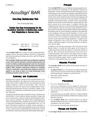
P-5217-A<br />
BioSign ® <strong>Mono</strong><br />
For Whole Blood, Serum or Plasma<br />
Rapid Heterophile Antibody Test for<br />
Infectious <strong>Mono</strong>nucleosis<br />
For in vitro Diagnostic Use<br />
Immunoassay for the Qualitative Detection of<br />
Infectious <strong>Mono</strong>nucleosis Heterophile Antibodies<br />
in Whole Blood, Serum or Plasma<br />
Serum/Plasma Whole-Blood<br />
CLIA Complexity: Moderate Waived<br />
CDC Analyte Identifier Code: 2809 2809<br />
CDC Test System Identifier Code: 37122 37143<br />
Catalog No. BSP-402WB-35 35 Test Kit<br />
BSP-402WB-10 10 Test Kit<br />
Intended Use<br />
<strong>BioSign®</strong> <strong>Mono</strong> test qualitatively detects infectious mononucleosis antibodies in<br />
human whole blood, serum or plasma specimens. This test is intended for use as<br />
an aid in the diagnosis of infectious mononucleosis.<br />
Summary and Explanation<br />
Infectious mononucleosis (IM) is an acute, self-limited, lymphoproliferative<br />
disease caused by the Epstein-Barr virus (EBV). Infection with EBV usually occurs<br />
early in life with no recognizable disease. When primary infection is delayed until<br />
young adulthood and adolescence, however, there is about a 50% chance that it will<br />
occur with the classic clinical manifestations associated with IM (1,2).<br />
The diagnosis of IM is usually based on the evaluation of characteristic clinical,<br />
hematological, and serological changes. In most cases of IM, clinical diagnosis can<br />
be made from the characteristic triad of fever, pharyngitis, and cervical lymphadenopathy,<br />
lasting for 1 to 4 weeks. IM may be complicated by splenomegaly,<br />
hepatitis, pericarditis, or central nervous system involvement(3). Rare fatal<br />
primary infections occur in patients with histiocytic hemophagocytic syndrome(4)<br />
or with a genetic X-linked lymphoproliferative syndrome(5). Hematologic<br />
features of IM include lymphocytosis with prominent atypical lymphocytes.<br />
Because other diseases may mimic the clinical and hematological symptoms of IM,<br />
serological testing is essential for the most accurate diagnosis. Serological diagnosis<br />
of IM is demonstrated by the presence of heterophile and EBV antibodies in the<br />
sera of patients (2, 6, 7).<br />
It has been well established that most individuals exposed to EBV develop a<br />
heterophile antibody response. Heterophile antibodies make up a broad class of<br />
antibodies which are characterized by the ability to react with surface antigens<br />
present on erythrocytes of different mammalian species. It is not known which<br />
specific antigen stimulates their production. It has been a common practice for<br />
physicians to use the detection of IM heterophile antibodies in the blood of patients<br />
as an aid in the diagnosis of IM. <strong>BioSign®</strong> <strong>Mono</strong> assay utilizes an extract of bovine<br />
erythrocytes which gives a greater sensitivity and specificity than similar extracts<br />
prepared from sheep and horse erythrocytes. The Forssman antibody interference<br />
has been known to be minimized by using the bovine erythrocyte extract (8,9).<br />
Principle<br />
<strong>BioSign®</strong> <strong>Mono</strong> one-step antibody test for IM uses direct solid-phase immunoassay<br />
technology for the qualitative detection of IM heterophile antibodies in human<br />
serum, plasma or whole blood. In the test procedure, 10 µl serum or plasma are added<br />
in the Sample Well (S) located below the result window. For finger-tip or whole<br />
blood, 25 µl of blood is added in the Sample Well (S). If any IM-specific<br />
heterophile antibody is present in the sample, it will be captured by the antigen band<br />
(bovine erythrocyte extracts) impregnated in the test membrane. The developer<br />
solution is then added in Sample Well (S). As the specimen followed by the<br />
developer moves by capillary action to the antigen band, the solution mobilizes the<br />
dye conjugated to anti-human IgM antibodies. Visualization of the antigen band<br />
at the Test position (T) in the result window will occur only when the IM-specific<br />
heterophile antibody which has been bound to the extracted antigen obtained from<br />
bovine erythrocytes. As the antibody-dye conjugate continues to move along the<br />
test membrane, it will bind to another band located at the Control position (C)<br />
to generate a colored band regardless of the presence of IM heterophile antibodies<br />
in the sample. Therefore, the presence of two colored bands, one at the Test<br />
position (T) and the other at the Control position (C), indicates a positive result,<br />
while the absence of a colored band at the Test position (T) indicates a negative<br />
result.<br />
Reagents and Materials Provided<br />
• <strong>BioSign®</strong> <strong>Mono</strong> test devices containing a membrane strip coated with bovine<br />
erythrocyte extract and a pad impregnated with the monoclonal mouse antihuman<br />
IgM antibody-dye conjugate in a protein matrix containing 0.1%<br />
sodium azide.<br />
• Developer Solution: Phosphate saline buffer containing 0.2% sodium azide as<br />
preservative.<br />
• Package insert<br />
Materials required but not provided:<br />
• Centrifuge capable of 1500 x g<br />
• Micropipette (200 µl)<br />
Precautions<br />
• The reagents in this kit contain sodium azide. Sodium azide may react with lead<br />
and copper plumbing to form highly explosive metal azides. Upon disposal, flush<br />
with a large amount of water to prevent azide buildup. Decontamination<br />
procedures for azide contaminated plumbing are available upon request from<br />
PBM Technical Services.<br />
• Human blood and its products are potentially infectious; handle with appropriate<br />
precautions.<br />
• For in vitro diagnostic use<br />
• Do not interchange reagents from different kit lots or use beyond the<br />
expiration date. The reagent in each kit are tested by Quality Control to<br />
function as a unit to assure proper sensitivity and maximum accuracy.<br />
• Use <strong>BioSign®</strong> <strong>Mono</strong> test only in accordance with instructions supplied with<br />
the kit.<br />
Storage and Stability<br />
<strong>BioSign®</strong> <strong>Mono</strong> test kit should be stored at 2°–30°C (36°–86°F) in its sealed pouch.<br />
Do not freeze. The storage conditions and stability dating given were established<br />
under these conditions.<br />
Specimen Collection and Preparation<br />
Whole Blood:<br />
a). Anticoagulated Blood:<br />
Whole blood collected over CPDA-1, heparin or EDTA can be used in this test. Mix<br />
whole blood by inversion and use in the test as outlined in the Test Procedure. Whole<br />
blood can be stored at 2°-8°C for 24 hours. If testing is anticipated after 24 hours,<br />
separate plasma, as outlined above, and freeze at or below -20°C.<br />
Caution: Do not freeze & thaw whole blood; hemolyzed blood can not be used in<br />
this test.<br />
b). Fingertip Blood:<br />
For fingertip blood, prick the finger and discard the first drop. Wipe the finger and<br />
use a micropipette to collect 25 µl of blood from the second drop. Immediately<br />
transfer the blood on to the upper end of the Sample Well (S) of the test device<br />
as outlined in the “Test Procedure”.<br />
Serum or Plasma:<br />
Use serum or plasma obtained from blood collected aseptically by venipuncture into<br />
a clean tube. If serum or plasma filter isolates are used, follow the manufacturer’s<br />
instructions.<br />
For serum, no anticoagulant should be used. For plasma, collect the whole blood<br />
specimen into a tube containing anticoagulant such as CPDA-1, heparin, or EDTA.<br />
For serum, blood should be allowed to clot at room temperature (18°-24 o C) and then<br />
centrifuged at 1500 x g for ten minutes at room temperature. The serum should<br />
be separated as soon as possible and may be tested immediately.
Remove the serum or plasma from the clot or red cells as soon as possible to avoid<br />
hemolysis. When possible, clear, nonhemolyzed specimens should be used. Mildly<br />
hemolyzed specimens do not affect the test result, but may create an undesirable<br />
reddish background in the result window. Specimens containing any particulate<br />
matter may give inconsistent test results. Such specimens should be clarified by<br />
centrifugation prior to testing.<br />
Storage of specimens - Refrigerate all specimens at 2°- 8°C until ready for testing.<br />
If serum or plasma specimens will not be tested within 48 hours of collection, they<br />
should be stored at or below -20°C. Specimens should not be repeatedly frozen and<br />
thawed. If specimens are to be mailed, they should be packed in appropriate shipping<br />
containers as currently described by the carrier services for handling of potentially<br />
infectious materials.<br />
Procedure<br />
Procedural notes<br />
• The test protocol must be followed in order to achieve optimal test reactivity<br />
with specimens. Follow the assay procedure and always perform the test under<br />
carefully controlled conditions.<br />
• Allow <strong>BioSign®</strong> <strong>Mono</strong> test devices, reagents and specimens to warm to room<br />
temperature before testing.<br />
• <strong>BioSign®</strong> <strong>Mono</strong> test device should remain in the sealed pouch prior to testing.<br />
• To avoid cross-contamination, use a new micropipette tip for each specimen.<br />
• Label the device with the patient’s name or control number.<br />
• When specimen is dispensed using a micropipet, allow the tip of the micropipet<br />
to touch lightly to the membrane at the UPPER END of the Sample Well<br />
(S) and then release the contents by pressing the micropipet lever.<br />
• When the finger-tip blood is dispensed, allow a free flow drop to form. Do not<br />
squeeze the finger too hard. Follow instructions under “Specimen Collection<br />
and Preparation.”<br />
• To add the Developer Solution, hold the dropper bottle in a vertical position<br />
above the LOWER END of the Sample Well (S) and dispense 2-3 drops in<br />
the well.<br />
• Mildly hemolyzed whole blood specimens do not affect the test result, but may<br />
create an undesirable reddish background in the result window.<br />
• To avoid contamination, do not touch the tip of the Developer Solution<br />
dropper bottle to skin or <strong>BioSign®</strong> <strong>Mono</strong> test device.<br />
• Use accepted microbiological practices for proper disinfection of potentially<br />
infectious test materials and contaminated equipment disposal.<br />
• After testing, dispose of <strong>BioSign®</strong> <strong>Mono</strong> test devices, micropipette tip and<br />
specimens in approved biohazard containers.<br />
Quality Control<br />
There are two internal control features in <strong>BioSign®</strong> <strong>Mono</strong> test. A colored control<br />
band will always appear at the Control position (C) if the test has been performed<br />
correctly and if the device is working properly. This is considered an internal<br />
positive procedural control. A clear background in the result window is considered<br />
an internal negative procedural control. If the test has been performed correctly<br />
and <strong>BioSign®</strong> <strong>Mono</strong> device is working properly, the background in the result<br />
window will be clear, providing a distinct result.<br />
If the colored control band does not appear at the Control position (C), the test<br />
is invalid and a new test should be performed. If the problem persists, contact PBM<br />
Technical Services immediately for assistance.<br />
External controls may also be used to assure that the reagents are working properly<br />
and the assay procedure is followed correctly. It is recommended that a control be<br />
tested at regular intervals as good laboratory testing process. For information on<br />
how to obtain controls, contact PBM’s Technical Services.<br />
Test Procedure<br />
NOTE: A positive test result may be read as soon as a distinct pink-purple colored<br />
band appears at the Test position (T) and at the Control position (C). Any shade<br />
of pink-purple colored horizontal band at the Test position (T) should be reported<br />
as a positive result. The intensity of the colored band at the Test position (T) may<br />
be different than the intensity of the band at the Control position (C).<br />
A. For Serum or Plasma:<br />
Step 1. Using a micropipette, add 10<br />
µl of serum or plasma to the UPPER<br />
END of the Sample Well (S).<br />
Step 2. Add 2-3 drops of Developer<br />
Solution into the LOWER END of<br />
the Sample Well (S).<br />
Step 3. Read test results at 8 minutes.<br />
A strong positive result may<br />
appear in less than 3 minutes. Waiting<br />
8 minutes is required to report a<br />
negative result. Results are stable up<br />
to 15 minutes after the addition of<br />
the Developer Solution.<br />
Developer<br />
Solution<br />
S<br />
T<br />
C<br />
<strong>Mono</strong><br />
S<br />
C<br />
T<br />
<strong>Mono</strong><br />
B. For Whole Blood:<br />
Step 1. Using a micropipette, add 25 µl of whole blood to the UPPER END<br />
of the Sample Well (S) of the device.<br />
Step 2. Add 2-3 drops of Developer Solution into the LOWER END of the<br />
Sample Well (S).<br />
Step 3. Read test results at 8 minutes. A strong positive result may appear<br />
in less than 3 minutes. Waiting 8 minutes is required to report a negative result.<br />
Results are stable up to 15 minutes after the addition of the Developer Solution.<br />
Positive:<br />
One pink-purple colored horizontal band each at the Test position (T) and at the<br />
Control position (C) indicate that IM-specific heterophile antibodies have been<br />
detected.<br />
<strong>Mono</strong><br />
S<br />
Interpretation of Results<br />
C<br />
T<br />
Negative:<br />
One pink-purple colored band at the Control<br />
position (C), with no distinct colored<br />
horizontal band at the Test position<br />
(T) other than the normal faint<br />
background color, indicates the IM-specific<br />
heterophile antibodies have not been<br />
detected.<br />
C<br />
T<br />
C<br />
T
Invalid:<br />
A distinct colored horizontal<br />
band at the Control position<br />
(C) should always appear. The<br />
test is invalid if no such band<br />
forms at the Control position<br />
(C).<br />
<strong>Mono</strong><br />
<strong>BioSign®</strong> <strong>Mono</strong> test is optimized to have a minimal prozone effect.<br />
Therefore, specimens containing a very high titer of antibody may produce<br />
a somewhat weaker signal but would still produce a positive result. The test<br />
does not require any specimen dilution, but it is recommended that the<br />
specimen be diluted and retested to confirm the result in case a prozone effect<br />
is suspected. The test should be used only for the qualitative detection of<br />
heterophile antibody.<br />
Limitations of the Procedure<br />
• The results obtained by this kit yield data which must be used only as<br />
adjunct to other information available to the physician.<br />
• Although most patients will have a detectable heterophile antibody level<br />
within three weeks of infection, occasionally a patient with strong clinical<br />
signs of IM may take longer than three months to develop a detectable<br />
level (10). If further testing is desired, collect additional specimens every<br />
few days and retest.<br />
• Some segments of the population who contract IM do not produce<br />
measurable levels of heterophile antibody. Approximately 50% of<br />
children under 4 years of age who have IM may test as IM heterophile<br />
antibody negative (11). EBV-specific laboratory diagnosis may be helpful<br />
in these cases.<br />
• Some individuals are reported to maintain a low but persistent level of<br />
heterophile antibodies long after their primary illness. Heterophile<br />
antibodies have been detected in blood specimens taken more than one<br />
year after the onset of the illness (12). Such false positive test results<br />
occurring in 2-3% of patients can be excluded by EBV-specific serology<br />
(3).<br />
• The IM heterophile antibody has been associated with disease states other<br />
than IM, such as leukemia, cytomegalovirus, Burkitt’s lymphoma,<br />
rheumatoid arthritis, adenovirus, viral hepatitis, and Toxoplasma gondii<br />
(13). In primary infections of adults with clinically atypical diseases,<br />
EBV-specific laboratory diagnosis may also be helpful.<br />
• <strong>BioSign®</strong> <strong>Mono</strong> for serum and plasma is classified as moderately<br />
complex under the CLIA ’88 regulations. <strong>BioSign®</strong> <strong>Mono</strong> for whole<br />
blood test is classified as waived under the CLIA ’88 regulations.<br />
• Open or broken/damaged pouches may produce erroneous results due to<br />
kit instability from exposure to moisture and should be discarded - Do Not<br />
Use.<br />
Expected Values<br />
1. In patients with symptoms indicating IM, a positive heterophile antibody<br />
result is diagnostic, and no further testing is necessary. During the acute<br />
phase of illness, IM-specific heterophile antibodies are detectable in 80-<br />
85% of IM cases. Humoral responses to primary infections appear to be<br />
quite rapid. Moderate to high levels of heterophile antibodies are seen<br />
during the first month of illness and decrease rapidly after week four (3).<br />
2. Positive test results may persist for months or even years due to the<br />
presence of persistent IM heterophile antibodies (14). This may occur with<br />
or without any clinical symptoms or hematological evidence of IM (12,<br />
15-17). Conversely, a confirmed heterophile antibody test may indicate<br />
an occult infection (18, 19). In fact, detection of IM prior to onset of<br />
clinical symptoms has been reported (20, 21).<br />
3. Some patients remain persistently negative, even though there may exist<br />
hematological and clinical evidence of IM (13, 22). In some of these<br />
patients, serological evidence for a diagnosis of cytomegalovirus infection,<br />
toxoplasmosis, or viral hepatitis, as well as others, have been found (13,<br />
S<br />
C<br />
T<br />
23) as well as others, have been found (13,23).<br />
Specificity<br />
Performance Characteristics<br />
The following potentially interfering substances do not interfere with infectious<br />
mononucleosis heterophile antibody determinations in <strong>BioSign®</strong> <strong>Mono</strong> Assay up<br />
to the levels shown below:<br />
Human Albumin<br />
15 g/dL<br />
Bilirubin<br />
60 mg/dL<br />
Hemoglobin<br />
1 g/dL<br />
Triglycerides<br />
1,300 mg/dL<br />
Proficiency <strong>Testing</strong> Results<br />
Venous blood was taken from 20 individuals. Five samples out of twenty were spiked<br />
with mononucleosis positive serum. Plasma was separated from these samples to<br />
test with <strong>BioSign®</strong> <strong>Mono</strong> kit. These spiked and unspiked samples were provided<br />
to a clinical POL site for blind testing. The results showed 100% correlation.<br />
Clinical <strong>Testing</strong> Results<br />
A total of 432 whole blood clinical samples (152 finger-stick and 280 venous blood)<br />
were tested at 7 different Physician Office Laboratory (POL) clinical sites, a<br />
reference laboratory, and in-house. Concurrently, serum or plasma samples from<br />
the same patients were obtained and tested at the same sites. In addition, a total<br />
of 144 serum/plasma samples were tested at a reference laboratory clinical site<br />
(Table 1).<br />
Table 1: Clinical Sample <strong>Testing</strong> Arrangement<br />
Finger Stick Venous Whole Serum<br />
Site Blood Blood /Plasma Total<br />
POL No. 1 0 50 0 50<br />
POL No. 2 0 50 0 50<br />
POL No. 3 6 42 0 48<br />
POL No. 4 20 13 0 33<br />
POL No. 5 31 31 0 62<br />
POL No. 6 51 0 0 51<br />
POL No. 7 17 17 0 34<br />
Reference Lab 0 50 144 194<br />
In-house 27 27 0 54<br />
Total 152 280 144 576<br />
Venous whole blood samples were tested with <strong>BioSign®</strong> <strong>Mono</strong>,and the corresponding<br />
serum/plasma samples were tested with a commercially available<br />
immunochromatographic heterophile antibody assay (Predicate) kit. When a<br />
finger stick blood sample was tested with <strong>BioSign®</strong> <strong>Mono</strong>, venous whole blood was<br />
drawn from the same patient at the same time. The plasma or serum was then<br />
prepared from each venous whole blood sample and run on a <strong>BioSign®</strong> <strong>Mono</strong><br />
device. <strong>BioSign®</strong> <strong>Mono</strong> results were compared with the commercially available<br />
immunochromatographic heterophile antibody assay (Predicate) test results (Table<br />
3). In the case of serum/plasma samples, each sample was run on both <strong>BioSign®</strong><br />
<strong>Mono</strong> and the commercially available immunochromatographic heterophile antibody<br />
assay devices, and the results were compared (Table 4). Table 2 combines<br />
both results shown in Tables 3 and 4.<br />
Table 2 shows that the agreement between two tests was 99.0% (570/576).<br />
<strong>BioSign®</strong> <strong>Mono</strong> demonstrated a relative specificity of 98.8% (479/485) and a<br />
relative sensitivity of >99.9% (91/91). The results obtained with the <strong>BioSign®</strong><br />
<strong>Mono</strong> test correlated well to the results obtained with the commercially available<br />
immunochromatographic heterophile antibody assay test.<br />
Table 2: Total Specimens<br />
<strong>BioSign®</strong> <strong>Mono</strong><br />
Positive Negative Total<br />
Commercially available Positive 91 0 91<br />
immunochromatographic Negative 6 479 485<br />
heterophile antibody assay<br />
Total 97 479 576
Table 3: Whole Blood (Finger Stick and Venous)<br />
<strong>BioSign®</strong> <strong>Mono</strong><br />
Positive Negative Total<br />
Commercially available Positive 77 0 77<br />
immunochromatographic Negative 6 349 355<br />
heterophile antibody assay<br />
Total 83 349 432<br />
Table 4: Serum or Plasma Specimens<br />
<strong>BioSign®</strong> <strong>Mono</strong><br />
Positive Negative Total<br />
Commercially available Positive 14 0 14<br />
immunochromatographic Negative 0 130 130<br />
heterophile antibody assay<br />
Total 14 130 144<br />
References<br />
1. Davidson I. Serologic <strong>Testing</strong> of Infectious <strong>Mono</strong>nucleosis. J. Am. Med. Assoc.<br />
183:289, 1937<br />
2. Evans, A.S. History of Infectious <strong>Mono</strong>nucleosis. Am J Med Sci 267:189, 1974<br />
3. Lennette, E.T. Epstein-Barr Virus. Manual of Clinical Microbiology, 5th ed.,<br />
Balows, A., et al (ed.) American Society for Microbiology, Washington DC, pp.<br />
847-852, 1991.<br />
4. Grierson, H. and Purtillo, D.T. Epstein-Barr Virus infections in Males with X-<br />
linked Lymphoproliferative Syndrome. Ann Intern Med 106:538, 19887.<br />
5. Wilson, E.R., et al. Fetal Epstein-Barr Associated Hemophagocystic Syndrome<br />
J Pediatr 98:260, 1981.<br />
6. Paul J.R. and Bunnel, W.W. The Presence of Heterophile Antibodies inInfectious<br />
<strong>Mono</strong>nucleosis. Am J. Med Sci 183:91, 1932.<br />
7. Lennette, E. and Henle, W. Epstein-Barr Virus Infections: Clinical and Serological<br />
features. Lab Manager 25:23, 1987.<br />
8. Baily, G.H. and Raffel, S. Hemolytic Antibodies for Sheep and Ox Erythrocytes<br />
in Infectious <strong>Mono</strong>nucleosis. J. Clin Invest 14:228, 1935.<br />
9. Fletcher, M.A. and Woodfolk, B.J. Immunological Studies of Infectious <strong>Mono</strong>nucleosis:<br />
Isolation and Characterization of Heterophile Antigens from Hemoglobin-free<br />
Stroma. J. Immunol 107:842, 1971.<br />
10. Penman, H.G. Seronegative Glandular Fever. J. Clin Path 21;50, 1968.<br />
11. Fleisher, G.R. Textbook of Human Virology, Belshe, R.B. (ed) Littleton, Mass.,<br />
PSG Publishing Co., pp 853-886, 1984.<br />
12. Evans, A.S., et al. A prospective Evaluation of Heterophile and Epstein-Barr<br />
Virus-Specific IgM Antibody tests in Clinical and Subclinical Infectious <strong>Mono</strong>nucleosis:<br />
Specificity and Sensitivity of Tests and Persistence of Antibody. J<br />
Infect Dis 132:546, 1975.<br />
13. Chin, T.D.Y. Diagnostic Criteria and Differential Diagnosis: Infectious <strong>Mono</strong>nucleosis,<br />
2nd ed. Schlossberg, D. (ed) Springer-Verlag, New York, 1990.<br />
14. Henle, W.G., et al. Infectious <strong>Mono</strong>nucleosis and Epstein-Barr Virus Associated<br />
Malignancies: Diagnostic Procedures for Viral, Rickettsial and Chlamydial<br />
Infections, 5th ed. Lennette, E. H. and Schmidt, N.J. (ed) American Public Health<br />
Association, Inc., Washington D.C. 1979.<br />
15. Henle, G., et al. Relation of Burkitt’s Tumor Associated Herpes-type Virus to<br />
Infectious <strong>Mono</strong>nucleosis. Proc Natl Acad Sci U.S.A. 59:94, 1968.<br />
16. Askinazi, C., et al. Positive Differential Heterophile Antibody Test. Persistence<br />
in a Symptomatic Patient. J Am Med. assoc 236:1492, 1976.<br />
17. Horwitz, C.A., et al. The Specificity of Heterophile Antibodies in Patients and<br />
Healthy Donors with No or Minimal Signs of Infectious <strong>Mono</strong>nucleosis. Blood<br />
47:91, 1976.<br />
18. Hallee, T.J., et al. Infectious <strong>Mono</strong>nucleosis at the United States Military<br />
Academy: A Prospective Study of a Single Class Over Four Years. Yale J Biol<br />
Med 3:182, 1974.<br />
19. Infectious <strong>Mono</strong>nucleosis and Its Relationship to EB Virus Antibody. A Joint<br />
Investigation by University Health Physicians and P.H.L.S. Laboratories. Br.<br />
Med J 11:643, 1971.<br />
20. Bauer, S. and Holf, G. Test Detects <strong>Mono</strong>nucleosis in Incubation Period.<br />
Annual Meeting of ASCP and CAP, Chicago, Illinois, October 15-23, 1965.<br />
21. Baehner, R.L and Schuler, S.E. Infectious <strong>Mono</strong>nucleosis in Childhood.<br />
Clinical Expressions, Serologic Findings, Complications, Prognosis. Clin<br />
Pediatr 6:393, 1967.<br />
22. Henle, G. and Henle, W. Epstein-Barr Virus and Infectious <strong>Mono</strong>nucleosis. N<br />
Engl J Med 288:263, 1964.<br />
23. Cameron, D. and McBean, L.M. A Clinical Study of Infectious <strong>Mono</strong>nucleosis<br />
and Toxoplasmosis. Baltimore, The Williams and Wilkins Company, pp. 24-<br />
27, 1973.<br />
Symbols Key<br />
Manufactured by<br />
CE Mark<br />
Authorized Representative<br />
In Vitro Diagnostic Medical Device<br />
Catalog Number<br />
Consult Instructions for Use<br />
Batch Code<br />
“Use By” date in year-month-day format<br />
Temperature Limitation<br />
Contains sufficient for tests<br />
Do not reuse<br />
Contents<br />
Test Device<br />
Developer Solution<br />
Instructions for Use<br />
Infectious <strong>Mono</strong>nucleosis Test<br />
BioSign ® is a Registered Trademark of Princeton BioMeditech Corporation.<br />
Patent No.: 5,559,041<br />
© 2000 PBM<br />
Printed in U.S.A.<br />
Revised Oct 2003<br />
P-5217-A 1009BL<br />
MT Promedt Consulting GmbH<br />
Eisenbahnstrasse 2<br />
D-66386 St. Ingbert<br />
Germany<br />
+49-68 94-58 10 20<br />
Manufactured by<br />
Princeton BioMeditech Corporation<br />
Princeton, NJ 08543-7139 U.S.A.<br />
1-732-274–1000 www.pbmc.com