Association of Alveolar Hemorrhage with Amiodarone ... - Tanaffos
Association of Alveolar Hemorrhage with Amiodarone ... - Tanaffos
Association of Alveolar Hemorrhage with Amiodarone ... - Tanaffos

You also want an ePaper? Increase the reach of your titles
YUMPU automatically turns print PDFs into web optimized ePapers that Google loves.
CASE REPORT<br />
<strong>Tanaffos</strong> (2008) 7(2), 75-78<br />
©2008 NRITLD, National Research Institute <strong>of</strong> Tuberculosis and Lung Disease, Iran<br />
<strong>Association</strong> <strong>of</strong> <strong>Alveolar</strong> <strong>Hemorrhage</strong> <strong>with</strong> <strong>Amiodarone</strong>:<br />
Role <strong>of</strong> Bronchoscopy<br />
Mehrdad Behnia<br />
Medical College <strong>of</strong> Georgia, Augusta, Georgia-USA<br />
ABSTRACT<br />
A common pulmonary complication due to the toxicity <strong>of</strong> amiodarone is chronic interstitial pneumonitis. <strong>Alveolar</strong> hemorrhage,<br />
<strong>with</strong> or <strong>with</strong>out hemoptysis, is an exceedingly infrequent presentation <strong>of</strong> amiodarone toxicity. We report a 69-year old patient<br />
<strong>with</strong> dyspnea, hypoxemia and bilateral diffuse interstitial and alveolar infiltrates occurring four months after treatment <strong>with</strong><br />
amiodarone. An initial and comprehensive work-up did not reveal the cause <strong>of</strong> infiltrates. Bronchoalveolar lavage (BAL) fluid<br />
demonstrated foamy macrophages and alveolar hemorrhage, not caused by either vasculitis or autoimmune diseases. We<br />
speculate that amiodarone may have been associated <strong>with</strong> BAL findings since cessation <strong>of</strong> the drug resulted in resolution <strong>of</strong><br />
the infiltrates. In amiodarone-induced lung injury, diffuse interstitial and alveolar infiltrates can be suggestive <strong>of</strong> alveolar<br />
hemorrhage and should be further investigated by bronchoscopy and BAL.(<strong>Tanaffos</strong> 2008; 7(2): 75-78)<br />
Key words: <strong>Amiodarone</strong>, <strong>Alveolar</strong> hemorrhage, Capillaritis, Vasculitis, Bronchoscopy<br />
INTRODUCTION<br />
Pulmonary manifestations <strong>of</strong> amiodarone range<br />
from diffuse interstitial pneumonitis to acute<br />
respiratory distress syndrome (1-3). With the<br />
exception <strong>of</strong> a few cases (seen in the setting <strong>of</strong><br />
diffuse alveolar damage) (3), alveolar hemorrhage,<br />
and/ or hemoptysis, are extremely uncommon<br />
presentations <strong>of</strong> amiodarone-induced lung disease.<br />
Bronchoscopy <strong>with</strong> BAL is not commonly used in<br />
such patients.<br />
We report a patient <strong>with</strong> alveolar hemorrhage and<br />
a cumulative intake <strong>of</strong> 4.5 grams <strong>of</strong> amiodarone over<br />
a four-month period. The interstitial and<br />
alveolar presentation <strong>of</strong> his disease were suggestive<br />
Correspondence to: Behnia M<br />
Address: 3630 J. Dewey Gray Circle, Augusta, GA 30909<br />
Email address: doctor@mbehnia.com<br />
Received: 5 August 2007<br />
Accepted: 20 May 2008<br />
<strong>of</strong> interstitial pneumonitis. However, bronchoscopy<br />
and BAL showed diffuse alveolar hemorrhage in the<br />
absence <strong>of</strong> an auto-immune process or vasculitis. We<br />
suggest that bronchoscopy and BAL, although not<br />
routinely performed for patients on amiodarone<br />
therapy <strong>with</strong> radiographic abnormalities should be<br />
considered as part <strong>of</strong> the work-up for interstitial and<br />
alveolar infiltrates in such patients in order to ruleout<br />
alveolar hemorrhage.<br />
CASE REPORT<br />
A 69- year- old male presented to the emergency<br />
room <strong>with</strong> gradual progression <strong>of</strong> shortness <strong>of</strong> breath<br />
over a period <strong>of</strong> several weeks. He was unable to<br />
walk beyond 10 meters. Review <strong>of</strong> systems was<br />
negative for fever, chills, weight loss, hemoptysis,<br />
orthopnea, proxysmal nocturnal dyspnea, or chest
76 <strong>Amiodarone</strong> and <strong>Alveolar</strong> <strong>Hemorrhage</strong><br />
pain.<br />
A few weeks prior to his visit, he was admitted to<br />
hospital <strong>with</strong> similar symptoms. At the time, his<br />
dyspnea was attributed to heart failure, requiring a<br />
two- day support on mechanical ventilation.<br />
The patient's past medical history included a fourvessel<br />
coronary artery bypass graft conducted 4<br />
months earlier. The postoperative course was<br />
complicated by respiratory insufficiency and failure<br />
to wean, as well as renal failure, requiring<br />
tracheostomy and chronic hemodialysis. He was<br />
ventilator- dependent for 2 months. Subsequently, the<br />
patient was re-hospitalized on another occasion <strong>with</strong><br />
the diagnosis <strong>of</strong> congestive heart failure.<br />
Other medical problems included hypertension,<br />
hypercholesterolemia, and previous myocardial<br />
infarctions. He was on chronic oxygen<br />
supplementation and had a 60 pack/year history <strong>of</strong><br />
smoking.<br />
Medications included amiodarone 400 mg every<br />
day (started after the bypass surgery- cumulative<br />
dose <strong>of</strong> 4.5 grams), aspirin, beclomethasone and<br />
ipratropium inhalers, diltiazem, and isosorbide.<br />
Hospital course: In the emergency room, the<br />
patient had a respiratory rate <strong>of</strong> 40/min. Arterial<br />
blood gas showed pH: 7.1, PCO 2 :82 mm Hg, and<br />
PO 2 : 68 mm Hg. He was intubated and supported by<br />
mechanical ventilation for respiratory failure for 4<br />
days during which he was dialyzed several times<br />
below his dry weight.<br />
On physical examination after extubation, the<br />
patient was afebrile <strong>with</strong> a respiratory rate <strong>of</strong> 25/min,<br />
pulse rate <strong>of</strong> 100 beats/min, and blood pressure <strong>of</strong><br />
100/70. The skin had a tanned appearance. Heart had<br />
a regular rate and rhythm and a 2 over 6 systolic<br />
ejection murmur. Crackles were audible diffusely<br />
throughout the lungs; there was no dullness to<br />
percussion. Hepatosplenomegaly was not present.<br />
Lower extremities had trace edema.<br />
Laboratory data revealed a white blood cell count<br />
<strong>of</strong> 18,900/mm3, hemoglobin: 11.8 g/dl, blood urea<br />
nitrogen: 79 mg/dl, and creatinine: 2.4 mg/dl. Liver<br />
function tests (including transaminases), prothrombin<br />
time (PT), and partial thromboplastin time (PTT)<br />
were <strong>with</strong>in normal limits. Erythrocyte sedimentation<br />
rate was 48 mm/hr. Urinalysis did not show any red<br />
blood cell cast or proteinuria. Electrocardiogram<br />
displayed a left bundle branch block and a first<br />
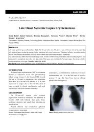
degree atrioventricular block. Chest radiograph and<br />
computed tomography (CT) displayed diffuse<br />
bilateral interstitial and alveolar infiltrates and slight<br />
pleural effusion (Figure 1).<br />
Figure 1. Radiograph (A) and CT (B) <strong>of</strong> the chest; significant findings<br />
are bilateral interstitial alveolar infiltrates and minimal pleural effusion.<br />
The tip <strong>of</strong> the hemodialysis access catheter is visible in the superior<br />
vena cave.<br />
A bronchoscopy <strong>with</strong> BAL was performed which<br />
revealed a sanguinolent specimen. Sequential<br />
instillation and aspiration <strong>of</strong> normal saline did not<br />
<strong>Tanaffos</strong> 2008; 7(2): 75-78
Behnia M. 77<br />
diminish the intensity <strong>of</strong> blood in the recovered<br />
aliquots, a finding highly indicative <strong>of</strong> alveolar<br />
hemorrhage. Due to renal impairment and high risk<br />
<strong>of</strong> fatal bleeding, transbronchial biopsies were not<br />
performed. The fluid white cell count had a normal<br />
differential; but an abundance <strong>of</strong> hemosiderin- laden<br />
and foamy macrophages (Figure 2). The fluid<br />
cytology was negative for malignancy. Bacterial,<br />
fungal, and viral cultures as well as special stains<br />
were non-contributory.<br />
Figure 2. Plate A) The bronchoalveolar lavage specimen demonstrating<br />
abundance <strong>of</strong> foamy marrophages (H&E stain). Plate B) Hemosiderin<br />
appears as dark spots <strong>with</strong>in the macrophages (iron stain).<br />
Work-up for alveolar hemorrhage included<br />
measurement <strong>of</strong> serum levels for complements (C3<br />
and C4), anti-nuclear and anti-DNA antibodies,<br />
rheumatoid factor, proteinase-3 and<br />
myeloperoxidase, anti-neutrophilic cytoplamic<br />
antibodies, and anti- glomerular basement membrane<br />
antibody. Their respective values were all <strong>with</strong>in the<br />
normal limits.<br />
<strong>Amiodarone</strong>, as the possible <strong>of</strong>fending agent, was<br />
stopped. The patient's dyspnea gradually resolved. A<br />
3-week follow-up radiograph showed near total<br />
resolution <strong>of</strong> the interstitial and alveolar infiltrates.<br />
DISCUSSION<br />
An iodinated benz<strong>of</strong>uran derivative, amiodarone<br />
is a frequently used antiarrhytmic drug that can cause<br />
considerable pulmonary toxicity in 5 to 15% <strong>of</strong><br />
patients (2). The most common toxicity is chronic<br />
interstitial pneumonitis (2), followed by organizing<br />
pneumonia <strong>with</strong> or <strong>with</strong>out bronchiolitis obliterans<br />
(3), and rarely respiratory distress syndrome (4). Risk<br />
factors for amiodarone-induced lung disease<br />
comprise drug dosage <strong>of</strong> greater than 400 mg per<br />
day, and intake <strong>of</strong> the drug for more than two months<br />
(2). Reports suggest that the total cumulative dose <strong>of</strong><br />
the drug may be a more substantial risk factor than<br />
the daily dose (5). Toxicity can also become evident<br />
in patients taking 200 mg <strong>of</strong> the drug per day over a<br />
span <strong>of</strong> years. Foamy alveolar macrophages, increase<br />
in both polymorphonuclear leukocytes, and T-<br />
suppressor/ T-cytotxic lymphocytes in the<br />
bronchoalveolar lavage fluid; all may indicate<br />
exposure to the drug (2).<br />
<strong>Alveolar</strong> hemorrhage is a very rare complication<br />
<strong>of</strong> amiodarone. Dean has reported the largest series<br />
<strong>of</strong> amiodorane-induced alveolar hemorrhage (3). In<br />
his study <strong>of</strong> 171 patients, minimal alveolar<br />
hemorrhage was noted in only a few patients on<br />
transbronchial, open lung, and post-mortem<br />
specimens (3). Only one patient had evidence <strong>of</strong><br />
significant alveolar hemorrhage on post-mortem<br />
study (an elderly male <strong>with</strong> chronic lung disease and<br />
cumulative amiodarone dose <strong>of</strong> 101 grams, fever,<br />
sedimentation rte <strong>of</strong> 150 mm/hr, and white blood cell<br />
count <strong>of</strong> 22,000/mm3). Of importance, diffuse<br />
alveolar damage in different stages <strong>of</strong> evolution was<br />
the common denominator in all the cases <strong>of</strong> alveolar<br />
<strong>Tanaffos</strong> 2008; 7(2): 75-78
78 <strong>Amiodarone</strong> and <strong>Alveolar</strong> <strong>Hemorrhage</strong><br />
hemorrhage in the series. It was therefore, difficult to<br />
determine if the alveolar hemorrhage could also be<br />
seen in the absence <strong>of</strong> diffuse alveolar damage.<br />
Other reports have also suggested amiodarone as<br />
the cause <strong>of</strong> hemoptysis and alveolar hemorrhage.<br />
Vizoli reported a case <strong>of</strong> a patient on 200 mg <strong>of</strong><br />
amiodarone twice daily for two months, who<br />
presented <strong>with</strong> severe hypoxemia and hemoptysis<br />
(6). On transbronchial biopsies, hemosiderin- laden<br />
macrophages and focal bronchiolitis obliterans<br />
organizing pneumonia (BOOP) were observed. In<br />
another report, a patient presented <strong>with</strong> hemoptysis<br />
and fever two weeks after treatment <strong>with</strong> amiodarone<br />
(7). Bronchoscopy demonstrated diffuse<br />
inflammatory changes but the source <strong>of</strong> bleeding<br />
could not be identified. Work-up for vasculitis and<br />
autoimmune diseases was inconclusive.<br />
In another report, acute alveolar hemorrhage and<br />
orthodeoxia were seen in a patient <strong>with</strong> severe<br />
ischemic cardiomyopathy receiving intravenous<br />
amiodarone for ventricular tachycardia (8). The<br />
patient was discharged after his first hospital stay<br />
during which he received 4 days <strong>of</strong> maintenance<br />
infusion <strong>of</strong> amiodarone. During his second admission<br />
one week later, he developed hemoptysis and had a 3<br />
g loss <strong>of</strong> hemoglobin.<br />
Due to platypnea and orthodeoxia, he required<br />
100 percent oxygen supplement. A BAL displayed<br />
abundant hemosiderin-laden macrophages. Heart<br />
failure and fluid overload were not the precipitating<br />
factors because <strong>of</strong> a normal pulmonary artery wedge<br />
pressure. Serologies for vasculitis and autoimmune<br />
diseases were negative. The patient subsequently<br />
required mechanical ventilation for hypoxemia. His<br />
condition improved upon cessation <strong>of</strong> amiodarone<br />
and administration <strong>of</strong> methylprednisolone (8). In the<br />
majority <strong>of</strong> the aforementioned cases, hemoptysis<br />
and/ or alveolar hemorrhage ceased upon termination<br />
<strong>of</strong> amiodarone, implicating it as the <strong>of</strong>fending cause.<br />
In conclusion, the present report demonstrated<br />
that amiodarone may be associated <strong>with</strong> alveolar<br />
hemorrhage in the absence <strong>of</strong> acute respiratory<br />
distress syndrome, renal-pulmonary syndrome, or<br />
auto-immune diseases. In our patient, infectious<br />
etiologies, i.e. fungal, bacterial, and viral, were not<br />
the precipitating cause <strong>of</strong> alveolar hemorrhage,<br />
either.<br />
Congestive heart failure and fluid overload were<br />
not present because the patient was dialyzed below<br />
his dry weight several times. <strong>Amiodarone</strong> can,<br />
therefore, be a cause <strong>of</strong> diffuse interstitial infliltrates<br />
and alveolar hemorrhage in patients who do not<br />
present <strong>with</strong> hemoptysis. Bronchoscopy <strong>with</strong> BAL,<br />
although not routinely performed, and possibly<br />
transbronchial biopsies, should be considered in<br />
establishing the above-mentioned diagnosis and for<br />
excluding vasculitides.<br />
REFERENCES<br />
1. Martin WJ 2nd, Rosenow EC 3rd. <strong>Amiodarone</strong> pulmonary<br />
toxicity. Recognition and pathogenesis (Part 2). Chest 1988;<br />
93 (6): 1242- 8.<br />
2. Martin WJ 2nd, Rosenow EC 3rd. <strong>Amiodarone</strong> pulmonary<br />
toxicity. Recognition and pathogenesis (Part I). Chest 1988;<br />
93 (5): 1067- 75.<br />
3. Dean PJ, Groshart KD, Porterfield JG, Iansmith DH, Golden<br />
EB Jr. <strong>Amiodarone</strong>-associated pulmonary toxicity. A clinical<br />
and pathologic study <strong>of</strong> eleven cases. Am J Clin Pathol<br />
1987; 87 (1): 7- 13.<br />
4. Greenspon AJ, Kidwell GA, Hurley W, Mannion J.<br />
<strong>Amiodarone</strong>-related postoperative adult respiratory distress<br />
syndrome. Circulation 1991; 84 (5 Suppl): III407- 15.<br />
5. Suárez LD, Poderoso JJ, Elsner B, Bunster AM, Esteva H,<br />
Bellotti M. Subacute pneumopathy during amiodarone<br />
therapy. Chest 1983; 83 (3): 566- 8.<br />
6. Vizioli LD, Cho S. <strong>Amiodarone</strong>-associated hemoptysis.<br />
Chest 1994; 105 (1): 305- 6.<br />
7. Goldstein I, Topilsky M, Segev D, Isakov A, Heller I. Very<br />
early onset <strong>of</strong> acute amiodarone pulmonary toxicity<br />
presenting <strong>with</strong> hemoptysis. Chest 1997; 111 (5): 1446- 7.<br />
8. Iskander S, Raible DG, Brozena SC, Gaitanaru DM, Ayala<br />
G, Iskandrian AE. Acute alveolar hemorrhage and<br />
orthodeoxia induced by intravenous amiodarone. Catheter<br />
Cardiovasc Interv 1999; 47 (1): 61- 3.<br />
<strong>Tanaffos</strong> 2008; 7(2): 75-78