MIDTERM EXAM
MIDTERM EXAM
MIDTERM EXAM

Create successful ePaper yourself
Turn your PDF publications into a flip-book with our unique Google optimized e-Paper software.
PSY 5065 - fMRI: Hands-on Training<br />
<strong>MIDTERM</strong> <strong>EXAM</strong><br />
Date assigned: March 9, 2015<br />
Date due: March 13, 2015<br />
Each problem: 5 points<br />
Total exam: 15% of course grade<br />
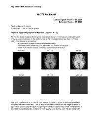
Problem 1: Basic NMR<br />
A) What magnetically active nucleus generates the signal we detect to make a typical<br />
MR image? (1 point)<br />
B) The gyromagnetic ratio of carbon-13 is 10.7 MHz/T. What is the resonant frequency<br />
of 13 C at 3 Tesla? How does this compare to the resonant frequency of 1 H at 3 Tesla?<br />
(1 point)<br />
C) In a static magnetic field oriented along the positive z- axis, what is the orientation of<br />
the net magnetic moment of a spin isochromat at equilibirum? (1/2 point)<br />
After application of a (perfect) inversion pulse, what is the orientation of the net<br />
magnetic moment of the isochromat? (1/2 point)<br />
D) After application of a 90 y degree excitation pulse (rotating M away from equilibrium,<br />
clockwise around the y-axis):<br />
What is the orientation of the net magnetic moment, M? (1/3 point )<br />
What is the relative magnitude of the longitudinal magnetization, M ||, if M (the length<br />
of M) is 10 units? (1/3 point)<br />
What is the relative magnitude of the transverse magnetization, M ⊥ (again, if M is 10<br />
units)? (1/3 point)<br />
E) After a 30 degree excitation pulse is applied to an isochromat at equilibrium, what<br />
are the relative magnitudes of M || and M ⊥ ?<br />
(1 point)
PSY 5065 - fMRI: Hands-on Training<br />
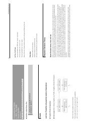
Problem 2: Relaxation<br />
A) Label, on the drawing below, the T 2 and T 2 * envelopes describing the magnitude of<br />
the net transverse magnetization generated by a collection of spin isochromats in an<br />
inhomogeneous magnetic field environment during a spin echo experiment. (1 point)<br />
Excitation<br />
pulse<br />
Refocusing<br />
pulse<br />
Echo<br />
Read-out<br />
M T<br />
S<br />
B) In the inversion-recovery experiment illustrated below, what would be the appropriate<br />
time to apply an excitation pulse if you want to acquire an imate with the signal from the<br />
gray matter nulled? (1 point)<br />
White matter<br />
Gray matter<br />
CSF
PSY 5065 - fMRI: Hands-on Training<br />
C) In the data shown above, which has the longest T 1 : gray matter, white matter or<br />
CSF? (1 point)<br />
D) In the acquisition described in (B) (TI set to null gray matter), what would be the<br />
relative image intensities of white matter and CSF in a magnitude image? (1 point)<br />
E) In any experiment, if the repetition time is very short, should the flip angle of the<br />
repeated RF pulse be large or small if you want to maximize image SNR? (1 point)<br />
Problem 3: Pulse sequences<br />
A) If a 45 degree RF pulse with 2 kHz bandwidth is applied in conjunction with a linear<br />
magnetic field gradient on the x-axis with a strength of 11.7 mT/m, what is the resulting<br />
slice thickness? (1/2 point).<br />
What is the orientation of the resulting slice – sagittal, axial or coronal? (1/2 point)<br />
B) What is the read-out direction in the MP-RAGE image below (up/down or front<br />
back)? Name 2 clues you can use to figure that out. (1 point)
PSY 5065 - fMRI: Hands-on Training<br />
B) Label the slice-select, read-out, and phase-encode gradient axes for the two pulse<br />
sequences illustrated below. Describe each pulse sequence as FLASH or EPI, Spin<br />
Echo or Gradient Echo. (2 points total - 1/6 each for gradient labels, 1/4 for each FLASH<br />
vs. EPI, and 1/4 for each SE vs. GE)<br />
Pulse sequence 3.B.1: _________________________________________<br />
N = 4<br />
RF<br />
G ______<br />
G ______<br />
G ______<br />
DAC<br />
Pulse sequence 3.B.2: _________________________________________<br />
RF<br />
G ______<br />
G ______<br />
G ______<br />
DAC
PSY 5065 - fMRI: Hands-on Training<br />
C) Draw on the grid provided below the k-space trajectory for the pulse sequence in<br />
3.B.2. Make sure to label the phase-encode direction. (It might help to number the<br />
gradients to match k-space excursions; no need to be precise once you get to the readout<br />
lines.) (1 point)<br />
Sequence 3.B.2
PSY 5065 - fMRI: Hands-on Training<br />
Extra Credit (1 point). For the EPI images shown below, indicate which was acquired<br />
with a spin echo pulse sequence, and which was acquired with a gradient echo pulse<br />
sequence. Assume both had the same echo time during the acquisition.