PSY 3031 Introduction to Sensation and Perception - Vision ...
PSY 3031 Introduction to Sensation and Perception - Vision ...
PSY 3031 Introduction to Sensation and Perception - Vision ...

Create successful ePaper yourself
Turn your PDF publications into a flip-book with our unique Google optimized e-Paper software.
Course: <strong>PSY</strong> <strong>3031</strong> <strong>Introduction</strong> <strong>to</strong> <strong>Sensation</strong> <strong>and</strong> <strong>Perception</strong> (sec 001) Spring 08<br />
https://moodle.umn.edu/course/view.php?id=1687<br />
1 of 31 7/29/2008 2:48 PM<br />
myMoodle | Email | myU | Library | One S<strong>to</strong>p | Support site<br />
You are logged in as Cheryl Olman (Logout)<br />
<strong>PSY</strong> <strong>3031</strong> <strong>Introduction</strong> <strong>to</strong> <strong>Sensation</strong> <strong>and</strong> <strong>Perception</strong> (sec 001) Spring 08<br />
Moodle Home ► <strong>PSY</strong><strong>3031</strong>_1S8<br />
Switch role <strong>to</strong>...<br />
Turn editing on<br />
Administration<br />
Turn editing on<br />
Settings<br />
Assign roles<br />
Au<strong>to</strong>matic<br />
Enrollment<br />
Groups<br />
Backup<br />
Res<strong>to</strong>re<br />
Import<br />
Reset<br />
Reports<br />
Questions<br />
Scales<br />
Files<br />
Grades<br />
Unenroll me<br />
from<br />
<strong>PSY</strong><strong>3031</strong>_1S8<br />
Quick links<br />
for students<br />
Watch online<br />
orientation<br />
User guides<br />
Student support<br />
forums<br />
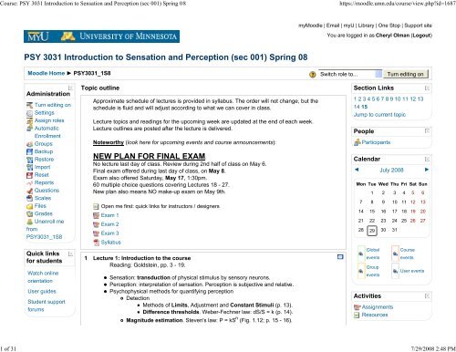
Topic outline<br />
Approximate schedule of lectures is provided in syllabus. The order will not change, but the<br />
schedule is fluid <strong>and</strong> will adjust according <strong>to</strong> what we can cover in class.<br />
Lecture <strong>to</strong>pics <strong>and</strong> readings for the upcoming week are updated at the end of each week.<br />
Lecture outlines are posted after the lecture is delivered.<br />
Noteworthy (look here for upcoming events <strong>and</strong> course announcements):<br />
NEW PLAN FOR FINAL EXAM.<br />
No lecture last day of class. Review during 2nd half of class on May 6.<br />
Final exam offered during last day of class, on May 8.<br />
Exam also offered Saturday, May 17, 1:30pm.<br />
60 multiple choice questions covering Lectures 18 - 27.<br />
New plan also means NO make-up exam on May 9th.<br />
Open me first: quick links for instruc<strong>to</strong>rs / designers<br />
Exam 1<br />
Exam 2<br />
Exam 3<br />
Syllabus<br />
1 Lecture 1: <strong>Introduction</strong> <strong>to</strong> the course<br />
Reading: Goldstein, pp. 3 - 19.<br />
<strong>Sensation</strong>: transduction of physical stimulus by sensory neurons.<br />
<strong>Perception</strong>: interpretation of sensation. <strong>Perception</strong> is subjective <strong>and</strong> relative.<br />
Psychophysical methods for quantifying perception<br />
Detection<br />
Methods of Limits, Adjustment <strong>and</strong> Constant Stimuli (p. 13).<br />
Difference thresholds. Weber-Fechner law: dS/S = k (p. 14).<br />
Magnitude estimation. Steven's law: P = kS n (Fig. 1.12; p. 15 - 16).<br />
Section Links<br />
1 2 3 4 5 6 7 8 9 10 11 12 13<br />
14 15<br />
Jump <strong>to</strong> current <strong>to</strong>pic<br />
People<br />
Participants<br />
Calendar<br />
◄ July 2008 ►<br />
Mon Tue Wed Thu Fri Sat Sun<br />
1 2 3 4 5 6<br />
7 8 9 10 11 12 13<br />
14 15 16 17 18 19 20<br />
21 22 23 24 25 26 27<br />
28 29 30 31<br />
Global<br />
events<br />
Group<br />
events<br />
Activities<br />
Assignments<br />
Resources<br />
Course<br />
events<br />
User events
Course: <strong>PSY</strong> <strong>3031</strong> <strong>Introduction</strong> <strong>to</strong> <strong>Sensation</strong> <strong>and</strong> <strong>Perception</strong> (sec 001) Spring 08<br />
https://moodle.umn.edu/course/view.php?id=1687<br />
2 of 31 7/29/2008 2:48 PM<br />
Email:<br />
moodle@umn.edu<br />
n < 1: compression<br />
n > 1: expansion<br />
Lecture 2: <strong>Introduction</strong> <strong>to</strong> neural mechanisms<br />
Reading: Goldstein, pp. 21 - 28.<br />
Basic properties of neurons<br />
Main divisions: dendrite, soma, axon, myelin (Fig. 2.3)<br />
Membrane potential = potential for communication (p. 25)<br />
Communication between neurons<br />
Action potentials are created/carried by ionic currents (Fig. 2.5)<br />
Information is encoded by the rate of action potentials (Fig. 2.6)<br />
Information is chemically transmitted at synapses (Fig. 2.7)<br />
Nerves are bundles of axons<br />
Sensory information travels in cranial nerves (the 12 nerves that enter the brainstem<br />
above the spinal cord) <strong>and</strong> peripheral nerves<br />
Conduction velocity is highest in large, myelinated axons<br />
Small, unmyelinated axons (C-fibers) transmit action potentials at ~1 m/s<br />
The largest myelinated axons (A-alpha fibers) conduct at ~100 m/s.<br />
Ana<strong>to</strong>my of the nervous system(s)<br />
Central nervous system: brain, spinal cord <strong>and</strong> retina.<br />
in general cannot regenerate after injury, e.g. spinal cord injury results in<br />
paralysis<br />
Peripheral nervous system: peripheral nerves & sensory neurons, cranial nerves.<br />
generally can re-grow after injury (a cut in the finger heals, <strong>and</strong> has normal<br />
sensation).<br />
ICE1<br />
2 Lecture 3: Touch<br />
Reading: Goldstein, pp. 305 - 318<br />
Soma<strong>to</strong>sensory system: sensation throughout the body (p. 305)<br />
Proprioception: balance <strong>and</strong> limb position<br />
Cutaneous senses: Mechanical, Thermal, Noxious<br />
Mechanorecep<strong>to</strong>rs: 4 primary types, we focus on 2 (Table 14.1)<br />
Merkel recep<strong>to</strong>r: slowly adapting, low frequency, perception of fine detail (Fig.<br />
14.10)<br />
Density is inversely related <strong>to</strong> receptive field size (Fig. 14.12)<br />
Pacinian corpuscle: rapidly adapting, high-frequency, texture <strong>and</strong> vibration
Course: <strong>PSY</strong> <strong>3031</strong> <strong>Introduction</strong> <strong>to</strong> <strong>Sensation</strong> <strong>and</strong> <strong>Perception</strong> (sec 001) Spring 08<br />
https://moodle.umn.edu/course/view.php?id=1687<br />
3 of 31 7/29/2008 2:48 PM<br />
Texture is perceived by a combination of Merkel <strong>and</strong> Pacinian recep<strong>to</strong>rs (pp 313-314;<br />
Fig. 14.15)<br />
Thermal recep<strong>to</strong>rs<br />
Separate recep<strong>to</strong>rs for warm (38 - 48 deg C) <strong>and</strong> cool (20 - 35 deg. C)<br />
For reference, body temperature is ~37 deg. C<br />
Non-uniformly distributed: cold sensation may not occur on all parts of the back of<br />
your h<strong>and</strong>.<br />
Pathways: spinothalamic (thermal) <strong>and</strong> dorsal column/medial lemniscal (mechanical)<br />
(Fig. 14.5)<br />
Thalamus: all sensory information goes <strong>to</strong> thalamus before cortex<br />
General principles (without memorizing details)<br />
Soma<strong>to</strong>sensory information is soma<strong>to</strong><strong>to</strong>pically organized like cortex<br />
Different pathways segregated in different nuclei (sub-regions)<br />
Response properties are more specialized, e.g. center-surround receptive field<br />
organization (Fig. 14.18)<br />
Cortical representation: soma<strong>to</strong><strong>to</strong>pic <strong>and</strong> non-uniform<br />
Primary soma<strong>to</strong>sensory cortex (S1) is on the post-central gyrus (Fig. 14.6)<br />
Body parts with higher tactile acuity have larger cortical representations.<br />
Attention. Unattended stimuli can fail <strong>to</strong> elicit neural response, even in primary<br />
soma<strong>to</strong>sensory cortex (Fig. 14.21)<br />
Object-selective responses in regions beyond S1, e.g. farther back in parietal cortex<br />
(Fig. 14.20)<br />
Lecture 4: Pain<br />
Reading: Goldstein, pp. 318 - 324<br />
Three types of pain<br />
Nociceptive: mediated by cutaneous nerves responding <strong>to</strong> chemical insult or<br />
extreme heat, cold or force (Fig. 14.22a). Travel in a separate pathway from other<br />
sensory information (<strong>to</strong>uch is in ipsilateral dorsal column - medial lemniscal path;<br />
nociception is in contralateral spinothalamic path, shown in Fig 14.5).<br />
Ipsilateral means that the neural representation is on the side of the body on<br />
which stimulus transduction occurred.<br />
Contralateral means that the neurons responding <strong>to</strong> a stimulus (or controlling<br />
an action) are on the opposite side of the body.<br />
Types of nocicep<strong>to</strong>rs: chemical, thermal (very hot <strong>and</strong> very cold), mechanical<br />
(respond <strong>to</strong> very sharp things or very strong pressure) <strong>and</strong> polymodal<br />
(respond <strong>to</strong> both thermal <strong>and</strong> mechanical stimulation)<br />
Inflamma<strong>to</strong>ry: mediated by prostagl<strong>and</strong>ins generated in response <strong>to</strong> injury (Fig<br />
14.22b). Prostagl<strong>and</strong>ins are chemical messengers that initiate platelet aggregation<br />
or increased or decreased blood flow where appropriate; they also sensitize sensory<br />
neurons.<br />
Neuropathic: caused by damage <strong>to</strong> or pressure on peripheral nerves or neurons in<br />
pain pathways (Fig. 14.22c). Examples:
Course: <strong>PSY</strong> <strong>3031</strong> <strong>Introduction</strong> <strong>to</strong> <strong>Sensation</strong> <strong>and</strong> <strong>Perception</strong> (sec 001) Spring 08<br />
https://moodle.umn.edu/course/view.php?id=1687<br />
4 of 31 7/29/2008 2:48 PM<br />
Carpal tunnel syndrome: the carpal ligament puts pressure on the medial<br />
nerve, causing weakness, numbness <strong>and</strong>/or pain in the h<strong>and</strong> (particularly<br />
thumb through 4th finger).<br />
Sciatica: pressure on the large sciatic nerve, where it goes through hip joints,<br />
causes burning or freezing pain in legs <strong>and</strong> but<strong>to</strong>cks.<br />
The importance of pain<br />
Social cost: $100 billion / year in US; 20% of American adults report chronic pain<br />
(most commonly lower back pain).<br />
Protective mechanism: lack of pain (e.g. CIPA) is life threatening.<br />
The experience of pain<br />
Nociception is the sensory component; pain is is the affective (emotional)<br />
component<br />
Fig. 14.24: decreasing intensity decreases unpleasantness, but<br />
unpleasantness can be decreased without decreasing intensity.<br />
Pain pathways project <strong>to</strong> the limbic system (amygdala, hippocampus, anterior<br />
cingulate gyrus, Fig. 14.23) as well as soma<strong>to</strong>sensory cortex.<br />
The treatment of pain<br />
Psychological fac<strong>to</strong>rs<br />
Expectation: surgical patients who know what <strong>to</strong> expect <strong>and</strong> are <strong>to</strong>ld <strong>to</strong> relax<br />
experience less pain <strong>and</strong> recover more quickly (p 320).<br />
Distraction: subjects looking at pleasant pictures can endure painfully cold<br />
stimuli longer than subjects looking at neutral or unpleasant pictures (Fig.<br />
14.25).<br />
Gating: although this model is still underdevelopment, Fig. 14.26 illustrates<br />
how sensory information can inhibit the pain response through T-cells in the<br />
substantia gelatinosa. Stimulation-produced analgesia illustrates this<br />
phenomennon.<br />
Physiological treatments<br />
NSAIDs. Non-steroidal anti-inflamma<strong>to</strong>ries inhibit the production of<br />
prostagl<strong>and</strong>ins (the molecules that trigger inflamma<strong>to</strong>ry responses <strong>and</strong><br />
potentiate nociception). Aspirin, Tylenol, ibuprofen (Advil) ...<br />
Opiods <strong>and</strong> cannabinoids: when endorphins (endogenous opiods:<br />
chemicals similar <strong>to</strong> opium that the body naturally produces) dock at opiod<br />
recep<strong>to</strong>rs in the CNS, pain is inhibited.<br />
Some opiod recep<strong>to</strong>rs/sites also induce pleasure.<br />
Exogenous opiods (heroin, morphine) activate the same pleasure<br />
pathways <strong>and</strong> suppress pain.<br />
Opiod blockers (naloxone) inhibit analgesic effects of endorphins (<strong>and</strong><br />
are used <strong>to</strong> treat heroin addiction).<br />
Capsaicin: a chemical produced by chile peppers that activates nociceptive<br />
neurons<br />
Burning sensation, because the heat nocicep<strong>to</strong>rs are activated.<br />
Role in nature: birds experience analgesia instead of pain when
Course: <strong>PSY</strong> <strong>3031</strong> <strong>Introduction</strong> <strong>to</strong> <strong>Sensation</strong> <strong>and</strong> <strong>Perception</strong> (sec 001) Spring 08<br />
https://moodle.umn.edu/course/view.php?id=1687<br />
5 of 31 7/29/2008 2:48 PM<br />
consuming capsaicin. Mammalian intestinal tracts destroy chile seeds,<br />
while they pass through birds unharmed <strong>and</strong> are distributed.<br />
Applied after a <strong>to</strong>pical analgesic, which blocks action potentials from<br />
traveling down sensory nerves, capsaicin overwhelms nociceptive<br />
response <strong>and</strong> causes thermal nociceptive neurons reduce the number<br />
of recep<strong>to</strong>rs <strong>and</strong> become less reactive. Capsaicin cream is used <strong>to</strong><br />
treat neuropathic pain.<br />
ICE2<br />
Principles of Neural Science (Google books)<br />
3 Lecture 5: Olfaction<br />
Reading: Goldstein, pp. 330 - 338<br />
Olfac<strong>to</strong>ry sensory neurons<br />
Cilia (Fig. 15.3) actually contact molecules<br />
Recep<strong>to</strong>rs are embedded in membrane of the tips of the cilia (Fig. 15.4)<br />
When the right molecule docks on the recep<strong>to</strong>r, a signaling cascade starts in the cell,<br />
resulting in:<br />
Opening of sodium channels<br />
Intracellular calcium increases (which can be detected by calcium imaging,<br />
Box, pg. 332)<br />
Initiation of action potential, which travels up (myelinated) axon through bone<br />
<strong>to</strong> glomerulus (Fig.15.4) in olfac<strong>to</strong>ry bulb on the base (ventral side) of the<br />
frontal lobe<br />
Each neuron has one recep<strong>to</strong>r type<br />
There are 350 recep<strong>to</strong>r types (pg. 332)<br />
Each recep<strong>to</strong>r responds <strong>to</strong> more than one molecule, but with different<br />
strengths (Fig. 15.6)<br />
Each molecule activates more than one recep<strong>to</strong>r<br />
Why different recep<strong>to</strong>rs respond <strong>to</strong> different molecules is not fully unders<strong>to</strong>od<br />
Olfac<strong>to</strong>ry mucosa<br />
This is where olfac<strong>to</strong>ry neurons are located<br />
Dime sized, at the <strong>to</strong>p of the nasal cavity<br />
Organized in<strong>to</strong> Zones (Fig. 15.8)<br />
Each zone contains many recep<strong>to</strong>r types<br />
Each recep<strong>to</strong>r type is only in one zone<br />
Olfac<strong>to</strong>ry bulb<br />
Recep<strong>to</strong>rs make their first synapse in clusters called glomeruli (singular:<br />
glomerulus)<br />
There is an orderly arrangement of odorant responses (Fig. 15. 9)<br />
Glomeruli responding <strong>to</strong> longer molecules are more anterior<br />
Glomeruli responding <strong>to</strong> different functional groups are in different places
Course: <strong>PSY</strong> <strong>3031</strong> <strong>Introduction</strong> <strong>to</strong> <strong>Sensation</strong> <strong>and</strong> <strong>Perception</strong> (sec 001) Spring 08<br />
https://moodle.umn.edu/course/view.php?id=1687<br />
6 of 31 7/29/2008 2:48 PM<br />
Distributed code<br />
Olfac<strong>to</strong>ry cortex (Fig. 15.5)<br />
Primary olfac<strong>to</strong>ry cortex is piriform cortex, on ventral aspect of temporal lobe<br />
Secondary olfac<strong>to</strong>ry cortex is in orbi<strong>to</strong>frontal cortex<br />
Lecture 6: Olfaction<br />
Reading: Goldstein, pp. 327 - 330<br />
ICE3<br />
Predicting Scent<br />
Unpredictable relationship between odorant molecular structure <strong>and</strong> individual<br />
recep<strong>to</strong>r activation: can't predict pattern of recep<strong>to</strong>r activation.<br />
Predictable relationship between pattern of recep<strong>to</strong>r activation, pattern of<br />
glomerular activation in olfac<strong>to</strong>ry bulb <strong>and</strong> odorant scent (Fig. 15.10).<br />
Quantifying Scent: the puzzle of olfac<strong>to</strong>ry quality (pgs. 330 - 331)<br />
Insufficient vocabulary<br />
Too many dimensions<br />
Experiencing Scent<br />
Smell is hardwired in<strong>to</strong> the limbic system<br />
Smell is the ONLY sense that bypasses thalamus<br />
Taste <strong>and</strong> smell offer important protective mechanisms<br />
Emotional content of memory evoked by smell is stronger than same memory<br />
evoked by verbal cues<br />
Recall of scent identities is generally poor (pg. 330)<br />
People can improve sensitivity with practice (experience)<br />
Subliminal scents (pheromones) mediate intra-species aggression <strong>and</strong> attraction<br />
in animals; probably humans.<br />
4 Lecture 7: Taste & Flavor<br />
Reading: Goldstein, pp. 338 - 348, 367 - 368<br />
Taste cells are located in taste buds (saliva gets in through taste pore)<br />
Taste buds are on papillae; papillae are on the <strong>to</strong>ngue(pp. 339 - 340; Fig. 15.13)<br />
There are 4 kinds of papillae, all of which have tactile <strong>and</strong> pain recep<strong>to</strong>rs.<br />
The 3 kinds of papillae that have taste buds are circuvilliate, foliate <strong>and</strong><br />
fungiform.<br />
Filiform papillae give the<strong>to</strong>ngue its hair-like appearance.<br />
The 4 primary dimensions of taste correspond <strong>to</strong> 4 taste recep<strong>to</strong>rs<br />
Sweet <strong>and</strong> bitter are similar <strong>to</strong> odorant recep<strong>to</strong>rs in olfac<strong>to</strong>ry mucosa:<br />
Molecules dock on trans-membrane recep<strong>to</strong>rs, changing membrane potential<br />
<strong>and</strong> initiating action potential
Course: <strong>PSY</strong> <strong>3031</strong> <strong>Introduction</strong> <strong>to</strong> <strong>Sensation</strong> <strong>and</strong> <strong>Perception</strong> (sec 001) Spring 08<br />
https://moodle.umn.edu/course/view.php?id=1687<br />
7 of 31 7/29/2008 2:48 PM<br />
Salty <strong>and</strong> sour are ionic sensors<br />
The 5th aspect of taste, umami, is detected by a recep<strong>to</strong>r that responds <strong>to</strong><br />
MSG<br />
Supertasters (p. 341 - 342) are people with a genetic difference that means they<br />
have an extra kind of taste cell in their taste buds, one which signals a bitter<br />
sensation in response <strong>to</strong> PROP (6-n-propylthiouracil),<strong>and</strong> a higher density of<br />
fungiform papillae.<br />
Taste pathways<br />
Tongue <strong>and</strong> mouth are enervated by four nerves (p. 339), in Cranial Nerves VII, IX &<br />
X<br />
Cell bodies are located in Nucleus of Solitary Tract in the brainstem (Fig. 15.15)<br />
Taste signals are relayed through thalamus <strong>to</strong> frontal operculum <strong>and</strong> insular<br />
cortex (primary taste area, Fig. 15.15).<br />
Flavor = Taste + Smell<br />
Appetite is more than taste & smell<br />
Taste response (NST reaction) is regulated by signals such as blood sugar<br />
Appetite is mediated by orbi<strong>to</strong>frontal cortex, which combines information<br />
from all senses (Fig. 15.22), as well as reward/desire (Fig. 15.23).<br />
Lecture 8: Lab day<br />
Spinal reflexes are behaviors initiated by sensation that do not require (involve) the brain<br />
Monosynaptic or disynaptic connections in the spinal cord au<strong>to</strong>matically connect<br />
afferent nerves <strong>to</strong> efferent neurons<br />
These hard-wired pathways mean that one particular sensation au<strong>to</strong>matically results<br />
in one particular action<br />
Example: knee jerk.<br />
Muscle spindle propriocep<strong>to</strong>rs in the quadriceps detect that the muscle is<br />
stretching. Usually this means the leg is bending.<br />
The afferent axon from the spindle propriocep<strong>to</strong>r is directly connected <strong>to</strong> the<br />
cell body of an efferent mo<strong>to</strong>r neuron.<br />
An action potential is therefore au<strong>to</strong>matically initiated in the mo<strong>to</strong>r neuron<br />
when the stretch recep<strong>to</strong>r is activated.<br />
The action potential in the efferent mo<strong>to</strong>r neuron axon contracts the<br />
quadriceps muscle.<br />
In-class demos<br />
Spinal reflex<br />
Cutaneous thermal recep<strong>to</strong>r density<br />
Taste test: chocolate<br />
Tasting without the nose: juju c<strong>and</strong>ies <strong>and</strong> sour gummy bears<br />
Supertasters<br />
Braille <strong>and</strong> Tactile Acuity<br />
Soma<strong>to</strong>sensory adaptation (thermal)<br />
Psychophysical methods: Method of Constant Stimuli <strong>and</strong> Method of Limits for
Course: <strong>PSY</strong> <strong>3031</strong> <strong>Introduction</strong> <strong>to</strong> <strong>Sensation</strong> <strong>and</strong> <strong>Perception</strong> (sec 001) Spring 08<br />
https://moodle.umn.edu/course/view.php?id=1687<br />
8 of 31 7/29/2008 2:48 PM<br />
measuring Two-Point Discrimination thresholds on finger tip <strong>and</strong> forearm.<br />
ICE4<br />
Exam 1 Study Guide<br />
5 Lecture 9: Balance <strong>and</strong> cue integration; exam review (2/19)<br />
Reading: Goldstein, pp. 220 - 221<br />
Balance is maintained by the interaction of three sensory systems:<br />
the vestibular system (see Study Guide for figures, information)<br />
vision (see text for description of optic flow)<br />
kinesthesis (sensation of the motions <strong>and</strong> positions of the limbs <strong>and</strong><br />
body).<br />
The Vestibular System<br />
The inner ear comprises the cochlea, the saccule, the utricle <strong>and</strong><br />
the semicircular canals<br />
Hair cells in the maculae of the saccule <strong>and</strong> utricle have their<br />
tips embedded in the o<strong>to</strong>lith organ, which shifts when the<br />
head undergoes linear acceleration<br />
Hair cells in the ampullae of the semicircular canals have<br />
their tips embedded in the cupula, which flexes when the<br />
head undergoes angular acceleration<br />
Bending the tips of hair cells causes membrane potential change<br />
Depending on the direction, deflection can generate both<br />
increases <strong>and</strong> decreases in action potential rates in sensory<br />
neurons<br />
Central pathways: brainstem nuclei process vestibular information<br />
<strong>and</strong> generate mo<strong>to</strong>r responses<br />
The Visual System <strong>and</strong> Balance<br />
Optic flow is the pattern of image motion received by the eye by<br />
stimuli in the environment when an observer moves relative <strong>to</strong> the<br />
environment (Fig. 10.10).<br />
Forward motion creates an exp<strong>and</strong>ing pattern of flow whereas<br />
backward motion creates a contracting pattern of flow.<br />
Visual cues of motion can override vestibular signals, causing<br />
humans <strong>to</strong> perceive that they are moving when in fact they are<br />
stationary, as demonstrated by the swinging room study by Lee
Course: <strong>PSY</strong> <strong>3031</strong> <strong>Introduction</strong> <strong>to</strong> <strong>Sensation</strong> <strong>and</strong> <strong>Perception</strong> (sec 001) Spring 08<br />
https://moodle.umn.edu/course/view.php?id=1687<br />
9 of 31 7/29/2008 2:48 PM<br />
<strong>and</strong> Aronson (Fig. 10.9).<br />
Kinesthesis <strong>and</strong> Balance<br />
Au<strong>to</strong>matic postural responses <strong>to</strong> vestibular input are no longer<br />
thought <strong>to</strong> be simple reflexes, but rather they are learned mo<strong>to</strong>r<br />
strategies that are influenced by the specific task at h<strong>and</strong> (walking<br />
while reading versus while holding a glass of water without spilling).<br />
(see Horak, Henry & Shumway-Cook. (1997). Postural<br />
perturbations: New insights for treatment of balance disorders.)<br />
Prior experience <strong>and</strong> the ability <strong>to</strong> predict the forces that will be<br />
exerted on the body allow au<strong>to</strong>matic postural responses <strong>to</strong> be more<br />
efficient.<br />
Integration of the Vestibular, Visual <strong>and</strong> Kinesthetic Senses: Motion<br />
Sickness<br />
Motion sickness is typically caused by real (as when riding in a<br />
car) or perceived (as when playing a video game) low frequency<br />
stimulation of the vestibular system.<br />
The sensory conflict theory of motion sickness claims that it<br />
is caused when the central nervous system receives<br />
discrepant information from different senses about the<br />
movement of the body.<br />
Another theory of motion sickness suggests that it is caused<br />
by postural instability. People who tend <strong>to</strong> get motion sick<br />
also sway more than people who do not get motion sick when<br />
st<strong>and</strong>ing inside a moving room (like in the Lee & Aronson<br />
study).<br />
Exam 1 (2/21)<br />
Practice exam<br />
Practice exam: with answers<br />
6 Lecture 10: Hearing (2/26)<br />
Reading: Goldstein, pp. 233 - 241<br />
The audi<strong>to</strong>ry system is crucial for navigating our environment,detecting threats in our<br />
environment, <strong>and</strong> communicating with people (<strong>and</strong> lots of other things, p. 234).<br />
Sound waves are pressure changes (p. 235)<br />
loudness is related <strong>to</strong> the amplitude of the wave (Fig. 11.2)<br />
The physical amplitude of the wave is measured in units of pressure, Pascals
Course: <strong>PSY</strong> <strong>3031</strong> <strong>Introduction</strong> <strong>to</strong> <strong>Sensation</strong> <strong>and</strong> <strong>Perception</strong> (sec 001) Spring 08<br />
https://moodle.umn.edu/course/view.php?id=1687<br />
10 of 31 7/29/2008 2:48 PM<br />
The most common unit of loudness is the sound pressure level, measured in<br />
decibels, which is a logarithmic (compressive) transformation of absolute<br />
pressure levels<br />
The dB scale is inherently a relative scale. Barely audible sounds are<br />
defined as 0 dB; conversation is ~60 dB; the pain threshold is 140 dB<br />
(Table 11.1).<br />
Perceived loudness is a compressive function of stimulus intensity.<br />
Steven's power law, exponent < 1.<br />
Fig. 11.4 is using a log transform (see Ch. 1) <strong>to</strong> convert the<br />
compressive function <strong>to</strong> a (roughly) straight line.<br />
SPL (dB) = 20log(P/P ref ). See practice problems <strong>to</strong> get good at using this<br />
equation.<br />
Pitch is related <strong>to</strong> frequency (the idea of a pure <strong>to</strong>ne, characterized by a sinusoidal<br />
waveform, is abstract but physically realistic)<br />
Lower frequency = longer wavelength = lower pitch<br />
Higher frequency = shorter wavelength = higher pitch<br />
A doubling in frequency is a one-octave increase in pitch<br />
Complex sounds are created by summing individual pure <strong>to</strong>nes (Fig. 11.8, 11.10)<br />
The typical human observer can hear frequencies between 20 Hz <strong>and</strong> 20,000 Hz<br />
We are most sensitive around 1,000 Hz (Fig. 11.7)<br />
Coincidentally, the frequencies used in speech are in this range<br />
Dogs can hear above 40,000 Hz; dolphins can hear up <strong>to</strong> 150,000 Hz (sound waves<br />
also propagate more efficiently in water, but our ears are not set up <strong>to</strong> detect them<br />
efficiently)<br />
Lecture 11: Ear <strong>and</strong> cochlea (2/28)<br />
Reading: Goldstein, pp. 241 - 252<br />
The ear is divided in<strong>to</strong> 3 major parts (Fig. 11.11)<br />
The outer ear comprises the pinna <strong>and</strong> audi<strong>to</strong>ry canal.<br />
Pinna collects sound <strong>and</strong> aids localization.<br />
The middle ear comprises the tympanic membrane (ear drum) <strong>and</strong> the ossicles (Fig.<br />
11.12)<br />
Mechanical advantage amplifies sound (Fig. 11.14)<br />
The inner ear comprises the cochlea <strong>and</strong> vestibular organs.<br />
Hair cells in cochlea transduce sound<br />
sound pressure waves vibrate the tympanic membrane, which is amplifed by the ossicles <strong>to</strong><br />
vibrate the oval window of the cochlea (Fig. 11.12), which sets the basilar membrane in<br />
motion (Fig. 11.19)<br />
The cochlea is a twirled-up cone, comprising: the fluid-filled spaces (scala tympani <strong>and</strong><br />
scala vestibuli) on either side of the basilar membrane, on which the organ of corti is found.<br />
Different parts of the basilar membrane move more in response <strong>to</strong> different<br />
frequencies
Course: <strong>PSY</strong> <strong>3031</strong> <strong>Introduction</strong> <strong>to</strong> <strong>Sensation</strong> <strong>and</strong> <strong>Perception</strong> (sec 001) Spring 08<br />
https://moodle.umn.edu/course/view.php?id=1687<br />
11 of 31 7/29/2008 2:48 PM<br />
Higher frequencies are represented closer <strong>to</strong> the base of the cochlea, on the<br />
skinny part of the basilar membrane (Figs. 11.20 & 11.23)<br />
When the basiilar membrane moves, cilia belonging <strong>to</strong> the hair cells in the organ of<br />
corti are stimulated<br />
the outer hair cells have a motile response, that amplifies the vibration of the<br />
basilar membrane (Figs 11.28 & 11.29)<br />
the inner hair cells change their membrane potential <strong>and</strong> transduce the<br />
physical vibration in<strong>to</strong> neural action potentials<br />
Neurons do not send action potentials fast enough <strong>to</strong> respond <strong>to</strong> 1,000 or 10,000 Hz<br />
stimulus<br />
The upper limit for a neuron is 500 - 800 impulses/sec (p. 27)<br />
A population code solves this problem: different neurons respond <strong>to</strong> different cycles.<br />
Added <strong>to</strong>gether, the waveform is represented<br />
Timing solves this problem: neurons initiate action potentials only in response <strong>to</strong><br />
peak deflection (phase-locking, Fig. 11.34).<br />
ICE5<br />
Log examples<br />
Log answers<br />
7 Lecture 12: Central audi<strong>to</strong>ry processing (3/4)<br />
Reading: Goldstein, pp. 253 - 261<br />
Basilar membrane as a frequency analyzer<br />
Different parts of the membrane move more when stimulated by different frequencies<br />
(Fig. 11.19)<br />
Frequency analysis converts a time-domain signal (e.g. pressure wave) in<strong>to</strong> a list of<br />
what frequency components are represented at what amplitudes (<strong>and</strong> phases, or<br />
relative onset times) in the signal. Fig. 11.32 is the same information as 11.8 & 11.9)<br />
Therefore, by measuring the vibration strength at different points on the basilar<br />
membrane (Fig. 11.22) you get a Fourier transform of the signal, which is frequency<br />
analysis.<br />
Frequency channels (Fig. 11.24)<br />
In a masking experiment (Fig. 11.25 & 11.26), a strong pure <strong>to</strong>ne only elevates<br />
audibility thresholds for neighboring <strong>to</strong>nes.<br />
Each channel has a b<strong>and</strong>width -- a range of frequencies that it "transmits"<br />
Cat audi<strong>to</strong>ry nerve fibers show finite b<strong>and</strong>width (equal on a log scale, Fig.<br />
11.24)<br />
Psychophysical functions show similar b<strong>and</strong>width (Fig. 11.31)<br />
The quality, or timbre, of a sound is determined by ...<br />
The different frequencies represented in the sound<br />
The relative amplitude of those frequency components<br />
The relative timing of those frequency components
Course: <strong>PSY</strong> <strong>3031</strong> <strong>Introduction</strong> <strong>to</strong> <strong>Sensation</strong> <strong>and</strong> <strong>Perception</strong> (sec 001) Spring 08<br />
https://moodle.umn.edu/course/view.php?id=1687<br />
12 of 31 7/29/2008 2:48 PM<br />
The pitch of a note is determined by the spacing between the harmonics (Fig. 11.44 &<br />
discussion on p. 257)<br />
The fundamental frequency is the lowest common denomina<strong>to</strong>r of all the frequency<br />
components.<br />
The harmonics are the higher frequencies, which are all multiples of the<br />
fundamental frequency (e.g. 880 Hz, 1320 Hz & 1760 Hz are all harmonics of a 440<br />
Hz fundamental frequency).<br />
Even when the fundamental is missing, we hear the fundamental pitch (which is<br />
created by a beat pattern between all the other harmonics).<br />
Brainstem & midbrain nuclei do lots of processing before sound information gets <strong>to</strong> the<br />
brain proper (primary audi<strong>to</strong>ry cortex on Heschl's gyrus, on the superior temproal lobe). Fig.<br />
11.36<br />
Cochlear nucleus in brainstem: signals from opposite ears are segregated<br />
Superior Olivary Nucleus: signals from ears are combined; timing information<br />
Inferior Colliculus: contributes <strong>to</strong> signal localization<br />
Medial Geniculate nucleus: in the thalamus<br />
Primary audi<strong>to</strong>ry cortex has <strong>to</strong>no<strong>to</strong>pic maps (Fig 11.42)<br />
There's a systematic progression through octaves as you move across cortex<br />
Plasticity means that cortical representation for important frequency b<strong>and</strong>s can<br />
exp<strong>and</strong> with experience<br />
A1 is required for pitch perception (Fig 11.43, patient A)<br />
Other regions of the brain are differentially involved in processing what (sound<br />
identification) <strong>and</strong> where (sound localization information) Fig. 11.38<br />
Dorsal <strong>and</strong> parietal areas are required for localization (Fig. 11.39, subject ES)<br />
Ventral <strong>and</strong> anterior parts of the temporal lobe are required for identification i(Fig.<br />
11.39, JG)<br />
Lecture 13: Audi<strong>to</strong>ry scene analysis (3/6)<br />
Reading: Goldstein, pp. 265 - 273; 278 - 282<br />
Deafness (absence of sound perception) <strong>and</strong> hearing loss (diminished sensitivity <strong>to</strong><br />
sound)<br />
usually fall in<strong>to</strong> one of two categories<br />
Sensorineural damage: unrecoverable damage <strong>to</strong> the inner ear (hair cells,<br />
audi<strong>to</strong>ry nerve)<br />
Conductive: often recoverable blockage of audi<strong>to</strong>ry canal or damage <strong>to</strong> the<br />
tympanic membrane<br />
have many causes<br />
heredity (severity of hearing loss with age can be predicted from family<br />
members)<br />
trauma (e.g. punctured tympanic membrane)<br />
o<strong>to</strong><strong>to</strong>xic medicines (e.g. quinine, some chemotherapy drugs)<br />
exposure <strong>to</strong> loud noise (damages inner hair cells) -- even just 85 dB if
Course: <strong>PSY</strong> <strong>3031</strong> <strong>Introduction</strong> <strong>to</strong> <strong>Sensation</strong> <strong>and</strong> <strong>Perception</strong> (sec 001) Spring 08<br />
https://moodle.umn.edu/course/view.php?id=1687<br />
13 of 31 7/29/2008 2:48 PM<br />
exposure is long term (e.g. snow-blowers, highway noise ...)<br />
Three examples of treatment:<br />
cochlear implant (p. 280 - 282, Fig 11.46): for severe damage <strong>to</strong> inner ear<br />
hearing aids: amplify <strong>and</strong> filter sounds <strong>to</strong> treat partial hearing loss<br />
tubes in ears: either <strong>to</strong> relieve pressure through tympanic membrane, or <strong>to</strong><br />
keep eustachian tubes open<br />
Sound transfer through bone: we (like dolphins) can receive audi<strong>to</strong>ry stimulus from sound<br />
that vibrates the basilar membrane after being transferred through our skulls instead of our<br />
audi<strong>to</strong>ry canals<br />
A tuning fork can be heard when the base is pressed against the temple or mas<strong>to</strong>id<br />
bone (behind the ear), even when there is complete conductive hearing loss. This is<br />
a good diagnostic <strong>to</strong>ol.<br />
Hasbro Tooth Tunes <strong>to</strong>othbrush.<br />
Sound localization, audi<strong>to</strong>ry scene analysis<br />
Sounds from all directions are mixed when they reach our ears, so we need tricks <strong>to</strong><br />
figure out what came from where.<br />
The coordinate system for sound localization is specified by distance, azimuth <strong>and</strong><br />
elevation (Fig. 12.1).<br />
There are three cues <strong>to</strong> the location of a sound source<br />
Interaural timing difference (ITD): if the same pitch reaches one ear before<br />
the other, we will interpret as being closer <strong>to</strong> the first ear. This is most useful<br />
for low frequencies (Fig. 12.4)<br />
Interaural level difference (ILD): for high frequencies, our heads cast<br />
acoustic shadows, so SPL is weaker in the more distant ear (Fig 12.5, 12.6)<br />
Spectral cues (head-related transfer function, HRTF): the shaped of each<br />
person's pinna acts as an individualized frequency filter, that slightly changes<br />
timbre of sound, depending on the elevation (Fig. 12.7)<br />
Remember the experiment in which people's ability <strong>to</strong> localize sounds with their eyes<br />
closed was measured before <strong>and</strong> after re-shaping the pinna (Fig 12.10).<br />
Hearing indoors: we know we're indoors because the same sound reaches us<br />
several times (echoes). Lots of time <strong>and</strong> money goes in<strong>to</strong> figuring out the acoustics<br />
of indoor spaces (Fig. 12.22).<br />
8 Lecture 14: Speech (3/11)<br />
Reading: Goldstein, pp. 286 - 300<br />
Time-frequency analysis shows how the frequencies that contribute <strong>to</strong> a sound change<br />
over time (Fig. 13.9)<br />
This is like the "graphic equalizer" on a stereo.<br />
This is what a spectrogram is: the temporal evolution of the pitch of a person's voice.<br />
Formants are the harmonics of a person's voice (Fig. 13.3).<br />
Vocal tract: the lips, teeth, alveolar ridge, <strong>to</strong>ngue, soft pallate, epiglottis, glottis (voxal<br />
cords) <strong>and</strong> larynx comprise the voxal tract -- these are the things we use <strong>to</strong> modulate our
Course: <strong>PSY</strong> <strong>3031</strong> <strong>Introduction</strong> <strong>to</strong> <strong>Sensation</strong> <strong>and</strong> <strong>Perception</strong> (sec 001) Spring 08<br />
https://moodle.umn.edu/course/view.php?id=1687<br />
14 of 31 7/29/2008 2:48 PM<br />
voice when we talk (Fig. 13.2)<br />
We tense or relax our vocal cords, <strong>and</strong> change the shape of our larynx, <strong>to</strong> affect the<br />
pitch of our voice.<br />
Throat singers create a second pitch using their pharynx (behind the <strong>to</strong>ngue, above<br />
the vocal cords <strong>and</strong> larynx).<br />
Phonemes (Table 13.1) are the basic unit of speech (the smallest part that will change a<br />
word's meaning when it is changed)<br />
Different languages have different phonemes.<br />
Computers have a difficult time underst<strong>and</strong>ing voices, even when we have no trouble<br />
Segmentation problem: it's not always clear where one phoneme s<strong>to</strong>ps <strong>and</strong> another<br />
starts (Fig. 13.5)<br />
Co-articulation problem: formants are different for the same phoneme, depending<br />
on what phoneme precedes or follows it (Fig. 13.06).<br />
Speaker problem: we all speak different dialects, with different pronunciations --<br />
often depending on where we grew up or where our parents grew up (Fig. 13.07).<br />
Practiced speakers/listeners hear phonemes categorically: a continuous range of sounds<br />
can be made between "da" <strong>and</strong> "ta," but we will hear one or the other (Fig. 13.11)<br />
This is a learned distinction. People in different language groups draw different<br />
phoneme boundaries.<br />
Babies are born with the ability <strong>to</strong> hear all phonemes. At about 12 mo. of age they<br />
lose the ability <strong>to</strong> distinguish between phonemes that don't affect word meaning.<br />
Specialized language areas in the brain (Fig. 13.16) were discovered originaly through<br />
lesion studies; now we have non-invasive neuroimaging techniques <strong>to</strong> study how the brain<br />
processes language. The following definitions are overly simplistic, but a useful starting<br />
framework.<br />
Wernicke's area is responsible for word meaning (lexicon, or dictionary). Damage <strong>to</strong><br />
Wernicke's area results in fluent aphasia, or word salad -- patients have no difficulty<br />
generating language, but it is often non-sensible ("colorless green ideas sleep<br />
furiously"). This part of language is called semantics.<br />
Broca's area is responsible for sentence structure (syntax -- how words go <strong>to</strong>gether).<br />
Damage <strong>to</strong> Broca's area results in very labored production of language, but<br />
meaningful words (appropriate <strong>to</strong> context) are used. Often called telegraphic speech,<br />
since patients generate a few meaningful words, as if paying by the length of the<br />
message. Phineas Gage was a famous patient with damage <strong>to</strong> Broca's area.<br />
when you read silently <strong>to</strong> yourself, Broca's area is active -- a counter-intuitive<br />
result from neuroimaging studies that shows us language is an inherently<br />
spoken thing (we re-generate the words in our head <strong>to</strong> underst<strong>and</strong> them)<br />
Conductive aphasia can be difficult <strong>to</strong> diagnose because it can share symp<strong>to</strong>ms<br />
with Wernicke's <strong>and</strong> Broca's aphasias, but it results from the loss of connection<br />
between crucial language areas.<br />
Lecture 15: Music & Lab Day (3/13)<br />
Reading: Goldstein, pp. 275 - 278
Course: <strong>PSY</strong> <strong>3031</strong> <strong>Introduction</strong> <strong>to</strong> <strong>Sensation</strong> <strong>and</strong> <strong>Perception</strong> (sec 001) Spring 08<br />
https://moodle.umn.edu/course/view.php?id=1687<br />
15 of 31 7/29/2008 2:48 PM<br />
Very brief introduction <strong>to</strong> music perception<br />
Pitch perception is not absolute (climbing pitch illusion, "Incredible Audio Illusion" on<br />
YouTube.com)<br />
We group pitches by proximity <strong>to</strong> hear a melody. Demonstration: interleaved violin<br />
parts in Tchaikovsky's 6th Symphony, 4th movement (<br />
http://lipscomb.umn.edu/music_cognition/)<br />
8 Stations<br />
McGurk Effect: the lips say "ga" without sound, the ears hear "ba" without sight, but<br />
watching <strong>and</strong> listening we hear "tha" or "da"<br />
Virtual Barbershop demonstration of sound localization when stereo signals (with<br />
ILD <strong>and</strong> ITD intact) are recorded using the HRTF <strong>and</strong> played back <strong>to</strong> the correct ears<br />
Tuning Forks: 256 Hz (middle C) transfers through the skull better than 512 Hz (an<br />
octave above)<br />
Hasbro Tooth Tunes: how cool is that!<br />
Decibel meter: snapping your fingers is 100dB if it's right close <strong>to</strong> your ear, but<br />
sound falls off with distance because the pressure waves spread out as they travel.<br />
Word scramble: we use <strong>to</strong>p-down knowledge <strong>to</strong> interpret both spoken <strong>and</strong> written<br />
language, so we only need sparse cues.<br />
Spectrogram reading: given a series of minimal pairs starting with "b" (short words<br />
or word-like things that are different by just one phoneme), look at a spectrogram of a<br />
short word starting with "d" <strong>and</strong> guess what the speaker is saying.<br />
Phonetic transcription: using Table 13.1, transcribe the phrase "linguistics is<br />
awesome" using the International Phonetic Alphabet<br />
ICE6<br />
ICE7<br />
9 Lecture 16: Light transduction (3/25)<br />
Reading: Goldstein, pp. 29 - 35<br />
Structure of the eye (Fig. 2.8)<br />
Terms <strong>to</strong> know<br />
Cornea - transparent but alive curved structure at the front of the eye, with<br />
embedded nerve endings (pain, <strong>to</strong>uch <strong>and</strong> thermal sensation) that provides<br />
80% of the focusing power of the eye<br />
Sclera: white, hard outside of the eyeball<br />
Aqueous humor: low viscosity fluid behind the cornea, in front of the lens<br />
Iris: muscular, colored tissue that surrounds (<strong>and</strong> shapes) the pupil<br />
Pupil: hole through which light enters the eye<br />
Lens: flexible, clear substance that provides 20% of the focusing power of the<br />
eye<br />
Ciliary (or lens) muscles: muscles that control shape (curvature) of the lens<br />
Vitreous humor: high viscosity fluid filling the eyeball
Course: <strong>PSY</strong> <strong>3031</strong> <strong>Introduction</strong> <strong>to</strong> <strong>Sensation</strong> <strong>and</strong> <strong>Perception</strong> (sec 001) Spring 08<br />
https://moodle.umn.edu/course/view.php?id=1687<br />
16 of 31 7/29/2008 2:48 PM<br />
Retina: sheet of neurons at the back of the eye<br />
Pigment epithelium: black layer behind the retina where visual pigments are<br />
replenished<br />
Optic nerve: collection of axons leaving the eye<br />
Blind spot: location on the retina where there are no pho<strong>to</strong>recep<strong>to</strong>rs<br />
(because axons heading for the optic nerve occupy that space)<br />
Fovea: where light from the center of gaze l<strong>and</strong>s on the retina<br />
Contribution of cornea <strong>to</strong> vision<br />
80% of focusing power (p. 30), but not flexible<br />
if the cornea focuses <strong>to</strong>o fast or the eyeball is <strong>to</strong>o long, you're<br />
near-sighted<br />
if the cornea does not focus strongly enough, light from far objects<br />
focuses behind the retina <strong>and</strong> is blurry: far-sighted<br />
LASIK can reshape the cornea <strong>to</strong> eliminate the need for corrective lenses for<br />
near-sightedness, far-sightedness, astygmatism <strong>and</strong> other issues<br />
The cornea can be replaced surgically in extreme cases.<br />
Lens<br />
Only 20% of focusing power of the eyes, but important because it is flexible<br />
<strong>and</strong> gives us the ability <strong>to</strong> accomodate<br />
Accomodation: the ability <strong>to</strong> focus on things that are near or far away (Fig.<br />
2.11)<br />
Presbyopia: "old eyes" (p. 31) the lens gets hard <strong>and</strong> can't be squished by<br />
the ciliary muscles <strong>to</strong> focus on things that are near. Eventually, your arms<br />
won't be long enough <strong>to</strong> hold reading material far enough away for your eyes<br />
<strong>to</strong> focus on it (Fig. 2.12).<br />
Cataracts: crystallization of the lens scatters light, making it hard <strong>to</strong> perceive<br />
detail. Solution: replace the lens.<br />
The retina<br />
In the eye backward, so light travels through several layers before it gets <strong>to</strong> the<br />
pho<strong>to</strong>recep<strong>to</strong>rs, specifically the outer segments of the rods <strong>and</strong> cones (Fig. 2.13).<br />
Rods<br />
More sensitive <strong>to</strong> light than cones<br />
Not present in the fovea<br />
Only one kind of visual pigment (therefore night vision is black-<strong>and</strong>-white)<br />
Cones<br />
Not as sensitive as rods<br />
Concentrated in the fovea but present every where<br />
Three kinds of visual pigments -- this is where we get color vision<br />
Light transduction<br />
Rods <strong>and</strong> cones have stacks of discs in their outer segments (Fig. 2.19)<br />
The disks are there <strong>to</strong> increase the surface area, so there can be lots of membrane<br />
area <strong>to</strong> carry pho<strong>to</strong>sensitive proteins<br />
The pho<strong>to</strong>sensitive protein in a rod is rhodopsin
Course: <strong>PSY</strong> <strong>3031</strong> <strong>Introduction</strong> <strong>to</strong> <strong>Sensation</strong> <strong>and</strong> <strong>Perception</strong> (sec 001) Spring 08<br />
https://moodle.umn.edu/course/view.php?id=1687<br />
17 of 31 7/29/2008 2:48 PM<br />
rhodopsin is a 7 transmembrane protein, with retinal attached <strong>to</strong> one of the<br />
transmembrane domains (Fig. 2.19)<br />
When light (of the right wavelength, or color) hits retinal, it changes<br />
conformation (Fig. 2.20), acting as a switch <strong>to</strong> start an enzyme cascade (Fig.<br />
2.22) in the cell, which eventually changes the rate at which rods release<br />
neurotransmitters<br />
The pho<strong>to</strong>sensitive protein in a cone will have one of three different pigments (like<br />
retinal, but slightly different molecular structure so it absorbs optimally at a different<br />
wavelength - see Fig. 2.29 for a comparison of rod pho<strong>to</strong>pigment with cone<br />
pho<strong>to</strong>pigments).<br />
After absorbing light, pho<strong>to</strong>pigments need <strong>to</strong> be regnerated at the pigment epithelium<br />
... using molecules derived from Vitamin A (which is half of beta-carotene).<br />
Lecture 17: Retina (3/27)<br />
Reading: Goldstein, pp. 36 - 41<br />
Light<br />
is electromagnetic radiation -- sinusoidal electric <strong>and</strong> magnetic fields that are<br />
oriented opposite <strong>to</strong> each other, out of phase (one gets large while the other gets<br />
small, then they trade off), <strong>and</strong> can propagate through a vacuum.<br />
speed = wavelength x frequency<br />
electromagnetic radiation propagates at a constant speed (300 million meters<br />
per second in a vacuum), so large wavelength corresponds <strong>to</strong> low frequency,<br />
<strong>and</strong> vice versa.<br />
is one small region of the electromagnetic spectrum (Fig. 2.19), <strong>and</strong> is visible <strong>to</strong> us<br />
only because we happen <strong>to</strong> have molecules in our retina that absorb that range of<br />
retinas<br />
a honeybee would consider UV <strong>to</strong> be visible (Fig. 2.30), where we just think of<br />
it as a sunburn hazzard.<br />
we define visible light as EM radiation in the range 400 - 700 nm.<br />
Even though light propagates as a continuous dance between electric <strong>and</strong> magnetic<br />
fields, the magnitude is quantized at very low light levels - a pho<strong>to</strong>n is the smallest<br />
amount of light that can be generated<br />
A blue pho<strong>to</strong>n has more energy than a red pho<strong>to</strong>n<br />
A pho<strong>to</strong>n in the gamma ray segment of the spectrum conveys much more<br />
energy than a pho<strong>to</strong>n in the visible portion ... which is much more energetic<br />
than a radio wave (which has a wavelength of tens or hundreds of meters)<br />
Foveal vs. peripheral vision<br />
Definition of fovea: the region of the retina where light is focused when it comes<br />
straight through the eyeballs optical axis (Fig. 2.13)<br />
The fovea contains only cones (Fig. 2.15).<br />
The fovea is a slight depression in the retina, which maximizes the number of<br />
cones that can be there.
Course: <strong>PSY</strong> <strong>3031</strong> <strong>Introduction</strong> <strong>to</strong> <strong>Sensation</strong> <strong>and</strong> <strong>Perception</strong> (sec 001) Spring 08<br />
https://moodle.umn.edu/course/view.php?id=1687<br />
18 of 31 7/29/2008 2:48 PM<br />
The fovea is the central 1 degree of visual angle.<br />
Definition of macula: the central 5 degrees of visual angle<br />
Larger than the fovea, but crucial <strong>to</strong> good visual perception<br />
Macular degeneration (p. 33 <strong>and</strong> Fig. 2.18) is the loss of the cones in the<br />
macula.<br />
10% of patients age 66 - 74 have macular degeneration.<br />
30% of patients age 75 - 85.<br />
The peripheral retina is dominated by rods, but also contains cones<br />
The blind spot (Fig. 2.17) exists in each eye, <strong>and</strong> is created by a lack of<br />
pho<strong>to</strong>recep<strong>to</strong>rs at the head of the optic nerve.<br />
Dark adaptation<br />
During daylight viewing, rods are almost completely pho<strong>to</strong>bleached, <strong>and</strong> cones are<br />
partially pho<strong>to</strong>bleached.<br />
pho<strong>to</strong>bleaching occurs because the pigment epithelium cannot regenerate<br />
11-cis retinal as fast as it is converted <strong>to</strong> all trans retinal (a process called<br />
isomeration) by arriving pho<strong>to</strong>ns (Fig. 2.25 shows pho<strong>to</strong>bleaching of a retina<br />
with no pigment epithelium around)<br />
In the dark visual pigments are replenished<br />
It takes about 7 min. for cone visual pigments <strong>to</strong> replenish (Fig. 2.24)<br />
It takes 20 - 30 min. for rod visual pigments <strong>to</strong> replenish<br />
A normal observer's threshold decreases with time in 2 phases<br />
for the first few minutes, the cones are more sensitive than the rods<br />
after 7 or 8 minutes, the rod pho<strong>to</strong>pigments have replenished <strong>and</strong> the<br />
rods become the most sensitive cells in the retina<br />
Practice Exam 2<br />
Practice Exam 2, with answers<br />
ICE8<br />
10 Exam 2 (4/1)<br />
Lecture 18: Low vision research (4/3)<br />
Reading: (see pdf linked below)<br />
I. What is low vision?<br />
a. Low vision can be defined as any chronic visual condition that impairs everyday<br />
activities <strong>and</strong> is not correctable by glasses or contact lenses.<br />
b. The distinction between low vision <strong>and</strong> blindness- "Blindness" is
Course: <strong>PSY</strong> <strong>3031</strong> <strong>Introduction</strong> <strong>to</strong> <strong>Sensation</strong> <strong>and</strong> <strong>Perception</strong> (sec 001) Spring 08<br />
https://moodle.umn.edu/course/view.php?id=1687<br />
19 of 31 7/29/2008 2:48 PM<br />
defined as the lack of any useful pattern vision.<br />
c. The most common causes of low vision <strong>and</strong> blindness in the U.S.<br />
are age-related eye diseases (macular degeneration, glaucoma,<br />
<strong>and</strong> cataract).<br />
II. Dimensions of vision loss<br />
a. Acuity<br />
i. Visual acuity refers <strong>to</strong> the ability of the eye <strong>to</strong> resolve fine detail.<br />
ii. Someone with a visual acuity of 20/200 needs letters <strong>to</strong> be 10x as big or 10x closer as<br />
someone with an acuity of 20/20 <strong>to</strong> be able <strong>to</strong> be able <strong>to</strong> read them.<br />
iii. There are several problems with using just acuity measurements <strong>to</strong> characterize low vision<br />
(Legge, 2005):<br />
b. Contrast<br />
1. St<strong>and</strong>ard eye charts cannot measure very low acuity.<br />
2. Testing conditions (such as lighting) can affect measurements.<br />
3. Acuity is not always a good predic<strong>to</strong>r of how well someone can<br />
perform everyday tasks (such as reading).<br />
i. Contrast sensitivity refers <strong>to</strong> the ability <strong>to</strong> see small differences in shades of gray that<br />
distinguish one pattern feature from another.<br />
ii. The contrast sensitivity function describes the relationship between<br />
detecting low contrast patterns with the size of patterns.<br />
c. Visual Field<br />
1. As the size of the pattern decreases, contrast<br />
sensitivity increases then decreases.<br />
i. Visual field is the area that can be seen with one fixation of the eyes.<br />
III. Common Visual Disorders (spend less time on- already covered some)<br />
a. Age-related Macular Degeneration (AMD)
Course: <strong>PSY</strong> <strong>3031</strong> <strong>Introduction</strong> <strong>to</strong> <strong>Sensation</strong> <strong>and</strong> <strong>Perception</strong> (sec 001) Spring 08<br />
https://moodle.umn.edu/course/view.php?id=1687<br />
20 of 31 7/29/2008 2:48 PM<br />
i. Dry type- yellow deposits collect in the retina, damaging the pho<strong>to</strong>recep<strong>to</strong>rs<br />
in the macula. This type of AMD develops gradually.<br />
ii. Wet type- occurs because of an abnormal growth of blood vessels in the<br />
retina.<br />
b. Glaucoma<br />
i. Occurs when the pressure inside the eye becomes excessively high<br />
because of an obstruction of the drainage of the fluid<br />
inside the anterior chamber. The increased pressure can<br />
result in damage <strong>to</strong> the cornea or optic nerve.<br />
ii. Damage <strong>to</strong> the optic nerve typically results in peripheral field loss.<br />
c. Cataracts<br />
i. Opacities in the lens of the eye that can result in blurred vision.<br />
ii. Ostrovsky, Andalman & Sinha (2006)<br />
1. Subject SRD had congenital cataracts until the age<br />
of 12, when she had surgery <strong>to</strong> remove the<br />
cataracts. This study showed that she was able <strong>to</strong><br />
perform basic visual tasks, such as matching<br />
shapes <strong>and</strong> counting objects, even though she did<br />
not gain useful vision until later in life.<br />
IV. Navigation Technology for the Visually-Impaired<br />
a. Navigation- the ability <strong>to</strong> plan <strong>and</strong> follow routes from one location<br />
<strong>to</strong> another in an environment.<br />
i. Not the same as obstacle avoidance.<br />
b. Information that is important for navigation:<br />
i. Current position in the environment<br />
ii. The distance <strong>to</strong> a location (or the next place <strong>to</strong> turn)<br />
iii. The direction <strong>to</strong> travel <strong>to</strong> a location
Course: <strong>PSY</strong> <strong>3031</strong> <strong>Introduction</strong> <strong>to</strong> <strong>Sensation</strong> <strong>and</strong> <strong>Perception</strong> (sec 001) Spring 08<br />
https://moodle.umn.edu/course/view.php?id=1687<br />
21 of 31 7/29/2008 2:48 PM<br />
c. <strong>Vision</strong> provides useful cues (l<strong>and</strong>marks, depth cues, optic flow) <strong>to</strong><br />
help determine current position, distance, <strong>and</strong> direction of travel.<br />
Navigation technology for the visually-impaired must convey the<br />
same information <strong>to</strong> users in a way that is practical <strong>and</strong> easy <strong>to</strong><br />
use.<br />
Article on low vision<br />
11 Lecture 19: Retinal processing<br />
Reading: Goldstein, pp. 48 - 66<br />
Architecture: 5 main cell types in 3 main layers. From back <strong>to</strong> front, these are:<br />
Input layer: pho<strong>to</strong>recep<strong>to</strong>rs (rods <strong>and</strong> cones)<br />
Processing layer: horizontal cells, amacrine cells & bipolar cells<br />
Output layer: retinal ganglion cells<br />
Convergence in the retina<br />
There are ~126 million pho<strong>to</strong>recep<strong>to</strong>rs in each eye (120M rods, 6M cones), but only<br />
1 million ganglion cells. Which means that, on average, the output of 120 rods is<br />
combined <strong>to</strong> determine the response of a single ganglion cell; 6 rods -> 1 ganglion<br />
cell (more convergence for rods than cones).<br />
In the fovea there is no convergence (1 cone -> 1 ganglion cell).<br />
Convergence plays several roles<br />
Increases sensitivity<br />
Decreases acuity<br />
Creates a network in which center-surround ganglion cell responses can be<br />
calculated.<br />
Visual acuity: the ability <strong>to</strong> see very fine detail<br />
Acuity is best in the fovea, where there are as many ganglion cells as there are<br />
cones.<br />
Acuity is best with good light levels<br />
Better response from cones<br />
Pupil diameter is 2 - 5 mm (instead off fully dilated~9mm which allows more<br />
off-axis light which blurs vision)<br />
Acuity is best with longer exposures (longer than ~100ms)<br />
Acuity is best when image is focused (contributing fac<strong>to</strong>rs: accommodation of lens<br />
<strong>and</strong> constriction of pupil)<br />
Eye movements<br />
A saccade is a fast motion of the eye, between fixations.<br />
When we study a picture, we move our eyes around it -- on average every few<br />
hundred milliseconds. It's actually impossible <strong>to</strong> hold your eyes perfectly still.<br />
Eye movements are often reflexive or au<strong>to</strong>matic<br />
Center/surround receptive fields <strong>and</strong> lateral inhibition.<br />
While the retinal circuits are very complicated, the effect is quite simple.
Course: <strong>PSY</strong> <strong>3031</strong> <strong>Introduction</strong> <strong>to</strong> <strong>Sensation</strong> <strong>and</strong> <strong>Perception</strong> (sec 001) Spring 08<br />
https://moodle.umn.edu/course/view.php?id=1687<br />
22 of 31 7/29/2008 2:48 PM<br />
Neighboring cells tend <strong>to</strong> inhibit each other<br />
A simple circuit with lateral inhibition explains Mach b<strong>and</strong>s, the illusory<br />
appearance of light <strong>and</strong> dark lines flanking an abrupt change in brightness<br />
(like a shadow).<br />
Retinal neurons have sustained response levels, so excitation can increase<br />
rate of neurotransmission, <strong>and</strong> inhibition can decrease the rate of<br />
neurotransmission.<br />
Definition of receptive field: for each visual neuron, this is the region of visual<br />
space in which a change in lightness or color will cause a change in the neuron's<br />
firing rate.<br />
Retinal ganglion cells have center-surround organization, with 4 main types<br />
Transient response, excita<strong>to</strong>ry center/inhibi<strong>to</strong>ry surround<br />
Transient response, inhibi<strong>to</strong>ry center/excita<strong>to</strong>ry surround<br />
Sustained response, excita<strong>to</strong>ry center/inhibi<strong>to</strong>ry surround<br />
Sustained response, inhibi<strong>to</strong>ry center/excita<strong>to</strong>ry surround<br />
Key function: when center <strong>and</strong> surround are balanced, the RGC will not change its<br />
firing rate in response <strong>to</strong> uniform illumination.<br />
Cortical receptive fields<br />
If you combine the receptive fields from a line of center/surround receptive fields, you<br />
get a receptive field that is an excita<strong>to</strong>ry bar flanked by inhibi<strong>to</strong>ry bars (or vice versa,<br />
depending on whether the centers were excita<strong>to</strong>ry or inhibi<strong>to</strong>ry.<br />
This is how oriented receptive fields are created in primary visual cortex.<br />
Lecture 20: Color perception; organization of lateral geniculate nucleus<br />
Reading: Goldstein, pp. 142 - 159<br />
Uses of color<br />
Scene segmentation; object detection; object recognition<br />
Mate selection<br />
Threat detection<br />
Aesthetic enjoyment<br />
Competing theories for color vision (proposed during mid-19th century; both correct)<br />
Trichromatic theory<br />
Basic idea: any color can be matched with a combination of 3 primary colors<br />
(<strong>and</strong> it's not important exactly which three colors)<br />
Remember the distinction between additive <strong>and</strong> subtractive color mixing.<br />
Opponent process theory<br />
We see colors as opponent pairs: red vs. green & blue vs. yellow.<br />
Resolution lies in the retina: we have 3 kinds of pho<strong>to</strong>recep<strong>to</strong>r pigments, but the<br />
circuity of the retina combines them so ganglion cells respond along a red/green axis<br />
or along a blue/yellow axis<br />
Ganglion cells therefore have center/surround receptive fields sensitive <strong>to</strong>:<br />
Luminance (bright/dark)
Course: <strong>PSY</strong> <strong>3031</strong> <strong>Introduction</strong> <strong>to</strong> <strong>Sensation</strong> <strong>and</strong> <strong>Perception</strong> (sec 001) Spring 08<br />
https://moodle.umn.edu/course/view.php?id=1687<br />
23 of 31 7/29/2008 2:48 PM<br />
The red/green axis (excited by red in the center, inhibited by green in<br />
the surround ... or vice versa)<br />
The blue/yellow axis. This circuit is created by adding the L & M<br />
responses, then subtracting the S (blue) response.<br />
Color deficiencies<br />
True color blindness (the lack of color sensation) is rare, found in rod<br />
monochromats <strong>and</strong> after particular brain injuries.<br />
Color deficiency is more common, <strong>and</strong> results from the lack of one of the cone<br />
pigments<br />
Protanopia: no L (long wavelength, or red) pigment. Hard <strong>to</strong> distinguish<br />
between red <strong>and</strong> green; reds look particularly dark. A few percent of the male<br />
population; a very small fraction of a percent of the female population (since<br />
the genes for the cone pigments are on the X chromosome).<br />
Deuteranopia: no M (medium wavelength, or green) pigment. Hard <strong>to</strong><br />
distinguish between red <strong>and</strong> green. A few percent of the male population; tiny<br />
fraction of a percent of the female population.<br />
Tritanopia: no S (short wavelength) pigment. Difficult <strong>to</strong> distinguish yellows,<br />
greens <strong>and</strong> blues. Very rare.<br />
Optic chiasm<br />
Where the 2 optic nerves (each with 1 million axons) cross<br />
Half of the information from each eye crosses<br />
This way, information from the left side of visual space reaches the right side of the<br />
brain, <strong>and</strong> vice versa.<br />
The lateral geniculate nucleus (LGN)<br />
A nucleus in the thalamus (named because of its location <strong>and</strong> shape).<br />
All ganglion cell outputs make synapses here with neurons that project <strong>to</strong> the cortex.<br />
Receptive cells are center/surround like the retinal ganglion cells<br />
6 layers, distinguishing<br />
Eye of origin: which eye the information is coming from<br />
On- or off-receptive field: whether the center is excita<strong>to</strong>ry or inhibi<strong>to</strong>ry<br />
Magnocellular or parvocellular pathway: see below<br />
Magnocellular <strong>and</strong> parvocellular systems<br />
Although the picture is more complicated, it is useful <strong>to</strong> think of 2 streams of<br />
information coming from the eyes <strong>to</strong> the brain.<br />
The magno <strong>and</strong> parvo systems are named for the size of their cell bodies in<br />
the retina. Magno for large, parvo for small.<br />
The magnocellular system carries information about large, fast things (low spatial<br />
frequency information; high temporal frequency information).<br />
magnocellular system is colorblind<br />
The parvocellular system carries information about small, slow things (high spatial<br />
frequency information; low temporal frequency information).<br />
parvocellular system is sensitive <strong>to</strong> color.
Course: <strong>PSY</strong> <strong>3031</strong> <strong>Introduction</strong> <strong>to</strong> <strong>Sensation</strong> <strong>and</strong> <strong>Perception</strong> (sec 001) Spring 08<br />
https://moodle.umn.edu/course/view.php?id=1687<br />
24 of 31 7/29/2008 2:48 PM<br />
ICE9<br />
12 Lecture 21: Visual system organization<br />
Reading: Goldstein, pp. 71 - 82<br />
Themes for organizing the visual system (Fig. 4.25)<br />
Microscopic: within a visual area, how are selective responses organized?<br />
Macroscopic: between visual areas, how does selectivity change?<br />
Visual areas vs. visual maps<br />
Many areas of the brain respond <strong>to</strong> visual stimuli (e.g. Halle Berry cell in en<strong>to</strong>rhinal<br />
cortex, Fig. 4.29), but a region that responds <strong>to</strong> words <strong>and</strong> ideas as strongly as it<br />
responds <strong>to</strong> visual stimuli should not be called a visual area.<br />
The most stringent test for whether a region is a visual area is whether it contains a<br />
regular map of some kind of stimulus attribute, e.g. in V1, different orientations are<br />
represented in different places on cortex.<br />
A looser test for "visual area" is whether the region responds better <strong>to</strong> visual stimuli<br />
than other stimuli (like sounds or words). This test is used for many higher visual<br />
areas, in the parietal <strong>and</strong> temporal cortex.<br />
Neuroimaging methods: non-invasive study of brain activity<br />
PET: positron emission <strong>to</strong>mography. Requires the use of small amounts of<br />
radioactive tracers, <strong>and</strong> each data point takes a long time <strong>to</strong> acquire, but this<br />
technique gives us good information about metabolic activity, or specific<br />
neurotransmitters (e.g. maps of dopamine concentrations in the brain).<br />
EEG: electroencephalography. This measures the electric fields in the scalp that are<br />
generated by clusters of neurons that are strongly stimulated.<br />
MEG: magne<strong>to</strong>encephalography. This measures the magnetic fields (perpendicular<br />
<strong>to</strong> the electric fields) that are generated by clusters of active neurons. MEG has<br />
slightly better spatial resolution (both techniques have millisecond temporal<br />
resolution), but is more difficult <strong>and</strong> much more expensive than EEG.<br />
fMRI: functional magnetic resonance imaging.<br />
MRI has been in clinical use since the '70's<br />
functional MRI is the use of MRI images <strong>to</strong> detect blood flow <strong>and</strong> blood<br />
oxygenation changes in the brain, which are the result of neural activity.<br />
Unlike the description in Goldstein on p. 74, MRI DOES NOT cause<br />
"the hemoglobin molecules <strong>to</strong> line up like tiny magnets."<br />
MRI is sensitive <strong>to</strong> the details of the magnetic field in your head, which<br />
is created by putting you in a very strong magnetic field, <strong>and</strong> which is<br />
changed when the hemoglobin loses its oxygen <strong>and</strong> becomes<br />
paramagnetic (the free electrons on deoxyhemoglobin cause<br />
microscopic amplifications of the local magnetic field). Thus<br />
hemoglobin affects the magnetic field <strong>and</strong> the signal, but DOES NOT<br />
ROTATE.<br />
Neuroimaging methods are useful, but must always be interpreted in the context of
Course: <strong>PSY</strong> <strong>3031</strong> <strong>Introduction</strong> <strong>to</strong> <strong>Sensation</strong> <strong>and</strong> <strong>Perception</strong> (sec 001) Spring 08<br />
https://moodle.umn.edu/course/view.php?id=1687<br />
25 of 31 7/29/2008 2:48 PM<br />
behavior: having a picture of a brain does not make you right!<br />
Organization of primary visual cortex (striate cortex, or V1). Relevant images/descriptions<br />
will be provided before the exam in a study guide. Some organizational principles are<br />
described on pg. 75 - 79.<br />
V1 is located in posterior occipital cortex<br />
it is named "striate" cortex because when the tissue is stained, the dense<br />
input from the LGN shows up as a dark b<strong>and</strong> in the input layers.<br />
V1 neurons respond well <strong>to</strong> short, oriented bars<br />
Receptive fields are small, which means neurons respond only <strong>to</strong> a small<br />
region of the visual field.<br />
Like retina <strong>and</strong> LGN, the receptive field center can be excita<strong>to</strong>ry or inhibi<strong>to</strong>ry.<br />
The flanking bars are the opposite -- an excita<strong>to</strong>ry center is flanked (not<br />
surrounded) by inhibi<strong>to</strong>ry regions.<br />
V1 has retino<strong>to</strong>pic organization, which means that neurons that respond <strong>to</strong><br />
neighboring regions of the visual field are located close <strong>to</strong> each other on the cortex.<br />
An aside on hypercolumns (gratui<strong>to</strong>us information): V1 is therefore tessellated<br />
by hypercolumns, each representing a little region of space. A hypercolumn is<br />
a chunk of cortex that has every kind of selective response represented once<br />
(one column for each eye, which contains an entire pinwheel for each eye ...).<br />
At least some part of the hypercolumn will respond <strong>to</strong> any kind of stimulus in<br />
that little region of space.<br />
Hypercolumns are the same size everywhere on cortex, which means<br />
that smaller regions of space are represented in the hypercolumns that<br />
represent the fovea ... receptive fields are smaller here ... <strong>and</strong><br />
hypercolumns in the cortex that represents peripheral V1 contain<br />
neurons with larger receptive fields.<br />
V1 has cortical magnification, which means that the region of cortex that<br />
represents stimuli in the cortex is disproportionately large.<br />
V1 has layers. Like the rest of cortex, input layers are in the middle. Local<br />
processing <strong>and</strong> connections <strong>to</strong> other parts of the brain are in the superficial layers.<br />
V1 has columns. Like the rest of cortex, neurons with similar response properties<br />
are clustered in columns that run throughout the depth of cortex. Inputs from the 2<br />
eyes are segregated in ocular dominance columns.<br />
V1 has pinwheels. Columns of neurons that with similar preferred orientations are<br />
generally next <strong>to</strong> each other, creating a spinning pattern of responses as stimuli of<br />
different orientations are presented.<br />
V1 has blobs. Blobs are regions of V1, scattered with no particular regard <strong>to</strong> column<br />
boundaries, where color-selective neurons are clustered. They're named blobs<br />
because of their shape, which shows up when you stain the cortex for cy<strong>to</strong>chrome<br />
oxidase, an enzyme involved in oxidative metabolism (which means that blobs<br />
consume more oxygen than other parts of cortex).<br />
What <strong>and</strong> where pathways. Similar <strong>to</strong> audi<strong>to</strong>ry stimuli, more dorsal regions of the posterior<br />
part of the brain (parietal cortex) are involved in processing information about location
Course: <strong>PSY</strong> <strong>3031</strong> <strong>Introduction</strong> <strong>to</strong> <strong>Sensation</strong> <strong>and</strong> <strong>Perception</strong> (sec 001) Spring 08<br />
https://moodle.umn.edu/course/view.php?id=1687<br />
26 of 31 7/29/2008 2:48 PM<br />
("Where/how pathway" Fig. 4.13); more ventral (temporal) regions are involved in<br />
recognizing objects ("what" pathway).<br />
Magnocellular information (colorblind neurons responding <strong>to</strong> large, fast things)<br />
tends <strong>to</strong> head up the "where/how" pathway (Fig. 4.15).<br />
Parvocellular information (color-sensitive neurons responding <strong>to</strong> small, slow things)<br />
tends <strong>to</strong> head down the "what" pathway (description, p. 79).<br />
Lecture 22: Visual system development<br />
Reading: Goldstein, pp. 82 - 90, 352 - 358<br />
Babies are born with underdeveloped neurons, which has many consequences<br />
Retinal pho<strong>to</strong>recep<strong>to</strong>rs have big inner segments <strong>and</strong> small outer segments, so visual<br />
information is undersampled (Fig. 16.4).<br />
Cortical neurons have relatively sparse connections. The first 6 months of<br />
development witnesses a great elaboration of connections (Fig. 16.3 -- note neural<br />
density is roughly the same, but connections -- axons & dendrites -- are much richer).<br />
Many aspects of vision change rapidly during the first 3 months<br />
Visual acuity is awful at birth -- 20/400 vision (legally blind). Acuity is reasonable at<br />
3 months, but still improving.<br />
Depth perception begins at 3 mo. (evidence from vergence -- eyes track objects as<br />
they get closer, <strong>and</strong> stereo vision (Fig. 16.11); infants underst<strong>and</strong> that closer things<br />
are larger ~ 7 mo.<br />
Object unity: infants underst<strong>and</strong> occlusion (Fig. 16.16, 16.17) <strong>and</strong> Gestalt principle<br />
of common fate at ~3 mo.<br />
Many aspects of vision continue <strong>to</strong> change for the first several years of life<br />
Contrast sensitivity function (Fig. 16.6 & study guide) makes good progress<br />
<strong>to</strong>ward "normal" (sensitivity <strong>to</strong> high spatial frequencies increases; this is related <strong>to</strong><br />
acuity) during the first several years of life, but does not reach adult form until ~10<br />
years old.<br />
Hyperacuity (the ability <strong>to</strong> perform better on a Vernier acuity task than predicted by<br />
normal visual acuity) develops after ~10 years.<br />
Some things are good from Day 1<br />
Color vision appears the same as for adults (Fig. 16.9)<br />
Infants show an immediate preference for faces (Fig. 16.13, 16.14), in spite of horrid<br />
visual acuity.<br />
UWO MedsWeb: early vision<br />
13 Lecture 23: Object perception, face perception<br />
Reading: Goldstein, pp. 93 - 116<br />
Gestalt principles: these are not absolute rules, but common sense principles explaining<br />
why we see what we see, developed during the 1st half of the 20th century. By contrast <strong>to</strong>
Course: <strong>PSY</strong> <strong>3031</strong> <strong>Introduction</strong> <strong>to</strong> <strong>Sensation</strong> <strong>and</strong> <strong>Perception</strong> (sec 001) Spring 08<br />
https://moodle.umn.edu/course/view.php?id=1687<br />
27 of 31 7/29/2008 2:48 PM<br />
structuralism (whole = sum of parts), Gestalt principles describe how the whole (our<br />
perception, or interpretation of an image) can be greater than the sum of the parts. Of the 9<br />
in the text (p. 99 - 103), the 4 in bold are required knowledge.<br />
Pragnanz, or simplicity. The simplest shape is usually the right explanation for an<br />
image.<br />
Similarity. Things that look alike probably come from the same source.<br />
Good continuation. Con<strong>to</strong>urs rarely change abruptly; curves are smooth, acute<br />
angles are rare.<br />
Proximity. Things that are close <strong>to</strong>gether belong <strong>to</strong>gether.<br />
Common fate. Things that move <strong>to</strong>gether belong <strong>to</strong>gether.<br />
Meaningfulness or familiarity. We cluster features in<strong>to</strong> familiar patterns (e.g. faces<br />
made out of rocks or branches in an image).<br />
Common region. Features that are circled or on the same background belong<br />
<strong>to</strong>gether.<br />
Connectedness. Connected features belong <strong>to</strong>gether.<br />
Synchrony. Features that appear <strong>and</strong> disappear at the same time belong <strong>to</strong>gether.<br />
Modern questions in vision research.<br />
How do neurons efficiently encode our environment? Neurons tend <strong>to</strong> be tuned <strong>to</strong><br />
(i.e. respond better <strong>to</strong>) features or shapes that are common in our environment. This<br />
is a consequence both of the architecture that we're born with <strong>and</strong> of our experience.<br />
Low-level example: oblique effect (Fig. 5.30)<br />
High-level example: fusiform face area (Fig. 4.19)<br />
Do we recognize objects or perform figure/ground segmentation first?<br />
Figure/ground segmentation relies on low-level cues (different features, like lines<br />
oriented in different directions, Fig. 5.29 ... but NOTE that this figure exaggerates the<br />
reduced response in (b)); shape or object recognition requires high-level knowledge.<br />
It's a chicken-egg problem that we haven't solved.<br />
Modulation of V1 responses (Fig. 5.29) requires awareness of the shape.<br />
Identification of foreground/background depends on shape recognition (Fig.<br />
5.31).<br />
How do we recognize objects from novel viewpoints? A computer would be<br />
flummoxed by the 3 views of the chair in Fig. 5.6. Humans aren't. There are 2<br />
dominant theories for how we recognize shapes<br />
3D models. Irv Biederman's geons are one instantiation of this type of theory,<br />
which holds that our object recognition areas (e.g. LOC) maintain ful 3d<br />
models of objects, which we can mentally rotate <strong>to</strong> compare against<br />
experiment.<br />
A set of 2D models. Our visual experience is inherently 2 dimensional,<br />
because the entire world is projected on<strong>to</strong> the retina, so it would make sense if<br />
what we learn about objects is a series of 2D projections (Fig. 5.37). Some<br />
experiments show that our viewpoint dependence acts like we're interpolating<br />
between s<strong>to</strong>red 2D memories.<br />
Where <strong>and</strong> how does the brain s<strong>to</strong>re <strong>and</strong> compute object models? This is the
Course: <strong>PSY</strong> <strong>3031</strong> <strong>Introduction</strong> <strong>to</strong> <strong>Sensation</strong> <strong>and</strong> <strong>Perception</strong> (sec 001) Spring 08<br />
https://moodle.umn.edu/course/view.php?id=1687<br />
28 of 31 7/29/2008 2:48 PM<br />
$100,000 question, <strong>and</strong> so far we know there are 2 pieces <strong>to</strong> the puzzle.<br />
Specialized areas. The list of specialized visual areas is growing: lateral<br />
occipital complex (LOC, shapes), extrastriate body area (EBA, body parts),<br />
fusiform face area (FFA, faces), parahippocampal place area (PPA),<br />
motion-selective complex (MST or MT, motion) ... Each responds<br />
preferentially (but not uniquely) <strong>to</strong> one type of stimulus.<br />
Coordination between areas.<br />
FFA responds better <strong>to</strong> faces than <strong>to</strong> chairs or houses ... but it also<br />
responds <strong>to</strong> houses. Perhaps the object code is distributed within <strong>and</strong><br />
between multiple areas.<br />
Multiple copies of each piece of visual information are processed in<br />
multiple visual areas (e.g. Patient DF, with damage <strong>to</strong> the ventral<br />
stream, who could not describe the orientation of the mail slot (form<br />
recognition), but could post a letter (action) -- so clearly she could see<br />
the slot, <strong>and</strong> could tell what angle it was at ... she just couldn't describe<br />
it).<br />
Lecture 24: Attention <strong>and</strong> stimulus selection<br />
Reading: Goldstein, pp. 121 - 140<br />
<strong>Vision</strong> without attention (inattentional blindness -- we don't see what we're not paying<br />
attention <strong>to</strong>.)<br />
Scene gist. In a very brief image presentation, we can't detect a lot of the details or<br />
name a lot of the objects, but we can tell if it is outdoors or indoors, <strong>and</strong> whether<br />
there are people or animals present.<br />
This very rapid processing is largely attributed <strong>to</strong> the magnocellular system --<br />
fast, but not much detail.<br />
We can get the scene gist in the absence of attention<br />
Change blindness. Many details can change without our awareness.<br />
In normal experience, change is accompanied by motion, which is a very<br />
salient cue.<br />
In the lab, we put white space between two images so there are no motion<br />
cues, <strong>and</strong> entire engines can disappear off of airplanes without our noticing it.<br />
<strong>Vision</strong> with attention (attention is the filter that decides which sensations get turned in<strong>to</strong><br />
perception).<br />
What grabs our attention?<br />
Salient image regions. When we start <strong>to</strong> look at an image, our eyes are<br />
attracted <strong>to</strong> points of high contrast or fast motion.<br />
Other parts of important objects. Experiments investigating how attention<br />
gives our vision a performance advantage (better detection threshold) find that<br />
all parts of a cued object have an advantage over a distrac<strong>to</strong>r object (Fig. 6.5).<br />
What does attention do for our vision?<br />
Attention binds <strong>to</strong>gether features that belong <strong>to</strong> the same object (Fig. 6.18)
Course: <strong>PSY</strong> <strong>3031</strong> <strong>Introduction</strong> <strong>to</strong> <strong>Sensation</strong> <strong>and</strong> <strong>Perception</strong> (sec 001) Spring 08<br />
https://moodle.umn.edu/course/view.php?id=1687<br />
29 of 31 7/29/2008 2:48 PM<br />
Without attention, people perform poorly at conjunction tasks (Fig. 6.21<br />
& 6.22).<br />
Without the parietal lobe (brain damage), people cannot perform<br />
conjunction tasks (Balint's syndrome, Table 6.1).<br />
... this doesn't mean that attention is in the parietal lobe. These are just<br />
2 aspects of the conjunction problem.<br />
Attention changes neural responses<br />
Object-selective responses (e.g. IT neuron that likes parrots, Fig. 6.26,<br />
or Harrison Ford response, Fig. 5.39) are only present when the<br />
observer is aware of the stimulus.<br />
Enhancement of early visual responses (e.g. figure/ground<br />
segmentation, Fig. 5.29 or simple light response, Fig. 6.24) is only<br />
present when monkey is aware of or attending <strong>to</strong> the stimulus<br />
ICE10<br />
Biological motion: adjustable individual<br />
Biological motion: many examples<br />
14 Lecture 25: Depth & Size perception<br />
Reading: Goldstein, pp. 169 - 192<br />
Three types of cues for depth perception<br />
Oculomo<strong>to</strong>r: proprioceptive information from oculomo<strong>to</strong>r muscles (which rotate<br />
the eyeballs <strong>to</strong> converge at a particular depth) <strong>and</strong> ciliary muscles (which<br />
compress the lens <strong>to</strong> change focal length)<br />
Monocular cues: these are cues that can be perceived without stereo vision, i.e. with<br />
just one eye.<br />
Occlusion: stuff gets in front of stuff<br />
Relative height: things on the ground at a distance look like their base is<br />
higher<br />
Relative size: things farther away are smaller<br />
Perspective convergence: parallel lines look like they get closer as they get<br />
farther away<br />
Familiar size: if we know how big something is, it will look farther away when it<br />
seems <strong>to</strong>o small, <strong>and</strong> close <strong>to</strong> us when it seems <strong>to</strong>o big.<br />
Atmospheric perspective: far away things look hazy (this cue can be<br />
misinterpreted when hiking on a clear day!)<br />
Texture gradient: similar <strong>to</strong> relative size, textures look finer as they recede<br />
Shadows: Kersten's ball-in-a-box demo<br />
(http://g<strong>and</strong>alf.psych.umn.edu/users/kersten/kersten-lab/demos/shadows.html)<br />
illustrates that shadows are a powerful cue for relative height <strong>and</strong> a depth -- as<br />
long as they're consistent with our "light from above" assumption (illustration<br />
at end of Ch. 6).
Course: <strong>PSY</strong> <strong>3031</strong> <strong>Introduction</strong> <strong>to</strong> <strong>Sensation</strong> <strong>and</strong> <strong>Perception</strong> (sec 001) Spring 08<br />
https://moodle.umn.edu/course/view.php?id=1687<br />
30 of 31 7/29/2008 2:48 PM<br />
Size<br />
Movement parallax: as we move, things at different depths cross over each<br />
other. YouTube video: http://www.youtube.com/watch?v=Jd3-eiid-Uw<br />
Accretion/deletion: when one thing moves in front of another, the amount of<br />
the thing in back that you can see gets deleted; when the front thing moves<br />
out of the way, there's accretion of the back object.<br />
Binocular cues: stereo vision<br />
Disparity is the relative location of images of the same object on each retina.<br />
The horopter is an imaginary arc drawn through the thing the eyes are<br />
converged on, which traces out the location of all other objects in the<br />
3D visual field that will l<strong>and</strong> on the retinae with zero disparity.<br />
In primary visual cortex, disparity neurons are tuned <strong>to</strong> the relative<br />
location of images of the same object on each retina. Some neurons<br />
are tuned <strong>to</strong> near<br />
The correspondence problem is the problem of figuring out how <strong>to</strong><br />
line up the images on each retina <strong>to</strong> perceive depth. It's a complex<br />
calculation <strong>and</strong> we're not sure how the brain does it.<br />
10% of the population is stereo-blind, lacking disparity neurons <strong>and</strong> unable <strong>to</strong><br />
perceive the depth cues in r<strong>and</strong>om dot stereograms (images without any<br />
monocular cues <strong>to</strong> depth, in which 90% of observers can still see depth ... with<br />
some training ... by looking through the image <strong>to</strong> a focal plane at which the<br />
dots in the stereogram line up <strong>to</strong> provide depth-from-disparity cues).<br />
Strabismus is the misalignment of the two eyes. Sometimes this is due<br />
<strong>to</strong> problems with oculomo<strong>to</strong>r muscles, which can be cured by surgery.<br />
Amblyopia occurs when the brain receives different images from the<br />
two eyes. This can be because of strabismus or because the optical<br />
path in one eye is much longer or shorter than the other, in which case<br />
one image will always be blurry<br />
The brain discards the information from the non-dominant eye,<br />
creating monocular vision instead of binocular.<br />
Usually this is not discovered in children until 3 or 4 years of<br />
age. If treated young, stereo vision can develop normally.<br />
We are horrid at judging size of unfamiliar objects without depth information.<br />
We are very good at perceiving constant size as an object changes depth.<br />
The Ames room demonstrates a situation in which size constancy breaks<br />
down because depth cues are wrong.<br />
The Ponzo illusion demonstrates how strongly our perception of size is<br />
affected by our perception of depth.<br />
Lab Day #3<br />
ICE11
Course: <strong>PSY</strong> <strong>3031</strong> <strong>Introduction</strong> <strong>to</strong> <strong>Sensation</strong> <strong>and</strong> <strong>Perception</strong> (sec 001) Spring 08<br />
https://moodle.umn.edu/course/view.php?id=1687<br />
31 of 31 7/29/2008 2:48 PM<br />
Strabismus & Amblyopia<br />
15 Lecture 27: <strong>Perception</strong> <strong>and</strong> action<br />
Reading: Goldstein, pp. 215 - 230<br />
Exam 3<br />
ICE12<br />
Study guide, Exam 3<br />
Practice Exam 3<br />
Answers, practice exam<br />
Moodle Home ► <strong>PSY</strong><strong>3031</strong>_1S8<br />
Moodle Docs for this page<br />
You are logged in as Cheryl Olman (Logout)<br />
Home