BECKER'S NEVUS WITH IPSILATERAL BREAST HYPOPLASIA: A ...
BECKER'S NEVUS WITH IPSILATERAL BREAST HYPOPLASIA: A ...
BECKER'S NEVUS WITH IPSILATERAL BREAST HYPOPLASIA: A ...

Create successful ePaper yourself
Turn your PDF publications into a flip-book with our unique Google optimized e-Paper software.
68<br />
Archives of Iranian Medicine, Volume 9, Number 1, January 2006<br />
Arch Iranian Med 2006; 9 (1): 68 – 71<br />
BECKER’S <strong>NEVUS</strong> <strong>WITH</strong> <strong>IPSILATERAL</strong> <strong>BREAST</strong><br />
<strong>HYPOPLASIA</strong>: A CASE REPORT AND<br />
REVIEW OF LITERATURE<br />
Abbas Rasi MD • , Alireza Taghizadeh MD, Babak Yaghmaii MD, Roya Setareh-Shenas MD<br />
Specific cutaneous associations in patients with Becker’s nevus have been reported. We<br />
present a patient with typical clinical and histopathological features clearly consistent with<br />
Becker’s nevus associated with ipsilateral breast hypoplasia. The changes were distinct and could<br />
be separated from smooth muscle hamartoma. We include clinical and histological illustrations of<br />
our case.<br />
Archives of Iranian Medicine, Volume 9, Number 1, 2006: 68 – 71.<br />
Keywords: Association • breast hypoplasia • nevus<br />
Introduction<br />
n 1949, Samuel Becker was the first to<br />
report a melanosis associated with<br />
hypertrichosis with a nevus unius lateristype<br />
distribution, later called Becker’s nevus. 1 I<br />
This<br />
nevus is also called Becker’s pigmented hairy<br />
melanosis. Although the lesion is called a nevus,<br />
there are no nevomelanocytic structures found. The<br />
diagnosis is mainly clinical, but it can be<br />
confirmed by histology.<br />
Case Report<br />
An 18-year-old girl presented to our<br />
dermatology section with a 15-year history of an<br />
irregular expanding patch of hypermelanosis with<br />
hypertrichosis over her chest wall on the left side<br />
associated with breast atrophy of the same side, but<br />
normal underlying pectoralis muscle, areola, and<br />
nipple of the hypoplastic breast. According to the<br />
patient’s mother, the first pigmented patch<br />
appeared when she was 3 years old, whereas the<br />
Authors’ affiliation: Department of Dermatology, Iran University<br />
of Medical Sciences, Tehran, Iran.<br />
•Corresponding author and reprints: Abbas Rasi MD,<br />
Department of Dermatology, Rasoul-e-Akram Hospital, Niayesh<br />
St., Sattarkhan Ave., Iran University of Medical Sciences, Tehran,<br />
Iran. Tel: +98-21-66516071-6, E-mail: dr_rasi2002@ yahoo.com.<br />
Accepted for publication: 16 January 2005<br />
Case Report<br />
breast hypoplasia only became noticeable at about<br />
13 years of age.<br />
Her medical history was not remarkable. No<br />
family members reported similar skin lesions or<br />
any other cutaneous or systemic disorders.<br />
Generally, the patient was in good health, and a<br />
review of systems revealed no abnormalities.<br />
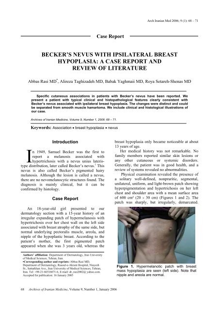
Physical examination revealed the presence of<br />
a solitary well-defined, nonpruritic, segmental,<br />
unilateral, uniform, and light-brown patch showing<br />
hyperpigmentation and hypertrichosis on her left<br />
chest and shoulder area with a mean surface area<br />
of 600 cm² (20 × 30 cm) (Figures 1 and 2). The<br />
patch was sharply, but irregularly, demarcated.<br />
Figure 1. Hypermelanotic patch with breast<br />
mass hypoplasia are seen (left side). Note that<br />
nipple and areola are normal.
Figure 2. A hypermelanotic patch with welldefined<br />
and irregular border is seen.<br />
There were no pustules, vesicles, perifollicular<br />
papular elevations, or indurations. The lesion did<br />
not show transient elevations on rubbing. Over the<br />
time, hypertrichosis gradually developed within it.<br />
There was no associated hypoplasia of underlying<br />
structures and asymmetry of the limbs.<br />
The patient’s blood pressure was 110/80<br />
mmHg. Radiographic studies of the entire spine<br />
and upper limbs did not show any alterations. The<br />
results of routine blood tests and urine analysis<br />
were within normal limits. The following hormone<br />
examinations were all within normal values: free<br />
testosterone, dihydrotestosterone, dehydroepiandrosterone,<br />
prolactin, follicular stimulating<br />
hormone, luteinizing hormone, and thyroid<br />
hormones. A biopsy specimen of the left chest<br />
pigmented lesion showed: hyperkeratosis,<br />
increased basal layer pigmentation, regular<br />
elongation, and clubbing of rete ridges (elongated<br />
Figure 3. A smooth muscle bundle independent<br />
of the hair follicle is seen (arrow).<br />
A. Rasi, A. Taghizadeh, B. Yaghmaii, et al<br />
rete ridges had flat tips). In the dermis, there was<br />
not any marked increase in smooth muscle fibers,<br />
but only one bundle of pilar smooth muscles was<br />
present, and no nevus cells were observed (Figure<br />
3). Examination with trichrome stain demonstrated<br />
no increase in smooth muscle fibers.<br />
On the basis of clinical and histopathologic<br />
findings, a diagnosis of Becker’s nevus associated<br />
with ipsilateral breast hypoplasia was made.<br />
Discussion<br />
Becker’s nevus is a unilateral (rarely bilateral)<br />
solitary, macular light brown hyperpigmentation of<br />
varying size, with geographic borders. The border<br />
is irregular and sharply demarcated. Occasionally,<br />
lesions resembling acne vulgaris, including<br />
papules, pustules, and cystic nodules, may occur<br />
within the affected area. 2 Over time, hypertrichosis<br />
(which is a characteristic feature of the lesion),<br />
develops within the lesion in about 50% of cases. 3<br />
Hairs, which are confined to the hyperpigmented<br />
area, are darker and coarser than normal. The<br />
lesion varies in size and may cover the entire upper<br />
arm or shoulder. A typical lesion is about 125 cm 2 ,<br />
although lesions up to 500 cm² have been reported.<br />
These lesions are usually asymptomatic and are<br />
usually localized on the shoulder, anterior chest, or<br />
upper arms, but there have been reports in other<br />
areas (e.g., proximal upper extremities, lower<br />
extremities, face, forearm, wrist, neck, and upper<br />
and lower back). 2<br />
In a study of 19,302 men aged 17 to 26 years, a<br />
prevalence of 0.52% was observed. 3 Reports of this<br />
condition in women are much rarer in the<br />
literature. The male-to-female ratio, approximated<br />
from case studies, has been reported to be<br />
anywhere from 4:1 to 6:1. 4, 5 There is no racial<br />
predilection, but it is more common in young<br />
people with fair skin. 6 Becker’s nevus is usually<br />
acquired in adolescence (the majority of cases are<br />
first noticed sharply before, at, or after puberty),<br />
but a congenital onset has been recorded, 7 as have<br />
familial cases in siblings, and in an uncle and a<br />
nephew.<br />
The lesion is a developmental anomaly, but<br />
occasionally, lesions have been said to follow<br />
severe sunburn. Lesional tissue has been found to<br />
have an increased level of androgen receptors,<br />
suggesting that heightened local androgen<br />
sensitivity may result in the hypertrichosis.<br />
Many abnormalities have been associated with<br />
Archives of Iranian Medicine, Volume 9, Number 1, January 2006 69
Becker’s nevus such as:<br />
• Smooth muscle hamartoma. This is the most<br />
frequent finding. In such cases, the area of<br />
Becker’s nevus may exhibit slight<br />
perifollicular papular elevations or slight<br />
induration. 8<br />
• Unilateral breast hypoplasia. This is possibly<br />
70<br />
as a result of enhanced androgen sensitivity.<br />
• Hypoplasia of underlying structures and<br />
asymmetry of limbs. 10<br />
Other abnormalities include: various skeletal<br />
malformations, connective tissue nevus, 9 aplasia of<br />
ipsilateral pectoralis major muscle, ipsilateral limb<br />
shortening, localized lipoatrophy, spina bifida,<br />
scoliosis pectus carinatum, congenital adrenal<br />
hyperplasia, and accessory scrotum. 2<br />
Unless associated with a hamartomatous<br />
process, histologic changes in Becker’s nevus are<br />
minimal. The epidermal changes are variable, but<br />
usually there is hyperkeratosis, subtle acanthosis,<br />
papillomatosis, 8 and basal layer hyperpigmentation.<br />
There are regular elongation of rete<br />
ridges, and elongated rete ridges tend to have flat,<br />
rather than pointed tips. Melanocyte proliferation<br />
is usually mild and not always obvious in routine<br />
sections; this increase is particularly evident when<br />
melanocytes are stained for dopa-oxidase activity<br />
in both involved and uninvolved skin nearby. 8 The<br />
dermis is thickened and contains numerous, but<br />
often inconspicuous bundles of smooth muscle<br />
fibers, unrelated to hair follicles or blood vessels. 8<br />
Controversy exists about the relationship of these<br />
cases to smooth muscle hamartoma. Some authors<br />
believe that, in cases associated with smooth<br />
muscle hamartoma, irregularly arranged, thick<br />
bundles of smooth muscle are present in the<br />
dermis. 8 There may also be an underlying increase<br />
in the number and size of hair follicles and<br />
sebaceous glands. 11 Some melanophages are seen<br />
in the dermis. 1 There should be no problem in<br />
making the diagnosis in a well-developed Becker’s<br />
nevus. In early lesions, the age of onset and,<br />
generally, the site are perhaps the most helpful<br />
diagnostic features.<br />
Clinically, congenital Becker’s nevus may be<br />
confused with a congenital melanocytic nevus.<br />
Histologically, Becker’s nevus does not have<br />
nevocellular nevus cells.<br />
Albright’s syndrome may be easily confused<br />
with Becker’s nevus, but the pigmented macule in<br />
Albright’s syndrome is present at birth.<br />
Theoretically, Becker’s nevus and smooth<br />
Archives of Iranian Medicine, Volume 9, Number 1, January 2006<br />
2, 9<br />
Becker’s nevus with ipsilateral breast hypoplasia<br />
muscle hamartoma are two quite separate entities.<br />
In fact, there are cases which could be considered<br />
as intermediate. As a matter of fact, a slight<br />
underlying smooth muscle hyperplasia can be seen<br />
in Becker’s nevus; on the other hand,<br />
hypermelanosis of basal layer and hypertrichosis<br />
may be encountered in smooth muscle hamartoma.<br />
Both conditions can be considered polar forms of a<br />
spectrum of dermal smooth muscle hyperplasia. 12,<br />
13 Becker’s nevus may enlarge slowly a year or two<br />
after presentation and then stabilizes and appears<br />
to persist indefinitely. The hyperpigmentation<br />
usually remains stable, although there have been<br />
reports of fading over many years. 4, 14 Becker’s<br />
nevus is usually too large to remove and is best left<br />
untouched. The hair may be shaved or permanently<br />
removed. The skin hyperpigmentation may<br />
respond to therapy with Q-switched ruby laser,<br />
although the results are unpredictable and<br />
recurrences are common.<br />
Although Becker’s nevus is most common in<br />
males, ipsilateral breast hypoplasia in Becker’s<br />
nevus occurs infrequently. Cases associated with<br />
breast hypoplasia are more frequently reported in<br />
women. In fact, 13 cases have been reported in the<br />
literature in whom the onset of Becker’s nevus in<br />
the mammary area during the prepubertal age was<br />
followed by breast hypoplasia. 8, 9, 15 – 17 For this<br />
reason, the androgen skin receptors were<br />
examined, and the results revealed high levels of<br />
these receptors in the affected skin. 9 This explains<br />
the mammary hypoplasia of the specific cases, as<br />
well as the frequent hypertrichosis, the possible<br />
presence of acneiform lesions, and the thick dermis<br />
with which the nevus is associated. We thought it<br />
would be interesting to report this case, not only<br />
because of the rare association of breast hypoplasia<br />
with Becker’s nevus, but due to this case’s early<br />
age of onset and the large size of the lesion.<br />
References<br />
1 Becker SW. Concurrent melanosis and hypertrichosis in<br />
distribution of nevus unius lateris. Arch Dermatol Syph.<br />
1949; 60: 155 – 160.<br />
2 Rook A, Wilkinson DS, Ebling FJG, Champion RH.<br />
Rook/Wilkinson/Ebling Textbook of Dermatology. 6th ed.<br />
Oxford; Malden, MA: Blackwell Science; 1998;<br />
542 – 543.<br />
3 Tymen R, Forestier J-F, Boutel B, Colomb D. Nevus tardif<br />
Becker. A propos d, une serie de 100 observations. Ann<br />
Dermatol Venereol. 1981; 108: 41 – 46.<br />
4 Chima KN, Janniger CK, Schwartz RA. Becker’s<br />
melanosis. Cutis. 1996; 57: 311 – 314.<br />
5 Happle R, Koopman RJ. Becker nevus syndrome. Am J
Med Genet. 1997; 68: 357 – 361.<br />
6 Ballone E, Fazii P, Lappa G, Di Mascio R, Di Mascio C,<br />
Schinoppa F. Prevalence of Becker’s nevi in a population<br />
of young men in central Italy. J Am Acad Dermatol. 2003;<br />
48: 795.<br />
7 Picascia DD, Esterly NB. Congenital Becker’s melanosis.<br />
Int J Dermatol. 1989; 28: 127 – 128.<br />
8 Freedberg IM, Fitzpatrick TB. Fitzpatrick’s Dermatology<br />
in General Medicine. 5th ed. New York: Mc Graw-Hill,<br />
Health Profession Division; 1999: 880 – 881, 882, 903,<br />
994 – 995.<br />
9 Formigon M, Alsina MM, Mascara JM, Rivera F.<br />
Becker’s nevus and ipsilateral breast hypoplasia.<br />
Androgen receptor-study in two patients. Arch Dermatol.<br />
1992; 128: 992 – 993.<br />
10 Lucky AW, Saruk M, Lerner AB. Becker’s nevus<br />
associated with limb asymmetry. Arch Dermatol. 1981;<br />
117: 243.<br />
11 Chapel TA, Tavafoghi V, Mehregan AH, Gagliardi C.<br />
Becker’s melanosis: an organoid hamartoma. Cutis. 1981;<br />
A. Rasi, A. Taghizadeh, B. Yaghmaii, et al<br />
27: 405 – 415.<br />
12 De la Espriella J, Grossin M, Marinho E, Belaich S.<br />
Smooth muscle hamartoma. anatomoclinical<br />
characteristics and nosological limits [in French]. Ann<br />
Dermatol Venereol. 1993; 120: 879 – 883.<br />
13 Civatte J, Marinho E, Oliver-Santos R. Smooth muscle<br />
hamartoma or nevus of Becker? A propos of 4 cases [in<br />
Spanish]. Med Cutan Ibero Lat Am.1988; 16: 145 – 148.<br />
14 Kopf AW, Yuppa J. Becker’s nevus. Arch Dermatol.<br />
1968; 98: 97 – 98.<br />
15 Cambiaghi S, Brusasco A, Tadini G, et al. Nevo di Becker<br />
congenito con ipoplasia mammaria ipsilaterale. G Ital<br />
Dermatol Venereol. 1994; 129: 169 – 172.<br />
16 Sharma R, Mishra A. Becker’s nevus with ipsilateral<br />
areolar hypoplasia in three males. Br J Dermatol. 1997;<br />
136: 471 – 472.<br />
17 Hoon-Jung J, Chan-Kim Y, Joon-Park H, Woo-Cinn Y.<br />
Becker’s nevus with ipsilateral breast hypoplasia:<br />
improvement with spironolactone. J Dermatol. 2003; 30:<br />
154 – 156.<br />
Archives of Iranian Medicine, Volume 9, Number 1, January 2006 71