Principal Component Analysis for Detection and ... - IEEE Xplore
Principal Component Analysis for Detection and ... - IEEE Xplore
Principal Component Analysis for Detection and ... - IEEE Xplore

Create successful ePaper yourself
Turn your PDF publications into a flip-book with our unique Google optimized e-Paper software.
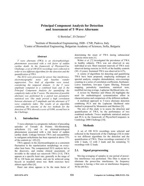
<strong>Principal</strong> <strong>Component</strong> <strong>Analysis</strong> <strong>for</strong> <strong>Detection</strong><br />
<strong>and</strong> Assessment of T-Wave Alternans<br />
G Bortolan 1 , II Christov 2<br />
1 Institute of Biomedical Enginnnering, ISIB - CNR, Padova, Italy<br />
2 Centre of Biomedical Engineering, Bulgarian Academy of Sciences, Sofia, Bulgaria<br />
Abstract<br />
T wave alternans (TWA) is an electrophisiologic<br />
phenomenon associated with a risk factor of sudden<br />
cardiac death. In the framework of Physionet/Cinc<br />
Challenge, a set of 100 ECG recordings were collected in<br />
order to test different algorithms <strong>for</strong> the detection <strong>and</strong> the<br />
quantification of TWA.<br />
The ECG were processed <strong>for</strong> power line interference,<br />
electromyographic noise <strong>and</strong> baseline w<strong>and</strong>er<br />
suppression. Two kind of algorithm were tested,<br />
considering the temporal domain: 1) the T wave<br />
amplitude computed in a combined lead, <strong>and</strong> 2) the<br />
<strong>Principal</strong> <strong>Component</strong> <strong>Analysis</strong> <strong>for</strong> quantifying the<br />
complexity index of the T waves. The detection of possible<br />
alternans was per<strong>for</strong>med by a paired non parametric<br />
statistical test. Our study proved a high correlation<br />
between alternans of T amplitude <strong>and</strong> the alternans of T<br />
wave complexity index. The results of an algorithm<br />
combining the outcome of the two methods <strong>for</strong> the<br />
detection of TWA showed a score of 0.890, second best in<br />
the Challenge.<br />
1. Introduction<br />
T-wave alternans is a prognostic indicator of preceding<br />
episodes of Torsade de Pointes life-threatening<br />
arrhythmia [1], <strong>and</strong> is an electrophysiological<br />
phenomenon associated with a risk factor of sudden<br />
cardiac death. Linkage between TWA <strong>and</strong> susceptibility<br />
to malignant arrhythmias has been reported at above 75%<br />
of the controls (p
A smoothing procedure <strong>for</strong> EMG noise suppression is<br />
applied [11]. It uses the least-squares approximation<br />
method, applied <strong>for</strong> defining the weighting coefficients<br />
<strong>for</strong> each sample of the selected smoothing interval of 60<br />
ms.<br />
A high-pass recursive filter <strong>for</strong> drift suppression with a<br />
cutoff frequency of 0.64 Hz has been used [12].<br />
QRS detection using combined adaptive thresholding<br />
was applied [13].<br />
T wave onset <strong>and</strong> offset delineations were<br />
automatically per<strong>for</strong>med [14], <strong>and</strong> the T amplitude was<br />
calculated, in a combined lead simulating the spatial<br />
vector [12] or another all-inclusive signal, rather than in<br />
the separate leads. The trans<strong>for</strong>m to the orthogonal leads<br />
(X,Y,Z) was per<strong>for</strong>med in [12] using ‘primary leads’, i.e.<br />
the 8 potential differences referred to the electrode of the<br />
left leg F. These primary leads were obtained from the<br />
12-lead ECG recordings, following the conversion<br />
<strong>for</strong>mulae in the [15]:<br />
R F = -II; L F = -III; Ci F = Vi – (II+III)/3<br />
The orthogonal leads were evaluated by:<br />
X=0.5*abs(C4 F -C1 F );<br />
Y=abs(R F );<br />
Z=abs(R F -C2 F );<br />
The combined lead (CL), which is the spatial vector in<br />
this case, is calculated by:<br />
CL=0.5(X+Y+Z+0.25(abs(X-Y)+abs(X-Z)+abs(Y-Z)));<br />
In cases of 3-leads ECG: X=Lead1; Y=Lead2;<br />
Z=Lead3.<br />
In cases of 2-leads ECG: X=Lead1; Y=Lead2; Z=0.<br />
global <strong>and</strong> local (see Figure 1). In the global method, the<br />
entire ECG recording was processed, producing a unique<br />
time series, which feed the detection block. The local<br />
method considers a set of variable length windows of RR<br />
intervals, <strong>and</strong> per<strong>for</strong>ming the parameter extraction in each<br />
of them. In particular this method considers a window of<br />
128 RR intervals, shifting it in the entire ECG recording.<br />
The detection block per<strong>for</strong>ms first, the separation of<br />
the parameters from odd <strong>and</strong> even RR intervals, <strong>and</strong> then<br />
the two series was compared in order to discover the<br />
presence of significative differences. The statistical<br />
analysis which provides the significance of the<br />
differences between odd <strong>and</strong> even samples was<br />
per<strong>for</strong>med by the non parametric paired-sampled<br />
Wilcoxon signed rank test.<br />
In the case of Global methods, the statistical test<br />
produces a single binary index, which represents the<br />
presence or absence of TWA. In the case of Local<br />
methods, this process is repeated <strong>for</strong> every interval,<br />
producing a set of binary indices, <strong>and</strong> the number of<br />
intervals with positive indices is considered. In case it is<br />
higher/lower of a predetermined threshold, it signifies the<br />
presence/absence of TWA.<br />
Global methods<br />
2.2. TWA detection<br />
The proposed approach takes into consideration three<br />
aspects of the TWA detection task: the parameter<br />
selection, the interval selection <strong>and</strong> the classification<br />
bock.<br />
In selecting the more appropriate parameter <strong>for</strong> TWA<br />
classification, the multi-lead approach has been followed,<br />
in order to extract a single index from the entire ECG<br />
record. Two parameters were chosen, considering the<br />
temporal domain: 1) the amplitude <strong>and</strong> 2) the complexity<br />
index of T waves. In the first case, a combined lead was<br />
determined with all the leads at disposal (2, 3 or 12),<br />
simulating the spatial vector, in which T wave onset <strong>and</strong><br />
offset delineations were per<strong>for</strong>med, <strong>and</strong> the amplitudes<br />
were computed.<br />
The second parameter <strong>for</strong> TWA discrimination<br />
considers the use of <strong>Principal</strong> <strong>Component</strong> <strong>Analysis</strong><br />
(PCA) <strong>for</strong> quantifying the complexity index of the T<br />
waves. In particular the PCA has been applied to the<br />
intervals of T waves, <strong>and</strong> the ratio of 2nd/1st eigenvalues<br />
characterizes the complexity index.<br />
In the interval selection two methods were applied:<br />
Local methods<br />
Fig.1. Methods <strong>for</strong> the interval selection <strong>for</strong> the<br />
detection of TWA: global (entire record) <strong>and</strong> local (a set<br />
of windows)<br />
All the RR intervals were analyzed, independently by<br />
the presence of noise or artifact. In addition, the heart rate<br />
was not considered <strong>for</strong> the determination of the<br />
presence/absence of TWA.<br />
3. Results<br />
The Computers in Cardiology Challenge made<br />
available a score system in order to rank the various<br />
methods. The score takes values in the range -1, +1.<br />
522
Five methods have been tested <strong>for</strong> the detection of<br />
TWA:<br />
- GLB1: a global index considering the alternans of the<br />
T amplitude on the entire ECG recording<br />
- GLB2: a global index considering the PCA index on<br />
the entire recording<br />
- LOC1: a local index considering the alternans of the<br />
T amplitude in various windows of 128 RR intervals<br />
- LOC2: a local index considering the alternans of the<br />
PCA index in the individual intervals<br />
- COMB: an index combining the previous indices,<br />
<strong>and</strong> requiring essentially a positive test in both the global<br />
<strong>and</strong> the local in dices.<br />
Table 1 reports a cross tabulation of the number of<br />
records with the presence of TWA with the 5 considered<br />
methods.<br />
Table 2 reports the percentage of agreement in the<br />
detection results between all the combinations of two<br />
methods, while Table 3 reports the correlation coefficient<br />
between them.<br />
Table 1. Number of TWA determined by the 5 methods:<br />
Comb (Combined), GLB1 (Global T amplitude), .GLB2<br />
(Global PCA_T), LOC1 (Local T amplitude), <strong>and</strong> LOC2<br />
(Local PCA_T).<br />
Comb GLB1 GLB2 LOC1 LOC2<br />
Comb 25<br />
GLB1 25 29<br />
GLB2 20 20 28<br />
LOC1 23 27 18 31<br />
LOC2 19 19 20 17 24<br />
Table 2. Agreement between the 5 different methods of<br />
TWA detection: Comb (Combined), GLB1 (Global T<br />
amplitude), .GLB2 (Global PCA_T), LOC1 (Local T<br />
amplitude), <strong>and</strong> LOC2 (Local PCA_T).<br />
Comb GLB1 GLB2 LOC1 LOC2<br />
Comb 100%<br />
GLB1 96% 100%<br />
GLB2 87% 83% 100%<br />
LOC1 90% 94% 77% 100%<br />
LOC2 89% 85% 88% 79% 100%<br />
Our study proved a high correlation between alternans<br />
of T amplitude (GLB1 <strong>and</strong> LOC1) <strong>and</strong> the alternans of T<br />
wave complexity index (GLB2 <strong>and</strong> LOC2). For example<br />
the two indices were in agreement in 83% <strong>and</strong> 79% in the<br />
global <strong>and</strong> local detection respectively.<br />
Table 4 shows the Challenge scores of the 5 methods.<br />
The Combined method reach the highest score, 0.890,<br />
which is the second best in the Challenge. It turns out that<br />
all the tested methods reach a higher score than 0.800,<br />
which is a satisfiable per<strong>for</strong>mance. In addition, in the<br />
Table 3. Correlation coefficient between the 5 different<br />
methods of TWA detection: Comb (Combined), GLB1<br />
(Global T amplitude), .GLB2 (Global PCA_T), LOC1<br />
(Local T amplitude), <strong>and</strong> LOC2 (Local PCA_T).<br />
Comb GLB1 GLB2 LOC1 LOC2<br />
Comb 1.00<br />
GLB1 0.90 1.00<br />
GLB2 0.67 0.58 1.00<br />
LOC1 0.70 0.80 0.40 1.00<br />
LOC2 0.66 0.56 0.63 0.46 1.00<br />
Table 4. Scores of the 5 methods of TWA detection:<br />
Comb (Combined), GLB1 (Global T amplitude), .GLB2<br />
(Global PCA_T), LOC1 (Local T amplitude), <strong>and</strong> LOC2<br />
(Local PCA_T).<br />
Method<br />
Score<br />
Comb 0.890<br />
GLB1 0.861<br />
GLB2 0.802<br />
LOC1 0.831<br />
LOC2 0.812<br />
Table 5.List of records with positive TWA detection with<br />
the Combined (Comb) method.<br />
twa01 twa32 twa73<br />
twa06 twa33 twa79<br />
twa09 twa34 twa82<br />
twa13 twa50 twa88<br />
twa15 twa51 twa91<br />
twa17 twa64 twa97<br />
twa21 twa67 twa98<br />
twa25 twa69<br />
twa29 twa72<br />
25<br />
20<br />
15<br />
10<br />
5<br />
0<br />
40 60 80 100 120 140<br />
Heart Rate<br />
Figure 2. Histogram of heart rate in patients with (blue<br />
bars) <strong>and</strong> without (red bars) the presence of TWA<br />
determined by the Combined (Comb) method.<br />
523
case of T amplitude, the global method achieves a better<br />
result than the local method, while the contrary is true <strong>for</strong><br />
the PCA index. Table 5 reports the list of all the ECG<br />
records with the presence of TWA considering the<br />
Combined method, <strong>and</strong> Figure 2 shows the histogram of<br />
the heart rate.<br />
4. Discussion <strong>and</strong> conclusions<br />
The task of TWA detection has been studied<br />
considering two parameters in the time domain: the T<br />
wave amplitude computed in a combined lead, <strong>and</strong> the<br />
complexity index of the T waves. The classification has<br />
been per<strong>for</strong>med analyzing the entire ECG records or a set<br />
of intervals. The results show that the combined<br />
algorithm has the best per<strong>for</strong>mance, as it could be<br />
expected in general. The parameters considering the T<br />
amplitude have a higher per<strong>for</strong>mance compared to the T<br />
wave complexity index based on PCA. However, the two<br />
parameters showed a good agreement between them, <strong>and</strong><br />
there was less score differences in the local methods.<br />
Acknowledgements<br />
The authors are greatly indebted <strong>for</strong> personal communications<br />
about the physiological aspects of TWA to:<br />
Velislav Batchvarov, MD, PhD, Department of<br />
Cardiac <strong>and</strong> Vascular Sciences, St. George’s University<br />
of London, London, United Kingdom<br />
Iana Simova, MD, Clinic of Cardiology, University<br />
Hospital ‘Aleks<strong>and</strong>rovska’, Medical University, Sofia,<br />
Bulgaria<br />
References<br />
[1] Surawicz B, Fisch C. Cardiac alternans: diverse<br />
mechanisms <strong>and</strong> clinical manifestations. J Am Coll Cardiol<br />
1992;20(2):483–99.<br />
[2] Verrier R. Ambulatory ECG monitoring of T-Wave<br />
alternans <strong>for</strong> arrhythmia risk assessment. J of Electrocard.<br />
2003;36:193-7.<br />
[3] Adam DR, Akselrod S, Cohen RJ. Estimation of<br />
ventricular vulnerability to fibrillation through T-wave<br />
time series analysis. Comp. in Card. 1981;8:307-10<br />
[4] Adam DR, Powell AO, Gordon H, Cohen RJ. Ventricular<br />
fibrillation <strong>and</strong> fluctuations in the magnitude of the<br />
repolarizacion vector. Comp. in Card. 1982;9:241-4.<br />
[5] de Luna AB, Batchvarov VN, Malik M. The morphology<br />
of the electrocardiogram. In: Camm AJ, Lüscher TF,<br />
Serruys T, editors. The ESC Textbook of Cardiovascular<br />
Medicine Blackwell Publishing, 2006: 1-35.<br />
[6] Hohnloser SH, Klingenheben T, Zavel M et al. T wave<br />
alternans during exercise <strong>and</strong> atrial pacing in humans. J<br />
Cardiovasc Electrophysiol 1997;8:987-93.<br />
[7] Weber S, Tillmanns H, Waldecker B. Prevalence of T<br />
wave alternans in healthy subjects. Pacing <strong>and</strong> Clin.<br />
Electrophys. 2003;25:49-51.<br />
[8] Martínez JP, Olmos S. Methodological principles of T<br />
wave alternans analysis: A unified framework. <strong>IEEE</strong><br />
Trans. on Biom. Eng. 2005;52:599-613.<br />
[9] Monasterio V, Martínez JP. A multilead approach to T-<br />
wave alternans detection combining principal component<br />
analysis <strong>and</strong> the Laplacian likelihood ratio method. Comp.<br />
in Card. 2007;34:5−8.<br />
[10] The PhysioNet / Computers in Cardiology Challenge 2008:<br />
Detecting <strong>and</strong> Quantifying T-Wave Alternans. Comp in<br />
Card 2008;35: http://physionet.org/challenge/2008/<br />
[11] Christov I, Daskalov IK. Filtering of electromyogram<br />
artifacts from the electrocardiogram. Med. Eng. & Phys.<br />
1999;21:731-36.<br />
[12] Daskalov IK, Dotsinsky IA, Christov II. Developments in<br />
ECG acquisition preprocessing parameter measurement<br />
<strong>and</strong> recording. <strong>IEEE</strong> Eng. in Med. & Biol. 1998;17:50-8.<br />
[13] Christov II. Real time electrocardiogram QRS detection<br />
using combined adaptive threshold. Biomed. Eng. Online<br />
2004;3(28) http://www.biomedicalengineeringonline.com/content/3/1/28.<br />
[14] Christov I, Simova I. Q-onset <strong>and</strong> T-end delineation:<br />
Assessment of the per<strong>for</strong>mance of an automated method<br />
with the use of a reference database. Physiol. Meas.<br />
2007;28(2):213-21.<br />
[15] Dotsinsky I, Christov I, Daskalov I. Twelve-lead<br />
electrocardiogram obtained by eight channels.<br />
Elektrotechnica & Elektronica E+E. 2002;1-2:10-2<br />
http://www.clbme.bas.bg/pwp/Ivaylo_Christov/Publication<br />
s/Leads_2002_ee.pdf<br />
Address <strong>for</strong> correspondence<br />
Giovanni Bortolan<br />
ISIB-CNR<br />
Corso Stati Uniti, 4<br />
35129 Padova , Italy<br />
bortolan@isib.cnr.it<br />
524