ABSTRACTS â ORAL PRESENTATIONS - AMCA, spol. s r.o.
ABSTRACTS â ORAL PRESENTATIONS - AMCA, spol. s r.o.
ABSTRACTS â ORAL PRESENTATIONS - AMCA, spol. s r.o.
You also want an ePaper? Increase the reach of your titles
YUMPU automatically turns print PDFs into web optimized ePapers that Google loves.
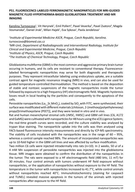
P51. FLUORESCENCE-LABELED FERROMAGNETIC NANOPARTICLES FOR MRI-GUIDED<br />
MAGNETIC FLUID HYPERTHERMIA-BASED GLIOBLASTOMA TREATMENT AND MR<br />
IMAGING<br />
Karolina Turnovcova 1 , Vit Herynek 2 , Emil Pollert 3 , Pavel Veverka 3 , Pavel Zvatora 4 , Magda<br />
Vosmanska 4 , Daniel Jirak 2 , Milan Hajek 2 , Eva Sykova 1 , Pavla Jendelova 1<br />
1<br />
Institute of Experimental Medicine ASCR, Prague, Czech Republic, karolina.<br />
turnovcova@biomed.cas.cz<br />
2<br />
MR-Unit, Department of Radiodiagnostic and Interventional Radiology, Institute for<br />
Clinical and Experimental Medicine, Prague, Czech Republic<br />
3<br />
Institute of Physics, ASCR, Prague, Czech Republic<br />
4<br />
The Institute of Chemical Technology, Prague, Czech Republic<br />
Glioblastoma multiforme (GBM) is the most common and aggressive primary brain tumor<br />
occuring in humans, and its cells are resistant to conventional therapy. Fluorescencelabeled<br />
ferromagnetic nanoparticles may serve for both diagnostic and therapeutic<br />
purposes. They represent intracellular labeling using endocytosis uptake, are a suitable<br />
contrast agent for magnetic resonance imaging (MRI) in vivo, and can also be used for<br />
guided temperature-controlled thermoablation. The method is based on the deposition<br />
of stable and nontoxic suspensions of the magnetic nanoparticles inside the tumor<br />
followed by exposure to a high frequency (HF) electromagnetic field. Magnetic hysteresis<br />
losses result in local heating by the particles and consequently to the apoptosis of the<br />
cells.<br />
Perovskite nanoparticles (La 1-x<br />
Sr x<br />
MnO 3<br />
), coated by SiO 2<br />
with FITC, were synthesized, their<br />
surface was modificated with different materials [chitosan, 2-[methoxy(polyethyleneoxy)<br />
propyl]trimethoxysilane (PEET)], and they were tested in vitro and in vivo.<br />
Rat and human mesenchymal stromal cells (rMSC, hMSC) and GBM cell lines (C6, A172<br />
and GaMG) were cultivated with nanoparticles for 48 hours using the xCELLigence System;<br />
the real-time growth curves were recorded, and the culture viability was analysed by<br />
trypan blue staining. The nanoparticle uptake into the cells was then analysed using<br />
FACS-based fluorescence intensity measurements and directly by ICP-MS spectrometry.<br />
The viability of cells incubated with the nanoparticles was in the range of 60 – 95%,<br />
whereas a control sample reached 98%. The amount of incorporated nanoparticles was<br />
up to 10-fold higher in the GBM cell lines and was affected by surface modification.<br />
Two million C6 cells were injected intradermally into rats (n=10). In 2 weeks, 50 ul of a<br />
4 mM Mn suspension of perovskite nanoparticles was injected into the glioblastoma<br />
tissue. MRI images were obtained to confirm the distribution of the nanoparticles in<br />
the tumor. The rats were exposed to a HF electromagnetic field (480 kHz, 11 mT) for<br />
30 minutes. Four control animals with tumors underwent HF field exposure without<br />
nanoparticles, or the application of nanoparticles without exposure to the HF field. The<br />
temperature in the tumor with nanoparticles increased to 41.7°C, while control animals<br />
without nanoparticles reached 40°C. Immunohistochemistry (staining for caspase3<br />
and TUNEL) revealed massive apoptosis in the tumors of the animals with injected<br />
nanoparticles after exposure to the HF field.<br />
148 Analytical Cytometry VII


