ABSTRACTS â ORAL PRESENTATIONS - AMCA, spol. s r.o.
ABSTRACTS â ORAL PRESENTATIONS - AMCA, spol. s r.o.
ABSTRACTS â ORAL PRESENTATIONS - AMCA, spol. s r.o.

Create successful ePaper yourself
Turn your PDF publications into a flip-book with our unique Google optimized e-Paper software.
<strong>ABSTRACTS</strong> – <strong>ORAL</strong> <strong>PRESENTATIONS</strong><br />
14. FLOW CYTOMETRY AND PLANT GENOMICS: A HAPPY MARRIAGE<br />
Jaroslav Doležel, Hana Šimková, Jan Šafář, Jan Vrána, Petr Cápal, David Kopecký,<br />
Veronika Burešová, Jarmila Číhalíková, Marie Kubaláková, Jan Bartoš<br />
Centre of the Region Haná for Biotechnological and Agricultural Research, Institute of<br />
Experimental Botany AS CR, Šlechtitelů 31, CZ-78371 Olomouc-Holice, Czech Republic;<br />
Email: dolezel@ueb.cas.cz<br />
The use of flow cytometry in plant science has been largely limited to the analysis<br />
of ploidy, nuclear genome size, and cell cycle. Other applications are less frequent,<br />
probably due to difficulties with preparation of samples suitable for flow cytometry from<br />
plant tissues and cells with rigid cell walls. However, one application has been gaining<br />
a growing attention and that is the chromosome sorting, which turned out to be very<br />
useful in plant genome analysis. Genomics aims to sequence and analyze the structure<br />
and function of complete genomes. However, this may be difficult in a number of species,<br />
including important agricultural crops, whose genomes are large and composed mostly of<br />
repetitive DNA. Moreover, many species are recent polyploids and interspecific hybrids.<br />
Sequence redundancy and presence of homologous and homoeologous sequences<br />
hampers gene mapping and cloning, construction of physical maps and sequence<br />
assembly. These tasks can be simplified after reducing sample complexity by dissecting<br />
nuclear genomes to single chromosomes and chromosome arms. We have shown<br />
previously that it is possible to prepare suspensions of intact mitotic chromosomes from<br />
synchronized root tips. Following this, we have developed a portfolio of applications of<br />
flow-sorted plant chromosomes, that includes physical mapping using PCR and FISH,<br />
construction of BAC libraries to facilitate construction of physical maps, and targeted<br />
development of molecular markers to saturate genetic maps. The International Wheat<br />
Genome Sequencing Consortium choose chromosome based strategy to develop ready<br />
to sequence physical map of hexaploid bread wheat, whose genome is almost 6-fold<br />
larger than that of human. The advent of next generation sequencing technology opened<br />
avenues for rapid sequencing DNA of isolated chromosomes, providing easy access to<br />
DNA composition of chromosomes. For example, this approach enabled identification<br />
of most of genes in barley and establishment of their order along the seven barley<br />
chromosomes. Sequencing flow-sorted rye supernumerary B chromosomes clarified their<br />
molecular structure and evolution from rye A chromosomes. Until recently, the potential<br />
of chromosome sorting was limited by the inability to discriminate chromosomes<br />
with the same DNA amount. A solution was to use special cytogenetic stocks such as<br />
chromosome deletion, addition or translocation lines, which are not readily available<br />
in many species. The constraint was overcome after Giorgi and colleagues developed<br />
a method called FISHIS for fluorescent labeling of DNA sequences on chromosomes in<br />
suspension (PLoS ONE 8: e57994, 2013). Importantly, this method provides opportunity<br />
46 Analytical Cytometry VII
to employ flow cytometry for estimation of the number of particular DNA sequences on<br />
chromosomes of interest. The numerous and important contributions to the analysis<br />
of complex plant genomes document how fruitful and happy the marriage between<br />
flow cytometry and plant genomics has been. Considering the recent methodological<br />
advances, one may expect that this relationship will flourish even more than before.<br />
18. THE PLANAR CELL POLARITY PATHWAY DRIVES PATHOGENESIS OF CHRONIC<br />
LYMPHOCYTIC LEUKEMIA BY THE REGULATION OF B-LYMPHOCYTE MIGRATION<br />
Markéta Kaucká 1 , Šárka Pavlová 2,3 , Pavlína Janovská 1 , Karla Plevová 2,3 , Lucie Poppová 2,3 ,<br />
Hana Mádrová 2,3 , Jan Verner 2 , Jiřina Procházková 1 , Šárka Pospíšilová 2,3 , Alois Kozubík 1,4 ,<br />
Gunnar Schulte 1,5 and Vítězslav Bryja 1,4<br />
1<br />
Institute of Experimental Biology, Faculty of Science, Masaryk University, Kotlářská 2,<br />
611 37, Brno, Czech Republic; bryja@sci.muni.cz<br />
2<br />
Center of Molecular Biology and Gene Therapy, Department of Internal Medicine–<br />
Hematology and Oncology, University Hospital Brno and Medical Faculty MU, Brno,<br />
Czech Republic<br />
3<br />
CEITEC - Central European Institute of Technology, Masaryk University, Brno, Czech<br />
Republic<br />
4<br />
Department of Cytokinetics, Institute of Biophysics, Academy of Sciences of the Czech<br />
Republic, Brno, Czech Republic<br />
5<br />
Department of Physiology and Pharmacology, Karolinska Institutet, Stockholm, Sweden<br />
Chronic Lymphocytic Leukemia (CLL) is the most common form of leukemia in<br />
Western countries found in adults. CLL is characterized by monoclonal expansion and<br />
accumulation of functionally incompetent B lymphocytes. Most patients are diagnosed<br />
with no symptoms using a routine blood test that shows a high white blood cell count.<br />
CD5 + lymphocytes show impaired migration and often invade into such organs as<br />
lymph nodes, spleen, liver and bone marrow which results in disrupted organ function,<br />
hematopoiesis (anemia) and weaken immunity. The prognosis of patients with CLL varies<br />
widely at diagnosis and the pathology of CLL remains still unclear.<br />
In our study, we show that core components of Wnt/planar cell polarity (PCP) pathway<br />
are upregulated in B lymphocytes from patients suffering from CLL. PCP is highly<br />
conserved signaling machinery, which acts as key regulator of cell polarity and migration<br />
throughout evolution. Several players of the PCP pathway, such as Prickle1, Celsr1,<br />
Vangl2, casein kinase 1 (Ck1ε), Dvl2, Dvl3, Fzd3, Fzd7 and Wnt5a, are increased in CLL<br />
cells both on mRNA and protein levels. The expression levels of PCP genes and proteins<br />
often correlate with the aggressiveness of the disease and some of the PCP genes have<br />
prognostic value.<br />
Further, our analysis of the migratory capacity of CLL model cell line Mec1 and human<br />
primary CLL lymphocytes in the chemokine gradient (Transwell Assay) suggests that<br />
PCP pathway is required for migration of leukemic cells. We were able to decrease<br />
CLL lymphocyte migration using (i) Ror1 blocking antibody, (ii) CK1 inhibitor D4476<br />
Analytical Cytometry VII 47
and (iii) siRNA-mediated knockdown of Dvl2. These findings were confirmed by the in<br />
vivo analysis of CLL homing (using human primary CLL lymphocytes transplantations<br />
into immunodeficient NOD SCID gammaC-/- mice) following treatment with the Ror1<br />
blocking antibody and CK1-specific inhibitors. Moreover, using confocal microscopy<br />
and live cell imaging we demonstrate that PCP proteins are polarized in migrating Mec1<br />
cells in CCL19 chemokine gradient. Mec1 cells treated with CK1 inhibitor show impaired<br />
chemokine-driven migration in the Dunn Chamber system in comparison to untreated<br />
cells.<br />
In summary, our data demonstrate that Wnt/PCP pathway acts as the key regulator of<br />
impaired CLL migration and can serve as a potential target for CLL therapy.<br />
Acknowledgements<br />
This work was supported by grants from the Czech Science Foundation (301/11/0747),<br />
Ministry of Health of the Czech Republic (NT11217-5/2010, NS10439-3/2009,<br />
FNBr 65269705), Masaryk University (MUNI/A/0723/2012) and European Regional<br />
Development Fund (CZ.1.07/2.3.00/20.0180, CZ.1.05/1.1.00/02.0068).<br />
24. ANALYSIS OF DNA REPAIR FOCI BY FLOW CYTOMETRY AND MICROSCOPY AFTER<br />
IRRADIATION TO LOW-DOSE IONIZING RADIATION<br />
Matus Durdik 1 , Jan Gursky 1 , Lenka Vokalova 1 , Miroslav Kubes 2 , Eva Markova 1 , Igor<br />
Belyaev 1<br />
1<br />
Cancer Research Institute, Bratislava, Slovak Republic; matusdurdik17@gmail.com<br />
2<br />
Eurocord-Slovakia, Bratislava, Slovak Republic<br />
It is known that DNA double strand breaks (DSB) is the most severe DNA damage involved<br />
in many effects of ionizing radiation and other genotoxic factors. Nowadays, the most<br />
common method for DSB analysis is enumeration of DNA repair foci, which are formed<br />
at the locations of DSB. Phosphorylated histone H2AX variant (γH2AX) and p53 binding<br />
protein 1 (53BP1) are established as appropriate molecular markers for DSB (Bekker-<br />
Jensen et al., 2006).<br />
Emerging evidence suggests that irradiation in low doses increases cancer risks<br />
(Wakeford, 2013). Exposure to low doses of ionizing radiation in the range of 1 mGy<br />
-5 cGy commonly occurs at medical examinations, like RTG or CT, in airplanes during<br />
ordinary flights, or at security controls. Radiosensitive subjects, which represent<br />
approximately 5% of patients irradiated with therapeutical doses of ionizing radiations,<br />
may be especially sensitive to low doses (Goodarzi and Jeggo, 2012). In different studies,<br />
it was reported that doses about 1cGy induce DSB measured with DNA repair foci<br />
(Belyaev, 2010). The main problem in analysis of low-dose effects is poor reproducibility<br />
of data in various laboratories and lack of sensitive standardized methods. In previous<br />
experiments, analysis of γH2AX by flow cytometry was not as sensitive as microscopic<br />
analysis.<br />
We analyzed ionizing radiation induced foci (IRIF) foci, 53BP1 and γH2AX, foci induced by<br />
low dose gamma-rays (2, 5, 10, 20, 50cGy) in lymphocytes from umbilical cord blood using<br />
48 Analytical Cytometry VII
two sophisticated systems: Metafer (Metasystems, Germany) and ImageStream (Amnis,<br />
USA). Metafer is automatized microscopic system that has recently been introduced<br />
for analyzing DNA repair foci. ImageStream is a combination of flow cytometer with<br />
fluorescent microscope. Thus, we compare ImageStream with Metafer in analyzing<br />
IRIF. We found that sensitivity of both systems to low-dose-induced γH2AX foci was<br />
very similar: ImageStream show statistical significant induction at the dose of 5cGy and<br />
Metafer at the dose of 10cGy while prior analysis using classical flow cytometry showed<br />
statistical significance at doses ≥ 50cGy. Analysis of 53BP1 foci by ImageStream was less<br />
sensitive then by Metafer.<br />
In conclusion, we show for the first time that analysis of low-dose-induced γH2AX foci by<br />
ImageStream system is very appropriate method, that reach sensitivity of automatized<br />
Metafer microscopic system and is more sensitive than measuring γH2AX fluorescence<br />
by classical flow cytometry. Thus, ImageStream is the system that has great potential for<br />
analyzing DNA repair foci.<br />
Acknowledgements<br />
This work was supported by the Slovak Research and Development Agency (APVV 0669-<br />
10) and the VEGA Grant Agency (2/0150/11) of the Slovak Republic.<br />
References<br />
Bekker-Jensen, S., Lukas, C., Kitagawa, R., Melander, F., Kastan, M. B., Bartek, J. and Lukas,<br />
J.: Spatial organization of the mammalian genome surveillance machinery in response to<br />
DNA strand breaks. - Journal of Cell Biology 173, 195-206, 2006.<br />
Belyaev, I. Y.: Radiation-induced DNA repair foci: Spatio-temporal aspects of formation,<br />
application for assessment of radiosensitivity and biological dosimetry. - Mutation<br />
Research-Reviews in Mutation Research 704, 132-141, 2010.<br />
Goodarzi, A. A. and Jeggo, P. A.: Irradiation induced foci (IRIF) as a biomarker for<br />
radiosensitivity. - Mutat Res 736, 39-47, 2012.<br />
Wakeford, R.: The risk of childhood leukaemia following exposure to ionising radiation--a<br />
review. - J Radiol Prot 33, 1-25, 2013.<br />
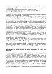
Legend to figure<br />
Typical data obtained by ImageStream<br />
after irradiation with the dose of 50cGy<br />
(transformed in black and white).<br />
There are 6 channels shown, from the<br />
left: brightfield, γH2AX stained with<br />
monoclonal mouse antibody and FITC.,<br />
granularity, DNA stained with DAPI, 53BP1<br />
stained with polyclonal rabbit antibody<br />
and AF647 and colocalization of γH2AX<br />
and 53BP1.<br />
Analytical Cytometry VII 49
25. PROFILING OF CHILDHOOD ACUTE LEUKAEMIA CELLS USING A NOVEL FLOW<br />
CYTOMETRY-BASED METHOD OF AFFINITY PROTEOMICS<br />
Daniela Černá 1 , Veronika Kanderová 1 , Jan Stuchlý 1 , Karel Fišer 1 , WeiWei Wu 2 , Anders<br />
Holm 2 , Ondřej Hrušák 1 , Fridtjof Lund-Johansen 2 , Tomáš Kalina 1<br />
1<br />
Pediatric Hematology and Oncology, Charles University Prague, 2 nd Medical Faculty,<br />
Praha 5, Czech Republic; cerna.daniela@lfmotol.cuni.cz<br />
2<br />
Dpt. of Haematology, Rikshospitalet, Oslo, 02770, Norway<br />
Acute leukaemia (AL) is the most common childhood malignancy driven by a number<br />
of aberrations at the DNA and mRNA level. Current research is mainly focused on<br />
the detection of mutations at the DNA level while functional consequences of these<br />
alterations on cellular level are not fully understood. Proteins are entities that form<br />
connection between gene expression and cell physiology, therefore more effective and<br />
sensitive approaches that could search for new prognostic markers on protein level are<br />
in development.<br />
Using SEC-MAP technology (Size-exclusion Chromatography - Microsphere-based Affinity<br />
Proteomics), we are searching for the expression and activation (e.g. phosphorylation) of<br />
differentially expressed proteins in AL cells regarding to cell type (B-cell, T-cell, myeloid),<br />
genotype (fusion genes, aneuploidy) and early response to treatment detected as<br />
minimal residual disease.<br />
SEC-MAP is a set of 1728 populations of fluorescently-labeled latex microbeads, each<br />
carrying an antibody against a human protein. We isolate the cellular proteins from<br />
membranes, cytoplasm and nuclei using detergents, label them with biotin and separate<br />
them using gel chromatography into 24 fractions. These fractions are incubated with<br />
SEC-MAP microbeads and the antibody-protein binding is detected using fluorescentlylabeled<br />
streptavidin by flow cytometry.<br />
We have examined the expression of cytoplasmic (n=980) and membrane (plus DNAbinding)<br />
(n=769) proteins in 69 diagnostic samples of AL. The analysis was performed<br />
using automatic software created in R-project. For the normalization of protein<br />
expression we have used Loess normalization commonly used in mRNA profiling studies.<br />
Due to ability of SEC-MAP to separate proteins according their molecular weight we have<br />
identified not only the expression of particular proteins but also the size that could serve<br />
as a control of proteolysis. We have detected proteolysis in 12 samples, which have been<br />
therefore excluded from the analysis. We have revealed the sensitivity to proteolysis of<br />
four standard house-keeping proteins (Akt, Abl, β-actin and β2-microglobulin). Abl and<br />
Akt have been cleaved while β-actin and β2-microglobulin have not been detected in<br />
their cleaved forms in the diagnostic AL samples. Therefore we have decided to use Abl<br />
and Akt as house-keeping proteins to reveal the proteolysis. So far we have identified 44<br />
proteins (e.g. SH2D1A, FAS, LAT, KIT, CD72) differentially expressed in different subtypes<br />
of AL. We have discovered e.g. higher expression of cAMP-dependent protein kinase<br />
PRKACA in TEL-AML1 positive AL. Recently we are verifying SEC-MAP data using other<br />
proteomic approaches.<br />
SEC-MAP is a novel method of functional proteomics combining the capacity of DNA<br />
50 Analytical Cytometry VII
microarrays and high-throughput evaluation by flow cytometry, while detecting also<br />
changes in posttranslational modification and cellular localization.<br />
Acknowledgements<br />
This work was supported by GAUK 596912, IGA NT13462, P302/12/G101, UNCE 204012,<br />
00064203, IGA NT12397.<br />
26. THE EFFECTS OF POTASSIUM CHANNEL INHIBITION ON CALCIUM INFLUX OF<br />
PERIPHERAL T LYMPHOCYTES IN RHEUMATOID ARTHRITIS VERIFIED USING KINETIC<br />
FLOW CYTOMETRY ANALYSIS<br />
Gergely Toldi 1 *, Anna Bajnok 1 , Diána Dobi 2 , Ambrus Kaposi 1 , László Kovács 2 , Barna<br />
Vásárhelyi 3 , Attila Balog 2<br />
*toldigergely@yahoo.com<br />
1<br />
First Department of Pediatrics, Semmelweis University, Budapest, Hungary<br />
2<br />
Department of Rheumatology, University of Szeged, Szeged, Hungary<br />
3<br />
Department of Laboratory Medicine, Semmelweis University, Budapest, Hungary<br />
Background: The transient increase of the cytoplasmic free calcium level plays a key<br />
role in the process of lymphocyte activation. Kv1.3 and IKCa1 potassium channels are<br />
important regulators of the maintenance of calcium influx during lymphocyte activation<br />
and present a possible target for selective immunomodulation. We aimed to characterize<br />
the effects of lymphocyte potassium channel inhibition on peripheral blood T lymphocyte<br />
activation in rheumatoid arthritis (RA) compared to healthy individuals.<br />
Methods: We isolated peripheral lymphocytes from 10 healthy controls and 9 recently<br />
diagnosed RA patients receiving no anti-rheumatic treatment. We evaluated calcium<br />
influx kinetics following activation in CD4, Th1, Th2 and CD8 cells. We also assessed the<br />
sensitivity of the above subsets to specific inhibition of the Kv1.3 and IKCa1 potassium<br />
channels.<br />
Cells were stained with intracellular calcium binding dyes (Fluo-3 and Fura Red) and flow<br />
cytometry analysis was performed in a kinetic manner (BD FACSAria). Data acquired from<br />
the measurements were evaluated using a novel algorithm based on the calculation of<br />
a double-logistic function for each recording (www.facskin.com). Specific parameter<br />
values describing each function were also calculated and used to compare individual<br />
measurements in an objective manner.<br />
Results: The peak of calcium influx in lymphocytes isolated from RA patients is reached<br />
more rapidly, indicating that they respond more quickly to stimulation compared to<br />
controls. In healthy individuals, the inhibition of the IKCa1 channel decreased calcium<br />
influx in Th2 and CD4 cells to a lower extent than in Th1 and CD8 cells. On the contrary,<br />
the inhibition of Kv1.3 channels resulted in a larger decrease of calcium entry in Th2 and<br />
CD4 than in Th1 and CD8 cells. No difference was detected between Th1 and Th2 or CD4<br />
and CD8 cells in the sensitivity to IKCa1 channel inhibition among lymphocytes of RA<br />
patients. However, specific inhibition of the Kv1.3 channel acts differentially on calcium<br />
influx kinetics in RA lymphocyte subsets. Th2 and particularly CD8 cells are inhibited<br />
Analytical Cytometry VII 51
more dominantly than Th1 and CD4 cells.<br />
Conclusions: Inhibition of Kv1.3 channels does not seem to be specific enough in<br />
peripheral RA lymphocytes, since anti-inflammatory Th2 cells are also affected to a<br />
noteworthy extent. Further studies are needed to evaluate the therapeutic potential of<br />
lymphocyte potassium channel inhibition in RA.<br />
27. FLOW CYTOMETRY-BASED GENETIC SCREEN IDENTIFIES TCF3/E2A AND TRIAP1 AS<br />
PATHWAY-SPECIFIC REGULATORS OF THE CELLULAR RESPONSE TO P53 ACTIVATION<br />
Zdenek Andrysik 1 , Jihye Kim 2 , Aik Choon Tan 2 and Joaquín M. Espinosa 1<br />
1<br />
Howard Hughes Medical Institute & Department of Molecular, Cellular and<br />
Developmental Biology, University of Colorado at Boulder, Boulder, Colorado 80309,<br />
U.S.A.<br />
2<br />
Department of Medicine/Medical Oncology, University of Colorado at Denver Anschutz<br />
Medical Campus, Aurora, Colorado 80045, U.S.A.<br />
SUMMARY<br />
The p53 transcription factor participates in diverse cellular responses to stress including<br />
cell cycle arrest, apoptosis, senescence and autophagy. The molecular mechanisms<br />
defining the ultimate outcome to p53 activation remain poorly characterized. We<br />
developed a protocol for flow cytometric detection of the p53 target genes CDKN1A<br />
(p21), an inhibitor of cell cycle progression, versus BBC3 (PUMA), a key mediator of<br />
apoptosis and performed a genome wide genetic screen to identify pathway-specific<br />
co-regulators p21 and PUMA. Using genome-wide shRNA library our screen identified<br />
numerous factors whose inactivation creates an imbalance in the p21:PUMA ratio upon<br />
p53 activation. The transcription factor TCF3/E2A drives p21 expression while repressing<br />
PUMA across cancer cell types of multiple origins. Accordingly, TCF3/E2A depletion<br />
impairs the cell cycle arrest response and promotes apoptosis upon p53 activation<br />
by chemotherapeutic agents. In contrast, TRIAP1 is a specific repressor of p21 whose<br />
depletion slows down cell cycle progression. Our results reveal a wealth of strategies<br />
to drive cells into specific p53-dependent responses. Confirming cellular effects of<br />
candidate genes predicted by our screen we also validated an innovative approach for<br />
identification of co-regulators of two or more proteins.<br />
52 Analytical Cytometry VII
28. S-ADENOSYLHOMOCYSTEINE HYDROLASE REGULATES C2-CERAMIDE INDUCED<br />
CELL DEATH VIA INHIBITION OF THE INDUCTION OF THE INTRINSIC APOPTOTIC<br />
PATHWAY<br />
Roman Hudec 1,3,* , Kozo Hamada 1,2 , Hideaki Ando 1,2 and Katsuhiko Mikoshiba 1,2<br />
1<br />
Laboratory for Developmental Neurobiology, Brain Science Institute, RIKEN, 2-1<br />
Hirosawa, Wako-shi, Saitama 351-0198, Japan<br />
2<br />
Calcium Oscillation Project, ICORP-SORST, Japan Science and Technology Agency (JST),<br />
4-1-8 Honcho, Kawaguchi, Saitama 332-0012, Japan<br />
3<br />
Institute of Biochemistry, Nutrition and Health Protection, Department of Biochemistry<br />
and Microbiology, Faculty of Chemical and Food Technology, Slovak University of<br />
Technology, Radlinskeho 9, 812 37 Bratislava, Slovak Republic<br />
*roman.hudec@stuba.sk<br />
S-adenosylhomocysteine hydrolase (SAHH) is the rate limited enzyme of the global<br />
S-adenosylmethionin (SAM) dependent transmethylation of various biological substrates.<br />
Here we report, that SAHH and in particular the SAHH enzymatic activity, is essential for<br />
the C2-ceramide induced apoptosis. SAHH knock down by the specific siRNA, disabled<br />
the C2-ceramide induced methylation of the catalytic subunit of protein phosphatase 2A<br />
(PP2Ac) and therefore its activation. Such negative regulation led to inhibition of the C2-<br />
ceramide induced apoptosis via mitochondrial intrinsic pathway. The opposite situation<br />
has been observed by the transient transfection with the plasmid carried SAHH coding<br />
sequence. Introduced SAHH protein caused augmentation of the endogenous SAHH<br />
activity and subsequently caused elevation of the C2-ceramide induced apoptosis level.<br />
SAHH protein levels are in general low in the neoplastic tissues; establishment or<br />
reestablishment of its physiological level might be beneficial for the chemotherapy<br />
successfulness, since many of currently used chemotherapeutics act also via mechanisms<br />
involving the elevation of the endogenous ceramide levels and apoptosis induction.<br />
Acknowledgements<br />
This work was supported by grants from the Japan Society for the Promotion of Science<br />
(JSPS), the Ministry of Education, Science, Sports and Culture of Japan to K.M (20220007),<br />
and ICORP-SORST of JST. R.H. was a JSPS postdoctoral fellow.<br />
Analytical Cytometry VII 53
29. INFLUENCE OF THE LEVEL OF CD133 EXPRESSION ON ADHESION AND PROLIFERATION<br />
OF CANCER STEM-LIKE CELLS<br />
Radek Fedr 1# , Zuzana Pernicová 1,2# , Šárka Šimečková 1,3 , Veronika Toporcerová 1,3 , Alois<br />
Kozubík 1,3 , Karel Souček 1,2 *<br />
1<br />
Department of Cytokinetics, Institute of Biophysics, Academy of Sciences of the<br />
Czech Republic, Brno, Czech Republic;<br />
2<br />
Center of Biomolecular and Cellular Engineering, International Clinical Research<br />
Center, St. Anne´s University Hospital Brno, Brno, Czech Republic;<br />
3<br />
Department of Experimental Biology, Faculty of Sciences, Masaryk University,<br />
Brno, Czech Republic<br />
#<br />
equal contribution, *ksoucek@ibp.cz<br />
CD133 is a transmembrane glycoprotein that has been described as one of the typical<br />
cancer stem cells (CSC) marker in many types of tissue. However, the fact if the expression<br />
of CD133 unambiguously defines subpopulation of CSC is still controversial. Interestingly<br />
the function of CD133 is poorly described. Clonogenic assay is widely used in vitro for<br />
elucidating the proliferative capacity of every single cell in the population. Here, we<br />
used this assay to determine, if expression of cancer stem cell markers CD133 and<br />
CD44 correlates also with functional properties of cancer cells derived from conditional<br />
PTEN-deletion mouse model from androgen-independent tumor. Moreover, label-free<br />
real time cell analyzer xCELLigence was used to compare adhesion and proliferation of<br />
subpopulations with different expression of cancer stem cell markers.<br />
We found that CD44+/CD133high subpopulation of prostate cancer cells has significantly<br />
higher clonogenic capacity in comparison to CD44+/CD133low subpopulation. Moreover<br />
increased clonogenic capacity correlates with both increased adhesion and proliferation<br />
rate in CD133high subpopulation, as confirmed using label-free measurement with<br />
real time cell analyzer xCELLigence. Interestingly, fibronectin coating significantly<br />
increases adhesion of CD44+/CD133low and more significantly of CD44+/CD133high<br />
subpopulation. Blocking fibronectin motif by pre-treatment with synthetic RGDS peptide<br />
decreases ability of CD133high cells to adhere to fibronectin.<br />
We conclude that CD133 expression might contribute to the regulation of cell cycle and<br />
adhesion of prostate cancer cells. However further studies are necessary to describe<br />
mechanisms of these features.<br />
Acknowledgements<br />
This work was supported by grants IGA MZD NT13573-4/2012, AV ČR M200041203,<br />
OrganoNET (CZ.1.07/2.4.00/31.0245), HistoPARK (CZ.1.07/2.3.00/20.0185) and<br />
by project FNUSA-ICRC (no. CZ.1.05/1.1.00/02.0123) from the European Regional<br />
Development Fund. Institutional support was provided by the Academy of Sciences of<br />
the Czech Republic.<br />
54 Analytical Cytometry VII
30. AN AUTOMATED ANALYSIS OF HIGHLY COMPLEX FLOW CYTOMETRY-BASED<br />
PROTEOMIC DATA<br />
Jan Stuchlý 1,2 , Veronika Kanderová 1,2 , Karel Fišer 1,2 , Daniela Černá 1,2 , Anders Holm 4 ,<br />
WeiWei Wu 4 , Ondřej Hrušák 1,2 , Fridtjof Lund-Johansen 4 , Tomáš Kalina 1,2<br />
1<br />
Department of Pediatric Hematology and Oncology, 2 nd Faculty of Medicine, Charles<br />
University Prague and University Hospital Motol, Prague, Czech Republic<br />
2<br />
CLIP - Childhood Leukemia Investigation Prague<br />
3<br />
Department of Numerical Mathematics, Faculty of Mathematics and Physics, Charles<br />
University, Prague, Czech Republic<br />
4<br />
Department of Immunology, Rikshospitalet Medical Center and the University of Oslo,<br />
Oslo, Norway<br />
Correspondence to: jan.stuchly@lfmotol.cuni.cz<br />
The combination of color-coded microspheres as carriers and flow cytometry as a<br />
detection platform provides new opportunities for multiplexed measurement of biomolecules.<br />
Here, we developed a software tool capable of automated gating of colorcoded<br />
microspheres, automatic extraction of statistics from all subsets and validation,<br />
normalization, and cross-sample analysis. The approach presented in this article<br />
enabled us to harness the power of high-content cellular proteomics. In size exclusion<br />
chromatography-resolved microsphere-based affinity proteomics (Size - MAP), antibodycoupled<br />
microspheres are used to measure biotinylated proteins that have been<br />
separated by size exclusion chromatography. The captured proteins are labeled with<br />
streptavidin phycoerythrin and detected by multicolor flow cytometry. When the results<br />
from multiple size exclusion chromatography fractions are combined, binding is detected<br />
as discrete reactivity peaks (entities). The information obtained might be approximated<br />
to a multiplexed western blot. We used a microsphere set with >1000 subsets, presenting<br />
an approach to extract biologically relevant information. The R-project environment<br />
was used to sequentially recognize subsets in two-dimensional space and gate them.<br />
The aim was to extract the median streptavidin phycoerythrin fluorescence intensity<br />
for all 1000+ microsphere subsets from a series of 96 measured samples. The resulting<br />
text files were subjected to algorithms that identified entities across the 24 fractions.<br />
Thus, the original 24 data points for each antibody were compressed to 1–4 integrated<br />
values representing the areas of individual antibody reactivity peaks. Finally, we provide<br />
experimental data on cellular protein changes induced by treatment of leukemia cells<br />
with imatinib mesylate. The approach presented here exemplifies how large-scale flow<br />
cytometry data analysis can be efficiently processed to employ flow cytometry as a highcontent<br />
proteomics method.<br />
Acknowledgements<br />
This work was supported by IGA NT/13462 and GAUK 596912<br />
Analytical Cytometry VII 55
36. B-CELL IMMUNOPHENOTYPING OF LARGE GROUP OF COMMON VARIABLE<br />
IMMUNODEFICIENCY PATIENTS REVEALED SUBCLUSTER WITH DISTINCT CLINICAL<br />
FEATURES AND SIMILAR T-CELL, CYTOKINE AND GENOMIC ABNORMALITIES<br />
Veronika Kanderova 1 , Jan Stuchly 1 , Marcela Vlkova 2 , Ivana Hermanova 1 , Ladislav Krol 1 ,<br />
Marie Trkova 4 , Ondrej Hrusak 1 , Anna Sediva 3 , Jiri Litzman 2 , Eva Fronkova 1 , Tomas Kalina 1<br />
1<br />
Charles University, 2 nd Faculty of Medicine, Dpt. of Pediatric Hematology / Oncology,<br />
CLIP-Cytometry, Prague, Czech Republic, veronika.kanderova@lfmotol.cuni.cz<br />
2<br />
Department of Clinical Immunology and Allergology, St Anne’s University Hospital and<br />
Faculty of Medicine, Masaryk University, Brno, Czech Republic<br />
3<br />
Charles University, 2 nd Faculty of Medicine, Dpt. of Immunology, Prague, Czech Republic<br />
4<br />
Gennet, Genetics and Reproduction Medicine Center, Prague, Czech Republic<br />
Common Variable Immunodeficiency (CVID) is a heterogeneous disorder of unknown<br />
etiology. The hallmark of the disease is a humoral deficiency characterized by low<br />
levels of serum IgG, IgA, and/or IgM, impaired specific antibody response after antigen<br />
challenge and the resulting bacterial infections. Investigation of peripheral blood<br />
cellular compartments revealed abnormalities in B- and T-cells. Due to a high number of<br />
analysed parameters it is difficult to define CVID subgroups that possibly share the same<br />
pathological mechanisms.<br />
We have investigated detailed immunophenotype of B-cells and T-cells in CVID patients<br />
by 8 color flow cytometry. We have used previously reported unsupervised method of<br />
probability binning to compare distribution of cells within all possible phenotypes. Ninetyeight<br />
patients‘ and 47 healthy donors‘ samples were used to create hierarchical tree<br />
according to B-cell immunophenotype similarities. The cohort split into 11 phenotype<br />
clusters and additional one with missing B-cells. Only one B-cell cluster (further<br />
referenced as cluster_5) presented with characteristically aberrant immunophenotype<br />
of T-cells. Cluster_5 CD4+ T-cells were reduced and presented with decreased proportion<br />
of naive cells and increased proportion of intermediate effector memory cells (CD27-<br />
CD28+). Increased expression of marker of exhaustion CD57, marker of anergy PD-1<br />
and activation markers CD69 and CD70 suggested a chronic activation but activating<br />
plasma cytokine levels (e.g. IFNg, IL-2, IL-4, IL-5 measured by bead assay) were not<br />
elevated. Moreover, cluster_5 B-cells contained increased numbers of CD27-CD21- cells,<br />
low number of follicular FO II cells and reduced transitional cells as compared to other<br />
clusters.<br />
Similar clinical presentation (autoimmunity and splenomegaly) and phenotypic profile<br />
supported the idea of similar pathological mechanism. Whole-exome sequencing<br />
yielded 23 possibly damaging gene alterations (single-nucleotide polymorphisms, SNP)<br />
present in cluster_5 patients but not in controls. Minor allele variant of SNPs rs17615<br />
and rs1048971 in CR2 (CD21) gene causing alternative splicing of exon 11 was present in<br />
all investigated cluster_5 patients (n=6) although the frequency of minor allele in healthy<br />
population is 27% (p
et al. 2006), we speculate that defects in interferon induced plasma cell differentiation<br />
is defective in cluster_5 patients. Biological consequences of these genomic alterations<br />
are currently being investigated.<br />
The results demonstrate the usefulness of standardised flow cytometry profiling of large<br />
group of patients samples.<br />
Acknowledgements<br />
This work was supported by IGA NT/11414-5, IGA NT/13271, P302/12/G101, UNCE<br />
204012, 00064203.<br />
References<br />
Asokan et. al. Characterization of Human Complement Receptor Type 2 (CR2/CD21) as<br />
a Receptor for IFN-α: A Potential Role in Systemic Lupus Erythematosus, The Journal of<br />
Immunology vol. 177 no. 1 383-394, July 1, 2006<br />
37. EFFECT OF GENETIC RISK FACTORS ON IMMUNE CELL PHENOTYPES IN PATIENTS<br />
WITH RHEUMATOID ARTHRITIS<br />
L. Chovanova 1,2 , M. Vlcek 1,2 , K. Krskova 1 , F. Spoutil 3 , J. Rovensky 4 , R. Brownlie 5 ,<br />
R. Zamoyska 5 , R. Imrich 1,2<br />
1<br />
Institute of Experimental Endocrinology; lucia.chovanova@savba.sk<br />
2<br />
Center for Molecular Medicine, Slovak Academy of Sciences;<br />
3<br />
Institute of Experimental Medicine, Academy of Sciences, Prague, Czech Republic;<br />
4<br />
National Institute of Rheumatic Diseases, Piestany, Slovak Republic<br />
5<br />
Institute of Immunology and Infection Research, University of Edinburgh, Edinburgh,<br />
United Kingdom<br />
The involvement of genetics, environment and autoimmunity in the pathogenesis of<br />
rheumatoid arthritis (RA) has been proposed recently. The most important genetic factor<br />
is the shared epitope (SE) sequence in HLA-DRB1 molecule, followed by more than 30<br />
variants in potentially pathogenic non-MHC genes. The knowledge about their actual<br />
effect on immune cell function and related mechanisms is relatively poor.<br />
The aim of our study was to obtain a broader picture of relations between circulating<br />
immune cell phenotype, cytokine production after stimulation and the genetic<br />
background.<br />
PBMC were isolated from 35 healthy individuals and 36 RA patients with known genotype<br />
in the genes: PTPN22, STAT4, CTLA4, PADI4, AFF3, IRF5, TRAF1/C5 and HLA-DRB1. The<br />
proportions of selected PBMC subsets were analysed by flow cytometry. TNF-α, IFN-γ<br />
and IL-17 production was assessed after stimulation with PMA/ionomycin. Redundancy<br />
analysis (RDA) was applied to analyse the data.<br />
RDA analysis showed that higher proportions of memory B cells and TCRγδ cells were<br />
associated with the presence of risk allele in PTPN22 and AFF3. Risk alleles in IRF5, STAT4<br />
and TRAF1/C5 genes were associated with increased production of TNF-α and IFN-γ by<br />
NKT cells regardless of the diagnosis.<br />
Alterations in immune cell proportions and cytokine secretion are suggested as the<br />
Analytical Cytometry VII 57
possible mechanism behind risk allele association.<br />
Keywords: PBMC, cytokines, rheumatoid arthritis, genetics<br />
Acknowledgments: ATMOS N00024, VEGA 2/0018/12, ITMS 26240220058<br />
38. THE EFFECT OF A DIPEPTIDYL PEPTIDASE-IV INHIBITOR JANUVIA (SITAGLIPTIN) ON<br />
THE IMMUNE FUNCTIONS IN PATIENTS WITH TYPE 2 DIABETES<br />
Lucie Šromová 1 , Petr Bušek 1 , Helena Marečková 2 , Michal Anděl 3 and Aleksi Šedo 1*<br />
1<br />
Laboratory of Cancer Cell Biology, Institute of Biochemistry and Experimental<br />
Oncology, 1 st Faculty of Medicine, Charles University in Prague<br />
2<br />
Institute of Immunology and Microbiology, 1 st Faculty of Medicine, Charles University<br />
in Prague<br />
3<br />
2 nd Dept. of Internal Medicine, 3 rd Faculty of Medicine, Charles University in Prague and<br />
Faculty Hospital Královské Vinohrady, Czech Republic<br />
*Corresponding Author: aleksi@cesnet.cz<br />
DPP-IV is a multifunctional membrane bound serine protease identical with the marker<br />
of activated lymphocytes CD26 and its soluble form is found in blood plasma. Gliptins<br />
(e.g. sitagliptin, vildagliptin) are novel anti-diabetic drugs that inhibit the enzymatic<br />
activity of DPP-IV, increase the bioavailability of incretins and thus facilitate insulin<br />
secretion. In addition to incretins, DPP-IV may be involved in the breakdown of several<br />
other biologically active peptides, such as neuropeptides and chemokines. Given that<br />
DPP-IV has diverse effects on the modulation of cell growth and immune functions, its<br />
long-term inhibition could lead to unfavorable effects including immune dysregulation<br />
(Stulc and Sedo 2010).<br />
The aim of this study is to assess the possible differences in lymphocyte differentiation<br />
and cytokine production in patients with type 2 diabetes mellitus treated with Januvia<br />
(sitagliptin).<br />
Patients are examined before and 4 weeks after the enrolment in the study. The DPP-IV<br />
enzymatic activity in heparinized blood plasma is measured to confirm its inhibition in<br />
the treatment group. Immunophenotypisation of Treg (CD4+CD25+FoxP3+CD127-) and<br />
T cells (CD4+, CD8+) is performed in freshly collected peripheral blood, and Th1 (CD4+Tbet+IL-12Rbeta2+IFNgamma+),<br />
Th2 (CD4+STAT6+IL-4R+IL4+), Th17 (CD4+IL-17+IL-22+IL-<br />
23R+) populations and the presence and activation of NK cells (CD3-CD56+ lymphocytes)<br />
are analyzed after stimulation with phytohemaglutinine, phorbol myristate 13-acetate<br />
and ionomycine in the presence of brefeldin A.<br />
Our data show that short term (4 week) sitagliptin treatment may influence the<br />
proportion of lymphocyte populations in the peripheral blood. A further follow-up<br />
examination is planned after one year of sitagliptin treatement. This study should<br />
improve our understanding of the possible immunological implications of the long-term<br />
inhibition of the DPP-IV enzymatic activity.<br />
Acknowledgements<br />
This work was supported by IGA 12407-4/2011, PRVOUK-P27/LF1/1, SVV-266 516 and<br />
58 Analytical Cytometry VII
UNCE 204013.<br />
References<br />
Stulc T., Sedo A.: Inhibition of multifunctional dipeptidyl peptidase-IV: Is there a risk of<br />
oncological and immunological adverse effects? Diabetes Res Clin Pract. 88(2): 125-3,<br />
2010<br />
40. NONCLASSICAL CD16 + PERIPHERAL BLOOD MONOCYTES EXPRESS CD11C WITH<br />
HIGH INTENSITY IN EARLY RHEUMATOID ARTHRITIS<br />
Olga Kryštůfková, Heřman Mann, Hana Hulejová, Ladislav Šenolt, Jiří Vencovský<br />
Institute of Rheumatology, Prague, Czech Republic and Dept. of Rheumatology,<br />
1 st Faculty of Medicine, Charles University, Prague, Czech Republic, krys@revma.cz<br />
Three populations of human monocytes have been described based on the relative<br />
expression of surface markers CD14 and CD16, as classical (CM; CD14 + CD16 - ), nonclassical<br />
(NCM; CD14 dim CD16 ++ ) and intermediate (IM; CD14 + CD16 +/++ ) [1]. NCM are<br />
potent antigen presenting cells and producers of proinflammatory cytokines, with high<br />
migratory ability and are involved in Fc mediated killing. They were enriched in peripheral<br />
blood (PB) of patients with rheumatoid arthritis (RA) and correlated with disease activity<br />
and titers of rheumatoid factor [2-5]. Response to therapy with methotrexate (MTX)<br />
was associated with reduction of CD16 expression [4,5]. Higher expression of CD16 on<br />
RA synovial fluid monocytes compared to PB and in RA synovium was reported. CD11c<br />
expressed by human monocytes, is involved in clearance of immune complexes, plays<br />
a role in antigen presentation and in production of proinflammatory cytokines. Higher<br />
expression of CD11c on mononuclear blood cells (PBMC) in patients with RA, expansion<br />
of CD11c positive inflammatory macrophages in RA synovium and a predictive role of<br />
higher CD11c mRNA expression for response to TNFα blocking treatment in RA were<br />
shown [6-8].<br />
The aim of this study was to evaluate the expression of CD11c on monocyte<br />
subpopulations in patients with early rheumatoid arthritis (ERA) and to compare them<br />
to patients with osteoarthritis (OA). We also studied changes of CD11c expression after<br />
three months of treatment.<br />
Thirty one patients with Early Rheumatoid Arthritis (ERA) and 9 gender and age-matched<br />
patients with osteoarthritis (OA) were included. Patients in the ERA cohort had symptom<br />
duration < 6 months and either fulfilled the 2010 ACR/EULAR classification criteria for RA<br />
or had undifferentiated inflammatory arthritis. Serum CRP and anti-CCP autoantibody<br />
levels and rheumatoid factor positivity were recorded. Disease activity was assessed<br />
using DAS28.<br />
Monocytes were selected using a sequential gating approach by exclusion of doublets<br />
(high FSc Area plotted in versus FSc Lin), granulocytes (SSc high granular events), CD45 -<br />
events and HLADR - /CD3 + /CD19 + lymphocytes. Their subpopulations were defined<br />
based on the positivity of CD14 and CD16 according to IUIS nomenclature [1]. Classical<br />
monocytes were defined as CD14 + CD16 - and non-classical as CD14 dim CD16 ++ . With use of<br />
Analytical Cytometry VII 59
isotype controls and FMO for CD14 and CD16; two intermediate (IM) populations were<br />
evaluated separately as CD14 + CD16 + and CD14 + CD16 ++ . In these monocyte populations,<br />
intensity of CD11c expression was analysed as the median intensity of fluorescence<br />
(MFI).<br />
ERA patients had lower proportions of intermediate monocyte subsets (IM-CD16 + p=0.02<br />
and IM-CD16 ++ p=0.15) and more classical monocytes (p=0.009) compared to OA. The<br />
presence of thyroiditis (n=3) was associated with higher proportion of non-classical and<br />
intermediate monocytes (p=0.01 and p=0.04).<br />
Majority of monocytes expressed CD11c (median 98.2%). NC monocytes had highest<br />
expression of CD11c (MFI 558.9 ± 117) compared to CM (MFI 331.7 ± 93.13; p
al., Adhesion molecule expression on peripheral blood mononuclear cells in rheumatoid<br />
arthritis: positive correlation between the proportion of L-selectin and disease activity.<br />
Clin Rheumatol, 1995. 14(3): p. 335-41.<br />
7. Tanaka, M., et al., Expansion of a unique macrophage subset in rheumatoid arthritis<br />
synovial lining layer. Clin Exp Immunol, 2008. 154(1): p. 38-47.<br />
8. Stuhlmuller, B., et al., CD11c as a transcriptional biomarker to predict response to<br />
anti-TNF monotherapy with adalimumab in patients with rheumatoid arthritis. Clin<br />
Pharmacol Ther, 2010. 87(3): p. 311-21.<br />
41. IMMUNOPHENOTYPE ANALYSIS OF NUCLEATED ERYTHROID PROGENITORS.<br />
APPLICATION OF OBTAINED RESULTS IN LEUKAEMIA DIAGNOSTICS<br />
Michaela Fajtová 1,2 , Anna Kovariková 1,2 , Jan Sedlák 1<br />
1<br />
Cancer Research Institute<br />
2<br />
Centre for Molecular Medicine, Slovak Academy of Sciences, Bratislava, Slovak<br />
Republic; e-mail: michaela.fajtova@savba.sk<br />
Introduction: Nucleated erythroid progenitors (NEPs) in normal regenerating bone<br />
marrow (BM) comprised of 4 developmental stages – pro-erythroblasts, basophilic<br />
erythroblasts, polychromatophilic erythroblasts, and orthochromatophilic erythroblasts.<br />
In clinical laboratories, the erythroid phenotype is conventionally characterised by the<br />
expression of CD36, CD71, and CD235a antigens, however the analysis of these antigens<br />
is not sufficient for the distinction of NEPs into 4 developmental stages. We supplemented<br />
previous NEP phenotype analysis (Wood, 2004) with the analysis of additional markers<br />
on NEPs, including CD105, CD117, CD45, CD38, we proposed NEPs gating strategy and<br />
determined the percentage of NEPs developmental stages in regenerating BM (Fajtova<br />
et al., 2013). The knowledge of exact erythroid phenotype is helpful for uncovering<br />
neoplastic erythroid population in diagnosis and follow-up of acute erythroid leukaemia<br />
(AML-M6) and myelodysplastic syndrome (MDS).<br />
Material and methods: NEPs phenotype was analysed on normal BM samples from 6<br />
non-leukemic and 14 leukemic patients (mainly B-ALL) in complete remission. Aberrant<br />
NEPs phenotype was analysed on BM samples from AML-M6 patients in diagnosis and<br />
follow-up. Flow cytometric measurement was performed on a BDCantoII Flow Cytometer<br />
using 6-colour staining. NEPs phenotype profile was examined with antibodies: anti-<br />
CD36-FITC, anti-CD36-FITC/CD235a-PE, anti-CD71-PE, anti-CD105-PerCP-Cy5.5, anti-<br />
CD34-PE-Cy7, anti-CD117-APC, anti-HLA-DR-FITC, anti-CD38-PE, anti-CD45-VioGreen<br />
and nucleic dye Syto.<br />
Results: All NEP developmental stages were gated as cells with the highest CD71 intensity<br />
and were stained with the nucleic acid dye Syto. Alternatively, NEPs in all stages could<br />
be also gated as CD36 + CD45 -/low Syto + cells, as CD45 -/low gating excluded the monocytic<br />
population and Syto + gating excluded the denucleated erythro debris (CD36 + Syto - ).<br />
CD36 + CD45 -/low Syto + gating is necessary in samples lacking CD71 antigen expression on<br />
NEP population.<br />
Analytical Cytometry VII 61
NEP developmental stages were distinguished on the basis of different values of CD105,<br />
CD117, FSC parameters. Pro-erythroblasts were characterized by the expression of<br />
CD71 + , CD105 + , CD117 + and the highest value of FSC; basophilic erythroblasts by the<br />
expression of CD71 + , CD105 + and decreased FSC; polychromatophilic erythroblasts by<br />
the expression of CD71 + and medium FSC; orthochromatophilic erythroblasts by the<br />
CD71 + and the lowest FSC value. NEP developmental stages expressed CD36, CD38 and<br />
CD45 in various intensity and did not express CD34 and HLA-DR.<br />
Knowledge of the expression profiles of normal NEPs, we used for the analysis of<br />
erythroid population in two cases of AML-M6 subtype erythroleukaemia, which we were<br />
analysed in the years 2008-2013.<br />
In first case of erythroleukaemia we identified the presence of myeloblasts (9% from<br />
BM nucleated cells) with aberrant phenotype (CD7 + , CD22 + ) and NEPs (75%). NEPs<br />
comprised of amplified cells of different developmental stages (7% pro-erythroblasts,<br />
5.4% basophilic erythroblasts, 21.5% polychromatophilic erythroblasts, and 38.8%<br />
orthochromatophilic erythroblasts). All stages displayed aberrantly reduced CD71<br />
expression (decreased intensity) and orthochromatophilic erythroblast expressed<br />
slightly decreased intensity of CD36 (90%).<br />
Patient underwent treatment, achieved complete remission and subsequently<br />
underwent allogeneic stem cell transplantation. Nine months after diagnosis, this<br />
patient relapsed, showing the presence of myeloblasts (21%) and NEPs (49%) in the<br />
BM. Pro-erythroblasts and basophilic erythroblasts possessed normal phenotype,<br />
polychromatophilic erythroblasts displayed aberrantly reduced CD71 expression (50%),<br />
and orthochromatophilic erythroblasts lost CD71 expression (1%) and showed reduced<br />
CD235a expression (30%).<br />
In second case of erythroleukaemia we identified in diagnosis the presence of myeloblasts<br />
(6%) with aberrant phenotype (CD38 dim , CD15 dim , CD13 - ) and NEPs (82%) in the BM.<br />
Neoplastic NEPs comprised of mass of cells with aberrant phenotype (CD36 neg-bright ,<br />
CD71 medium , CD235a + , CD105 dim , HLA-DR dim , CD38 dim a CD117 - ), aberrantly increased SSC<br />
characteristic and without the possibility to distinguish different NEPs developmental<br />
stages. Detection of minimal residual disease by flow cytometry on day 33 and 64 was<br />
based on identification of residual NEPs with aberrant phenotype. However, during<br />
follow-up we identified NEPs (2%), in which it was possible to identify all 4 developmental<br />
stages with normal phenotype.<br />
Conclusions: AML-M6 is very rare type of AML that accounts for less than 5% of adult<br />
AML cases. MDS is more frequent clonal malignant disorder, in which also NEPs could<br />
possess phenotypic abnormalities, such as asynchronous expression of CD71 versus<br />
CD235a (Malcovati et al., 2005), altered distribution of nucleated red blood progenitors<br />
and increased numbers of CD36 -/low NEPs (Matarraz et al., 2010). In clinical laboratories,<br />
the phenotype of neoplastic NEPs in AML-M6 or MDS is evaluated as a one whole that<br />
causes losing information about aberrant phenotypes on different NEP developmental<br />
stages - important information for leukaemia follow-up. Our proposed NEP gating scheme<br />
allows for the exact assessment of phenotype of different NEP developmental stages in<br />
BM. Evaluation of CD71, CD235a, CD117, CD105, CD36, CD45 antigen expression is also<br />
recommended in standardized Euroflow panels for AML/MDS.<br />
62 Analytical Cytometry VII
Acknowledgements<br />
This work was supported by a grants from the Slovak Grant Agency VEGA (No.2/0041/10),<br />
(No.2/0134/13) and Grant NFM/EEA SK0095. The authors acknowledge the physicians<br />
(Peter Švec, MD PhD., Halina Vargová, PhD.) from the Department of Pediatric Oncology<br />
of University Children’s Hospital, Bratislava, Slovakia, for submitting patient bone marrow<br />
samples and referrals.<br />
References<br />
Wood, B. Cytometry, 4th Edition New Developments. Elsevier Academic Press 2004;<br />
Methods in Cell Biology 75:559–76.<br />
Fajtova, M., et al. Leuk Lymphoma. 2013 Apr 23. [Epub ahead of print]<br />
Malcovati, L., et al. Leukemia 2005; 19:776–783.<br />
Matarraz, S., et al. Cytometry Part B. 2010; 78B:154–168.<br />
42. EFFECTS OF NATALIZUMAB ON CD4+CD25HIGH LYMPHOCYTES<br />
Helena Mareckova 1 , Zdenka Hruskova 1 , Lucia Skacikova 1 , Eva Havrdova 2<br />
1<br />
Inst. Immunology and Microbiology, 1 st Medical Faculty, Charles University, Prague<br />
2<br />
MS Centrum Neurology Clinic, 1 st Medical Faculty, Charles University, Prague<br />
Background: Natalizumab is a humanized monoclonal antibody directed against very<br />
late activation antigen 4 (VLA-4) and has a potent effect on disease activity in multiple<br />
sclerosis (MS). Blockade of VLA-4 with natalizumab may not only interfere<br />
with autoimmune mechanisms but also with central nervous system immune surveillance.<br />
Methods: Longitudinal study (follow-up between 2 and 5 years) to determine the<br />
effect of natalizumab on the frequency of CD4+CD25high regulatory T cells (Tregs) in<br />
peripheral blood (PB) and in cerebrospinal fluid (CSF). We examined 58 patients with MS<br />
treated with Tysabri (Biogen, Idec) at baseline and during therapy. Using flow cytometry<br />
with six-color imaging (Canto, BD Biosciences) following markers were assessed: CD4,<br />
CD8, CD25, CD28, CCR5 (BD) and CXCR3 (RD) in CSF and PB.<br />
Results: There was a gradual increase in circulating CD4+CD25high lymphocytes in PB<br />
during whole follow-up and marked increase in all patients in CSF between baseline and<br />
control examination. Results from CSF analysis revealed that natalizumab blocks CD4+<br />
more than CD8+ T cells from transmigration<br />
Conclusion: The evidence for the use of natalizumab in MS from clinical trials is strong,<br />
but knowledge on effects on different immune cell populations is limited, especially for<br />
CSF. Impairment of the inhibitory function of Tregs seems to play a role in MS disease<br />
pathogenesis. Direct effects of natalizumab on T cell function have been proposed to<br />
promote some of the effects in MS, and VLA-4 blockade has been reported to modulate<br />
the activation state of CD4+ T cells. Thus, natalizumab may positively influence frequency<br />
of Tregs.<br />
Supported by IGA MZ NT / 13108 – 4<br />
Analytical Cytometry VII 63
50. THE PROGRESSION OF HIV INFECTION IN TERMS OF LABORATORY IMMUNOLOGY<br />
Alexandra Lochmanová 1 , Lenka Olbrechtová 2 , Jitka Kolčáková 2 , Alena Zjevíková 2<br />
1<br />
Institute of Public Health, Department of Immunology and Allergy, Ostrava, Czech<br />
Republic; alexandra.lochmanova@zuova.cz<br />
2<br />
Faculty Hospital Ostrava, Clinic of Infectious Medicine, Czech Republic<br />
The pathogenesis of HIV infection includes depletion of the total body CD4+ T-cell pool,<br />
leading to immunodeficiency. This effect is accompanied by activation of numerous<br />
elements of immune system. Immune activation appears to be driven by both<br />
homeostatic response to CD4+ T/cell depletion and an inflammatory response to HIV<br />
infection.<br />
The correlation between peripheral blood CD4+ counts and the spectrum of clinical<br />
manifestations of HIV disease is well defined and has been recognized for many years.<br />
Despite its value, the peripheral blood CD4+ count is recognized as an imperfect marker<br />
of HIV disease progression. In some cases, the CD4+ percentage is a better marker of<br />
immune competence than CD4+ count.<br />
Measurements of T-cell function seems to be more effective and central to understanding<br />
HIV disease. Three types of assays are typically used to measure T-cell function:<br />
cytotoxicity, proliferation and cytokine secretion. Nonproliferation-base assessments<br />
of immune activation include measuring expression of so-called “early” cell surface<br />
markers such as HLA-DR, CD38, CD69 or CD25. A proliferative state as a definitive<br />
indicator of activation accompanied by DNA synthesis can be assessed by 3 H-thymidine<br />
uptake, measurements of intracellular proteins such as Ki67 or staining with tracking<br />
dyes (CFSE).<br />
Chronic HIV infection is accompanied by permanent activation of immune system a<br />
persistent inflammation. The intensity of this process is associated with serious outcome<br />
and poor prognosis. One of the most effective cytokine in thi sproces is IL-2, which is a<br />
potent inducer of T-cell proliferation and participates in both induction and suppression<br />
of inflammatory process.<br />
Routine laboratory monitoring of HIV patients involves determining peripheral blood<br />
CD3+, CD3+ CD4+ and CD3+CD8+ absolute count and T-cell proliferation measured by<br />
3<br />
H-thymidine incorporation after stimulation with PHA.<br />
It was show that the degree of cell activation, resp. functional activity, clearly reflects<br />
the depth of immunodeficiency. Reduction of functional activity indicates impairment<br />
of immune competence, whose improvement can be achieved therapeutically. Longterm<br />
unresponsiveness suggests a definite loss of immune competence and appears<br />
in the terminal stage of the disease. Impaired ability of lymphocyte proliferation may<br />
be caused by a lack of synthesis of ribonucleotides due to defect of relevant metabolic<br />
pathways. However, IL-2 seems to be one of the most important components of this<br />
process because IL2 is essential for lymphocyte proliferation. In accordance with these<br />
findings correspond clinical studies which indicate that administration of recombinant<br />
IL2 leads to a permanent increase in the number and function of CD4 + T cells in patients<br />
in both early-and late-stage of HIV infection.<br />
64 Analytical Cytometry VII
Our data demonstrate that simultaneous determination of the proliferative activity of<br />
cells with CD4+ count seems to be more effective marker for assessing the degree of<br />
functional immunodeficiency monitoring HIV patients than CD4+ count itself.<br />
References<br />
Streeck H, Nixon DF. T cell immunity in acute HIV-1 infection. J Infect Dis. 2010 Oct<br />
15;202 Suppl 2:S302-8.<br />
Chattopadhyay PK, Roederer M. Good cell, bad cell: flow cytometry reveals T-cell<br />
subsets important in HIV disease. Cytometry A. 2010 Jul;77(7):614-22.<br />
Vrisekoop N, Mandl JN, Germain RN. Life and death as a T lymphocyte: from immune<br />
protection to HIV pathogenesis. J Biol. 2009;8(10):91.<br />
Boasso A, Shearer GM, Chougnet C. Immune dysregulation in human<br />
immunodeficiency virus infection: know it, fix it, prevent it? J Intern Med. 2009<br />
Jan;265(1):78-96.<br />
51. MYSTERIES IN THE LABORATORY OF CELLULAR IMMUNOLOGY OR<br />
WHAT WE CAN ENCOUNTER WHEN ANALYZING DATA FROM FLOW CYTOMETER<br />
Doris Vokurková 1 , Pavlína Králíčková 1 , Eva Malá 1 , Jakub Novosad 1 , Pavel Rozsíval 2 , Eva<br />
Pařízková 2<br />
vokurkovad@lfhk.cuni.cz<br />
1<br />
Institute of Clinical Immunology and Allergology, Medical Faculty and Faculty Hospital,<br />
Hradec Kralove, Charles University in Prague, Czech Republic<br />
2<br />
Department of Pediatrics, University Hospital, Charles University, Hradec Kralove,<br />
Czech Republic<br />
One of the advanced techniques that in recent years achieved significant technological<br />
development is the method of flow cytometry. It plays a vital role in the immunological<br />
and haematological laboratory and the number of tests for examining patients continues<br />
to grow. A number of methods are validated, there are commercial kits for functional<br />
tests, regular calibration is carried out, protocols for the methods are set, but still<br />
sometimes we get results that deviate from the norm for other reasons than because of<br />
pathological value of the result.<br />
For example, how to interpret normal percentage values of metabolic flare in granulocytes<br />
with a substantially lower MFI? How to explain a relatively good clinical condition of<br />
a patient who according to the results of burstest and cytoplasmatic myeloperoxidase<br />
should suffer from serious bacterial infections? Why, from time to time, during the<br />
measurement of basic lymphocyte populations there appears „unbalanced“ twoparameter<br />
dot-plot histograms and the compensation matrix of the given sample must<br />
be adjusted? How to evaluate patients with recurrent high negative control in bazotest?<br />
Even after several years of work with a flow cytometer we encounter very baffling cases<br />
that are difficult to interpret and therefore they deserve to be discussed in more detail.<br />
Analytical Cytometry VII 65
52. PRŮKAZ REGULAČNÍCH T LYMFOCYTŮ – METODICKÉ ASPEKTY<br />
Jitka Pohořská 1 , Jan Laštovička 2 , Vlastimil Král 1<br />
1<br />
Zdravotní ústav se sídlem v Ústí nad Labem, Centrum imunologie a mikrobiologie, Ústí<br />
nad Labem, jitka.pohorska@zuusti.cz<br />
2<br />
Fakultní nemocnice Motol, Ústav imunologie, 2. Lékařská fakulta UK, Praha<br />
Snaha o identifikaci populace buněk se „supresorovou“ aktivitou se datuje již do<br />
počátku 90. let minulého století. Významným průlomem byl průkaz supresivní aktivity<br />
populace CD4 + CD25 ++ T buněk v myším systému Sakaguchim a <strong>spol</strong>. v r. 1995. Identifikace<br />
regulačních T lymfocytů jako CD4 + CD25 ++ lymfocytů se v lidském systému brzy ukazovala<br />
jako nedostatečná a byly hledány další diferenciační/aktivační znaky T buněk pro přesnější<br />
identifikaci Treg. Rozvoj poznatků o úloze transkripčních faktorů a výsledky experimentů<br />
v myším systému znamenaly zásadní posun pro identifikaci Treg. Byl objeven diferenciační<br />
znak myších Treg - transkripční faktor FoxP3 s výraznou expresí v populaci CD4 + CD25 hi ,<br />
který byl díky analogii u myší považovaný za jednoznačně určující i pro člověka. Přibližně<br />
ve stejné době byly prováděny experimenty s průkazem Treg in situ v nádorové tkáni,<br />
kde byla zaměřována pozornost na molekuly ICOS (inducibilní kostimulátor indukující<br />
syntézu IL10) a GITR (receptor pro TNF indukovaný glukokortikoidy, jehož exprese se<br />
zvyšuje u aktivovaných T lymfocytů). Pokroky ve výzkumu Treg v lidském systému utlumily<br />
počáteční optimismus spojovaný s FoxP3 jako jednoznačným diferenciačním znakem této<br />
subpopulace T lymfocytů u lidí. Exprese FoxP3 není u člověka omezena pouze na Treg –<br />
tento transkripční faktor není (na rozdíl od myší) markerem konečného diferenciačního<br />
stadia. Důležitým krokem k přesnější identifikaci Treg u člověka bylo poznání fyziologické<br />
funkce receptoru pro IL7 (molekula CD127, alfa řetězec IL-7R). V práci Mazzucchelliho a<br />
<strong>spol</strong>. (2007) byla prokázána nepřímá závislost exprese CD127 a FoxP3 u aktivovaných a<br />
regulačních T lymfocytů.<br />
Dalšími slibnými kandidáty pro průkaz tlumivé funkce Treg se jeví CD152 (indukce CTLA-<br />
4 u revmatoidní artritidy obnovuje supresivní kapacitu Treg). V současnosti nachází<br />
využití v klinické praxi sledování exprese CTLA-4/CD152 v diagnostice imunologicky<br />
podmíněných neplodností. Pro rozlišení přirozených a indukovaných Treg subpopulací<br />
byl v posledních letech zmiňován transkripční faktor Helios (odlišení přirozených a<br />
indukovaných Treg), jehož nevýhodou je podobně jako u FoxP3 intracelulární značení.<br />
V současnosti se ale objevují práce, které prokazují i přirozené Treg s negativitou Helios.<br />
Podobně jako CD127 je zvažován další „negativní“ marker pro Treg - extracelulární<br />
serinová protéza s dipeptidyl-peptidázovou aktivitou (CD26). Exprese CD26 identifikuje<br />
Th1 efektorovou odpověď (rozdíly v expresi mezi Treg a efektorovými T lymfocyty jsou<br />
stabilní).<br />
Komplikace v metodických přístupech mohou do značné míry souviset s aktuálně<br />
uznávaným konceptem „diferenciační plasticity“ populace CD4+ T lymfocytů u člověka,<br />
která předpokládá určitou reversibilitu diferenciačních stupňů v závislosti na cytokinovém<br />
prostředí a expresi transkripčních faktorů.<br />
Budou zmíněny základní metodické přístupy stanovení Treg a jejich vliv na získaná data,<br />
modifikace vhodné pro detekci Treg v rutinním provozu klinické laboratoře (porovnání<br />
66 Analytical Cytometry VII
výsledků získaných ze separovaných lymfocytů a plné krve).Hladiny regulačních T<br />
lymfocytů byly sledovány u různých imunopatologických stavů (infekce MTB, alergie,<br />
autoimunitní choroby léčené biologickou terapií, atd.).<br />
Reference<br />
Bluestone JA, Zang Q., Curr. Opin. Immunol. 2005, 17:638-642, Liu W., Putnam, AL., Xuyu<br />
Z., et al., JEM, 2006, 203(7), 1701-1711)<br />
Liu W, Putnam AL, Xu-Yu Z, Szot GL, Lee MR, Zhu S, Gottlieb PA, Kapranov P, Gingeras<br />
TR, Fazekas de St Groth B, Clayberger C, Soper DM, Ziegler SF, Bluestone JA. CD127<br />
expression inversely correlates with FoxP3 and suppressive function of human CD4+ T<br />
reg cells. J Exp Med. 2006 Jul 10; 203(7):1701-11<br />
Mazzucchelli M. and Scott K. Durum, Interleukin-7 receptor expression: intelligent<br />
design. Nature reviews Immunology Vol 7, No 6, June 2007)<br />
53. THE RESULTS INTERPRETATION OF BASIC LYMPHOCYTE SUBPOPULATIONS BY THE<br />
VIEW OF FLOW CYTOMETRIST AND CLINICAL IMMUNOLOGIST<br />
Marcela Vlková, Zdenka Pikulová<br />
Department of Clinical Immunology and Allergology, St Anne’s University Hospital,<br />
Faculty of Medicine, Masaryk University, Brno, Czech Republic<br />
marcela.vlkova@fnusa.cz<br />
Results of immunophenotyping of peripheral blood lymphocyte subsets may<br />
give important information for the diagnosis and treatment of haematological or<br />
immunological disorders as well as infectious diseases. For example, the number of<br />
CD4+T-cells are used to define the stage of the human immunodeficiency virus infection,<br />
the number of cytotoxic T-cells and their activation status may be an important tool for<br />
the determination of viral infections such as cytomegalovirus or Epstein–Barr virus. It<br />
is necessary to keep some basic terms of sample manipulation to be able to correctly<br />
interpret the results of immunophenotyping of peripheral blood lymphocyte subsets<br />
from flow cytometer.<br />
The measurement process should meet the requirements of internal quality control.<br />
First condition is properly collected blood: proper storage of sample, transportation<br />
conditions, timely delivery to the laboratory. The second condition is the right<br />
preparation and processing of samples for measurement. Third condition involves<br />
functional and properly configured flow cytometer. Fourth important requirement is a<br />
correctly measured sample.<br />
If all these conditions are met we can proceed to the actual interpretation of cytometric<br />
data. For this we need the determination of a range of reference values of lymphocyte<br />
subpopulations depending on the patient’s age. At the birth the immune system is<br />
functionally immature and undergoes a sequential development which is followed<br />
as changing leukocytes and lymphocytes levels and percentage of lymphocyte<br />
subpopulations. This development is genetically determinated and depends on<br />
subsequent stimulation by antigen. These changes necessitate age-matched reference<br />
Analytical Cytometry VII 67
values when analysing paediatric lymphocyte subsets. To present the development of<br />
the lymphocyte subset, the relative as well as the absolute cell count is needed.<br />
The other changes on the numbers and percentage proportions are depend on<br />
actual or long-term treatment of patient. Immunophenotyping of basic lymphocyte<br />
subpopulations is performed most often in patients with suspected immunodeficiency.<br />
The results are also affected by other diagnoses such as viral or bacterial infection,<br />
leukaemia or cancer. For those reasons it is necessary to interpret the flow cytometric<br />
immunophenotyping results of patients in different age groups with different diagnoses<br />
from a perspective of a cytometrist and a clinical immunologist althogether.<br />
Acknowledgements<br />
This work was supported by IGA NT/11414-5, IGA NT/13271<br />
54. KDY POMÝŠLET NA HEMATOLOGICKOU MALIGNITU V RUTINNÍM<br />
IMUNOLOGICKÉM VYŠETŘENÍ PRŮTOKOVOU CYTOMETRIÍ<br />
Ester Mejstříková, Ondřej Hrušák<br />
2 nd Faculty of Medicine, Charles University and University Hospital Motol, Prague, Czech<br />
Republic<br />
Hodnocení zastoupení lymfocytárních subpopulací průtokovou cytometrií v rámci<br />
imunologického vyšetření je základní metodou k vyloučení závažných defektů<br />
buněčné imunity. Vyšetření zpravidla porovnává procentuální zastoupení jednotlivých<br />
lymfocytárních subpopulací s věkově definovanou normou.<br />
Obecně podezření na hematologickou malignitu vzniká v situaci, kdy v periferní krvi je<br />
zřetelná populace s netypickými optickými vlastnostmi nebo když nacházíme populaci<br />
s odlišnou intezitou fluorescence klíčového znaku (např. CD45, CD19, CD3, CD8,<br />
CD4 a jiných). Typické změny nacházíme u chronické lymfatické leukémie, leukémie<br />
z velkých granulárních buněk (T, resp. NK buněčného původu), akutní leukémie,<br />
myelodysplastického syndromu (MDS). Typické změny lze nalézt rovněž u pacientů<br />
s pre-maligními stavy, jako je např. monoklonální B lymfocytóza či lymfocytární varianta<br />
eosinofilního syndromu. Významnou aberací v periferní krvi (nikoli v kostní dřeni) svědčící<br />
pro přítomnost atypických buněk, je záchyt populace s nízkou granularitou a expresí<br />
antigenu CD45 nižší než mají nemaligní lymfocyty. Abnormální granulaci granulocytů,<br />
která může vypadat obdobně jako MDS, nacházíme u vzácného primárního imunodeficitu<br />
selektivní deficience granul na podkladě mutace v transkripčním faktoru CEBPε.<br />
Klíčové pro správné zhodnocení lymfocytárních subpopulací je, že jednotlivé populace<br />
přibližně dají dohromady 100% v rámci lymfocytů a že součet CD4 a CD8 se rovná<br />
procentu TCRαβ pozitivních T lymfocytů.<br />
Některé primární imunodeficience jsou asociované s rizikem rozvoje hematologické<br />
malignity, u části je typický nález v předchorobí. Známá je asociace autoimunitního<br />
lymfoproliferativního syndromu a lymfómu či deficience transkripčního faktoru GATA-<br />
2 a myeloidní malignity. Imunologické vyšetření může vést ke správné diagnóze a<br />
monitorování pacienta.<br />
Podpořeno granty UNCE 204012, GAČR P/301/10/1877, NT/12425-4, NT/14534, NT13462<br />
68 Analytical Cytometry VII
55. ACTIVITY OF ALDEHYDE-DEHYDROGENASE IN B-CELL AND PLASMA CELL SUBSETS<br />
OF MULTIPLE MYELOMA PATIENTS<br />
Pavla Vsianska 1,2,3 , Lucie Rihova 1,2 , Fedor Kryukov 1,2 , Miroslav Penka 1 , Roman Hajek 1,2<br />
1<br />
Dept. of Clinical Haematology, University Hospital Brno, Brno, Czech Republic,<br />
ZarbochovaP@seznam.cz<br />
2<br />
Babak Myeloma Group by Dept. of Pathological Physiology, Faculty of Medicine,<br />
Masaryk University, Brno, Czech Republic<br />
3<br />
Dept. of Experimental Biology, Faculty of Science, Masaryk University, Brno,<br />
Czech Republic<br />
Multiple myeloma (MM) is characterized by the presence of clonal plasma cells (PC)<br />
arising from malignant transformed B-cells. It is still not clear which stage of B-cell<br />
differentiation is responsible for the development of MM and for eventual relapse after<br />
treatment, so nowadays there is an effort to identify the source of myeloma-initiating<br />
cells. Aldehyde-dehydrogenase (ALDH) is an intracellular enzyme catalysing degradation<br />
of aldehydes to protect the cell against a toxic damage (Russo and Hilton, 1988). This<br />
enzyme is active in hematopoietic stem and progenitor cells and its increased activity<br />
was detected also in some types of cancer stem cells (Shenoy et al., 2012; Rappa et al.,<br />
2013).<br />
The aim of this study was monitoring of ALDH activity in B-cell and plasma cell<br />
subpopulations in MM patients to identify potential source population of myeloma<br />
progenitors.<br />
Bone marrow (BM) of 10 newly diagnosed MM patients were analysed by 8-color flow<br />
cytometry. Identification of immature, transitional, naïve, memory (with/without isotype<br />
switch) and switched CD27 - B-cells, as well as plasmablasts and PCs (CD19 + PCs, CD19 - PCs<br />
and CD138 -/dim+ PCs) was performed according to expression of surface markers CD38,<br />
CD45, CD20, CD138, CD19, CD27 and IgM. Aldefluor assay was used to identify activity<br />
of ALDH in individual subsets and negative control with ALDH inhibitor was used in each<br />
sample. Rate of ALDH activity was assessed based on percentage of ALDH positive cells<br />
(%pos) and ratio of median fluorescence intensity (MFI) of ALDH and MFI of negative<br />
control. Statistical significance of differences in continuous variables among groups of<br />
patients was analysed using nonparametric Kruskal-Wallis or Mann-Whitney U test. For<br />
the robust analysis of continuous parameters relationship the Spearman correlation<br />
coefficient was adopted.<br />
There was found an decreasing trend of ALDH activity in B-cell development from<br />
immature to mature naïve B-cells, then the activity is stable until the stage of PC, where<br />
it increases again. Higher activity of ALDH in immature B-cells in comparison with naïve<br />
B-cells was found according to MFI ratio [1,42 (1,15-1,79) vs. 1,03 (0,72-1,59), p
42,0) vs. 1,5 (0,0-28), p
extrinsic type of apoptosis, associated with an increase in DR5 expression, the formation<br />
of DR5-containing death inducing signaling complex (DISC) and subsequent activation of<br />
caspase-8. Supportive evidences were generated by stable overexpression of FADD-DN<br />
(FAS-associating death domain-containing protein-dominant negative) or c-FLIP (cellular<br />
FLICE-like inhibitory protein), potent inhibitors of DISC aggregation. A similar but<br />
significantly delayed death signaling pathway was observed in HCT116 cells lacking p53,<br />
as demonstrated by less prominent specific cleavage of caspases and PARP, and reduced<br />
mitochondrial release of cytochrome c, compared to their p53 expressing counterparts.<br />
More recently, we have focused on the primary trigger point of FU and our preliminary<br />
data indicate that the elicited death response is emerging from transcriptional but not<br />
from DNA lesions in some CRC cell lines. We believe that FU indeed misincorporates into<br />
DNA but fails to induce a stress response. Therefore, current aims involve modulation of<br />
DNA-repair systems in order to further augment cell death by stimulating DNA stress in<br />
parallel to RNA toxicity. Interestingly, in FU-treated cells suppression of PARP activity, an<br />
important factor in base excision repair, specifically induces DNA damage and enhances<br />
apoptosis in p53 deficient HCT116 cells. This response pattern has been verified also<br />
in other cell systems. In conclusion, lack of p53 reduces the apoptotic response to FU<br />
alone but allows for sensitivity to combinatorial treatment including a PARP inhibitor.<br />
Ongoing experiments are trying to reveal the molecular mechanism underlying these<br />
observations.<br />
57. RELATIVE BIOLOGICAL EFFICIENCY OF PROTONS AT LOW AND THERAPEUTIC<br />
DOSES IN INDUCTION OF γH2AX FOCI AND APOPTOSIS<br />
Lucian Zastko 1,2 , S. Sorokina 2,3 , P. Plavckova 1,2 , E. Markova 2 , J. Gursky 2 , J. Dobrovodsky 4 ,<br />
I. Y. Belyaev 2<br />
1<br />
Proton Therapy Complex, Central Military Hospital, Ružomberok, Slovak Republic;<br />
2<br />
Cancer Research Institute, Slovak Academy of Sciences, Bratislava,<br />
Slovak Republic; exonzast@savba.sk<br />
3<br />
Institute of Theoretical and Experimental Biophysics, Russian Academy of Science,<br />
Pushchino, Russia<br />
4<br />
Slovak Institute of Metrology, Bratislava, Slovak Republic<br />
Currently, the radiation therapy of tumors is mostly performed with high-energy photon<br />
beams generated by clinical linear electron accelerators. High-energy proton or ion<br />
beams have recently been advanced as a promising alternative to photon radiotherapy<br />
treatment and is a rapidly emerging treatment modality. The unique properties of<br />
protons, which lose their energy forming a Bragg peak, enable precise delivery of a high<br />
dose of radiation to the tumor region, while also sparing critical organs and healthy<br />
tissues. However, this type of radiotherapy has not been widely accepted mainly because<br />
of high costs and related secondary neutron irradiation associated with increased risks<br />
of secondary cancers (Sorokina et al., 2013).<br />
New ProTom proton therapy technology, which may overcome some major obstacles<br />
Analytical Cytometry VII 71
for application of proton therapy, has recently become available at the Central Military<br />
Hospital Ružomberok, Slovak Republic. This technology uses pencil proton beams of<br />
different energies in the range of 30–300 MeV during the same treatment. Under ProTom,<br />
the combination of proton beams/energies is determined that covers the tumor by the<br />
correspondent Bragg peaks, while sparing normal tissue from damage. Such treatment<br />
will kill selectively cancer cells within the tumor while cells in normal tissues around<br />
the tumor are not significantly damaged. It is important to pinpoint the actual relative<br />
biological effectiveness (RBE) value that is defined relative to photon radiation (most<br />
often γ 60 Co-rays or linear accelerator X-rays).<br />
We measured RBE of 200 MeV protons in the Bragg peak relative to γ 60 Co-rays in<br />
human umbilical cord blood cells (UCBC). UCBC were obtained from healthy newborns.<br />
UCBC represent subsets of mononuclear cells: hematopoietic stem cells, precursor<br />
lymphocytes, mature lymphocytes, erythroblasts, and monocytes.<br />
Cellular FITC (histone γH2AX) was measured and analyzed with a BD FACSCanto II flow<br />
cytometer (BD Biosciences) and automatic fluorescent microscope Metafer Scanning<br />
System (Metasystems). Apoptosis was measured with Accuri C6 flow cytometer (BD<br />
Biosciences) and analyzed by quantifying the annexin V-FITC and propidium iodide (PI)<br />
signals. Statistically significant induction of γH2AX foci was seen at doses of 50 cGy and<br />
higher both for γ-rays and protons using flow cytometry (Fig. 1). Similar to data obtained<br />
with focus enumeration by Metafer, protons induced slightly higher effects at doses<br />
lower 20 cGy, but these effects were not statistically significant. While no statistically<br />
significant effect of radiation quality was seen by analysis of variance, increased RBE<br />
for protons at low doses < 20 cGy was consistently observed by all end-points. Level<br />
of early apoptotic cells 24 hours after γ-irradiation was similar at low (2, 20 cGy) and<br />
therapeutical (2 Gy) dose which is usually used in one fraction for treatment of various<br />
tumors. For late apoptosis, the data have shown almost 2-fold difference between low<br />
and therapeutical doses. Whether these data are comparable to proton-irradiation<br />
apoptotic effects, will be investigated later.<br />
In conclusion, our data provide evidence that γ-rays and protons induce similar number<br />
of ionizing radiation-induced foci in UCBC, as measured after therapeutic doses by<br />
fluorescence microscopy and flow cytometry. Further experiments will reveal if these<br />
findings match with apoptosis induction.<br />
Acknowledgements<br />
This work was supported by the Structural Funds of EU (Agency) by the Ministry of<br />
Education, Science, Research and Sport of the Slovak Republic (Protonbeam, ITMS:<br />
26220220129), the Slovak Research and Development Agency (APVV 0669-10), the<br />
National Scholarship Program of the Slovak Republic (SAIA); and the VEGA Grant<br />
Agency (2/0150/11) of the Slovak Republic.<br />
References<br />
Sorokina, S., Markova, E., Gursky, J., Dobrovodsky, J., and Belyaev, I.: Relative biological<br />
efficiency of protons at low and therapeutic doses in induction of 53BP1/γH2AX foci<br />
in lymphocytes from umbilical cord blood, International Journal of Radiation Biology,<br />
2013<br />
Legend to figure<br />
Fig. 1. Radiation-induced γH2AX in UCBC irradiated with protons at doses of 5, 50 and<br />
72 Analytical Cytometry VII
200 cGy and fixed 30 min after irradiation for measurements using flow cytometry.<br />
Sectors Q3 represent control level of γH2AX, sectors Q4 contain cells with increased<br />
γH2AX intensity. γH2AX signal was measured as FITC intensity (X-values). Y-axis shows<br />
side scatter of cells.<br />
58. TUMOR-DRIVEN CHANGES IN HUMAN MESENCHYMAL STROMAL CELLS CAN<br />
GENERATE PHENOTYPE OF CARCINOMA-ASSOCIATED FIBROBLASTS<br />
Lucia Kucerova 1 , Jakub Zmajkovic 2 , Lenka Baranovicova 1 , Svetlana Skolekova 1 , Miroslava<br />
Matuskova 1<br />
1<br />
Laboratory of Molecular Oncology, Cancer Research Institute, Slovak Academy of<br />
Sciences, Vlarska 7, 833 91 Bratislava, Slovak Republic, lucia.kucerova@savba.sk<br />
2<br />
Arsanis Biosciences GmbH, Helmut-Qualtinger-Gasse 2, 1030 Vienna, Austria<br />
Background: Human mesenchymal stromal cells have been added to the list of numerous<br />
non-malignant cells contributing to tumor microenvironment thereby influencing tumor<br />
growth and progression. Tumor-produced paracrine factors are capable of driving<br />
molecular changes leading towards the differentiation of MSC into cells with the<br />
properties of carcinoma associated fibroblasts. MSC interaction with particular tumor<br />
cells is either tumor supportive or suppressive, and the underlying mechanisms remain<br />
to be elucidated [1-3]. We hypothesize that it is the tumor-dictated process of molecular<br />
changes in MSC responsible for the tumor growth support/inhibition.<br />
Methods: Project should unravel effects of tumor produced paracrine factors on<br />
MSC properties, such as differentiation capacity, immunophenotype and cytokine<br />
expression profile. More importantly, changes in production of proteins affecting tumor<br />
vascularization, aggressiveness, invasiveness and other sequence of events leading<br />
toward acquired carcinoma associated fibroblast phenotype in MSC dictated by tumor<br />
microenvironment were evaluated. These events were mimicked by long-term culture<br />
of MSC under the influence tumor-derived paracrine factors in vitro. Moreover, these<br />
Analytical Cytometry VII 73
effects will be also characterized upon MSC coimplantation with the tumor cells in vivo<br />
in the future. (Fig. 1).<br />
Figure 1. Experimental design and the outcomes to be evaluated in the process of<br />
tumor cell-driven MSC differentiation.<br />
Results: Our data previously documented both tumor-promoting and tumor-suppressive<br />
effects of MSC on different human tumor cells both in vitro and in vivo. Human melanoma<br />
cells A375 represented the promoting features and induced expression of VEGFR2,<br />
FSP, vimentin, αSMA and endosialin in MSC, which were indicative of differentiation<br />
towards carcinoma-associated fibroblasts. Human glioblastoma cells 8-MG-BA<br />
represented the inhibitory features and did not upregulate these proteins. On the<br />
contrary, paracrine stimulation with these cells led to upregulation of genes involved in<br />
adipocyte differentiation leading to the idea, that these cells promote aberrant terminal<br />
differentiation of MSC with the tumor-suppressive outcome. Several surface markers<br />
characteristic for the MSC were evaluated and multiple changes in CD73, CD90, CD105,<br />
CD133, CXCR4, CD166, CD146, BCRPI, HLA-DR, CD44, CD31 and CD34 III were detected.<br />
Tumor cells also affected migratory properties of MSC and altered the secretome profile<br />
of the treated MSC. Many proinflamatory cytokines were upregulated significantly by<br />
incubation in the A375-conditioned medium, including VEGF secretion and VEGFR2<br />
expression.<br />
Conclusion: Our data attempted to contribute to the understanding of tumor biology by<br />
unraveling mechanisms involved in tumor-driven effects on non-neoplastic stromal cells.<br />
74 Analytical Cytometry VII
Furthermore, they may unravel targets to interfere with in order to affect tumor stromal<br />
interaction and/or inhibit tumor growth. Moreover, counteracting inhibitory effects<br />
may enable to grow tumors and provide novel in vivo models as well as combination of<br />
differentiated tumor associated fibroblasts with tumor cells may produce more relevant<br />
tumor models for evaluation of tumor behavior and responses in vivo.<br />
Acknowledgements<br />
This work was supported by the Slovak Research and Development Agency under the<br />
contract No.APVV-0230-11, VEGA grants 2/0088/11 and 2/0171/13. The experiments on<br />
the IncuCyte ZOOM were enabled with the kind help and the financial support from<br />
the Cancer Research Foundation.<br />
1. Kucerova L, Matuskova M, Hlubinova K, Altanerova V, Altaner C: Tumor cell behaviour<br />
modulation by mesenchymal stromal cells. Mol Cancer 2010, 9:129.<br />
2. Kucerova L, Kovacovicova M, Polak S, Bohac M, Fedeles J, Palencar D, Matuskova M:<br />
Interaction of human adipose tissue-derived mesenchymal stromal cells with breast<br />
cancer cells. Neoplasma 2011, 58:361-370.<br />
3. Kucerova L, Skolekova S: Tumor microenvironment and the role of mesenchymal<br />
stromal cells. Neoplasma 2013, 60:1-10.<br />
59. USE OF NANO-SIZED REALGAR PARTICLES AND SOLUBLE ARSENIC IN TREATMENT<br />
OF CANCER: KNOWLEDGE FROM TRADITIONAL CHINESE MEDICINE<br />
Pastorek M. 1 , Balaz P. 2 , Bujnakova Z. 2 , Jakubikova J. 1,3 , Cholujova D. 1 , Gronesova P. 1 ,<br />
Duraj J. 1 , Hunakova L. 1 , Sedlak J. 1<br />
1<br />
Cancer Research Institute, Slovak Academy of Sciences<br />
2<br />
Institute of Geotechnics, Slovak Academy of Sciences<br />
3<br />
Dana Farber Cancer Institute, Department of Medical Oncology, Boston, USA<br />
Despite significant progress that has been made in field of cancer treatment, it still<br />
remains an incurable disease. The use of natural compounds from traditional chinese<br />
medicine and their application along with western knowledge is among a new emerging<br />
efforts in cancer patient treatment. Xionghuang is a traditional remedy containing realgar<br />
(As 4<br />
S 4<br />
) prepared by special procedure. Because of its very low solubility we have used<br />
high-energy milling to prepare realgar in form of nanoparticles. Therefore the effect of<br />
realgar was compared with ATO (arsenic trioxide) using the panel of melanoma cell lines.<br />
Flow cytometry analysis showed time- and concentration-dependent cytotoxicity<br />
of realgar nanoparticles and ATO solution that was proportional to concentration of<br />
arsenic. Both tested compounds influenced cell cycle distribution with significant<br />
induction of G2/M block. Despite postive and negative regulation of glutathione level<br />
observed within different ranges of arsenic concentration, the use of BSO (inhibitor of<br />
GSH synthesis) increased toxicity of realgar and ATO within whole concentration scale of<br />
arsenic. Combined treatment of realgar or ATO with sulforaphane, the chemopreventive<br />
compound known for modulation of GSH level, achieved synergistic toxic effect.<br />
Analytical Cytometry VII 75
Although multiple signalling pathways are known to be modulated by arsenic<br />
compounds, precise mechanism of action of realgar nanoparticles remains unclear. Even<br />
though physical properties of soluble ATO and realgar nanoparticles differ significantly,<br />
suprisingly substantial similarities in their effect on tested cells were observed.<br />
Acknowledgements<br />
This study is the result of the project implementation: TRANSMED 2, ITMS 26240120030<br />
supported by the Research & Development Operational Programme funded by the ERDF<br />
and the Joint Research Projects Cooperation of SAS-NSC No. 2010/03<br />
60. EPIGENETIC EFFECTS OF VPA ON EXPRESSION OF CD133 IN NEUROBLASTOMA<br />
CELL LINES IN NORMOXIA AND HYPOXIA<br />
Ashraf M. Khalil, Jan Hraběta, Helena Maříková, Tomáš Groh, Šimon Cipro, Tomáš<br />
Eckschlager<br />
Department of Paediatric Haematology and Oncology, 2 nd Faculty of Medicine and<br />
University Hospital Motol, Charles University, Prague, Czech Republic<br />
CD133, also known as Prominin-1, is a pentaspan transmembrane glycoprotein<br />
commonly found in the stem and progenitor cells in various tissues particularly in the<br />
CNS. Many authors have shown the cancer stem cell (CSC) characters of CD133 positive<br />
cells in different solid tumours (Qianga et al., 2009). The value of studying such cells is<br />
related to the assumption they likely can initiate the tumour, relapse and metastasis.<br />
This is due to their ability for self-renewal, high proliferative potential, maintaining and<br />
regulating the neoplastic clone. Targeting CSCs by conventional therapy isn’t efficient to<br />
eradicate them because they are generally resistant to conventional chemotherapy and<br />
radiotherapy.<br />
Increased CD133 expression is described as a sign associated with worse prognosis<br />
in Neuroblastoma (NBL) and other tumours (Tong et al., 2008). NBL, a tumour of the<br />
peripheral sympathetic nervous system, is the most common extracranial solid tumour<br />
in children. Prognosis of high risk tumors is still poor, because drug resistance arises in<br />
the majority of those patients with a little improvement in therapeutic options during<br />
the last decade. Nowadays, epigenetic therapy is considered as a promising approach to<br />
target CSC. Histon deacetylase inhibitors (HDACi) are tested as a new class of anticancer<br />
agents which can induce chromatin remolding with subsequent re-expression of<br />
epigenetically silenced genes in tumours. Valproic acid (VPA) is an established drug in the<br />
long-term therapy of epilepsy. It has demonstrated antitumor activity as a (HDACi) such<br />
as inhibition of the cell cycle as well as induction of apoptosis. Epigenetic modifications<br />
such as Histon acetylation and promoter methylation have been proved to control CD133<br />
transcription (Tabu et al., 2008). Hence, we were interested in evaluating the effect of<br />
VPA on the expression of CD133 in normoxia and hypoxia<br />
In this study, we have examined 4 neuroblastoma cell lines. Neither IMR-32 nor UKF-<br />
NB4 were expressing CD133 protein whether in the control or sample treated with VPA<br />
as reveled by westernblot. Interestingly, CD133 transcription has been reactivated and<br />
76 Analytical Cytometry VII
CD133 protein was detected after using demethylating agent 5-aza-2’-deoxycytidine.<br />
Flowcytometric and western blot analysis showed that VPA treated culture increased<br />
expression of CD133 protein to several folds and maintained it high as far as VPA is<br />
included in the culture of SHSY5Y, UKF-NB3 NBL cell lines. On the other hand, Valpromid,<br />
a carboxamide derivative of VPA that lack the histone deacetylase inhibition effect<br />
doesn‘t induce CD133 expression in UKF-NB3 which confirms the regulation of CD133<br />
by the state of histone.<br />
We also highlight that hypoxia (1% oxygen) induced robust down regulation of CD133<br />
up to disappearance of the protein expression in UKF-NB3 cell line. Hypoxia-inducible<br />
factors seem to be involved in this regulation because the same effect was obtained<br />
when cobalt chloride, that mimic hypoxia by inducing hypoxia-inducible factors, was<br />
added to culture in normoxia. Moreover, the great effect of VPA on expansion of<br />
CD133 in normoxia, was diminished in hypoxic condition and is regained again when<br />
VPA is combined with 5-aza-2’-deoxycytidine, suggesting that part of the cells acquired<br />
methylation of the CD133 promoter in hypoxic condition.<br />
The regressive response of the amount of CD133 in hypoxia is unusual to CSCs behavior<br />
in comparison to CD133 positive cells in other solid tumours (Soeda et al., 2009) that<br />
is of great importance to understand the tumor microenvironment and the different<br />
reaction of CD133 expression in various cell types in response to hypoxia. Obviously,<br />
CD133 gene expression is directly controlled by epigenetic regulation.<br />
Acknowledgments<br />
This work was supported by GAUK grant No 620612 and GAČR No No P301/10/0356.<br />
Refrences<br />
Qianga L, Yanga Y, Ma YJ, et al.,: Isolation and characterization of cancer stem like cells in<br />
human glioblastoma cell lines. Cancer Letters 279: 13–21 (2009).<br />
Soeda A, Park M, Lee D, et al.: Hypoxia promotes expansion of the CD133-positive glioma<br />
stem cells through activation of HIF-1a. Oncogene 28: 3949–3959 (2009).<br />
Tabu K, Sasai K, Kimura T, et al.: Promoter hypomethylation regulates CD133 expression<br />
in human gliomas. Cell Research 18:1037-1046 (2008).<br />
Tong QS, Zheng LD, Tang ST, et al.: Expression and clinical significance of stem cell marker<br />
CD133 in human neuroblastoma. World J Pediatr 4(1):58-62 (2008)<br />
61. POPULATION DYNAMICS OF SP-PHENOTYPE UNDER IN VITRO CONDITIONS<br />
Jaromír Mikeš, Jana Vargová, Lucia Mikešová, Zuzana Zsihovicsová, Ján Kovaľ, Rastislav<br />
Jendželovský, Peter Fedoročko<br />
Institute of Biology and Ecology, University of Pavol Jozef Šafárik in Košice, Slovak Republic;<br />
jaromirmikes@yahoo.com<br />
A concept of cancer cells with “mighty” powers of stem cells was primarily identified and<br />
issued in haematological malignancies in 1990’s and it seems to be valid in solid tumour,<br />
too. The main consequence accrued from this concept is the existence of rare cells,<br />
aka “cancer stem-like cells” (cSCs), with a high resistance to anti-cancer therapy and<br />
Analytical Cytometry VII 77
an ability to recreate the tumour from one or limited number of surviving cells. These<br />
subpopulations, therefore, are hold responsible for relapses. Accordingly, it is presumed<br />
that therapy targeted against these cells should lead to better therapeutic outcome.<br />
Since these cells can be extremely rare, they are hard to identify or study. Moreover,<br />
there is a model of “hierarchical structure of tumours” that puzzles the whole situation as<br />
it proposes existence of multiple subpopulation with different phenotypes having different<br />
potential to prosper under particular conditions within the same tumour. There are multiple<br />
approaches to identify these cells based on molecular and/or physiological phenotype.<br />
One of these approaches is based on intensive efflux of specific substrates in cells<br />
with highly expressed/active ABC-transporters, which are often linked with multi-drug<br />
resistance (MDR) phenotype. Method based on efflux of DNA-binding fluorescent dye<br />
Hoechst 33342 allows to identify so called “side population” (SP) that can be found in<br />
multiple species (Goodell,1997). Although not all of these cells are exclusively presenting<br />
attributes of the stem cells or cSCs, this SP-population tends to be enriched with<br />
hematopoietic stem cells (Goodell, 1996) or these cells tend to be, at least, resistant to<br />
multiple drugs in case of tumour-originating cells (Hadnagy, 2006). As the method itself<br />
is rather empirically established, we focused the particular steps in order to more closely<br />
understand the basic principles.<br />
Comparing multiple cancer cell lines, we found significant variations in percentage of<br />
SP-cells ranging from less than 0.1 % to more than 50 %. Interestingly, we found as well,<br />
that the cultivation conditions, especially actual cell population density, are significantly<br />
affecting the final results. At the same time, we optimized the staining conditions and<br />
proved that thermal stability during staining significantly increases viability as well<br />
as ratio of SP-cells. However, we also found that variations in staining density of cells<br />
(100.000 – 5.000.000 cells/ml) results in enormous differences of SP-cells ratio (~2 – 80<br />
%). Changes in cultivation medium evoked by purposeful modifications or unintentionally<br />
by intensive usage of the incubator affected the overall outcome significantly as well.<br />
All these findings indicate that variable results presented in literature can be, at least<br />
partially, affected by any of the factors or their combination.<br />
Our results also demonstrates another important conclusion that the SP-phenotype in<br />
lung adenocarcinoma A549 cells cannot be “inherited” but it is rather given by multiple<br />
factors defined by various culturing conditions, yet primarily by actual cell density. This<br />
definitely distinguishes the cSCs from physiological subpopulations of stem cells.<br />
Acknowledgements<br />
This work was supported by the Slovak Research and Development Agency under<br />
contract Nos. APVV-0040-10 and VVCE-0001-07 and the Scientific Grant Agency of the<br />
Ministry of Education of the Slovak Republic under contract No. VEGA 1/0626/11.<br />
References<br />
Goodell MA, et al.: Isolation and functional properties of murine hematopoietic stem<br />
cells that are replicating in vivo. - J Exp Med 183(4):1797-806, 1996.<br />
Goodell MA, et al.: Dye efflux studies suggest that hematopoietic stem cells expressing<br />
low or undetectable levels of CD34 antigen exist in multiple species. - Nat Med 3(12):<br />
1337-45, 1997.<br />
Hadnagy A, et al.: SP analysis may be used to identify cancer stem cell populations. - Exp<br />
Cell Res 312(19):3701-10, 2006.<br />
78 Analytical Cytometry VII
62. GREEN FLUORESCENCE PROTEIN (GFP) MICE IN HEMATOPOIETIC STEM CELL<br />
TRANSPLANTATION EXPERIMENTS: THE POSSIBLE LIMITATIONS IN CHIMERISM<br />
QUANTIFICATION<br />
Filipp Savvulidi, Katarina Forgacova, Emanuel Necas, Ludek Sefc<br />
Charles University in Prague, 1 st Faculty of Medicine, Prague, Czech Republic,<br />
immunophi@gmail.com<br />
Transgenic mice expressing enhanced Green Fluorescent Protein (GFP) under the<br />
direction of the human ubiqutin C promoter (C57BL/6-Tg(UBC-GFP)30Scha/J mice) are<br />
frequently used in hematopoietic stem cell (HSC) transplantation experiments. GFP is<br />
expressed in the leukocytes, platelets, and erythrocytes of these mice. Bone marrow<br />
cells are transplanted into congenic C57BL/6 recipients and chimerism of white blood<br />
cells based on GFP expression is determined in lysed peripheral blood of recipient at<br />
different intervals after transplantation. Another congenic mouse transplantation model<br />
uses functionally identical isoforms of CD45 antigen - Ly5.1 (CD45.1) or Ly5.2 (CD45.2).<br />
GFP and their congenic C57BL/6 recipients both express just Ly5.2 isoform.<br />
In order to compare the accuracy of both models, as well as to establish a new model<br />
for tracking of mutual competition of three hematopoietic tissues together, we cotransplanted<br />
GFP(Ly5.2) and Ly5.1 bone marrow cells to sublethally irradiated wild-type<br />
C57BL/6 (Ly5.2) recipients. We expected to distinguish GFP + Ly5.1 - Ly5.2 + , GFP - Ly5.1 -<br />
Ly5.2 + and GFP - Ly5.1 + Ly5.2 - populations representing all three mouse strains involved.<br />
Surprisingly, we found also GFP + Ly5.1 + Ly5.2 - cells (5-10%) which were not doublets nor<br />
cells of any used strain. These cells were detected in lower number also in bone marrow<br />
of transplanted mice which was not lysed in contrary to peripheral blood.<br />
In order to explain the presence of evidently false, Ly5.1/GFP double-positive cells in<br />
our observation, we incubated non-GFP Ly5.1 peripheral blood cells with supernatant<br />
from lysed GFP blood. A significant portion of Ly5.1 cells gained GFP positivity (Fig. 1B).<br />
Filtering of GFP supernatant with 0.22 µm filter abolished its GFP staining capacity (Fig. 1C).<br />
We conclude that fragments of GFP cells after ammonium chloride lysis, presumably of<br />
erythrocyte ghosts, attach at the surface of Ly5.1 cells generating artificial Ly5.1/GFP<br />
double-positive particles. This cellular debris is relatively large in size (larger than 0.22µm).<br />
Our results demonstrate that using a standard GFP - C57BL/6 congenic transplantation<br />
model, a GFP derived chimerism can be overestimated by flow cytometry detection.<br />
Acknowledgements<br />
We thank M. Molik for his excellent technical help.<br />
This work was supported by the Ministry of Education, Youth and Sports of the Czech<br />
Republic (project PRVOUK-P24/LF1/3), and by Charles University in Prague (grant SVV-<br />
2012-264507)<br />
Legend to Fig.1: Peripheral<br />
blood cells, singlets gated. (A)<br />
Ly5.1 cells stained with anti-<br />
Ly5.1 Ab; (B) Ly5.1 cells co-<br />
Analytical Cytometry VII 79
incubated with non-filtered supernatant of lysed GFP cells; (C) Ly5.1 cells co-incubated<br />
with 0.22um filtered supernatant of lysed GFP cells; (D) unstained GFP cells.<br />
63. NORMAL HEMATOPOIETIC STEM CELLS CAN BE DETECTED IN NEWLY DIAGNOSED<br />
CHRONIC MYELOID LEUKEMIA PATIENTS AND LEUKEMIC STEM CELLS CAN BE<br />
DETECTED IN CML PATIENTS AFTER THERAPY BY A MULTI-TECHNIQUE APPROACH<br />
USING ANTIGEN CD26<br />
Marek Borský 1 , Veronika Neméthová 1 , Filip Rázga 1 , Jiří Mayer, Zdeněk Ráčil 1,2<br />
1<br />
University Hospital of Brno, mborsky@fnbrno.cz<br />
2<br />
Masaryk University Brno<br />
Despite the success in the treatment of CML with tyrosine kinase inhibitors, these drugs<br />
cannot eradicate the disease because the most primitive, quiescent leukemic stem cells<br />
(LSC) survive. The LSCs as well as their offsprings are characterized by the presence of<br />
the fusion oncogene BCR-ABL. BCR-ABL is a specific cytogenetic abnormality known as<br />
the Philadelphia chromosome (Ph). Ph + cells constitute the majority of leukocytes in<br />
bone marrow (BM) in CML patients at diagnosis. How can we distinguish LSC and normal<br />
hematopoietic stem cells (HSC)? Several studies have shown differences in expression<br />
of some plasma membrane-associated genes between CML LSC and normal HSC. These<br />
include cell surface genes significantly upregulated in CML LCSs: CD26, CD25, PTPRD,<br />
CACNA1D, IL1RAP and other (Gerber et al. 2013). Investigation of CD26 disclosed that<br />
the engraftment of Lin - /CD26 + stem cells into NOD scid gamma mice resulted in BCR/<br />
ABL + cells expansion whereas the engraftment of Lin - /CD26 - stem cells lead to normal<br />
multilineage BCR/ABL - hematopoiesis.<br />
We aimed to identify CML LSC and HSC using anti-CD26 in BM of CML patients at diagnosis.<br />
For this purpose we established protocol combining MACS, FACS and FISH techniques.<br />
In the first step, neutrophils were depleted by anti-CD15 mAbs using MACS which lead<br />
to about ten times higher concentration of CD34 + cells in the sample. Subsequent cell<br />
sorting separated target subpopulations CD45 + 34 + 38 - , CD45 + 34 + 38 + , CD45 + 34 + 38 - 26 - and<br />
CD45 + 34 + 38 - 26 + for the purpose of Ph-positivity evaluation using FISH technique.<br />
Our preliminary data from 15 newly diagnosed chronic myeloid leukemia patients suggest<br />
the existence of two expression patterns. In pattern 1, antigen CD26 is a discriminatory<br />
marker for distinguishing Ph + cells from Ph - cells. This pattern is characterized by presence<br />
of at least 20% or more small cells (FSC/SSC low ) which are predominantly CD45 + 34 + 38 -<br />
26 - and, at the same time, Ph - (non-malignant HSC). In pattern 2, the antigen CD26 is<br />
expressed on both Ph + and Ph - cells and the proportion of FSC/SSC low cells is lower than<br />
20%. Our findings are similar to results of Holland’s research group (Janssen et al.2012).<br />
In this work, patients with pattern 2 appear to have higher risk due to higher Euro score.<br />
Following these observation we started separation of target subpopulations from BM<br />
of monitored patients after therapy. We aimed to find differences between patients<br />
with pattern 1 compared to patients with pattern 2 which develop after therapy. Our<br />
preliminary results show that CD45 + 34 + 38 - Ph - cells are exclusively CD26 - , whereas Ph +<br />
80 Analytical Cytometry VII
cells (LSC) are present in CD45+34+38-26+ subpopulation only. Loss of expression of<br />
CD26 on Ph - cells from patients with pattern 2 appears to be specific for after-therapy<br />
phase of the disease. On the other hand, not all CD45 + 34 + 38 - 26 + cells were found Ph<br />
positive.<br />
Our findings confirm that CD26 antigen is useful for discrimination between Ph + and<br />
Ph - cells in exact disease phase and/or pattern-dependent only. However, considering<br />
easy identification of chronic myeloid stem cells by Ph positivity in CD45 + 34 + 38 - 26 - or<br />
CD45 + 34 + 38 - 26 + populations, it is obvious that precise separation LSC from HSC in target<br />
populations using CD26 is not possible in all settings. Despite these facts, this approach<br />
opens up a new possibilities for CML study.<br />
Acknowledgements<br />
Supported by MH CZ - DRO (FNBr, 65269705)<br />
References<br />
Gerber JM, Gucwa JL, Esopi D, Gurel M, Haffner MC, Vala M, Nelson WG, Jones RJ,<br />
Yegnasubramanian S.: Genome-wide comparison of the transcriptomes of highly<br />
enriched normal and chronic myeloid leukemia stem and progenitor cell populations.<br />
Oncotarget. 2013<br />
Janssen JJ, Deenik W, Smolders KG, van Kuijk BJ, Pouwels W, Kelder A, Cornelissen JJ,<br />
Schuurhuis GJ, Ossenkoppele GJ.: Residual normal stem cells can be detected in newly<br />
diagnosed chronic myeloid leukemia patients by a new flow cytometric approach and<br />
predict for optimal response to imatinib. Leukemia 2012<br />
64. EPITHELIAL-TO-MESENCHYMAL TRANSITION IN SUBPOPULATION OF MOUSE<br />
PROSTATE STEM CELLS AND CANCER STEM-LIKE CELLS<br />
Zuzana Pernicová 1,2 , Eva Slabáková 1,2 , Radek Fedr 1 , Maximilian Marhold 3 , Erwin<br />
Tomasich 3 , Peter Horak 3 , Alois Kozubík 1,4 , Karel Souček 1,2<br />
1<br />
Department of Cytokinetics, Institute of Biophysics, Academy of Sciences of the Czech<br />
Republic, Brno, Czech Republic; E-mail: pernicova@ibp.cz<br />
2<br />
Center of Biomolecular and Cellular Engineering, International Clinical Research Center,<br />
St. Anne‘s University Hospital Brno, Brno, Czech Republic<br />
3<br />
Department of Internal Medicine I – Oncology, Comprehensive Cancer Center, Medical<br />
University of Vienna, Wien, Austria<br />
4<br />
Department of Experimental Biology, Faculty of Sciences, Masaryk University, Brno,<br />
Czech Republic<br />
Epithelial-to-mesenchymal transition (EMT) is an important process during<br />
embryogenesis, which may be re-activated in several pathophysiological processes<br />
including tumor progression. In several models, normal cells or cancer cells undergoing<br />
EMT display properties and characteristics of stem cells (Kong et al., 2011; Mani et al.,<br />
2008). In our work, we aimed to identify EMT transcription factors with altered expression<br />
in subpopulation of mouse prostate stem cells or cancer stem cells, respectively, to prove<br />
involvement of EMT in biology of stem cells also in mouse prostate. There are several<br />
Analytical Cytometry VII 81
approaches how to detect and isolate mouse prostate stem cells and cancer stem cells,<br />
respectively. According to published results (Goldstein et al., 2008), using multicolor<br />
flow cytometry we detected putative mouse prostate adult stem cells with phenotype<br />
Lin - /Sca1 + /CD49f + /Trop-2 + in normal prostate and prostate cancer.<br />
To elucidate whether subpopulation of cells expressing stem cells markers display<br />
changes in levels of EMT markers and regulators, using high speed sorter we isolated<br />
subpopulations of cells with phenotype Lin - /Sca1 + /CD49f + /Trop-2 + and Lin - /Sca-1 - /CD49f - .<br />
Further we introduced a PCR method for detection of mRNA level from limited number<br />
of sorted cells and compared mRNA levels of EMT regulators (SNAI1, SNAI2, TWIST2,<br />
ZEB1, ZEB2), and markers of differentiation (VIM and CDH1) in these subpopulations.<br />
Our results show that EMT transcription factor SNAI2/Slug is strongly up-regulated in<br />
subpopulation of both mouse prostate stem cells and cancer stem cells with phenotype<br />
Lin - /Sca1 + /CD49f + /Trop2 + . Interestingly, this was not accompanied with up-regulation of<br />
EMT markers, since vimentin was found to be strongly down-regulated in both mouse<br />
prostate stem cells and cancer stem cells subpopulations. Further we aimed to confirm<br />
this Slug up-regulation also on protein level using multicolor flow cytometry protocol for<br />
simultaneous detection of selected surface and intracellular markers.<br />
Taken together our results show that subpopulation of cells with stem cell characteristics<br />
can be detected in both wt prostate and prostate cancer using combination of surface<br />
markers Sca-1, CD49f and Trop-2. Moreover, we found out that transcription factor and<br />
regulator of EMT Slug is strongly up-regulated in this subpopulation of putative prostate<br />
stem cells / cancer stem cells.<br />
Acknowledgements<br />
This work was supported by grants IGA MZD NT13573-4/2012, AV ČR M200041203, GA<br />
ČR P301/12/P407, HistoPARK (CZ.1.07/2.3.00/20.0185), and by project FNUSA-ICRC (no.<br />
CZ.1.05/1.1.00/02.0123) from the European Regional Development Fund. Institutional<br />
support was provided by the Academy of Sciences of the Czech Republic.<br />
References<br />
Goldstein, A. S., Lawson, D. A., Cheng, D., Sun, W., Garraway, I. P., and Witte, O. N.Trop2<br />
identifies a subpopulation of murine and human prostate basal cells with stem cell<br />
characteristics: Proc Natl Acad Sci U S A, v. 105, p. 20882-7, 2008.<br />
Kong, D., Li, Y., Wang, Z., and Sarkar, F. H. Cancer Stem Cells and Epithelial-to-Mesenchymal<br />
Transition (EMT)-Phenotypic Cells: Are They Cousins or Twins?: Cancers (Basel), v. 3, p.<br />
716-729, 2011.<br />
Mani, S. A., Guo, W., Liao, M. J., Eaton, E. N., Ayyanan, A., Zhou, A. Y., Brooks, M.,<br />
Reinhard, F., Zhang, C. C., Shipitsin, M., Campbell, L. L., Polyak, K., Brisken, C., Yang, J., and<br />
Weinberg, R. A. The epithelial-mesenchymal transition generates cells with properties of<br />
stem cells: Cell, v. 133, p. 704-15, 2008.<br />
82 Analytical Cytometry VII
66. DNA REPAIR STUDIES IN LIVING CELLS BY THE USE OF CONFOCAL MICROSCOPY<br />
Eva Bártová, Soňa Legartová, Petra Sehnalová, Lenka Stixová, Stanislav Kozubek<br />
Institute of Biophysics, Academy of Science of Czech Republic, v.v.i., Brno, Czech Republic;<br />
bartova@ibp.cz<br />
The maintenance of genome integrity is fundamental for proper cellular functions.<br />
Genomes are continuously exposed to genotoxic injuries, including UV irradiation<br />
and oxidative stress caused by pollutants. Thus, starting an appropriate DNA repair<br />
signaling pathway is more than demanding for genome stability. Genotoxic stress<br />
generally leads to induction of strand breaks in DNA and especially double-stranded<br />
breaks are dangerous in terms of their faulty repair. The result of inappropriate DNA<br />
repair is the emergence of mutations or chromosomal translocations leading to cancer<br />
progression. Thus, here, we tried to address the function of epigenetic factors, including<br />
histone acetylation, phosphorylation and methylation during DNA damage response. In<br />
addition, in living cells, we analyzed histone code-related proteins and their functional<br />
properties in relationship to optimal DNA repair. In tumor cells, we found acetylationdependent<br />
recruitment of BMI1 and HP1β proteins into locally induced DNA lesions.<br />
Moreover, appearance of Polycomb group-related BMI1 protein and heterochromatin<br />
protein HP1β to DNA lesions was ATPdependent. Changes in histone acetylation and<br />
phosphorylation of H2AX, we also studied in embryonic stem cells, characterized by<br />
acetylation-dependent recruitment of OCT4 protein to DNA lesions, induced in living<br />
cells by local micro-irradiation. Besides OCT4, transcription factors UBFs were recruited<br />
to DNA lesions induced in both nucleoli and non-nucleolar compartment of interphase<br />
nuclei. These experiments showed us a new direction of how to study the fate of<br />
transcription factors after micro-irradiation of living cells.<br />
Acknowledgements<br />
Work was supported by Grant Agency of the Czech Republic, project No.: 13-07822S<br />
and by national COST-CZ project No.: LD11020.<br />
67. INTRODUCTION TO AUTOMATED MICROSCOPY FOR HIGH-THROUGHPUT AND<br />
HIGH-CONTENT SCREENING<br />
Eva Vesela 1 , Tomas Furst 2 , Lenka Radova 3 and Martin Mistrik 1<br />
1<br />
Laboratory of Integrity of Genome, Institute of Molecular and Translational Medicine,<br />
Palacky University, Olomouc, Czech Republic; eva.vesela@upol.cz<br />
2<br />
Department of Matematical Analysis and Math Apllications, Faculty of Science, Palacky<br />
University,Olomouc, Czech Republic<br />
3<br />
Laboratory of Experimental Medicine, Institute of Molecular and Translational<br />
Medicine, Palacky University, Olomouc, Czech Republic<br />
Microscopy-based readout provides the most detailed single cell evaluation. Recent<br />
Analytical Cytometry VII 83
progress in development of automated microscopes and specialized image analysis<br />
software opens new possibilities for application of microscopes in high-content (HCS)<br />
and high-throughput screenings (HTS). In this presentation an overview of current<br />
technologies will be introduced including practical experience using three different<br />
automated microscopes currently available in our institute. Automated high-content<br />
measurements are frequently used for evaluations of a wide range of parameters related<br />
to whole cells and/or its parts. Selected commercial and open-source software for image<br />
acquisition and analysis will be briefly introduced. Last but not least, several practical<br />
problems guiding sample preparation and data handling will be discussed.<br />
Acknowledgement:<br />
This work was supported by grant provided by Ministry of the Interior of Czech Republic<br />
No.VG2010201400<br />
70. DOWNREGULATION OF WIP1 PHOSPHATASE MODULATES THE CELLULAR<br />
THRESHOLD OF DNA DAMAGE SIGNALING IN MITOSIS<br />
Jan Benada 1,2 , Libor Macurek 1,2 , Erik Müllers 3 , Vincentius A. Halim 4 , Kateřina Krejčíková 2 ,<br />
Kamila Burdová 2 , Soňa Pecháčková 1,2 , Zdeněk Hodný 2 , Arne Lindqvist 3 ,<br />
René H. Medema 4 and Jiri Bartek 2,5<br />
1<br />
Department of Cancer Cell Biology and 2 Department of Genome Integrity, Institute of<br />
Molecular Genetics, Academy of Sciences of the Czech Republic, CZ14200 Prague, Czech<br />
Republic; jan.benada@img.cas.cz<br />
3<br />
Department of Cell and Molecular Biology, Karolinska Institutet, SE-17177 Stockholm,<br />
Sweden;<br />
4<br />
Division of Cell Biology, The Netherlands Cancer Institute, 1066 CX Amsterdam,<br />
Netherlands;<br />
5<br />
Danish Cancer Society Research Center, Copenhagen, Denmark<br />
Cells are constantly challenged by DNA damage and protect their genome integrity<br />
by activation of an evolutionary conserved DNA damage response pathway (DDR). A<br />
central core of DDR is composed of a spatiotemporally ordered net of posttranslational<br />
modifications among which protein phosphorylation plays a major role. Activation of<br />
checkpoint kinases ATM/ATR and Chk1/2 leads to stabilization of tumor suppressor p53,<br />
which results in a temporal arrest of cell cycle progression (checkpoint) and allows time<br />
for DNA repair (Bartek and Lukas, 2007).<br />
Several key proteins of DDR machinery localize directly to the site of DNA breaks and<br />
form distinct nuclear foci (Lukas et al., 2011). Their number reflects the level of DDR<br />
activation in a quantitative manner. The most commonly used markers of these foci<br />
are phosphorylated histone H2AX (γH2AX) and repair mediator protein 53BP1, which<br />
is recruited as a result of phosphorylation- and ubiquitination-dependent events. To be<br />
able to precisely determine the DDR foci number per cell, we applied the high-content<br />
image analysis approach using automated imaging station Scan^R (Olympus).<br />
Following DNA repair, cells re-enter the cell cycle by checkpoint recovery. Wip1<br />
84 Analytical Cytometry VII
phosphatase (also called PPM1D) dephosphorylates multiple proteins involved in DDR<br />
and is essential for timely termination of the DDR (Medema and Macurek, 2012). Here<br />
we have investigated how Wip1 is regulated in the context of the cell cycle (Macurek<br />
et al., 2013). Wip1 protein level increases from G1 phase to G2 and declines in mitosis.<br />
Decreased level of Wip1 during mitosis is caused by proteasomal degradation in APC/<br />
C-cdc20-dependent manner. In addition, Wip1 is phosphorylated at multiple residues<br />
during mitosis and this leads to inhibition of its enzymatic activity. Importantly, ectopic<br />
expression of Wip1 reduced γH2AX foci number in mitotic cells and decreased the<br />
number of 53BP1 nuclear bodies in G1 cells. We propose that the combined degradation<br />
and inhibition of Wip1 in mitosis decreases the threshold necessary for DDR activation<br />
and enables cells to react even to modest levels of DNA damage encountered during<br />
unperturbed mitotic progression.<br />
Acknowledgements:<br />
We are thankful to Dr. Staněk (IMG, Prague) for making the Scan^R imaging station<br />
available and all members of our laboratories for helpful discussions. This work was<br />
supported by the Grant Agency of the Czech Republic (projects P305/10/P420 and<br />
P301/10/1525), the Netherlands Genomic Initiative of NWO (CGC), the Danish Cancer<br />
Society and the European Commission (project DDResponse). AL was supported by<br />
Swedish Cancer Foundation and Swedish Childhood Cancer Foundation.<br />
References:<br />
Bartek, J. & Lukas, J. (2007), ‚DNA damage checkpoints: from initiation to recovery or<br />
adaptation.‘, Curr. Opin. Cell Biol. 19(2), 238-245.<br />
Lukas, J.; Lukas, C. & Bartek, J. (2011), ‚More than just a focus: The chromatin response<br />
to DNA damage and its role in genome integrity maintenance.‘, Nat Cell Biol 13(10),<br />
1161-1169.<br />
Macurek L, Benada J, Müllers E, Halim VA, Krejčíková K, Burdová K, Soňa Pecháčková,<br />
Zdeněk Hodný, Arne Lindqvist, René H Medema and Jiri Bartek (2013), ‚Downregulation<br />
of Wip1 phosphatase modulates the cellular threshold of DNA damage signaling in<br />
mitosis.‘ Cell Cycle; 12(2): 251–262.<br />
Medema, R. H. and Macurek, L. (2012), ‚Checkpoint control and cancer.’ Oncogene<br />
31(21), 2601-2613.<br />
71. ANALYSIS OF MAST CELL MOTILITY, MORPHOLOGY AND ACTIVATION USING<br />
QUANTITATIVE IMAGE-BASED CYTOMETRY<br />
Monika Bambousková, Ivana Hálová, Petr Dráber<br />
Institute of Molecular Genetics, Academy of Sciences of the Czech Republic, Prague,<br />
Czech Republic; monika.bambouskova@img.cas.cz<br />
Mast cells are important component of innate and adaptive host immune system. They<br />
contribute to protection against helminths and bacterial infection and play a role in<br />
angiogenesis and autoimmunity. Under pathological conditions mast cells are involved in<br />
allergic and inflammatory diseases (1). Enhanced accumulation of mast cells in inflamed<br />
Analytical Cytometry VII 85
tissues is characteristic for a number of these pathological states. Thus, chemoattractantdirected<br />
migration of mature mast cells or their progenitors might be one of the key<br />
mechanisms responsible for local accumulation of these cells under physiological or<br />
pathological conditions.<br />
Aggregation of high-affinity receptors for IgE (FcεRI), expressed in the plasma membrane<br />
of mast cells, with polyvalent antigen (Ag) leads to cell activation and release of a<br />
broad spectrum of chemical mediators. FcεRI is also involved in mast cell chemotaxis<br />
towards Ag. Mast cells express a variety of chemokine receptors which bind various<br />
chemoattractants, including stem cell factor, sphingosin-1-phosphate, and arachidonic<br />
acid metabolites as leukotriens and prostaglandins (2). Prostaglandin E 2<br />
(PGE 2<br />
) is one<br />
of the major eicosanoids generated during inflammation and potent mediator which<br />
substantially influence mast cell responses, including chemotaxis (3). PGE 2<br />
binds to<br />
E-prostanoid receptors and engage cAMP/PKA pathway (4). However, detail signaling<br />
process leading to mast cell chemotaxis mediated by PGE 2<br />
is incompletely understood.<br />
In this study we analyzed the effect of different drugs affecting cAMP/PKA signaling<br />
in PGE 2<br />
stimulated mouse bone marrow derived-mast cells (BMMCs). We focused on<br />
characterisation of cell spreading on fibronectin as well as changes in cell motility after<br />
stimulation. For analysis of these responses we introduced several quantitative imagebased<br />
cytometry techniques which facilitate large scale image acquisition and data<br />
processing. Using these and other methods we found that potentiation or blocking of<br />
cAMP/PKA signaling has signifcant effect on mast cell signaling pathways induced by<br />
PGE 2<br />
. Consequently, these signaling pathways might contribute to pathological states<br />
occuring in allergy and inflammatory diseases.<br />
Acknowledgements<br />
This work was supported by COST CZ project No. LD12073.<br />
References<br />
Galli, S. J., Tsai, M., and Piliponsky, A. M.: The development of allergic inflammation. –<br />
Nature 454: 445-454, 2008.<br />
Hálová, I., Dráberová, L., and Dráber, P.: Mast cell chemotaxis – chemoattractants and<br />
signaling pathways. - Frontiers in Immunology 3: 1-19, 2012.<br />
Kuehn, H. S., Rådinger, M., Brown, J. M., Ali, K., Vanhaesebroeck, B., Beaven, M.<br />
A., Metcalfe, D. D., and Gilfillan, A. M.: Btk-dependent Rac activation and actin<br />
rearrangement following FcεRI aggregation promotes enhanced chemotactic responses<br />
of mast cells. – Journal of Cell Science 123: 2576-2585, 2010.<br />
Serra-Pages, M., Olivera, A., Torres, R., Picado, C., de Mora, F., and Rivera, J.: E-prostanoid<br />
2 receptors dampen mast cell degranulation via cAMP/PKA-mediated suppression of<br />
IgE-dependent signaling. - Journal of Leukocyte Biology 92: 1155-1165. 2010.<br />
Legend to figure<br />
Fig. 1. Images and quantification of changes in mast cell morphology after activation<br />
with Ag or PGE 2<br />
. BMMCs were sensitized with IgE and attached to fibronectin. Cells<br />
were activated with Ag (B), PGE 2<br />
(C) or were not activated (A). After fixation and<br />
permeabilization the cells were stained with Alexa Fluor 488-phalloidin for filamentous<br />
actin (F-Actin; green) and with Hoechst stain for nuclei (blue). Images were acquired<br />
on Olympus Scan^R system and analyzed using CellProfiler image analysis software<br />
86 Analytical Cytometry VII
(Broad Institute, Boston MA). Ag-activated cells spread<br />
on fibronectin whereas upon PGE 2<br />
stimulation the<br />
cells created F-Actin rich protrusions. Cell area was<br />
determined and normalized to non-activated cells (D).<br />
At least 300 cells were evaluated in each condition.<br />
Means ±SD were calculated from duplicates in one<br />
representative experiment. Bars 20 mm.<br />
<strong>ABSTRACTS</strong> – POSTERS<br />
P1. NITRO-OLEIC ACID ATTENUATES THE INNATE IMMUNE RESPONSES OF<br />
MACROPHAGES STIMULATED WITH BACTERIAL ENDOTOXIN<br />
Gabriela Ambrozova 1,2 , Lukas Kubala 1,2 , Tanja K. Rudolph 3 , Antonin Lojek 1 , Thorben<br />
Ravekes 3 , Michaela Pekarova 1<br />
1<br />
Institute of Biophysics AS CR, Brno, Czech Republic; ambrozova@ibp.cz<br />
2<br />
ICRC/FNUSA, Brno, Czech Republic;<br />
3<br />
Hamburg University Heart Center, Hamburg, Germany<br />
Nitrated fatty acids (NO 2<br />
-FAs) represent a novel group of pluripotent signalling molecules,<br />
endogenously generated as an adaptive response of organism to oxidative stress (Baker<br />
et al., 2009). NO 2<br />
-FAs were shown to exert significant anti-inflammatory signalling action<br />
in immune and vascular models and so are hypothesized as protective compounds that<br />
can effectively down regulate ineligible activation of innate immune cells, particularly<br />
professional phagocytes (Trostchansky and Rubbo, 2008). Protective effects of NO 2<br />
-FAs<br />
on severe diseases including pulmonary hypertension and atherosclerosis have been<br />
described (Rudolph et al., 2010). Nevertheless, detailed mechanisms of their action in<br />
regulation of innate immune responses are not completely understood. Therefore, we<br />
investigated the role and mechanism of action of nitro-oleic acid (OANO 2<br />
) in endotoxinstimulated<br />
RAW 264.7 macrophages.<br />
Our results showed that OANO 2<br />
prevented the endotoxin-induced activation of<br />
macrophages in a dose-dependent manner. It was able to down-regulate phagocytic<br />
capability, production of reactive oxygen and nitrogen species as well as inflammatory<br />
cytokines. Importantly, changes in the production of inflammatory mediators were<br />
associated with reduction of endotoxin-induced MAPK activation and expression of NF-<br />
Analytical Cytometry VII 87
kappaB. Beside that, we found OANO 2<br />
-dependent inhibition of NADPH oxidase, iNOS,<br />
CD36, and TLR2/4 expression in endotoxin-stimulated RAW 264.7 cells.<br />
We can conclude that OANO 2<br />
is able to significantly reduce the oxidative and nitrative<br />
stress, generated within the macrophage-dependent innate immune responses, via the<br />
regulation of crucial intracellular signalling pathways connected with MAPK and NFkappaB<br />
activation.<br />
Acknowledgements: This work was supported by the grant of GAČR 13-40824P.<br />
References:<br />
Baker, P. R., Schopfer, F. J., O’Donnell, V. B., and Freeman, B. A.: Convergence of nitric<br />
oxide and lipid signaling: anti-inflammatory nitro-fatty acids, Free Radic Biol Med 46,<br />
989-1003, 2009.<br />
Rudolph, T. K., Rudolph, V., Edreira, M. M., Cole, M. P., Bonacci, G., Schopfer, F. J.,<br />
Woodcock, S. R., Franek, A., Pekarova, M., Khoo, N. K., Hasty, A. H., Baldus, S., and<br />
Freeman, B. A.: Nitro-fatty acids reduce atherosclerosis in apolipoprotein E-deficient<br />
mice, Arterioscler Thromb Vasc Biol 30, 938-945, 2010.<br />
Trostchansky, A., and Rubbo, H.: Nitrated fatty acids: mechanisms of formation, chemical<br />
characterization, and biological properties, Free Radic Biol Med 44, 1887-1896, 2008.<br />
P2. THE ROLE OF AUTOPHAGY IN MODULATION OF 5-FU-INDUCED DEATH RESPONSE<br />
IN COLON CANCER CELLS WITH DIFFERENT P53 STATUS<br />
Barbora Bujokova 1,2 , Iva Jelinkova 1,2 , Birce Akpinar 3 , Alois Kozubik 1,2 , Boris Zhivotovsky 3 ,<br />
Magnus Olsson 3 , Alena Hyrslova Vaculova 1<br />
1<br />
Department of Cytokinetics, Institute of Biophysics, Academy of Sciences of the Czech<br />
Republic, v.v.i., Brno, Czech Republic<br />
2<br />
Department of Animal Physiology and Immunology, Faculty of Science, Masaryk<br />
University, Brno, Czech Republic<br />
3<br />
Division of Toxicology, Institute of Environmental Medicine, Karolinska Institutet,<br />
Stockholm, Sweden<br />
Autophagy is a cellular catabolic pathway by which macromolecules and organelles<br />
are sequestered in autophagosomes and delivered to the lysosomes for degradation.<br />
Autophagy may serve both, to promote cell survival or execute cellular demise. Autophagy<br />
plays an important role in carcinogenesis, and can also be induced by chemotherapeutic<br />
drugs in various tumor types. The role of autophagy in cancer therapy still needs to<br />
be clarified, as it has been shown either counteract or mediate the cytotoxic effects of<br />
some anticancer agents. Thus, selective modulation of autophagy may be considered as<br />
a novel and effective approach for enhancing the efficacy of existing anticancer therapy.<br />
5-Fluorouracil (5-FU) is a well-known chemotherapeutic drug used in the treatment of<br />
patients with colorectal cancer. Intracellular metabolites of 5-FU can exert their cytotoxic<br />
effects via inhibition of thymidylate synthase, or through incorporation into DNA and<br />
RNA, events that finally induce cell cycle arrest or apoptosis. In addition, autophagy<br />
has also been reported as an important mechanism in the cellular response to 5-FU.<br />
88 Analytical Cytometry VII
However, a precise mode of the regulation of 5-FU-elicited autophagy and its functional<br />
role in general cytotoxic response of colon cancer cells as well as its relationship with<br />
p53 signaling still remains poorly defined.<br />
We investigated the ability of 5-FU to trigger autophagy in human colon cancer cells with<br />
different p53 status, the kinetics of the response, and the essential molecules involved.<br />
Using chemical inhibitors of autophagy (e.g. 3-methyladenine, bafilomycin A1) or specific<br />
siRNAs against crucial autophagy regulators of the Atg family, we studied the functional<br />
role of autophagy in modulation of the overall death response of colon cancer cells to<br />
5-FU. The crosstalk of 5-FU-induced autophagy and apoptosis was also examined and<br />
compared in cells with different status of p53.<br />
We showed that the ability of 5-FU to trigger autophagic response was significantly<br />
enhanced in colon cancer cells deficient for p53, which were simultaneously less<br />
sensitive to 5-FU-induced apoptosis compared to their wt counterparts. Chemical<br />
inhibitor-mediated blockage of autophagy in its early (3-MA) or later (bafilomycin<br />
A1) stages resulted in suppression or stimulation of 5-FU-induced apoptosis and/or<br />
overall death especially in p53-deficient colon cancer cells, respectively. At the same<br />
time, siRNA-mediated silencing of selected Atg family regulators (e.g. Beclin 1, Atg7)<br />
exerted weaker effects. In addition, inhibition of selected MAPKs impaired 5-FU-induced<br />
autophagy preferentially in cells lacking p53.<br />
Our results suggest that targetting autophagy could contribute to the modulation of the<br />
general cytotoxic response to 5-FU especially in colon cancer cells with non-functional<br />
p53.<br />
This work was supported by the grants from IGA of the Ministry of Health of the Czech<br />
republic No. NT11201-5, Ministry of Education, Youth and Sports of the Czech Republic,<br />
European Regional Development Fund (CZ.1.07/2.3.00/20.0180).<br />
P3. EFFECT OF HSP90 INHIBITION ON CELL CYCLE REGULATION VIA ITS CLIENT<br />
PROTEINS<br />
Ľubomír Čulka, Jaromír Mikeš, Lenka Kundeková, Peter Fedoročko<br />
P. J. Šafárik University in Košice, Faculty of Science, Institute of Biology and Ecology,<br />
Košice, Slovak Republic; lubo.culka@gmail.com<br />
Heat shock protein 90 (HSP90) is an abundant protein crucial for many cellular processes.<br />
It fulfils a protective function for the cell under stress conditions and it also plays an<br />
essential role as molecular chaperone. In addition, HSP90 exerts its function via specific<br />
subset of proteins called client proteins, as the HSP90 function is essential for their<br />
conformational maturation and stability. Regulation of client proteins by the chaperone<br />
plays a crucial role in processes such as cell cycle control, differentiation and apoptosis<br />
(Mahalingam et al., 2009)<br />
In cancer cells, HSP90 overexpression is regularly observed in many types of tumours<br />
and its higher levels are typically associated with poorer prognosis and resistance to<br />
Analytical Cytometry VII 89
therapy in patients (Calderwood et al., 2006). Altered function of HSP90 as well as its<br />
higher levels seem to be essential for survival of cancer cells. Deregulation of HSP90<br />
and destabilization of its client proteins, which are known to be components of complex<br />
signalling pathways, contribute to „hallmarks of cancer“ (Bagatell and Whitesell, 2004).<br />
HSP90 is involved in regulation of cell cycle transition which, among others, is mediated<br />
by client proteins- Aurora B and CHK1 kinase. Both client proteins are characteristic<br />
for G2/M and M phase of cell cycle and they are often found to be overexpressed in<br />
colon cancers. As cancer cells require higher levels of HSP90, the chaperone has become<br />
attractive target for anticancer drug and several strategies to suppression of HSP90 level<br />
have been documented, including specific inhibitors of HSP90 such as geldanamycin<br />
or its derivatives (17-AAG, 17-DMAG), the agents targeting the conserved ATP-binding<br />
domain in molecular structure of the chaperone, leading to disruption of HSP90 and its<br />
client proteins (Li et al., 2012).<br />
Photodynamic therapy (PDT) is an alternative approach used as anti-cancer treatment<br />
with selective toxicity towards tumour cells utilizing oxygen, photosensitizer and light<br />
of specific wave length. Singlet oxygen and reactive oxygen species (ROS) produced<br />
during photodynamic reaction evoke destruction on molecular level targeting, among<br />
others, also the HSP90 protein (Dougherty et al., 1998). Hypericin is naturally occurring<br />
photosensitizer found in Hypericum perforatum plant and is used in PDT.<br />
Involvement of HSP90 and its client proteins (Aurora B, CHK1) in regulation of cell cycle<br />
in cancer cells in context of photodynamic therapy with hypericin or 17-DMAG is the aim<br />
of our study.<br />
Initially, we performed an extensive screening using a panel of tumour cell lines that<br />
were subjected to hypericin-mediated PDT (HY-PDT) with various doses of hypericin and<br />
time intervals. The treatment revealed alterations in distribution of cells in each phase<br />
and also cell cycle arrests in several cell lines. Based on significant and relevant changes<br />
in cell cycle, we selected the cell lines arrested in G2/M phase (colon and colorectal cell<br />
lines) in order to examine changes in levels of HSP90 protein and its client proteins.<br />
Surprisingly, analyses of three cancer cell lines (HCT116, HT29, SW620) exposed to HY-<br />
PDT or 17-DMAG did not show significant alterations in levels of HSP90. Similarly, levels<br />
of Aurora B seemed to be moderately influenced. But the treatments markedly lowered<br />
the CHK1 level.<br />
Further analyses revealed significant decrease in metabolic activity of experimental<br />
groups treated either with HY-PDT or specific inhibitor 17-DMAG.<br />
Our results confirmed inhibitory effect on CHK1 and Aurora B evoked by both therapeutic<br />
modalities, HY-PDT as well as 17-DMAG. Combination of both treatments may potentially<br />
bring enhanced effect. Therefore it will be studied in the future.<br />
Acknowledgements<br />
This work was supported by the Slovak Research and Development Agency under the<br />
contract No. APVV-0040-10 and the Scientific Grant Agency of the Ministry of Education<br />
of the Slovak Republic under the contract No. VEGA 1/0626/11.<br />
References<br />
Bagatell, R., Whitesell, L.: Altered Hsp90 function: A unique therapeutic opportunity. - In:<br />
Mol Cancer Ther., vol. 3, pp. 1021-1030, 2004.<br />
Calderwood, S.K., Khaleque, M.A., Sawyer, D.B., Ciocca, D.R.: Heat shock proteins in<br />
90 Analytical Cytometry VII
cancer: chaperones of tumorigenesis. - In: Trends in Biochemical Sciences, Vol.31, No.3:<br />
pp.164-172, 2006.<br />
Dougherty, T.J., Charles, J.G., Henderson, B.W.: Photodynamic therapy. - In Journal of the<br />
National Cancer Institute., Vol. 90, No. 12: pp. 889-905, 1998.<br />
Li, Y., Zhang, D., Xu, J., Shi, J., Jiang, L., Yao, N., Ye, W.: Discovery and development of<br />
natural heat shock protein 90 inhibitors in cancer treatment. – In: Acta Pharmaceutica<br />
Sinica B, 2(3): pp. 238–245, 2012.<br />
Mahalingam, D., Sword, R., Carew, J.S., Nawrocki, S.T., Bhalla, K., Giles, F.J.: Targeting<br />
HSP90 for cancer therapy. - In: Br J Cancer; 100 (10): pp.1523-1529, 2009.<br />
P4. EFFICIENCY AND TRANSPORT OF TAXANES IN HUMAN BREAST CANCER CELLS<br />
Marie Ehrlichova 1 , Radka Vaclavikova 1 , Katerina Kloudova 1,2 , Veronika Brynychova 1,2 ,<br />
Vlasta Nemcova 3 , Jana Voborilova 3 , Stanislav Horsky 1 , Iwao Ojima 4 , Ivan Gut 1 and Pavel<br />
Soucek 1<br />
1<br />
Laboratory of Toxicogenomics, National Institute of Public Health in Prague, Czech<br />
Republic; ehrlichova@szu.cz<br />
2<br />
3 rd Faculty of Medicine, Charles University, Prague, Czech Republic;<br />
3<br />
Department of Cell and Molecular Biology, 3 rd Faculty of Medicine, Charles University,<br />
Prague, Czech Republic;<br />
4<br />
Institute of Chemical Biology & Drug Discovery, State University of New York at Stony<br />
Brook, Stony Brook, New York, 11794-3400, USA<br />
As breast cancer is the most common cancer in women worldwide, infallible methods<br />
for diagnosis and treatment are needed. Mitotic poisons taxanes are among the most<br />
successfully used drugs in chemotherapy of breast cancer. Taxanes bind to microtubules<br />
and cause degradation of mitotic spindle and prevent cell division. However, inherited or<br />
acquired multidrug drug resistance (MDR) of tumor cells may occur. MDR is connected<br />
with changes in expression levels of ABC and SLC transporters, biotransformation of<br />
xenobiotic compounds and cell cycle modifications, too. Due to MDR, the effectiveness<br />
of breast cancer chemotherapy is decreased or even dramatically suppressed. The aim<br />
of this project was to explore, if the success rate of breast cancer treatment by taxanes<br />
can be enhanced by the use of novel synthetic derivatives – second generation taxanes<br />
and fluorinated taxanes.<br />
Efficiency of classical, second generation and fluorinated taxanes was investigated. The<br />
experiment was carried out with the use of human breast cancer cell lines MDA-MB-231<br />
(taxane-sensitive) and MT-3 (taxane-resistant). First, cytotoxicity was measured by the<br />
use of MTT kit and LC50 values were determined. HPLC analysis and liquid scintillation<br />
were performed to show transport of paclitaxel (classical taxane) and some fluorinated<br />
taxanes. Next, flow cytometer BD FACSVersa was used to explore changes in cell cycle<br />
progression and to detect induction of cell death after incubation with taxanes.<br />
The effectiveness of classical and novel taxanes in sensitive breast cancer cells was similar,<br />
while it differed in resistant cells. Cytotoxicity test revealed that there was no observable<br />
Analytical Cytometry VII 91
difference in the effect caused either by paclitaxel or fluorinated taxanes in MDA-MB-231<br />
cells. On the other hand, novel taxanes were significantly more toxic for taxane-resistant<br />
MT-3 cells than paclitaxel. Accumulation of paclitaxel and fluorinated taxanes in taxanesensitive<br />
cells was also comparable, while in resistant cells, accumulation of novel<br />
taxanes was higher than accumulation of paclitaxel. Changes in cell cycle progression<br />
induced by novel and classical taxanes were determined by flow cytometry. The drugs<br />
caused G2/M block in both cell lines. Higher concentration of paclitaxel was needed to<br />
cause the same effect in resistant cells as in the sensitive ones. The effective dose of<br />
paclitaxel was also higher than that of fluorinated taxanes, which was more potent in<br />
cell cycle disruption. Paclitaxel was also shown to induce apoptosis. Late-apoptotic and<br />
necrotic cells occurred 24 hours after application of the drug; the effective concentration<br />
was 10-times higher in resistant cells than in the sensitive cells.<br />
In conclusion, novel taxanes act more effectively in resistant breast cancer cells than the<br />
classical taxane paclitaxel. The novel taxanes are therefore new potential drugs to be<br />
used in breast cancer therapy of patients with multidrug resistance phenotype.<br />
Acknowledgements<br />
This project is supported by grants GACR 301/09/0362 and IGA NT13679-4, NT14055-3.<br />
P5. INHIBITION OF XIAP AND SURVIVIN SENSITIZES HUMAN COLORECTAL<br />
ADENOCARCINOMA CELLS TO CYTOTOXIC EFFECT OF HYPERICIN-MEDIATED<br />
PHOTODYNAMIC THERAPY<br />
Katarína Gyurászová, Jaromír Mikeš, Ján Kovaľ, Rastislav Jendželovský, Peter Fedoročko<br />
P. J. Šafárik University in Košice, Faculty of Science, Institute of Biology and Ecology,<br />
Košice, Slovak Republic; katarina.gyuraszova@student.upjs.sk<br />
Photodynamic therapy is a minimally invasive alternative form of anticancer therapy<br />
characterized by selective cytotoxicity toward tumour tissue. The procedure involves<br />
administration of a photosensitizing agent followed by irradiation at appropriate<br />
wavelength (Dougherty et al., 1998). Hypericin is one of the most potent naturally<br />
occurring photosensitizer synthesized as the secondary metabolite of St. John’s wort<br />
(Hypericum perforatum L.). During irradiation with light of appropriate wavelength<br />
excitation of hypericin to triplet state and its subsequent relaxation back to singlet<br />
ground state accompanied with reactive oxygen species generation occurs. A series<br />
of subsequent photochemical and photobiological processes lead to apoptosis and/or<br />
necrosis, damage of tumour vasculature and host immune response activation (Agostinis<br />
et al., 2002).<br />
Apoptosis has been accepted as a fundamental component in the pathogenesis of<br />
cancer. Molecules involved in regulation of apoptotic signaling pathways represent<br />
potential therapeutic targets (Viktorsson et al., 2005). XIAP and survivin belong to the<br />
family of apoptosis inhibitor proteins (IAPs). They are frequently overexpressed in cancer<br />
and participate in oncogenesis by assisting in resistance to apoptotic cell death. They are<br />
involved in several antiapoptotic and regulatory processes, thus contribute to tumour<br />
92 Analytical Cytometry VII
cell survival, resistance to anticancer therapy and disease progression (Srinivasula and<br />
Ashwell, 2008). Based on these findings we considered XIAP and survivin as factors with<br />
potentially negative impact on efficiency of hypericin-mediated photodynamic therapy<br />
(HY-PDT).<br />
Impact of specific XIAP and survivin inhibition on human colorectal adenocarcinoma HT-<br />
29 cells themselves and after their exposition to HY-PDT were the primary objectives<br />
of presented study. In our experimental system, specific inhibitors embelin (XIAP) and<br />
YM155 (survivin) were applied as pre-treatment as well as post-treatment to HY-PDT.<br />
We observed that inhibition of XIAP and survivin resulted in significant decrease in the<br />
metabolic activity of HT-29 cells. Further, inhibition of XIAP and survivin in combination<br />
with HY-PDT decreased percentage of cells with normal mitochondrial membrane<br />
potential, increased the number of death cells and caused accumulation of cells in G2<br />
phase of cell cycle.<br />
Considering our results, we may suggest that application of IAPs antagonists stimulates<br />
HY-PDT efficiency, therefore it may contribute to a more favorable outcome than the use<br />
of single-modality HY-PDT.<br />
Acknowledgements<br />
This work was supported by the Slovak Research and Development Agency under the<br />
contract No. APVV-0040-10; the Scientific Grant Agency of the Ministry of Education of<br />
the Slovak Republic under the contract No. VEGA 1/0626/11.<br />
References<br />
Agostinis, P., Vantieghem, A., Merlevede, W., and de Witte, P. A.: Hypericin in cancer<br />
treatment: more light on the way. - International Journal of Biochemistry & Cell Biology<br />
34: 221-241, 2002.<br />
Dougherty, T. J., Gomer, C. J., Henderson, B. W., Jori, G., Kessel, D., Korbelik, M., Moan,<br />
J., and Peng, Q.: Photodynamic therapy. - Journal of the National Cancer Institute 90:<br />
889-905, 1998.<br />
Srinivasula, S. M., and Ashwell, J. D.: IAPs: what´s in a name? - Molecular Cell 30: 123-<br />
135, 2008.<br />
Viktorsson, K., Lewensohn, R., and Zhivotovsky, B.: Apoptotic pathways and therapy<br />
resistance in human malignancies. - Advances in Cancer Research 94: 143-196, 2005.<br />
P6. L-ASPARAGINASE STRONGLY AFFECTS BIOENERGETICS IN LEUKEMIC CELLS<br />
Ivana Hermanova 1 , Hana Nuskova 2 , Karel Valis 3 , Karel Fiser 1 , Sonia Fernandez 4 , Josef<br />
Houstěk 2 , Arkaitz Carracedo 4 , Jan Trka 1 and Julia Starkova 1<br />
1<br />
Childhood Leukaemia Investigation Prague, Department of Pediatric Hematology/<br />
Oncology, 2 nd Faculty of Medicine, Charles University Prague, Czech Republic, Ivana.<br />
hermanova@lfmotol.cuni.cz<br />
2<br />
Department of Bioenergetics, Institute of Physiology, Academy of Sciences of the Czech<br />
Republic, Prague, Czech Republic<br />
3<br />
Laboratory of Molecular Structure Characterization Institute of Microbiology, Academy<br />
of Sciences of the Czech Republic, Prague, Czech Republic<br />
Analytical Cytometry VII 93
4<br />
CIC bioGUNE Technology Park of Bizkaia, Derio, Bizkaia, Spain<br />
L-asparaginase (L-asp) is an important component of childhood acute lymphoblastic<br />
leukemia (ALL) therapy. Its cytotoxic effect is based on the depletion of extracellular<br />
asparagine and glutamine. Leukemic cells are sensitive to this depletion due to the<br />
lower ability to synthesize asparagine compared to healthy cells. However, the exact<br />
mechanism of cytotoxic effect and resistance development remains unclear.<br />
We established cell lines resistant to L-asp derived from the B-cell precursor ALL REH (TEL/<br />
AML1[+]; very sensitive) and NALM6 (TEL/PDGFRB[+]; medium sensitive) cells by longterm<br />
incubation with L-asp. We used the GEP data published by Holleman (Holleman<br />
et al, 2004) of ALL patients´ samples sensitive/resistant to L-Asp. Pathway analysis of<br />
GEP data from the cell lines and patient samples revealed that protein translation and<br />
glucose/lipid metabolic pathways are strongly involved in the effect of L-asp.<br />
Next to glucose, glutamine is the other major cellular energy source, it is also important<br />
for the activation of PI3K/Akt/mTOR pathway - the key regulator of translation. Since<br />
L-asp has also glutaminase activity we focused on the effect of L-asp treatment on<br />
translation and bioenergetics in leukemic cells.<br />
The amount of p-P70S6K expression fell in REH and NALM6 after treatment with L-asp.<br />
Therefore we were interested whether PI3K/mTOR inhibition affects the sensitivity<br />
of resistant cell lines. We measured the cytostatic effect of co-treatment: L-asp with<br />
mTORC1 inhibitor (rapamycin); with dual inhibitor of mTORC1 and PI3K (LY294002); and<br />
with specific inhibitor of PI3K (PX866) on REH, res-REH, NALM6 and res-NALM6. The<br />
MTS assay proved the L-asp/rapamycin combination is the most effective. It strongly<br />
diminished viability of all cell lines. The effect was even more eminent in resistant cells.<br />
Next we determined the impact of L-asp on metabolism of leukemic cells. The<br />
protein level of c-myc, the activator of glucose and glutamine catabolism, and glucose<br />
transporter 1 (Glut-1) was decreased in REH and NALM6 after L-asp treatment. The<br />
inhibition of glycolysis was confirmed by decrease of glucose uptake and decrease of<br />
lactate production in cells treated with L-asp.<br />
Beside glutamine and glucose metabolism, fatty acids are next substrates for ATP<br />
production. Fatty acid oxidation (FAO) was significantly increased in both cell lines after<br />
L-asp. The capacity of cell respiration was also increased.<br />
Our results indicate that leukemic cells, which normally depend on glycolysis, are able<br />
to switch to fatty acid oxidation followed by activation of respiratory chain under amino<br />
acid deprivation stress. Respiration of healthy lymphocytes was not changed after<br />
incubation with L-asp.<br />
L-asp inhibits mTORC1, glycolysis and activates FAO and respiration. We believe that<br />
these changes are mainly caused by the effort of the cells to mobilize metabolism with the<br />
aim to supply depleted amino acids. We conclude from these data that resistance of the<br />
cells is caused by better biochemical adaptability to the nutrient deprived environment.<br />
Acknowledgements<br />
This work was supported by IGA NT/12429-5, UNCE204012, GAUK 632513<br />
References<br />
Holleman A, Cheok MH, den Boer ML, et al.: Gene-expression patterns in drug-resistant acute<br />
lymphoblastic leukemia cells and response to treatment. N Engl J Med. 2004;351:533–542<br />
94 Analytical Cytometry VII
P7. MOLECULAR MECHANISMS RESPONSIBLE FOR DIFFERENT MODULATION OF<br />
THE CELL CYCLE PROGRESSION IN COLON CANCER CELLS TREATED WITH LA-12 AND<br />
OXALIPLATIN<br />
Iva Jelinkova 1,2 , Olga Vondalova Blanarova 1,2 , Jarmila Laukova 1,2 , Jirina Hofmanova 1,2 ,<br />
Petr Sova 3 , Alois Kozubik 1,2 , Alena Hyrslova Vaculova 1<br />
1<br />
Department of Cytokinetics, Institute of Biophysics, Academy of Sciences of the Czech<br />
Republic, v.v.i., Kralovopolska 135, 612 65 Brno, Czech Republic,<br />
2<br />
Institute of Experimental Biology, Faculty of Science, Masaryk University, Terezy<br />
Novakove 64, 621 00 Brno, Czech Republic,<br />
3<br />
Platinum Pharmaceuticals a.s., Brno, Czech Republic<br />
ivina@ibp.cz, vaculova@ibp.cz<br />
Platinum-based antitumor agents such as oxaliplatin are used in the therapy of many<br />
solid cancers including colorectum, but their use is limited by serious side effects and<br />
intrinsic or acquired resistance. Therefore, an intensive search for novel more suitable<br />
candidates is still ongoing. Recently, platinum (IV) adamantylamine ligand-containing<br />
complex LA-12 has been introduced and shown as highly effective in many cancer cells<br />
including cisplatin resistant. Platinum-based drugs express their cytotoxicity by creating<br />
adducts on DNA that may result in initiation of DNA damage pathways. Activation of<br />
these pathways leads to halting the progression of the cell cycle, providing the time<br />
for DNA repair or induction of cell death. The key downstream target of DNA damage<br />
signaling pathways is p53 protein, which is essential for the cell cycle arrest, apoptosis<br />
and DNA repair. As a transcriptional target of p53, p21 protein is an important regulator<br />
of cyclins and cyclin dependent kinases.<br />
In our study, we compared the ability of oxaliplatin and LA-12 to modulate cell cycle and<br />
induce cell death in colon carcinoma cell lines HCT116 and RKO, and investigated the<br />
molecular mechanisms responsible for the differences in their response. We observed<br />
that LA-12 exerted cytotoxicity independently on p53 and p21 status, and in significantly<br />
lower concentration than oxaliplatin. Using p53/p21 deficient HCT116 cells, we also<br />
confirmed the indispensable role of these regulators in oxaliplatin-induced G2 arrest<br />
that was accompanied by downregulation of cyclin B1 and active Cdk1 (Flow cytometry,<br />
western blotting). On the other hand, LA-12-induced toxicity was not associated with<br />
p53- and p21-dependent G2-phase arrest and block in M phase entry observed in<br />
oxaliplatin-treated cells. The higher cytotoxicity together with its ability to bypass the<br />
cell cycle arrest important for the cellular damage repair suggest LA-12 as more effective<br />
candidate for elimination of colon tumors with various genetic backgrounds when<br />
compared to oxaliplatin.<br />
This work was supported by IGA Ministry of Health of the Czech Republic NT 11201-<br />
5, Czech Science Foundation P301/11/1730 and European Regional Development Fund<br />
(CZ.1.07/2.3.00/20.0180).<br />
Analytical Cytometry VII 95
P8. PROADIFEN (SKF-525A) ATTENUATES CISPLATIN RESISTANCE IN A2780CIS<br />
OVARIAN CARCINOMA CELLS<br />
Rastislav Jendželovský, Zuzana Papčová, Lucia Hiľovská, Jaromír Mikeš, Ján Kovaľ, Peter<br />
Fedoročko<br />
P. J. Šafárik University in Košice, Faculty of Science, Institute of Biology and Ecology,<br />
Košice, Slovak Republic; rastislav.jendzelovsky@upjs.sk<br />
Cytochrome P450 monooxygenases represent a large family of proteins involved primarily<br />
in phase I metabolism of xenobiotics. Some of these enzymes catalyze the conversion of<br />
arachidonic acid to epoxyeicosatrienoic acids (EETs), hydroxyeicosatrienoic acids (HETEs)<br />
and diols. Proadifen is known as a broad-spectrum inhibitor of P450 monooxygenases.<br />
Because of this ability proadifen should be included in the large class of non-steroidal<br />
anti-inflammatory drugs (NSAIDs) together with inhibitors of cyclooxygenases and<br />
lipoxygenases. Analogous to most of these NSAIDs proadifen possess also antiproliferative<br />
and pro-apoptotic activities in cancer cells originating from different tissues<br />
(Hoferová et al., 2004; Jendželovský et al., 2012).<br />
In our previous study, we have found that pre-treatment of HT-29 colon adenocarcinoma<br />
cells with proadifen administered prior to hypericin-mediated photodynamic therapy<br />
(HY-PDT) increased hypericin content and enhanced oxidative stress in the cells, which<br />
led to the onset of apoptosis. We also found that proadifen as P450 monooxygenase<br />
inhibitor affected the activity of transport proteins MRP1 and BCRP (Jendželovský et<br />
al., 2009). Based on these results we asked, whether proadifen is able to increase the<br />
toxicity of well-known chemotherapeutic agent, cisplatin, whose anticancer effect may<br />
be affected among other mechanisms either by enhanced efflux or increased inactivation<br />
(Materna et al., 2005; Wang et al., 2013).<br />
In this study IC20 values of proadifen in combination with IC20 values of cisplatin were<br />
used to treat both cisplatin-sensitive (A2780) and cisplatin-resistant (A2780cis) ovarian<br />
carcinoma cells. Estimated IC20 values were extrapolated from an exponential (cisplatin)<br />
and polynomial fit (proadifen) to the metabolic activity data. Metabolic activity was<br />
established by MTT assay. Expression of drug efflux transporters (MRP1, MRP2, BCRP),<br />
CYP3A4 and proteins engaged in apoptosis and/or tumour progression was analysed by<br />
Western blot. Phosphatidylserine externalization and cell viability analysis, mitochondrial<br />
membrane depolarization, histone H2AX phosphorylation (pS139) and transport activity<br />
of ABC proteins were examined by flow cytometry.<br />
Combined proadifen and cisplatin treatment inhibited metabolic activity of A2780cis<br />
cells, which was accompanied by significant onset of cell death. Moreover, flow cytometry<br />
analyses revealed that proadifen affected mainly the function of drug efflux protein<br />
MRP2 but also the MRP1 in smaller extend. In agreement with these results, histone<br />
H2AX phosphorylation was also reversed by proadifen pre-treatment. Downregulation<br />
of MRP1, MRP2, CYP3A4 and anti-apoptotic proteins with most pronounced effect on<br />
survivin expression was also observed.<br />
Our findings demonstrate that downregulation of MRP2 and survivin by proadifen<br />
should be primarily responsible for the enhanced cytotoxic effect of cisplatin in A2780cis<br />
96 Analytical Cytometry VII
carcinoma cells. Since proadifen is a P450 monooxygenase inhibitor, the possibility that<br />
cisplatin effect is influenced by drug-metabolizing enzymes could not be excluded. We<br />
hypothesize that proadifen may be used as a strategy for the sensitization of resistant<br />
ovarian carcinomas to cisplatin treatment. However, future studies will be needed to<br />
establish the impact of proadifen on cisplatin action in vivo.<br />
Acknowledgements<br />
This work was supported by the Slovak Research and Development Agency under<br />
contract Nos. APVV-0040-10 and VVCE-0001-07 and the Scientific Grant Agency of the<br />
Ministry of Education of the Slovak Republic under contract No. VEGA 1/0626/11.<br />
References<br />
Hoferová, Z., Souček, K., Hofmanová, J., Hofer, M., Chramostová, K., Fedoročko, P., and<br />
Kozubík, A.: In vitro proliferation of fibrosarcoma cells depends on intact functions of<br />
lipoxygenases and cytochrome P-450-monooxygenase. - Cancer Investigation 22: 234-<br />
247, 2004.<br />
Jendželovský, R., Kovaľ, J., Mikeš, J., Papčová, Z., Plšíková, J., and Fedoročko, P.: Inhibition<br />
of GSK-3β reverses the pro-apoptotic effect of proadifen (SKF-525A) in HT-29 colon<br />
adenocarcinoma cells. - Toxicology in Vitro 26: 775-782, 2012.<br />
Jendželovský, R., Mikeš, J., Kovaľ, J., Souček, K., Procházková, J., Kello, M., Sačková,<br />
V., Hofmanová, J., Kozubík, A., and Fedoročko, P.: Drug efflux transporters, MRP1<br />
and BCRP, affect the outcome of hypericin-mediated photodynamic therapy in HT-29<br />
adenocarcinoma cells. - Photochemical & Photobiological Sciences 8: 1716-1723, 2009.<br />
Materna, V., Liedert, V., Thomale, J., and Lage, H.: Protection of platinum-DNA adduct<br />
formation and reversal of cisplatin resistance by anti-MRP2 hammerhead ribozymes in<br />
human cancer cells. - International Journal of Cancer 115: 393-402, 2005.<br />
Wang, H., Li, X., Chen, T., Wang, W., Liu, Q., Li, H., Yi, J., and Wang, J.: Mechanisms of<br />
verapamil-enhanced chemosensitivity of gallbladder cancer cells to platinum drugs:<br />
glutathione reduction and MRP1 downregulation. - Oncology Reports 29: 676-684, 2013.<br />
P9. MITOCHONDRIAL STATUS OF MHTT MINIATURE PIG SPERMATOZOA DETERMINED<br />
BY FLOWCYTOMETRY<br />
Jiri Klima, Ivona Valekova, Monika Macakova, Antonin Pavlok, Jan Motlik<br />
Institute of Animal Physiology and Genetics, Academy of Sciences of Czech Republic,<br />
Libechov, Czech Republic; klima@iapg.cas.cz<br />
Huntington´s disease (HD) is caused by CAG repeat expansion in huntingtin gene. Yet<br />
uncharacterized „gain of function“ of mutant huntingtin (mHtt) is responsible for this<br />
devastating disorder affecting not only particular neural cell populations of central neural<br />
system but also other cells, tissues and organs. MHtt affects various cellular processes<br />
among them are mitochondrial biogenesis, mitophagy and metabolism. Moreover,<br />
mitochondria are involved in mHtt driven adverse effects like excitotoxicity or elevated<br />
formation of reactive oxygen species (ROS).<br />
Reproductive defects have also been documented both in HD patients and small animal<br />
Analytical Cytometry VII 97
models. As manifested by compromised fertilization parameters, some transgenic boars<br />
of miniature pig line bearing mutated N-terminal part of human huntingtin produce<br />
low quality semen too. Functional spermatozoa mitochondrion plays a substantial role<br />
in oocyte fertilization, therefore the impact of mHtt expression on semen quality and<br />
spermatozoa parameters had been determined using flowcytometry approach.<br />
The mitochondrial mass was estimated by nonyl Acridine Orange (NAO) staining of<br />
cardiolipin. NAO signals do not differ between transgenic (Tg) and wild type (Wt)<br />
spermatozoa. Although defective in motility Tg spermatozoa displayed mitochondrial<br />
membrane potential similar to Wt as assessed by JC-1 staining.<br />
Both Tg and Wt spermatozoa produce ROS spontaneously as determined by MitoSOX TM<br />
Red (MSR) reagent. While there is no difference in maximal levels of MSR signal measured<br />
between Wt and Tg animals, higher ROS production is indicated by sooner development<br />
of detectable MSR signal during Tg spermatozoa staining procedure compared to Wt<br />
spermatozoa.<br />
Acknowledgement<br />
This work was supported by the CHDI Foundation (ID 1035, A5378), The Technical<br />
Agency of the Czech Republic (TA01011466), RVO 67985904, and European Regional<br />
Development Fund CZ.1.05/2.1.00/03.0124.<br />
P10. THE CELL DEATH INDUCED BY TAXANES IN PANCREATIC CANCER CELL LINES<br />
Katerina Kloudova 1,2 , Marie Ehrlichova 1 , Radka Vaclavikova 1 , Veronika Brynychova 1,2 ,<br />
Martin Oliverius 3 , Eva Honsova 3 , Matej Kocik 3 , Iwao Ojima 4 and Pavel Soucek 1<br />
1<br />
Laboratories of Toxicogenomics, National Institute of Public Health in Prague, Czech<br />
Republic; katerina.kloudova@szu.cz<br />
2<br />
3 rd Faculty of Medicine, Charles University in Prague, Czech Republic;<br />
3<br />
Institute for Clinical and Experimental Medicine, Prague, Czech Republic;<br />
4<br />
Institute of Chemical Biology & Drug Discovery and Department of Chemistry, State<br />
University of New York at Stony Brook, New York, USA.<br />
Pancreatic carcinoma is one of the most aggressive tumours with high mortality<br />
worldwide (Jemal et al., 2007). Gemcitabine is primary cytotoxic agent used in the<br />
therapy of pancreatic carcinoma. Unfortunately, treatment with gemcitabine has so far<br />
proved ineffective (Hildago, 2010). Combined therapy together with taxane presents<br />
effort to increase of antitumor effect of gemcitabine (Frese et al., 2012). Taxanes are<br />
successfully used in therapy of various human cancers, mainly of breast and ovarian<br />
carcinomas. However, inherited or acquired resistance of tumour cells to taxanes<br />
(paclitaxel, docetaxel) presents one of the major obstacles in successful therapy. Drug<br />
resistance is a multifactorial process that may be related to drug transport, metabolism<br />
or alterations in apoptosis induction by e.g. taxanes. Certain novel taxanes (i.e. taxanes<br />
of second generation or fluorinated taxanes) possess extremely high potency against<br />
drug-resistant cancer cells expressing the multidrug resistance (MDR) phenotype (Ojima<br />
et al., 1996; Ehrlichova et al., 2012) The aim of our study was to investigate the effect<br />
98 Analytical Cytometry VII
of classical and novel taxanes on cell death and cell cycle in pancreatic cancer cell lines.<br />
Our experimental model is composed of three tumour pancreatic cell lines; gemcitabine<br />
sensitive BxPC-3, resistant MIAPaCa-2 and cell line Paca-44. These cell lines also differ in<br />
mutational spectra of KRAS gene (Paca-44 and MIAPaCa-2 mutated vs. BXPc-3 wild type).<br />
Cytotoxicity of cell lines to taxanes (paclitaxel, SB-T-1214 and SB-T-1216) was measured<br />
by MTT. Modulation of cell cycle and induction of cell death by taxanes were followed<br />
by flow cytometry. In addition, gene expression of selected ABC transporters known to<br />
transport taxanes was quantified using real-time PCR with relative quantification.<br />
Pancreatic carcinoma cell lines Paca-44 and BxPC-3 were more sensitive than MIAPaCa-2<br />
to all examined taxanes (2-3-times). In general, classical taxane paclitaxel was more toxic<br />
than SB-T taxanes in all three cell lines. The effective concentrations for induction of G2/M<br />
block in pancreatic cells by taxanes were 100nM for paclitaxel and SB-T-1214 in Paca-<br />
44 and MIAPaCa-2 cells. Novel taxane SB-T-1216 was efficient in 100nM concentration<br />
in Paca-44 cells and 300nM in MIAPaCa-2. Furthermore, very low concentrations of<br />
paclitaxel (10nM), SB-T-1214, SB-T-1216 (30nM) induced subpeak G0/G1 in Paca-44<br />
cell. The induction of apoptosis by taxanes was measured by Annexin V assay kit in all<br />
three cell lines. In addition, gene expression profile of ABC transporters (ABCA1, ABCA3,<br />
ABCA7, ABCB1, ABCB2, ABCB3, ABCB4, ABCB11, ABCC1, ABCC2, ABCC3, ABCC5, ABCC5,<br />
ABCC6, ABCC7, ABCC8, ABCC10, ABCG1, ABCG2) was estimated. ABCC genes exerted<br />
higher expression levels than members of ABCB family in all cell lines used. Cells differed<br />
in gene expression of ABCA3, ABCB11, ABCC2, ABCC6, ABCG1 and ABCG2. All three cell<br />
lines did not expressed ABCB1 gene at all.<br />
In conclusion, Paca-44, MIAPaCa-2, BxPC-3 pancreatic cell lines seem to be very good<br />
experimental models for study of classical and novel taxane efficiency. These different<br />
types of pancreatic cell lines have variable sensitivity to classical and promising novel<br />
taxane analogs suggesting potential use in resistant types of solid tumours.<br />
Acknowledgements<br />
This work was supported by grants GACR P301/12/1734 and IGA NT/14055-3.<br />
References<br />
Ehrlichova, M., Ojima, I., Chen, J., Vaclavikova, R., Nemcova-Furstova, V., Voborilova, J.,<br />
Simek, P., Horsky, S., Soucek, P., Kovar, J., Brabec, M., and Gut, I.: Transport, metabolism,<br />
cytotoxicity and effects of novel taxanes on the cell cycle in MDA-MB-435 and NCI/ADR-<br />
RES cells. - Naunyn Schmiedebergs Arch Pharmacol. 385: 1035-1048, 2012.<br />
Frese, K.K., Neesse, A., Cook, N., Bapiro, T.E., Lolkema, M.P., Jodrell, D.I., and Tuveson,<br />
D.A.: nab-Paclitaxel potentiates gemcitabine activity by reducing cytidine deaminase<br />
levels in a mouse model of pancreatic cancer. - Cancer Discovery 2: 260-269, 2012.<br />
Hidalgo M.: Pancreatic cancer. - N Engl J Med 362: 1605–1617, 2010.<br />
Jemal, A., Siegel, R., Ward, E., Murray, T., Xu, J., and Thun, M.J.: Cancer statistics, 2007. -<br />
CA Cancer J Clin 57: 43-66, 2007.<br />
Ojima, I., Slater, J.C., Michaud, E., Kuduk, S.D., Bounaud, P.Y., Vrignaud, P., Bissery, M.C.,<br />
Veith, J.M., Pera, P., and Bernacki, R.J.: Syntheses and structure-activity relationships<br />
of the second-generation antitumor taxoids: exceptional activity against drug-resistant<br />
cancer cells. - J Med Chem. 39: 3889-3896, 1996.<br />
Analytical Cytometry VII 99
P11. NEW BIOACTIVE AND FLUORESCENT MACROLIDE COMPOUND ISOLATED FROM<br />
STREPTOMYCES DURMITORENSIS<br />
Koukalova Alena 1,2 , Cerny Jan 1 , Humpolickova Jana 2<br />
1<br />
Faculty of Science, Charles University in Prague, Prague, Czech Republic, alena.<br />
koukalova@natur.cuni.cz<br />
2<br />
J. Heyrovsky Institute of Physical-Chemistry, Academy of Sciences of the Czech Republic,<br />
Prague, Czech Republic<br />
One possibility how to discover new bioactive compounds is to focus on screening<br />
various bacterial secondary metabolites, especially isolated from newly described<br />
species. Actinomycetes and in particular the genus Streptomyces represents very<br />
important source of such naturally preselected biologically active compounds.<br />
Some of the polyene macrolides found in bacteria are very useful antibiotics<br />
and antifungal agents or experimental tools (Berdy, 2005). Association between<br />
macrolides and particular membrane components is a controversial issue, and there<br />
is only rare biophysical evidence for direct interactions. The mode of interaction with<br />
the membrane could be a key for understanding the molecular mechanism<br />
of bioactivity.<br />
We are thoroughly characterizing a new polyene macrolide<br />
32,33-didehydroroflamycoin (DDHR) produced by the bacteria Streptomyces<br />
durmitorensis found in soil samples in Montenegro (Savic et al., 2007) related to the<br />
macrolide filipin and closely related to the antibiotic roflamycoin (Stodulkova et al.,<br />
2011). Previous data suggested that DDHR has an ability to rearrange actin and tubulin<br />
cytoskeleton in treated cells and induce apoptosis (Stodulkova et al. 2011).<br />
On the contrary to published results we have found out that DDHR causes necrosis rather<br />
than apoptosis. Mode of toxicity was quantified by using flow cytometry (Annexin-V/<br />
DAPI) and confirmed by DNA electrophoresis.<br />
DDHR has a bioactivity and toxicity to various cell types. Prokaryotic cells represented<br />
by Escherichia coli are not sensitive. On contrary, yeast Saccharomyces cerevisiae and<br />
various types of human cells respond to micromolar concentrations. Biological activity<br />
assay performed on HeLa cells, T-lymphocyte cell line and primary skin fibroblasts has<br />
shown dissimilar toxic effect of DDHR to the different types of mammalian cells (IC 50<br />
= 25<br />
µM for Hela cells, 15 µM for primary skin fibroblasts and T-lymphocyte cell line).<br />
DDHR is not only a bioactive molecule, but also demonstrates interesting fluorescence<br />
properties, which allows visualization of particular internal membrane structures even<br />
at very low (
The pore-forming activity of DDHR was investigated on individual giant unilamellar<br />
phospholipid vesicles (GUVs) made of 1,2-dioleoyl-sn-glycero-3-phosphocholine (DOPC)<br />
and DOPC with 30% content of cholesterol. Using multiphoton fluorescence microscopy<br />
we observed formations of necklace-shaped structures or internal small particles inside<br />
GUVs made of DOPC. This behaviour is consistent with the theory of pore formation<br />
as mode of action explaining the bioactivity of some particular secondary metabolites<br />
(Zemel at al., 2005). Content of cholesterol in GUVs inhibits formation of these structures.<br />
Permeability of membranes after treatment with DDHR was proved in the standard dye<br />
leakage assay performed on the same vesicle types.<br />
We have also proved the direct interaction between cholesterol and DDHR using<br />
FTIR spectroscopy. This interaction could play a crucial role in its bioactivity and<br />
the ability to stain only certain membrane components in cells.<br />
Acknowledgements<br />
This work was supported by Long-Term Research Plans of the Ministry of Education,<br />
Youth and Sports of the Czech Republic No. MSM0021620858. Author is thankful to<br />
the laboratory of RNDr. Miroslav Flieger, CSc., Institute of Microbiology, Academy<br />
of Sciences of the Czech Republic.<br />
References<br />
Berdy, J. (2005). Bioactive Microbial Metabolites. The Journal of Antibiotics, 58(1):<br />
1-26.<br />
Savic, M., Bratic, I., & Vasiljevic, B. (2007). Streptomyces durmitorensis sp. nov., a<br />
producer of an FK506-like immunosuppressant. International journal of systematic and<br />
evolutionary microbiology, 57(Pt 9): 2119-24.<br />
Stodulková, E., Kuzma, M., Hench, I. B., Černý, J., Králová, J., Novák, P., Chudíčková,<br />
M., et al. (2011). New polyene macrolide family produced by submerged culture of<br />
Streptomyces durmitorensis. The Journal of antibiotics, 64(11): 717-22.<br />
Zemel, A., Ben-Shaul, A., May, S. (2005). Perturbation of a lipid membrane by amphipathic<br />
peptides and its role in pore formation. European biophysics journal, 34(3): 230-42.<br />
P12. MODULATION OF HYPERICIN-MEDIATED PHOTODYNAMIC THERAPY USING<br />
HISTONE DEACETYLASE INHIBITORS<br />
Ján Kovaľ, Andrea Halaburková, Rastislav Jendželovský, Jaromír Mikeš, Peter Fedoročko<br />
P. J. Šafárik University in Košice, Faculty of Science, Institute of Biology and Ecology,<br />
Košice, Slovak Republic; jan.koval@upjs.sk<br />
Photodynamic therapy (PDT) is a minimally invasive cancer treatment method which<br />
utilizes different tumor-localizing agents that are activated by irradiation with light<br />
of a specific wavelength. This triggers production of reactive oxygen species, which<br />
subsequently activate processes implicated in irreversible changes to various cellular<br />
targets leading to cell death (Dougherty et al., 1998). Hypericin (HY), a naturally occurring<br />
aromatic polycyclic diantraquinone from St. John’s wort (Hypericum perforatum L.), has<br />
unique photocytotoxic properties suitable for PDT.<br />
Analytical Cytometry VII 101
Acetylation of histones, along with other epigenetic modifications, has been described<br />
as the main epigenetic regulator controlling cell fate and therefore histone deacetylase<br />
inhibitors (HDIs) are being investigated as a new promising group of anticancer drugs<br />
(De Ruijter et al., 2003). HDIs induce hyperacetylation of proteins/histones, thus<br />
decondensing chromatine structure, which subsequently increases the accessibility of<br />
DNA to transcription factors and activates specific genes, tumor suppressor, oncogenes<br />
and non-histones proteins implicated in differentiation, proliferation, cell cycle and<br />
apoptosis. Moreover, HDIs may enhance, through processes described above, cytotoxicity<br />
of drugs targeting DNA. HDIs are of both natural and synthetic origin and they have<br />
demonstrated potent anticancer activity in many pre-clinical and clinical trials, either<br />
as monotherapies or in combination with conventional chemotherapy (Glaser, 2007).<br />
These include derivates of hydroxamic acid: suberoylanilid hydroxamic acid (Vorinostat,<br />
SAHA - treatment of cutaneous T cell lymphoma) and trichostatin A (TSA - antifungal<br />
antibiotic); and a group of short-chain fatty acids HDIs: valproic acid (VPA - epilepsy drug)<br />
and sodium phenylbutyrate (NaPB – disorders of the urea cycle).<br />
Remodeling of chromatin structure may contribute to enhanced sensitivity to<br />
photochemical and photobiological processes caused by PDT, increasing the ability of<br />
oxygen radicals to attack DNA thus influence cell differentiation and cell death. The aim<br />
of this study was therefore to evaluate the impact of HDIs alone and in combination with<br />
hypericin-mediated photodynamic therapy (HY-PDT) on the response of a human HT-29<br />
colon adenocarcinoma cell line.<br />
Based on extensive screening using the MTT assay, one hypericin and two concentrations<br />
for each HDI were chosen for further experiments. Analysis of total cell number<br />
demonstrated that pre-treatment with all HDIs alone and HY-PDT reduced the total cell<br />
number significantly. A more pronounced drop in this parameter was observed when<br />
combined treatment of HDIs with HY-PDT was applied. The impact of HDIs and of HY-PDT<br />
was further characterized using flow cytometry. Lower concentrations of TSA significantly<br />
decreased mitochondrial membrane potential (MMP) in comparison to other HDIs. Pretreatment<br />
with higher concentrations of SAHA and TSA made cells more vulnerable to<br />
damage resulting from HY-PDT and this led to a considerable dissipation of the MMP. A<br />
similar pattern of the effect on MMP between the two groups of HDIs was also observed.<br />
Subsequently, we analysed the effect of HDIs, HY-PDT and their combination on the<br />
viability/metabolic activity of cancer cells. Similarly to MMP, we observed a higher<br />
potential of the hydroxamic acid derivates (SAHA, TSA) to sensitize cancer cells to the<br />
effect of HY-PDT. We detected a significantly higher number of dead cells in combined<br />
treatment than when drugs were applied alone at both time intervals. The analysis of<br />
cell cycle distribution revealed an accumulation of cells in the S phase after treatment<br />
with higher concentrations of SAHA, TSA and VPA in combination with HY-PDT.<br />
In this study we observed that HDIs are able to modulate and enhance the cytotoxic effects<br />
of HY-PDT. However, the details of molecular mechanisms mediating photodynamicsensitization<br />
by HDIs are at present, not elucidated.<br />
Acknowledgements<br />
This work was supported by the Slovak Research and Development Agency under<br />
contract no. APVV-0040-10 and VVCE-0001-07 and the Scientific Grant Agency of the<br />
Ministry of Education of the Slovak Republic under contract No. VEGA 1/0626/11.<br />
102 Analytical Cytometry VII
References<br />
De Ruijter, A.J.M., van Gennip, A.H., Caron, H.N., Kemp, S., and van Kuilenburg,<br />
A.B.: Histone deacetylases (HDACs): characterization of the classical HDAC family. –<br />
Biochemical Journal 370(Pt 3): 737-749, 2003.<br />
Dougherty, T.J., Gomer, C.J., Henderson, B.W., Jori, G., Kessel, D., Korbelik, M., Moan, J.,<br />
and Peng, Q.: Photodynamic therapy – Journal of the National Cancer Institute 90(12):<br />
889-905, 1998.<br />
Glaser, K.B.: HDAC inhibitors: clinical update and mechanism-based potential. –<br />
Biochemical Pharmacology 74(5): 659-671, 2007.<br />
P13. MYELOPEROXIDASE DEFICIENCY ATTENUATES THE CELL DEATH OF NEUTROPHILS<br />
INDUCED BY OXIDATIVE BURST<br />
Silvie Kremserová 1,2 , Karel Souček 1,3 , Anna Klinke 4 , Hana Kolářová 1,3 , Stephan Baldus 4 ,<br />
Jason P. Eiserich 5 , Lukáš Kubala 1,3<br />
1<br />
Institute of Biophysics, Academy of Sciences of the Czech Republic;<br />
kremserova.s@gmail.com<br />
2<br />
Department of Animal Physiology, Faculty of Science, Masaryk University, Czech<br />
Republic;<br />
3<br />
International Clinical Research Center - Center of Biomolecular and Cellular<br />
Engineering, St. Anne‘s University Hospital Brno, Brno, Czech Republic;<br />
4<br />
Department of Internal Medicine, School of Medicine, University of California, Davis, CA;<br />
5<br />
Heart Center, University of Cologne, Cologne, Germany<br />
Neutrophils play a critical role in host defense. On the other hand, neutrophils<br />
accumulated at the site of inflammation contribute to tissue injury associated with acute<br />
and chronic inflammatory disease. Thus, a neutrophil clearance by apoptosis is a key<br />
mechanism for the inflammation resolution. (Klebanoff, 2005, Borregaard and Cowland,<br />
1997).<br />
Myeloperoxidase (MPO) is the most abundant enzyme in granules of neutrophils that<br />
is release upon their activation during degranulation process. Interestingly, in mouse<br />
models of acute and chronic inflammation, MPO deficiency is connected with a more<br />
severe course of the inflammatory process (Tsurubuchi et al., 2001). This can be due to<br />
reduced clearance of MPO-deficient neutrophils from the site of inflammation. Thus, the<br />
involvement of MPO in neutrophil cell death was studied.<br />
Neutrophils isolated from MPO-deficient mice exhibited a significantly lower rate of cell<br />
death compared to neutrophils isolated from wild type mice in response to the stimulation<br />
of oxidative burst by the activators phorbol-12-myristate-13-acetate and ionomycin.<br />
Interestingly, the spontaneous cell death rate was similar for neutrophils isolated from<br />
MPO-deficient and wild type mice. The cell death induced by the employed activators<br />
was connected with a massive increase in the surface expression of phosphatidylserine<br />
detected by Annexin V, which was significantly reduced in neutrophils isolated from<br />
MPO-deficient mice. Interestingly, the activation of caspase 3 was not detected in these<br />
Analytical Cytometry VII 103
stimulated cells. The physiological importance of this phenomenon is supported by the<br />
observation of a significant accumulation of neutrophils in the lungs of MPO-deficient<br />
mice compared to wild type mice in a model of acute pulmonary inflammation induced<br />
by the intranasal application of lipopolysaccharide.<br />
Collectively, these results suggest that neutrophil-derived MPO can control the course<br />
of the inflammation process through the ability of MPO to modulate the life span of<br />
neutrophils.<br />
Acknowledgements<br />
This work was supported by grants from the Czech Science Foundation No. P305/12/<br />
J038 and from DFG BA1870/9-1; KL 2516/1-1 and the European Social Fund - Project<br />
OrganoNET (CZ.1.07/2.4.00/31.0245) and HistoPARK (CZ.1.07/2.3.00/20.0185). LK and<br />
KS were supported by the European Regional Development Fund - Project FNUSA-ICRC<br />
(No. CZ.1.05/1.1.00/02.0123).<br />
References<br />
Borregaard, N., and Cowland, J.B.: Granules of the human neutrophilic<br />
polymorphonuclear leukocyte. - Blood 89, 3503-3521, 1997.<br />
Klebanoff, S.J.: Myeloperoxidase: friend and foe. - Journal of Leukocyte Biology,<br />
77(5):598-625, 2005.<br />
Tsurubuchi, T., Aratani, Y., Maeda, N., Koyama, H.: Retardation of early-onset PMAinduced<br />
apoptosis in mouse neutrophils deficient in myeloperoxidase. - Journal of<br />
Leukocyte Biology 70(1): 52-58, 2001.<br />
P14. THE ROLE OF B CELLS IN THE EARLY STAGES OF IMMUNE RESPONSE AGAINST<br />
INTRACELLULAR BACTERIAL PATHOGEN FRANCISELLA TULARENSIS<br />
Zuzana Krocova, Klara Kubelkova, Lenka Plzakova, Lenka Zarybnicka, Zuzana Sinkorova,<br />
Ales Macela<br />
University of Defence, Faculty of Military Health Sciences, Hradec Kralove, Czech<br />
Republic<br />
E-mail: krocova@pmfhk.cz<br />
Keywords: B cells, Francisella tularensis, Innate immunity<br />
Francisella tularensis, as an intracellular bacterial pathogen, adhere, interact, and enter<br />
the range of phagocytic and non-phagocytic cells. Recently, we have demonstrated,<br />
using in vitro model, that both, vaccine strain F. tularensis LVS and virulent F. tularensis<br />
strain FSC200 are able to adhere and even to enter the human (Ramos) and mouse<br />
(A20) B cells where survive in non-replicative state. The entrance of F. tularensis into B<br />
cells required active participation of bacterium and engagement of B cell receptor. We<br />
provided the in vivo analysis of the cellular response during early stages of F. tularensis<br />
FSC200 i.p. infection on murine model. The phenotyping of leukocyte populations, B<br />
cell populations (B-1a, B-1b and B-2), phenotype of infected cells, the expression of<br />
activating markers (MHC II, CD25, CD54, CD69 CD71, CD80 and CD86) on B-1a, B-1b and<br />
B-2 cells and production of cytokines by B cells sorted from peritoneal cavity and spleen<br />
104 Analytical Cytometry VII
was analyzed. We found out that, in the site of infection, B-1a cells are activated very<br />
early after infection (within 12 hours) and in this time B cells relatively broad spectrum of<br />
cytokines. Thus, considering the protective efficacy of circulating antibodies, production<br />
of cytokines, and potent antigen-presenting function of B cells, B cell-mediated, as well<br />
as T cell-mediated immunity plays an equivalent role in control of F. tularensis infection<br />
in mice.<br />
Acknowledgement<br />
The authors are supported by the grant from Grant Agency of Czech Republic No.<br />
P302/11/1631<br />
P15. SENSITIZATION OF CELLS TOWARDS ANTICANCER TREATMENT BY INHIBITORS OF<br />
SUCCINATE DEHYDROGENASE<br />
Björn Kruspig 1 , Belma Skender 2,3 , Boris Zhivotovsky 1 , and Vladimir Gogvadze 1<br />
1<br />
Institute of Environmental Medicine, Division of Toxicology, Karolinska Institutet, Box<br />
210, Stockholm, SE-171 77 Sweden<br />
2<br />
Department of Cytokinetics, Institute of Biophysics, Academy of Sciences of the Czech<br />
Republic, v.v.i., Královopolská 135, 612 65 Brno, Czech Republic<br />
3<br />
Department of Animal Physiology and Immunology, Institute of Experimental Biology,<br />
Faculty of Science, Masaryk University, Terezy Novákové 64, 621 00 Brno, Czech<br />
Republic<br />
Cancer and apoptosis are regarded as antagonistic processes. Apoptosis may be<br />
involved in spontaneous regression of tumors, whereas defects in apoptotic pathways<br />
may contribute to tumor progression and resistance to treatment. Therefore, the<br />
stimulation of cell death is the most widely used tools in cancer therapy. Considering<br />
mitochondria as key participants in various forms of cell death steps directed to<br />
mitochondrial destabilization, permeabilization of the outer mitochondrial membrane<br />
(OMM) and release of pro-apoptotic proteins represent a promising strategy in fighting<br />
cancer. Mitochondria are multifunctional organelles, playing different vital roles in<br />
cell metabolism, survival and death. One of their essential physiological functions is<br />
the generation of ATP for metabolic demands through the oxidative phosphorylation<br />
(OXPHOS) pathway. They are also known as a major site of reactive oxygen species (ROS)<br />
production, which participates in different signaling pathways, but might cause cell<br />
damage if produced excessively. In addition, mitochondria are involved in intracellular<br />
Ca 2+ homeostasis regulation. All these functions of mitochondria are essential for cell<br />
survival and also play an important role in cell death stimulation, thus making these<br />
organelles an attractive target for cancer therapy.<br />
In the present work we analyzed possible consequences of combined treatment of<br />
tumor cells with inhibitors of succinate dehydrogenase (complex II of the mitochondrial<br />
respiratory chain) – thenoyltrifluoroacetone (TTFA) and methyl-malonate (MM) – in<br />
combination with the conventionally used anticancer drug cisplatin.<br />
Incubation of neuroblastoma Tet21N and SK-N-BE(2) cells with 5 µg/ml cisplatin for 24<br />
Analytical Cytometry VII 105
h induced very minor manifestations of apoptosis, including morphological changes,<br />
release of cytochrome c from mitochondria, stimulation of caspase 3-like activity, and<br />
cleavage of poly (ADP-ribose) polymerase (PARP). However, in a combined treatment with<br />
cisplatin and 100 µM TTFA a strong increase of the drug-induced toxicity was observed,<br />
whereas TTFA by itself did not cause any toxicity. Measuring mitochondrially produced<br />
ROS with a superoxide-sensitive dye that is specifically targeted to the mitochondria,<br />
revealed a strong induction of ROS by TTFA in combination with cisplatin. Antioxidant<br />
N-acetyl-cysteine (NAC) partially reversed the stimulatory effect of TTFA on cisplatininduced<br />
apoptosis. Both results clearly demonstrate the importance of ROS produced by<br />
complex II for cellular response to anticancer treatment.<br />
Another inhibitor of complex II, methyl-malonate, failed to stimulate cisplatin-induced<br />
apoptosis. Interestingly, both drugs convey their effect by inhibiting different subunits<br />
of the succinate dehydrogenase complex, indicating an importance of the activity of<br />
individual subunits for the sensitivity of cells to drug induced cell death.<br />
P16. INTERACTION OF MYELOPEROXIDASE WITH ENDOTHELIAL CELLS<br />
Hana Kolářová 1,2 , Jan Víteček 1,2 , Silvie Kremserová 1,3 , Anna Klinke 4 , Stephan Baldus 4 ,<br />
Lukáš Kubala 1,2<br />
1<br />
Institute of Biophysics, Academy of Sciences of the Czech Republic;<br />
2<br />
International Clinical Research Center - Center of Biomolecular and Cellular<br />
Engineering, St. Anne‘s University Hospital Brno, Brno, Czech Republic;<br />
3<br />
Department of Animal Physiology, Faculty of Science, Masaryk University, Czech<br />
Republic;<br />
4<br />
Heart Center, University of Cologne, Cologne, Germany<br />
Activation of polymorphonuclear neutrophils (PMN) and release of myeloperoxidase<br />
(MPO), expressed in large amounts in PMN, are suggested to be important in the vascular<br />
inflammatory processes. It was shown that MPO can bind to vascular endothelium and<br />
undergo transcytosis. However, the importance of the interaction of highly cationic MPO<br />
with glycocalyx on endothelial surface layer, which is composed of glycosaminoglycans<br />
(GAGs) and proteoglycans, for the glycocalyx integrity and function has not been<br />
described. Herein we focused on characterization of MPO interaction with selected GAGs<br />
and proteins that can be found in extracellular matrix produced by vascular endothelium.<br />
We show that MPO binds to the ECM proteins collagen IV and fibronectin, and<br />
this association is enhanced by the pre-incubation of these proteins with GAGs.<br />
Correspondingly, an excess of GAGs in solution during incubation inhibits the binding of<br />
MPO to collagen IV and fibronectin. These observations were confirmed with cell-derived<br />
ECM. The oxidizing and chlorinating potential of MPO was preserved upon binding to<br />
collagen IV and fibronectin; even the potentiation of MPO activity in the presence of<br />
collagen IV and fibronectin was observed.<br />
Further, we aimed to characterize to which extent MPO modulates the charge and the<br />
three dimensional structure of the glycocalyx. The hypothesis of the project assumes<br />
106 Analytical Cytometry VII
that the interaction of MPO with glycocalyx GAGs can lead to collapse of the glycocalyx<br />
structure. In order to get an insight into the mechanism how MPO may affect this, MPO<br />
mediated modulation of viscosity of GAGs solution as a degree of modulation of GAG<br />
three dimensional structure was determined. Data show a remarkable MPO mediated<br />
increase of the viscosity of two model GAGs hyaluronate and chondroitin sulphate.<br />
Further, MPO mediated decrease of GAG intrinsic negative charge at physiological<br />
conditions was evaluated. Different experimental settings were tested to stabilize GAGs<br />
in three dimensional structures which allowed the determination of the charge change<br />
employing dyes alcian blue X8 or direct red. Results suggest that staining with both dyes<br />
corresponds in a linear manner to the amount of embedded GAGs and that cationic<br />
proteins decrease the overall charge of embedded GAGs. Interestingly, similar effect was<br />
observed in vivo in mouse after application of alcian blue into the carotid artery and<br />
consequent loss of staining after application of MPO.<br />
These findings extend the knowledge of MPO inflammatory properties in vascular<br />
inflammation.<br />
Acknowledgements<br />
This work was supported by grants from the Czech Science Foundation No. P305/12/<br />
J038 and from DFG BA1870/9-1; KL 2516/1-1 and the European Social Fund - Project<br />
OganoNET (CZ.1.07/2.4.00/31.0245) and HistoPARK (CZ.1.07/2.3.00/20.0185). LK was<br />
supported by the European Regional Development Fund - Project FNUSA-ICRC (No.<br />
CZ.1.05/1.1.00/02.0123).<br />
P17. ROSIGLITAZONE ACTS AS AN EFFICIENT MODULATOR OF LA-12-INDUCED<br />
CYTOTOXIC AND CYTOSTATIC RESPONSE OF COLON CANCER CELLS<br />
Jarmila Lauková 1,2,3 , Iva Jelínková 1,2 , Jiřina Hofmanová 1,2 , Nicol Straková 1,6 , Petr Sova 4 , Jiří<br />
Ehrmann 5 , Zdeněk Kolář 5 , Alois Kozubík 1,2 and Alena Hyršlová Vaculová 1,3<br />
1<br />
Department of Cytokinetics, Institute of Biophysics, Academy of Sciences of the Czech<br />
Republic, v.v..i., Brno, Czech Republic; laukova@ibp.cz, vaculova@ibp.cz<br />
2<br />
Institute of Experimental Biology, Faculty of Science, Masaryk University, Brno, Czech<br />
Republic<br />
3<br />
Center of Biomolecular and Cellular Engineering, International Clinical Research Center,<br />
St. Anne´s University Hospital Brno, Czech Republic<br />
4<br />
Platinum Pharmaceuticals, a.s., Brno, Czech Republic<br />
5<br />
Department of Clinical and Molecular Pathology, Faculty of Medicine and Dentistry,<br />
Palacky University Olomouc, Czech Republic<br />
Rosiglitazone, a synthetic ligand of peroxisome proliferator-activated receptor gamma<br />
(PPARgamma) can suppress proliferation and stimulate death of colon cancer cells.<br />
In addition, it may also act as an agent sensitizing the cancer cells to the action of<br />
selected chemotherapeutic drugs, which suggests its potential application in antitumor<br />
therapy. Our results showed that pretreatment of HCT116 human colon cancer cells<br />
with rosiglitazone resulted in enhancement of apoptosis induced by platinum (IV)<br />
Analytical Cytometry VII 107
chemotherapeutic drug LA-12. These effects were caspase-dependent, involved<br />
mitochondrial apoptotic pathway, and occurred independently on p53.<br />
Here we investigated the relationship between the apoptosis induction and the cell cycle<br />
modulation following the combined treatment of colon cancer cells with rosiglitazone<br />
and LA-12. The particular attention was paid to the S- and G2/M-related changes,<br />
and associated regulatory proteins (e.g. cyclins and cyclin-dependent kinases). The<br />
functional role of the molecules involved in the cell cycle and DNA damage response<br />
regulation in the combined cytotoxic/cytostatic action of rosiglitazone and LA-12 was<br />
examined using the specific colon cancer cell lines with their deficiency/silencing, and<br />
flow cytometry-based analyses. In addition, the involvement of selected kinases (MAPK,<br />
Akt) in modulation of apoptotosis induced by the drug combination was investigated,<br />
and these new findings will be presented in our contribution.<br />
Our results suggest that rosiglitazone can positively modulate LA-12-induced cytotoxic<br />
and cytostatic response of colon cancer cells, which highlights its possible therapeutic<br />
application in this tumor type.<br />
Acknowledgements<br />
This work was supported by the IGA of the Ministry of Health of the Czech Republic (NT<br />
11201-5), Czech Science Foundation P301/11/1730 and FNUSA-ICRC European Regional<br />
Development Fund No. CZ.1.05/1.1.00/02.0123, HistoPARK - CZ.1.07/2.3.00/20.0185<br />
P18. THE SIGNIFICANCE OF HYPOXIA AND HIF-1ΑLFA FOR THE DEVELOPMENT OF<br />
CHEMORESISTANCE IN NEUROBLASTOMA CELL LINES<br />
Marikova H. 1 , Hrabeta J. 1 , Groh T. 1,2 , Cipro Š. 1,3 , Rahman M. A. 1 , Eckschlager T. 1<br />
1<br />
Department of Paediatric Hematology and Oncology, 2 nd Faculty of Medicine, Charles<br />
University in Prague and University Hospital Motol, Czech Republic; h.marikova@gmail.<br />
com<br />
2<br />
Department of Biochemistry, Faculty of Science, Charles University in Prague, Czech<br />
Republic;<br />
3<br />
Department of Pathology and Molecular Medicine, 2 nd Faculty of Medicine, Charles<br />
University in Prague and University Hospital Motol, Czech Republic<br />
Hypoxia is the hallmark of the solid tumour microenvironment. Adaptation of tumour cells<br />
to hypoxic conditions has important biological effects and contributes to the aggressive<br />
attitude of tumours and their dedifferentiation. Hypoxia is one of the reasons for reduced<br />
apoptosis in cytostatic-treated cancer cells and increases their genetic instability. The<br />
main factor necessary for adaptation to hypoxic environment is hypoxia-inducible<br />
transcription factor (HIF). Based not only on our previous work, the inhibition of HIF-<br />
1α can suppress the chemoresistance of hypoxic tumour cells to various antineoplastic<br />
agents, but only partially. Therefore, it is still discussed whether the chemoresistance<br />
of tumor cells is induced by hypoxia itself or whether it is due to the expression of<br />
HIF-1α or also another factor. Beside HIF-1α, it seems that other factors also cause<br />
chemoresistance.<br />
108 Analytical Cytometry VII
In our work, we focus on exploring the other factors involved in hypoxia-induced<br />
chemoresistance, both dependent and independent on HIF-1α. We have supressed the<br />
expression of HIF-1α in UKF-NB-4 cell line by shRNA gene silencing. In this cell line, we<br />
observed lower hypoxia induced chemoresistance to cisplatin, but not its absolutely<br />
withdrawal, which is consistent with our previous results, when we used small-molecule<br />
HIF-1α inhibotors and siRNA gene silencing. We started to work with hypoxia-conditioned<br />
media and observed that the chemoresistance is still present also in cells incubated with<br />
media coming from the HIF-1α knockdown cell line. We also focused on a research on<br />
V-ATPase and its impact on chemoresistance.<br />
The research results will contribute to the understanding of the biological properties of<br />
neuroectodermal tumors, especially the hypoxia-induced resistance to cytostatics.<br />
Acknowledgements<br />
This work is supported by GA UK 620612.<br />
References<br />
Nishisho, T., Hata, K., Nakanishi, M., et al.: The a3 Isoform Vacuolar Type H+-ATPase<br />
Promotes Distant Metastasis in the Mouse B16 Melanoma Cells. - Mol Cancer Res.<br />
2011;9(7):845-55.<br />
Schnitzer, S. E., Schmid, T., Zhou, J., et al.: Hypoxia and HIF-1alpha protect A549 cells<br />
from drug-induced apoptosis. - Cell Death Differ. 2006;13(9):1611-3.<br />
Semenza, G. L.: HIF-1: upstream and downstream of cancer metabolism. Curr Opin<br />
Genet Dev. 2010 Feb; 20(1):51-6. Epub 2009 Nov 26.<br />
Yee Koh, M., et al.: HIF-1 regulation: not so easy come, easy go. - Trends Biochem Sci.<br />
2008 Nov; 33(11):526-34. Epub 2008 Sep 21.<br />
P19. EVALUATION OF CHEMOPREVENTIVE ACTIVITY OF PHYTOCHEMICALS USING<br />
NORMAL AND TUMORIGENIC RAT LIVER EPITHELIAL CELLS<br />
Premysl Mikula 1 , Zuzana Lencesova 1,2 , James Trosko 3 , Brad Upham 3 , Pavel Babica 1,2<br />
1<br />
Institute of Botany of the ASCR, Department of Experimental Phycology and<br />
Ecotoxicology, Lidicka 25/27, 602 00 Brno, Czech Republic; premysl.mikula@centrum.cz<br />
2<br />
Masaryk University, RECETOX – Research Centre for Toxic Compounds in the Environment,<br />
Kamenice 753/5, 625 00 Brno, Czech Republic<br />
3<br />
Michigan State University, Department of Pediatrics and Human Development, East<br />
Lansing, MI 48824, USA<br />
Cancer chemopreventive and anticancer potential of selected phytochemicals (e.g.<br />
metformin, silibinin and quercetin) was evaluated in WB-F344 rat liver epithelial cells as<br />
well as in WB-ras cell line derived from WB-F344 cells by stable transduction with H-ras<br />
oncogene.<br />
While WB-F344 cell line represents normal non-tumorigenic diploid cells, expressing<br />
gap-junctional protein connexin43 and communicating via gap-junctional channels,<br />
which may differentiate into functional hepatocytes or cardiomyocytes upon injection<br />
into a syngenic Fischer 344 rat, WB-ras cells are highly tumorigenic in vivo and show<br />
Analytical Cytometry VII 109
aberrant expression, localization and phosphorylation of connexin43 in vitro. They are<br />
also characterized by a loss of contact inhibition of growth and capability of anchorageindependent<br />
growth.<br />
Chemopreventive and anticancer effects of phytochemicals tested were evaluated on<br />
three different levels (endpoints). Restoration of oncogene-inhibited gap junctional<br />
intercellular communication (GJIC), which represents a key cellular event linked to<br />
tumor promotion and chemoprevention, was investigated in WB-ras cell line using<br />
scrape loading/dye transfer assay. Selective antiproliferative and/or cytotoxic effects of<br />
phytochemicals towards tumorigenic WB-ras cells were assessed by neutral red uptake<br />
assay and compared with phytochemical effects in normal non-tumorigenic WB-F344<br />
cell line. In addition to that, alterations of cell cycle induced by phytochemial treatment<br />
were also studied in both cell lines using propidium iodide staining and flow cytometry<br />
measurement.<br />
The results of conducted experiments suggested that tested phytochemicals were able<br />
to selectively affect evaluated parameters in tumorigenic WB-ras cells, indicating their<br />
chemopreventive and anticancer activity as well as possible alterations of ras-dependent<br />
signaling. The employed in vitro models/methods were thus demonstrated to be a<br />
valuable and effective tool for future phytochemical and dietary compound screening<br />
for cancer chemopreventive and anticancer activity and for further characterization of<br />
their mechanisms of action.<br />
Acknowledgements<br />
This work was supported by a research grant of the Ministry of Education, Youth and Sports<br />
LH12034 (CHEMOPREV) - Novel in vitro approach for identification of chemopreventive<br />
effects and mechanisms of phytochemicals (LH KONTAKT II funding programme).<br />
P20. IMMUNOMODULATORY PROPERTIES OF CANDIDA GLABRATA CELL WALL<br />
MANNAN<br />
Lucia Paulovičová 1 , Ema Paulovičová 1 , Eva Pericolini 2 , Elena Gabrielli 2 , Anna Vecchiarelli 2<br />
1<br />
Center of Excellence Glycomed, Department of Immunochemistry of Glycoconjugates,<br />
Institute of Chemistry, Slovak Academy of Sciences, Bratislava, Slovak Republic;<br />
chemluli@savba.sk<br />
2<br />
Microbiology Section, Department of Experimental Medicine and Biochemical Sciences,<br />
University of Perugia, Perugia, Italy<br />
The yeasts Candida glabrata is a human commensal fungus that resides on the skin,<br />
mucosa surfaces and gastrointestinal tract of healthy individuals. Although C. glabrata is<br />
not pathogenic under normal host conditions, it can cause infections when host defence<br />
is compromised. In Slovakia C. glabrata was the second abundant human pathogen<br />
according to the frequencies in clinical isolates from upper (16.3%), low respiratory<br />
tract (24.5%) and gynaecological swabs (6%) following C. albicans (Mycology Labs.,<br />
HPL ,Slovakia). This fungus is associated with a wide spectrum of diseases in humans,<br />
ranging from acute superficial infections of skin and mucosal membranes due to impaired<br />
110 Analytical Cytometry VII
epithelial barrier functions in immunocompetent individuals up to inflammatory diseases<br />
and severe life-threatening infections in immunocompromised patients. C. glabrata is of<br />
special importance because of a recent increase in its frequency and its innately reduced<br />
susceptibility to antifungal agents, specifically to azoles. The immunomodulation of<br />
fungal infections depends on the specific recognition of cell-wall antigens, representing<br />
the pathogen associated molecular patterns by immunocompetent cells. These antigens<br />
could be able to induce proliferation and activation of immune cells. The outermost layer<br />
of the cell wall of Candida species consists of mannoproteins containing O-glycosylated<br />
oligosaccharide and N-glycosylated polysaccharide moieties. Both carbohydrate<br />
moieties have been shown to play an essential role in host-fungal interactions, including<br />
the triggering and modulation of the anti-Candida host immune responses, which<br />
appear to rely on the interplay between the innate and adaptive immunities. Until<br />
yet, the role of mannan on immune system modulation is not sufficiently described.<br />
The dendritic cells (DCs) model represents a promising target for immunotherapy<br />
interventions and vaccine development. DCs increase an innate or specific response to<br />
fungi by producing inflammatory mediators, they are uniquely appropriate for decoding<br />
the fungus-associated information and next translation into a qualitatively different T<br />
helper responses. We observed, that C. glabrata mannan is able to induce activation<br />
of DCs and to increase the expression of co-stimulatory molecules CD80 and CD86. C.<br />
glabrata mannan was also able to stimulate proliferation of mouse splenocytes and<br />
increase production of TNF-α and IL-4 in concentration dependent manner.<br />
Acknowledgements<br />
This contribution is the result of project implementation: Centre of Excellence for<br />
Glycomics, ITMS 26240120031, supported by the Research & Development Operational<br />
Programme funded by ERDF.<br />
This work was supported by the Grant Agency of Slovak Academy of Sciences VEGA<br />
2/0026/13 and by FEMS Research Fellowship FRF 2010-2.<br />
Legend to figure<br />
Fig.1: Expression of CD80 and CD86<br />
DCs stimulated for 24 h with 10 μg/ml E. coli lipopolysaccharide (LPS10), 100μg/ml<br />
mannan C. glabrata (M100), 300μg/ml mannan C. glabrata (M300) and 600μg/ml<br />
mannan C. glabrata (M600).<br />
Analytical Cytometry VII 111
P21. ASSESSMENT OF MITOCHONDRIAL FUNCTIONS<br />
Barbora Pavlů, Jan Černý<br />
Faculty of Science, Charles University in Prague, Czech Republic;<br />
barbora.pavlu@natur.cuni.cz<br />
Mitochondria have become targets for activating specific killing of cancer cells and as<br />
a consequence this creates a need for assessment of drugs targeting mitochondria.<br />
To test effects of these substances - so called ”mitocans”( Ralph et al., 2006) - on<br />
mitochondria we employed MitoTracker ® Red CMXRos ( Molecular Probes® Invitrogen<br />
detection technologies), that is chloromethyl-X-rosamine. This fluorescent probe is<br />
retained in active mitochondria and its accumulation is dependent upon membrane<br />
potential. Results obtained with this mitochondrion selective probe by FACS analysis<br />
correspond with qualitative data from fluorescence microscopy. We found that working<br />
concentration for FACS analysis is between 2-5 nM.<br />
Suggested mechanism of induction of apoptosis by mitocans is a production of reactive<br />
oxygen species (ROS), which affect mitochondria in multiple ways (Neužil et al., 2006).<br />
To test production of ROS in cells treated by mitocans we used CellROX ® Green Reagent<br />
( Molecular Probes® Invitrogen detection technologies). This probe is non-fluorescent in<br />
reduced state. After oxidation it becomes fluorescent and binds to DNA. Once again data<br />
from FACS analysis were in agreement with fluorescence microscopy. We found that<br />
working concentration for FACS analysis is between 10-30 µM.<br />
These two probes are compatible for use in multicolour experiments and are suitable for<br />
assessment of mitochondrial functions in cells treated by mitocans.<br />
Acknowledgements<br />
This work was supported by Grant Agency of the Czech Republic (P302/13/16565S), the<br />
Czech Ministry of Education, Youth and Sports of the Czech Republic (MSM0021620858)<br />
and by the grant SVV (265211).<br />
References<br />
Neuzil J, Wang XF, Dong LF, Low P, Ralph SJ.: Molecular mechanism of ‘mitocan’-induced<br />
apoptosis in cancer cells epitomizes the multiple roles of reactive oxygen species and<br />
Bcl-2 family proteins. - FEBS Lett. 2006 Oct 2;580(22):5125-9.<br />
Ralph SJ, Low P, Dong L, Lawen A, Neuzil J.: Mitocans: mitochondrial targeted anti-cancer<br />
drugs as improved therapies and related patent documents. - Recent Pat Anticancer<br />
Drug Discov. 2006 Nov;1(3):327-46.<br />
112 Analytical Cytometry VII
P22. HUMAN INTERLEUKIN-23 RECEPTOR ANTAGONISTS DERIVED FROM AN<br />
ALBUMIN-BINDING DOMAIN SCAFFOLD INHIBIT IL-23-DEPENDENT EX VIVO<br />
EXPANSION OF IL-17-PRODUCING T-CELLS<br />
Ondřej Pelák 1 , Milan Kuchař 2 , Lucie Vaňková 2 , Hana Petroková 2 , Jiří Černý 3 , Radim<br />
Osička 4 , Hana Šípová 5 , Bohdan Schneider 3 , Jiří Homola 5 , Peter Šebo 2,4 , Tomáš Kalina 1<br />
and Petr Malý 2<br />
1<br />
Department of Pediatric Hematology and Oncology, 2 nd Faculty of Medicine, Charles<br />
University and University Hospital Motol, Prague, Czech Republic<br />
2<br />
Laboratory of Ligand Engineering and 3 Laboratory of Molecular Recognition, Institute<br />
of Biotechnology AS CR, v. v. i., Vídeňská 1083, 142 20 Prague, Czech Republic<br />
4<br />
Institute of Microbiology AS CR, v. v. i., Vídeňská 1083, 142 20 Prague, Czech Republic<br />
5<br />
Institute of Photonics and Electronics AS CR, v. v. i., Chaberská 57, 182 51, Prague,<br />
Czech Republic<br />
pelak.ondrej@gmail.com<br />
Autoimmune diseases such as psoriasis, Crohn’s disease, rheumatoid arthritis or multiple<br />
sclerosis, have recently been found to be associated with IL-23-mediated signaling and<br />
increased TH17 lymphocytes. Thus, inhibition of IL-23 might provide therapeutic effect.<br />
Engineered recombinant binders from small protein scaffolds of the albumin binding<br />
domain of streptococcal protein G can be generated with binding affinities similar to<br />
monoclonal antibodies. These binders can exert inhibitory function by blocking of the<br />
cytokine binding epitope on its receptor.<br />
We tested the inhibition effect of recombinant binders of human interleukin-23 receptor<br />
(IL-23R) on primary human CD4+ T-cells. Peripheral mononuclear cells (PBMNC) were<br />
isolated from blood of healthy donors and stimulated with anti-CD3 antibody and<br />
co-stimulated by activating antibodies CD28 and CD49d in presence of the TH-17<br />
conditioning cytokines Il-23 and Il-2 for 3 days. After 3 days the intracellular cytokine<br />
staining protocol was used to detect production of IL-17 and INF-γ to see the inhibition<br />
effect of novel IL-23R-binding proteins. We show that presence of REX009, REX115 and<br />
REX125 variants of the IL-23R-binding proteins inhibited the IL-23-driven expansion of<br />
IL-17- producing primary human CD4+ T-cells even by 75%.<br />
To confirm direct inhibitory effect of REX binders on T-cells, we repeated the same<br />
experiment in samples with separated T-cells with similar results.<br />
In summary, we show that REX variants inhibited IL-23-dependent TH-17+ cell expansion<br />
to the level of samples with no IL-23 added. While IL-23 was shown to promote the<br />
development of TH-17+ T-cells in man, the mode of this action might be fixing the TH-<br />
17 commitment rather than “de novo” TH-17 development, survival or proliferation<br />
enhancement (Stritesky et al, 2008).<br />
References<br />
Stritesky GL, Yeh N, Kaplan MH. IL-23 promotes maintenance but not commitment to the<br />
Th17 lineage. J Immunol 2008;181(9):5948-5955.<br />
Acknowledgement<br />
OP and TK are supported by MH CR, DRO 00064203<br />
Analytical Cytometry VII 113
P23. PTEROSTILBENE: COMPARISON OF ANTI-INFLAMMATORY EFFECTS IN VITRO AND<br />
IN VIVO<br />
Tomas Perecko 1,2 , Gabriela Ambrozova 2 , Antonin Lojek 2 , Milan Ciz 2 , Radomir Nosal 1 ,<br />
Juraj Harmatha 3 , Katarina Drabikova 1 , Viera Jancinova 1<br />
1<br />
Institute of Experimental Pharmacology and Toxicology, Slovak Academy of Sciences,<br />
Dubravska cesta 9, 841 04 Bratislava, Slovak Republic<br />
2<br />
Institute of Biophysics, Academy of Sciences of the Czech Republic, v. v. i., Kralovopolska<br />
135, 612 65 Brno, Czech Republic, tomas.perecko@ibp.cz<br />
3<br />
Institute of Organic Chemistry and Biochemistry, Academy of Sciences of the Czech<br />
Republic v.v.i., Flemingovo namesti 2, 166 10 Prague, Czech Republic<br />
Neutrophils and macrophages (professional phagocytes) are a crucial part of the innate<br />
immune system. In response to infection or foreign particles, they produce reactive<br />
oxygen and nitrogen species (ROS/RNS) to destroy the invading pathogens. The dark<br />
side of phagocyte activity is their contribution to tissue damage. This is due to overproduction<br />
of ROS/RNS seen in many inflammatory diseases, e.g. rheumatoid arthritis<br />
(Witko-Sarsat et al., 2000).<br />
Rheumatoid arthritis is a chronic autoimmune inflammatory disease characterised by<br />
bone erosion and cartilage damage with synovial hyperplasia. Primed neutrophils and<br />
macrophages are present in the synovial fluid and on the panus-cartilage interface in<br />
arthritis. Thus, activated phagocytes by producing ROS/RNS could contribute to joint<br />
destruction (Edwards and Hallett, 1997). This highlights the importance of searching for<br />
therapeutic agents capable to control the production of ROS/RNS in chronic inflammatory<br />
diseases.<br />
In this study, the effects of pterostilbene (stilbene type polyphenol) on neutrophil<br />
ROS production and macrophage RNS generation were investigated in isolated human<br />
neutrophils and murine macrophage RAW264.7 cell line in vitro. Since apoptosis<br />
of neutrophils and their subsequent phagocytosis by macrophages is important for<br />
resolution of inflammation (Fox et al., 2010), the effect of pterostilbene on neutrophil<br />
apoptosis was investigated by using annexin-V and propidium iodide staining. Lewis<br />
rat arthritis model was used for evaluation of pterostilbene effects on inflammation in<br />
vivo. Solvent agent was administered to the healthy control group and arthritic control<br />
group of animals. In the third group, arthritic animals received pterostilbene 30 mg/kg,<br />
daily, p.o. The number and activity of neutrophils in blood were measured at weekly<br />
basis during the whole experiment (21 days). Moreover, the GM-CSF in plasma of tested<br />
animals was analysed by using cytometric bead assay.<br />
Pterostilbene decreased the production of ROS and RNS in vitro. However, the<br />
mechanisms differ: pterostilbene scavenged ROS and down-regulated the iNOS<br />
expression without interacting with NO. There was no effect on neutrophil apoptosis in<br />
vitro. In the pterostilbene treated arthritic group, the treatment significantly lowered the<br />
number of neutrophils in blood on day 14 and 21 without significant down-regulation<br />
of neutrophil oxidative burst. Pterostilbene had no effect on GM-CSF level in arthritic<br />
animals.<br />
114 Analytical Cytometry VII
These results indicate that the promising in vitro effects of pterostilbene on ROS/RNS<br />
production operate by different mechanisms and its beneficial activity in experimental<br />
arthritis may be attributed to regulation of neutrophil numbers.<br />
Acknowledgement<br />
Supported by the grants APVV-0052-10 and 0315-07, VEGA-2/0010/13 and<br />
CZ.1.07/2.3.00/30.0030.<br />
References<br />
Edwards, S. W., and Hallett, M. B.: Seeing the wood for the trees: the forgotten role of<br />
neutrophils in rheumatoid arthritis. - Immunology Today 18: 320-324, 1997.<br />
Fox, S., Leitch, A. E., Duffin, R., Haslett, C., Rossi, A. G.: Neutrophil apoptosis: relevance<br />
to the innate immune response and inflammatory disease. – Journal of Innate Immunity<br />
2: 216-227, 2010.<br />
Witko-Sarsat, V., Rieu, P., Descamps-Latscha, B., Lesavre, P., Halbwachs-Mecarelli,<br />
L. Neutrophils: molecules, functions and pathophysiological aspects. - Laboratory<br />
Investigation 80: 617-653, 2000.<br />
P24. Β-ARRESTIN PROMOTES WNT-INDUCED LRP6 PHOSPHORYLATION VIA<br />
INCREASED MEMBRANE RECRUITMENT OF AMER1<br />
Vítězslav Kříž 1,2* , Vendula Pospíchalová 1*# , Jan Mašek 1,6 , Michaela Brita Christina<br />
Kilander 4 , Josef Slavík 5 , Kristina Tanneberger 3 , Gunnar Schulte 1,4 , Miroslav Machala 5 ,<br />
Alois Kozubík 1,2 , Juergen Behrens 3 and Vítězslav Bryja 1,2<br />
1<br />
Faculty of Science, Institute of Experimental Biology, Masaryk University, Brno, Czech<br />
Republic<br />
2<br />
Department of Cytokinetics, Institute of Biophysics, Academy of Science of the Czech<br />
Republic, Brno, Czech Republic<br />
3<br />
Nikolaus-Fiebiger-Center, University Erlangen-Nürnberg, Erlangen, Germany<br />
4<br />
Department of Physiology and Pharmacology, Karolinska Institutet, Stockholm, Sweden<br />
5<br />
Department of Toxicology, Pharmacology and Immunotherapy, Veterinary Research<br />
Institute, Brno, Czech Republic<br />
6<br />
Institute of Molecular Genetics, Academy of Science of the Czech Republic, Prague,<br />
Czech Republic<br />
*equal contribution<br />
#<br />
contact email: pospich@sci.muni.cz<br />
β-arrestin is a scaffold protein, which regulates signal transduction by seven<br />
transmembrane spanning receptors. Among other functions it is also critically required<br />
for Wnt/β-catenin signal transduction (for review see Schulte G, et al., 2010). In the<br />
present study we provide for the first time a mechanistic basis for the β-arrestin function<br />
in Wnt/β-catenin signaling. We demonstrate that β-arrestin is required for efficient<br />
Wnt3a-induced Lrp6 phosphorylation, a key event in downstream signaling (Pan W,<br />
et al., 2008). β-arrestin regulates Lrp6 phosphorylation via a novel interaction with<br />
phosphatidylinositol (4,5) bisphosphate (PtdIns(4,5)P 2<br />
)-binding protein Amer1/WTX/<br />
Analytical Cytometry VII 115
Fam123b. Amer1 has been shown very recently to bridge Wnt-induced and Dishevelled<br />
(Dvl)-associated PtdIns(4,5)P 2<br />
production to the phosphorylation of Lrp6 (Tanenberger<br />
K, et al., 2011). We show here that β-arrestin is required for the Wnt3a-induced Amer1<br />
membrane dynamics (Fig.1) and downstream signaling. Finally we show that β-arrestin<br />
interaction with PtdIns kinases is responsible for Wnt3a-induced PtdIns(4,5)P 2<br />
formation<br />
(PI4KIIα and PIP5KIβ). Importantly, cells lacking β-arrestin showed higher steady state<br />
levels of relevant PtdInsP and were unable to increase levels of these PtdInsP in response<br />
to Wnt3a. In summary, our data show that β-arrestins regulate Wnt3a-induced Lrp6<br />
phosphorylation by the regulation of the membrane dynamics of Amer1. We propose<br />
that β-arrestins via their scaffolding function facilitate Amer1 interaction with PtdIns(4,5)<br />
P 2<br />
, which is produced locally upon Wnt3a stimulation by β-arrestin- and Dvl-associated<br />
kinases.<br />
Figure 1. Wnt3a is unable to regulate AMER1 dynamics in the absence of β-arrestin<br />
and in the case of AMER1(2-838aa) construct, which lacks the β-arrestin interaction<br />
domain.<br />
Acknowledgements<br />
This work was supported by Czech Science Foundation (204/09/0498, 204/09/J030),<br />
EMBO Installation Grant, the Ministry of Education Youth and Sport of the Czech<br />
Republic (MSM0021622430). GS is supported by Knut and Alice Wallenberg Foundation<br />
(KAW2008.0149), Swedish Research Council (K2008-68P-20810-01-4, K2008-<br />
68K-20805-01-4) and The Swedish Foundation for International Cooperation in Research<br />
and Higher Education (STINT). JB is supported by grant Be1550/6-1 from Deutsche<br />
Forschungsgemeinschaft (DFG). The collaboration between Masaryk University and<br />
Karolinska Institutet is supported by by the European Regional Development Fund (KI-<br />
MU; CZ.1.07/2.3.00/20.0180).<br />
References<br />
1. Pan, W., S. C. Choi, H. Wang, Y. Qin, L. Volpicelli-Daley, L. Swan, L. Lucast, C. Khoo,<br />
X. Zhang, L. Li, C. S. Abrams, S. Y. Sokol, and D. Wu. 2008. Wnt3a-mediated formation<br />
of phosphatidylinositol 4,5-bisphosphate regulates LRP6 phosphorylation. Science<br />
321:1350-1353.<br />
2. Schulte, G., A. Schambony, and V. Bryja. 2010. beta-Arrestins - scaffolds and signalling<br />
elements essential for WNT/Frizzled signalling pathways? Br J Pharmacol 159:1051-1058.<br />
3. Tanneberger, K., A. S. Pfister, K. Brauburger, J. Schneikert, M. V. Hadjihannas, V. Kriz, G.<br />
Schulte, V. Bryja, and J. Behrens. 2011. Amer1/WTX couples Wnt-induced formation of<br />
PtdIns(4,5)P2 to LRP6 phosphorylation. Embo J 30:1433-1443.<br />
Legend to figure<br />
Fig. 1: Analysis of AMER1 membrane dynamics in presence and absence of β-arrestin<br />
116 Analytical Cytometry VII
in MEF cells reveals requirement of the interaction between AMER1 and β-arrestin for<br />
Wnt3a-induced AMER1 dynamics. (A-C) WT and β-arr1/2 DKO MEFs were transfected<br />
with EGFP-AMER1 (A, B) or EGFP-AMER1(2-838) (C) and membrane dynamics of the<br />
constructs was analyzed by Fluorescence Recovery After Photobleaching (FRAP) method.<br />
Example of cells subjected to FRAP is given on the left, squares indicate bleached regions.<br />
On the right statistical analysis of FRAP experiment using WT and β-arr1/2 DKO MEFs<br />
stimulated with PBS or Wnt3a is shown. The graphs show mean values + s.e.m., and<br />
the best fitting curve model, which was used for calculation of mobile pool of EGFP-<br />
AMER1 (% of fluorescence recovered) and of the recovery halftime (T1/2). N – number<br />
of analyzed cells.<br />
P25. DETECTION OF CANCER STEM CELL MARKERS AND ANALYSIS OF EPITHELIAL-TO-<br />
MESENCHYMAL TRANSITION IN PROSTATE BENIGN AND CANCER CELL LINES<br />
Eva Sedlmaierová 1,2* , Zuzana Pernicová 1,3 , Šárka Šimečková 1,2 , Eva Slabáková 1,3 , Radek<br />
Fedr 1 , Alois Kozubík 1,2 , Karel Souček 1,3*<br />
1<br />
Department of Cytokinetics, Institute of Biophysics, Academy of Sciences of the Czech<br />
Republic, Brno, Czech Republic;<br />
2<br />
Department of Experimental Biology, Faculty of Science, Masaryk University, Brno,<br />
Czech Republic;<br />
3<br />
Center of Biomolecular and Cellular Engineering, International Clinical Research Center,<br />
St. Anne´s University Hospital Brno, Brno, Czech Republic<br />
*Correspondence to: sedlmaierova@ibp.cz, ksoucek@ibp.cz<br />
Cancer stem cells (CSCs) have long been implicated in numerous tumour formations<br />
and therapy resistance including prostate cancer disease. Importantly, mechanisms of<br />
their origin have not been elucidated in prostate cancer yet. It has been demonstrated<br />
that epithelial-to-mesenchymal transition (EMT) is phenomenon which can lead to the<br />
acquisition of stem cell properties in various types of cancer. This was first discovered<br />
by research on immortalized human mammary epithelial cells, where induction of EMT<br />
resulted in the acquisition of mesenchymal properties and in the expression of stem<br />
cell markers. Furthermore those cells had properties similar to mammary epithelial<br />
stem cells (Mani et al., 2008). In prostate cancer, cells with EMT phenotype have been<br />
shown to express stemness factors and to have increased clonogenic capacity (Kong et<br />
al., 2010). However phenotypic heterogeneity and EMT status of routinely cultivated<br />
prostate cell lines remains unknown.<br />
In this study we aimed to screen surface expression of selected CSCs markers (Trop2,<br />
CD133, CD44, CD24 and CD49f) in a panel of prostate benign and cancer cell lines using<br />
multicolour flow cytometry. We found out that each cell line express different set and<br />
combinations of these CSCs markers. Next, to elucidate the EMT status in putative<br />
CSCs subpopulations, we combined analysis of selected surface stem cell markers with<br />
detection of regulators and markers of EMT (Snail, Slug, E-cadherin, N-cadherin) in<br />
CD24/CD44 subpopulation of PC-3 and DU-145 cells.<br />
Analytical Cytometry VII 117
In summary, we described different subpopulations of CSCs in a panel of prostate<br />
epithelial cell lines and determined EMT status of those subpopulations.<br />
Acknowledgements<br />
This work was supported by grants IGA MZD NT13573-4/2012, AV ČR M200041203,<br />
and by project FNUSA-ICRC (no. CZ.1.05/1.1.00/02.0123) from the European Regional<br />
Development Fund. Institutional support was provided by the Academy of Sciences of<br />
the Czech Republic.<br />
References<br />
Kong, D., S. Banerjee, et al. (2010). “Epithelial to Mesenchymal Transition Is Mechanistically<br />
Linked with Stem Cell Signatures in Prostate Cancer Cells.” PLoS ONE 5(8): e12445.<br />
Mani, S. A., W. Guo, et al. (2008). “The Epithelial-Mesenchymal Transition Generates<br />
Cells with Properties of Stem Cells.” Cell 133(4): 704-715.<br />
P26. POTENTIAL INHIBITORS OF GLUCOSYLCERAMIDE SYNTHASE AND THEIR EFFECT<br />
ON CELL VIABILITY AND CALCIUM TRANSPORT<br />
Boris Lakatoš* 1 , Katarína Turáková 1 , Daniela Moravčíková 2 , Andrej Duriš 2 , Dušan Berkeš 2<br />
1<br />
Department of Biochemistry and Microbiology, Faculty of Chemical and Food<br />
Technology, Slovak University of Technology, Bratislava, Slovak Republic;<br />
2<br />
Department of Organic Chemistry, Faculty of Chemical and Food Technology, Slovak<br />
University of Technology, Bratislava, Slovak Republic;<br />
*boris.lakatos@stuba.sk<br />
Glucosylceramide (GlcCer) is a fundamental glycosylated lipid found in organisms ranging<br />
from mammals to fungi. It is composed of a hydrophilic β-linked glucose and a hydrophobic<br />
ceramide. Mammalian GlcCer mainly contains sphingosine (d18:1), a sphingoid base<br />
that has one double bond at the C4 position in a trans conformation, and N linked C16–<br />
C24 fatty acids. GlcCer is the precursor of hundreds of different glycosphingolipids. This<br />
cerebroside is synthesized from uridine diphosphate-glucose and ceramide by a GlcCer<br />
synthase (UDP-glucose:ceramide glucosyltransferase; UGCG, GlcT-1, CGT, or GCS, EC<br />
2.4.1.80). GlcCer-based sphingolipids have been identified as important mediators of a<br />
variety of cellular functions, including proliferation, differentiation, development, and<br />
cell-cell recognition (1). There is several pathological situations which are related with<br />
disequilibrium of sphingolipids in cells, e.g. cancer, diabetes, neurological diseases (2).<br />
Aim of this work was to study effects of several newly synthesized derivatives of (±)-threo-<br />
1-phenyl-2-decanoylamino-3-morpholino-1-propanol (PDMP), which is known inhibitor<br />
of GlcCer synthase, on activity of this enzyme, cell viability and calcium transport. As<br />
a model cells we used thymocytes isolated from thymus of 3-7 weeks old mice (ICR<br />
strain). Changes of calcium transport were measured using Ca 2+ sensitive probe Fluo-3<br />
on spectrofluorometer FluoroMax-4. The activity of GlcCer synthase was assessed from<br />
lipid extracts of cells cultivated with derivatives by thin layer chromatography using NBD-<br />
C12-ceramide. HPTLC plates were analyzed using phosphoimager Typhoon 9210 and cell<br />
viability was determined by flow cytometry and direct cell counting.<br />
118 Analytical Cytometry VII
Effects of studied newly synthesized PDMP derivatives were dependent on their chemical<br />
structure and duration of cell cultivation. Several derivatives were able to change<br />
Ca 2+ transport already after 15 minutes of cultivation, but their effect of cell viability<br />
manifested only after 12 h of cultivation. Activity of GlcCer synthase was affected in case<br />
of 4 from 12 studied compounds after longer cultivation, but was not in relation with<br />
changes in calcium transport and cell viability.<br />
Acknowledgement<br />
This work was supported by grant agency VEGA No. 01/0471/11 and 01/0441/11<br />
References:<br />
1. Hakomori, S. and Igarashi, Y. (1993) Adv. Lipid Res. 23, 147–162<br />
2. Lahiri, S. and Futerman, A. H. (2007) Cell. Mol. Life Sci. 64 (2007) 2270 – 2284<br />
P27. DOCOSAHEXAENOIC ACID EFFECTIVELY STIMULATES TRAIL-INDUCED APOPTOSIS<br />
OF COLON CANCER CELLS AT THE LEVEL OF MITOCHONDRIA<br />
Belma Skender 1, 2 , Björn Kruspig 3 , Vladimir Gogvadze 3 , Jiřina Hofmanová 1, 2 , Alois<br />
Kozubík 1, 2 , Boris Zhivotovsky 3 , Alena Hyršlová Vaculová 1<br />
1<br />
Department of Cytokinetics, Institute of Biophysics, Academy of Sciences of the Czech<br />
Republic, v.v.i., Královopolská 135, 612 65 Brno, Czech Republic;<br />
2<br />
Department of Animal Physiology and Immunology, Faculty of Science, Masaryk<br />
University, Terezy Novákové 64, 621 00 Brno, Czech Republic<br />
3<br />
Institute of Environmental Medicine, Division of Toxicology, Karolinska Institutet, Box<br />
210, Stockholm, SE-171 77 Sweden<br />
belma@ibp.cz, vaculova@ibp.cz<br />
Docosahexaenoic acid (DHA), an essential n-3 polyunsaturated fatty acid, is known for<br />
its beneficial effect in a wide variety of diseases ranging from autoimmune disorders<br />
to several types of malignancies including colorectal cancer. During carcinogenesis,<br />
mitochondria-dependent apoptotic pathways are very frequently suppressed, which<br />
results in attenuation of cell death and enhanced proliferation. Mitochondria belong<br />
to the subcellular sites where n-3 fatty acids are rapidly incorporated. DHA containing<br />
six double bonds causes cardiolipin structural changes and increases mitochondrial<br />
membrane fatty acids unsaturation, their susceptibility to oxidative stress and alters<br />
membrane barrier properties. We hypothesize that DHA might facilitate the toxic effects<br />
of certain antitumour agents, which could directly and specifically reinforce apoptosis in<br />
cancer cells. Our aim was to elucidate a potential sensitizing effect of DHA on apoptosis<br />
induced by a cytokine of the tumor necrosis factor (TNF) family - TRAIL (TNF-related<br />
apoptosis inducing ligand) in colon cancer cells. TRAIL has been shown to selectively<br />
induce apoptosis in tumor, but not in normal cells. However, many tumor cells may still<br />
exhibit a TRAIL-resistant phenotype, which is currently a major obstacle in TRAIL-based<br />
therapy.<br />
Here, we demonstrate that DHA is capable to increase significantly apoptosis induced<br />
by this cytokine and highlight the importance of the mitochondrial apoptotic pathway in<br />
Analytical Cytometry VII 119
this process. Combined treatment with TRAIL and DHA markedly stimulated apoptosis<br />
in SW620 cells, which included the activation of proapoptotic Bcl-2 family proteins Bak<br />
and Bax, facilitated permeabilization of the outer mitochondrial membrane and release<br />
of cytochrome c, decrease in mitochondrial membrane potential and inhibition of<br />
mitochondrial respiration.<br />
In summary, our results show that DHA can be used to enhance TRAIL-induced apoptosis<br />
in resistant colon cancer cells by targeting mitochondria.<br />
This work was supported by grants Nos. P301/11/1730, 13-097665 of the Czech Science<br />
Foundation and MSM0021622430 Ministry of Education, Youth and Sports.<br />
P28. ANALYSIS OF MIGRATION AND INVASION PROPERTIES ASSOCIATED WITH<br />
CANCER TRANSFORMATION OF HUMAN EPITHELIAL CELLS<br />
Eva Slabáková 1,2 , Veronika Toporcerová 1,2,3 , Radek Fedr 1 , Antonín Hlaváček 4 , Petr<br />
Skládal 4 , Alois Kozubík 1,3 , Karel Souček 1,2<br />
1<br />
Academy of Science of the Czech Republic, Institute of Biophysics, Department of<br />
Cytokinetics, Královopolská 135, 61265 Brno, Czech Republic<br />
2<br />
International Clinical Research Center, St. Anne‘s University Hospital Brno, Brno, Czech<br />
Republic<br />
3<br />
Department of Experimental Biology, Masaryk University, Faculty of Science, Brno,<br />
Czech Republic<br />
4<br />
Centre for Structural Biology, Central-European Technology Institute, Brno, Czech<br />
Republic.<br />
Correspondence to: slabakova@ibp.cz<br />
Most cancer–related deaths are associated with advanced disease and metastasis.<br />
Epithelial-mesenchymal transition (EMT) facilitates cancer cell dissemination and<br />
metastasis by changes in cell polarity, cell-cell adhesions and organization of cytoskeleton.<br />
Moreover, EMT can also facilitate survival of metastasizing cells by endowing them with<br />
cancer stem cell properties.<br />
Cell adhesion, interaction with extracellular matrix (ECM) and reorganization of<br />
cytoskeleton are crucial processes implicated in cell migration and invasion. While<br />
certain cell types invade through the ECM using extracellular protein degradation by<br />
enzymatic activity of matrix metalloproteases (MMP), other cell types rely on their<br />
ability to reorganize the cytoskeleton and are characterized by an amoeboid mode of<br />
migration which does not involve ECM degradation.<br />
Analyzing paired benign and tumorigenic cell lines derived from benign human prostate<br />
or breast tissue, we observed that cancer transformation is often accompanied by EMT,<br />
as evidenced by decreased expression of epithelial markers and increased expression of<br />
mesenchymal markers and EMT regulating transcription factors. To confirm that these<br />
changes in gene and protein expression and localization are functionally implicated in cell<br />
motility, we compared the migration and invasion potential of benign and transformed<br />
120 Analytical Cytometry VII
cell lines using multiple methodical approaches. Human metastatic cancer cell lines<br />
were used as positive controls.<br />
The goal of this work was to find an optimal functional assay which could be used for<br />
subsequent functional assessment of specific molecules in cell migration and invasion.<br />
To study cell migration, we performed chemotactic migration assays using transwell<br />
chambers or real-time analysis with xCELLigence system. Alternatively, a “wound healing<br />
- scratch assay” or agarose spot invasion assay was tested. Cell invasion was studied in a<br />
similar setup to cell migration, after coating the membranes with ECM proteins such as<br />
collagen or Matrigel. Activity of ECM degrading enzymes was analyzed by zymography<br />
or a fluorescence microscopy-based gelatinase assay, expression of MMP proteins was<br />
evaluated by western blotting.<br />
Cell migration towards chemoattractant was enhanced by starving the cells in growth<br />
factor depleted media. We observed a specific and robust activation of TGF-beta<br />
signaling pathway under these conditions. These results were correlated with expression<br />
of a set of secreted proteins on a cytokine array. Altogether, we observed that cancer<br />
transformation of human benign prostate hyperplasia-derived BPH-1 cells is associated<br />
with increased migration potential, but not with significant enhancement of cell invasion<br />
associated with ECM protein degradation.<br />
This work was supported by grant no. P301/12/P407 of the Czech Science Foundation;<br />
by grant no. NT13573-4/2012 of the Ministry of Health of the Czech Republic; by the<br />
Academy of Sciences of the Czech Republic, grant no. AV0Z50040702, and by the project<br />
FNUSA-ICRC no. CZ.1.05/1.1.00/02.0123 from the European Regional Development<br />
Fund.<br />
References:<br />
Slabakova, E., et al., TGF-beta1-induced EMT of non-transformed prostate hyperplasia<br />
cells is characterized by early induction of SNAI2/Slug. Prostate, 2011.<br />
P29. NKD1- CREER T2 MOUSE STRAIN: A NEW TOOL FOR SITE-SPECIFIC<br />
RECOMBINATION IN WNT RESPONSIVE CELLS OF MOUSE INTESTINE AND LIVER<br />
Fafílek Bohumil 1 , Stančíková Jitka 1,2 , Hlavatá Adéla 1,2 , Kořínek Vladimír 1<br />
1<br />
Laboratory of Cell and Developmental Biology, IMG AV CR (jitka.stancikova@img.cas.cz)<br />
2<br />
Faculty of Science, Charles University in Prague<br />
Wnt signaling pathway plays a crucial role in ontogenesis and development of<br />
all metazoans. In adult mammals, the Wnt signaling pathway is required for the<br />
maintenance of the intestinal homeostasis and establishment of proper hepatic zonation.<br />
In contrary to that, aberrant activation of the Wnt pathway leads to neoplasia and cancer<br />
development, notably in the intestine and liver.<br />
To investigate the role of the Wnt pathway in gut epithelium homeostasis and its<br />
malignant transformation we employed chromatin immunoprecipitation method (ChIP)<br />
in combination with DNA microarrays (so-called ChIP-on-chip) to identify genes regulated<br />
by the Wnt signaling. One of the most prominent targets was the NKD1 (Naked Cuticle<br />
Analytical Cytometry VII 121
Homolog 1) gene; previously identified as a Wnt-induced intracellular negative regulator<br />
of the canonical Wnt signaling.<br />
With use of BAC recombineering, we generated mice with CreER T2 recombinase produced<br />
corresponding to the gene NKD1 (NKD1-CreER T2 ) expression. Comparing the natural<br />
NKD1 expression with transgenic Cre in NKD1-Cre x Rosa-lacZ reporter strain hybrids<br />
proved that the transgenic mouse produces Cre in NKD1 + cells only. Two of the most<br />
interesting sites of the NKD1-CreER T2 expression in adult mice are perivenous hepatocytes<br />
and intestinal crypt compartment, which was confirmed by the expression profiling. New<br />
mouse strain NKD1-Cre ER T2 is therefore a unique tool for gene manipulation particularly<br />
in hepatocytes localized in the perivenous zone.<br />
P30. EFFECTS OF CHOLESTANE BRASSINOSTEROID DERIVATIVES ON HORMONE-IN/<br />
SENSITIVE BREAST AND PROSTATE CANCER CELL LINES<br />
Steigerová J. 1,2 , Rárová L. 3 , Křížová K. 2 , Oklešťková J. 4 , Šváchová M. 5 , Levková M. 2 , Kolář<br />
Z. 1,2 a Strnad M. 3,4<br />
1<br />
Laboratory of Molecular Pathology, Department of Clinical and Molecular Pathology,<br />
Faculty of Medicine and Dentistry, Palacký University, Hněvotínská 3, 775 15 Olomouc,<br />
Czech Republic<br />
2<br />
Institute of Molecular and Translation Medicine, Faculty of Medicine and Dentistry,<br />
Palacký University and Faculty Hospital in Olomouc, Puškinova 6, 775 20 Olomouc,<br />
Czech Republic<br />
3<br />
Centre of the Region Haná for Biotechnological and Agricultural Research, Department<br />
of Growth Regulators, Faculty of Science, Palacký University, Šlechtitelů 11, 783 71<br />
Olomouc, Czech Republic<br />
4<br />
Laboratory of Growth Regulators, Palacký University & Institute of Experimental<br />
Botany ASCR, Šlechtitelů 11, 783 71, Olomouc, Czech Republic<br />
5<br />
Department of Clinical and Molecular Pathology, Faculty of Medicine and Dentistry,<br />
Palacký University and University Hospital Olomouc, I. P. Pavlova 6, 775 20 Olomouc,<br />
Czech Republic<br />
Brassinosteroids (BRs), polyhydroxylated sterol derivatives with close structural similarity<br />
to animal and insect steroid hormones, are plant growth regulators representing a<br />
group of newly-discovered agents with relatively wide-ranging effects in plants. Based<br />
on a structural domain similarity between steroids and BRs, the brassinosteroid<br />
cytotoxic activity could be, at least partially, related to brassinosteroid-steroid receptor<br />
interactions. Investigation of the mechanisms of action of BRs in human cancer cells<br />
using cellular and molecular techniques indicated the possible involvement of steroid<br />
receptors in BR action. Understanding the mechanisms of nuclear receptor action<br />
will enhance our knowledge of transcription and hormone influences on disease and<br />
facilitate the design of drugs with greater therapeutic value.<br />
Molecular and cellular effects of natural BRs and their synthetic cholestane derivatives<br />
were examined in human hormone-in/sensitive breast (MCF-7, MDA-MB-68) and<br />
122 Analytical Cytometry VII
prostate (LNCaP, DU-145) cancer cell lines. The effects of treatments with studied agents<br />
on cancer cells were surveyed using flow cytometry, reporter assay, Western blotting,<br />
TUNEL assay and immunofluorescence analyses.<br />
We found that these agents inhibited cell proliferation and induced G 1<br />
-phase cell cycle<br />
arrest in association with alterations of cell cycle related protein expression. In hormonedependent<br />
cell lines, treatment with studied compounds resulted in changes in the<br />
expression and subcellular distribution of nuclear steroid hormone receptors (ER-α, ER-β<br />
and AR). KAR6299 was by far the most potent compound from the whole set of tested<br />
cholestane derivatives and it activated ER-α, ER-β and AR. It showed a slight selectivity<br />
toward ER-α compared to ER-β. Changes in ER-α and ER-β localization patterns were<br />
observed in MCF-7 cells after 24 h treatment. KAR6299 downregulates ER-α in MCF-7<br />
cells through an unknown mechanism which probably compensates for its estrogenic<br />
activities in long term proliferation studies and results in inhibition of proliferation of<br />
both hormone-sensitive and hormone-insensitive cell lines.<br />
The mechanisms of action of BRs in animal cells are still largely unknown, but it seems<br />
possible that they may interact with one or more of the numerous steroid-binding<br />
proteins. Apparently there are multiple effects, both steroid receptor-dependent and<br />
-independent. Thus comparison of the effects of natural BRs and their analogues on<br />
receptors, and detailed characterization of their biological effects, could both provide<br />
extend fundamental knowledge of hormone-receptor interactions and could have<br />
valuable practical applications.<br />
Acknowledgements<br />
This work was supported by grant from the Ministry of Health of the Czech Republic<br />
(NT11060), grant ED0007/01/01 of Centre of the Region Haná for Biotechnological<br />
and Agricultural Research, grants No. IAA400550801 and 1M06030. Infrastructural<br />
part of this project (Institute of Molecular and Translational Medicine) was supported<br />
from the Operational Programme Research and Development for Innovations (project<br />
CZ.1.05/2.1.00/01.0030).<br />
P31. COMBINED TREATMENT OF PPAR LIGAND WITH OXALIPLATIN AFFECT THE<br />
CELL CYCLE AND DEATH IN COLON CELL LINE BOTH SENSITIVE AND RESISTANT TO<br />
OXALIPLATIN<br />
Strakova N. 1,3 , Hofmanova J. 1 , Zapletal O. 1 , Soucek K. 1,2 , Fedr R. 1 , Bouchal J. 3 ,<br />
Ehrmann J. 3 , Kolar Z. 3 , Hyrslova Vaculova A. 1 , Tylichova Z. 1 , Laukova J. 1 , Kozubik A. 1<br />
1<br />
Department of Cytokinetics, Institute of Biophysics, Academy of Sciences of the Czech<br />
Republic, v.v.i., Brno, Czech Republic; strakova@ibp.cz<br />
2<br />
Center of Biomolecular and Cellular Engineering, International Clinical Research Center,<br />
St. Anne´s University Hospital Brno, Brno, Czech Republic<br />
3<br />
Department of Clinical and Molecular Pathology, Faculty of Medicine and Dentistry,<br />
Palacky University Olomouc, Olomouc, Czech Republic<br />
PPAR (Peroxisome Proliferator - Activated Receptors) are transcription factors which<br />
Analytical Cytometry VII 123
egulate expression of different genes. PPAR isoforms (PPAR alpha, beta/delta, gamma)<br />
function through very specific mechanisms. PPAR alpha is predominantly expressed in<br />
liver, heart muscle and brown adipose tissues. It controls fatty acid oxidation and lipid<br />
metabolism by regulation of target genes, like ApoA1, the major apolipoprotein in highdensity<br />
lipoprotein. PPAR alpha agonist turns on ApoA1 expression. PPAR alpha also<br />
controls the expression of lipoprotein lipase, which hydrolyzes triglycerides. PPAR delta<br />
is the second member of this family. It is ubiquitously expressed and has crucial role<br />
in fatty acid oxidation. PPAR gamma is expressed mainly in white and brown adipose<br />
tissues where it functions as central regulator of adipogenesis but it also has important<br />
roles in glucose control and insulin sensitivity by regulation expression of target genes.<br />
In addition, PPAR gamma has been described in many different types of normal and<br />
cancer cells. All PPAR are activated by specific ligands and then they form complex with<br />
RXR alpha. Co-repressors are released and co-activators are recruited to this complex.<br />
Finally, they modify expression of different target genes. The ligand-binding domain for<br />
these receptors is exceptionally large. There are various ligands for the same receptor<br />
that would have different responses with different side effect profiles. Thiazolidinediones<br />
(e.g. rosiglitazone, pioglitazone) are selective PPAR gamma ligands, but their effects in<br />
cancer monotherapy is not very convincing.<br />
Oxaliplatin is the newest platinum derivative used in standard chemotherapy acting<br />
primarily through DNA damage. However, serious side-effects and resistance to<br />
treatment are the main disadvantages of oxaliplatin. Therefore, the new combined<br />
chemotherapeutic strategies are considered with the aim to suppress these unfavorable<br />
effects of platinum-based drugs.<br />
Our study was focused on the combination of oxaliplatin with PPAR gamma ligand<br />
(rosiglitazone; RGZ) in human colon cancer cell lines. The results confirmed that<br />
oxaliplatin and RGZ alone inhibit proliferation of human colon adenocarcinoma cell line<br />
HT-29 in dose - dependent manner. RGZ caused localization of PPAR gamma receptor<br />
into the nucleus after 0.5 - 1 hour treatment. Expression of PPAR gamma was decreased<br />
and localization was translocated perinuclearly after 5 – 48 hour RGZ treatment. Using<br />
PPAR gamma siRNA we confirmed that the decline of PPAR gamma expression is caused<br />
by protein degradation after longer time of RGZ application. This verified that RGZ<br />
decreased either protein expression or nuclear localization in longer time interval. RGZ<br />
alone slightly increased BrdU (5-bromo-2-deoxyuridine) incorporation into the nucleus<br />
after 24 hours. Combination of RGZ with oxaliplatin promoted antiproliferative effects<br />
and cell cycle arrest in the G2/M phase. At the same time we observed enhanced<br />
apoptosis and increased expression of cell cycle related and DNA damage proteins. We<br />
suggest that increased percentage of active BrdU cells induced by RGZ may support the<br />
effects of oxaliplatin.<br />
Because resistance to oxaliplatin is major problem in colorectal cancer treatment we<br />
established cell line derived from HT-29 that acquired resistance to 10mM oxaliplatin<br />
(HT-29 res). We confirmed that in those cells RGZ or oxaliplatin alone did not affect cell<br />
cycle progression. Interestingly, combination of RGZ and oxaliplatin arrested these cells<br />
in G2/M phase, similarly to sensitive cells treated only by oxaliplatin. In addition, single<br />
cell sorting of HT-29 res showed that combination of RGZ with oxaliplatin suppress their<br />
colony formation ability.<br />
124 Analytical Cytometry VII
In summary, our results demonstrated that combination of RGZ with oxaliplatin may be<br />
more effective then single drug treatment in both sensitive and resistant colon cancer<br />
cells. Thus, this combination seems to be promising in the treatment of colon cancer<br />
cells resistant to oxaliplatin.<br />
Acknowledgement<br />
This work was supported by grant of Internal Grant Agency of Ministry of Health of the<br />
Czech Republic No. NT 11201-5/2010, grant Czech Science Foundation No. P301/11/1730<br />
and FNUSA-ICRC European Regional Development Fund No. CZ.1.05/1.1.00/02.0123.<br />
P32. SYNTHETIC LETHALITY OF CHK1 INHIBITION AND DNA DAMAGE IN NORMAL<br />
AND TUMOR CELL LINES<br />
Tereza Suchánková 1 , Ondřej Hylse 2 , Kamil Paruch 2,3 , Zuzana Pernicová 1,3 , Alois Kozubík 1,4 ,<br />
Karel Souček 1,3<br />
1<br />
Academy of Sciences of the Czech Republic, Institute of Biophysics, Královopolská 135,<br />
61265 Brno, Czech Republic.<br />
2<br />
Department of Chemistry, Faculty of Science, Masaryk University, Brno, Czech Republic.<br />
3<br />
International Clinical Research Center, St. Anne‘s University Hospital Brno, Brno, Czech<br />
Republic<br />
4<br />
Department of Experimental Biology, Masaryk University, Faculty of Science, Brno,<br />
Czech Republic.<br />
Correspondence to: suchankovatereza@ibp.cz, ksoucek@ibp.cz<br />
Progress in understanding of the pathways affected by cancer specific mutations lead<br />
to novel therapeutic approach called the concept of synthetic lethality. Components of<br />
DNA damage response pathway were identified as suitable targets of synthetic lethal<br />
interactions with commonly lost tumor suppressor genes such as p53. Loss of tumor<br />
suppressor gene alone may not change the response to genotoxic stress induced by<br />
chemotherapy. However, the inhibition of a second pathway involved in a synthetic lethal<br />
interaction with this lost suppressor can result in a dramatic difference in sensitivity to<br />
treatment. In this context, the promising druggable targets are kinases regulating the<br />
cell cycle checkpoints.<br />
Checkpoint kinase 1 (CHK1) is an essential serine/threonine kinase that responds to<br />
DNA damage. CHK1 inhibitors sensitize tumors to a variety of DNA-damaging agents in<br />
preclinical models and are being evaluated in clinical trials. SCH 900776 was identified as<br />
a potent and selective CHK1 inhibitor (Guzi et al., 2011).<br />
In our work, we proved the hypothesis of synthetic lethality using inhibitors of CHK1 in<br />
combination with pre-treatment by gemcitabine and hydroxyurea in vitro in normal and<br />
tumor cells. Our results indicate that these inhibitors enable to reduce the concentration<br />
of chemotherapeutics to obtain the same growth inhibition. The most significant<br />
sensitization was observed in human prostate cancer cell lines PC3 and DU-145. The<br />
effect was also achieved in tumorigenic prostate cells BPH-1 CAFTD03, but only transient<br />
effect has occurred in their benign counterparts. Further, using the same amount of drug<br />
Analytical Cytometry VII 125
was harmful neither alone nor in combination to normal cells, which can be a possible<br />
rationale for future clinical trials.<br />
Acknowledgements<br />
This work was supported by MŠMT grant CZ.1.07/2.3.00/30.0030, by MZD grant<br />
NT13573-4/2012; by the AVCR grant AV0Z50040702, and by the project FNUSA-ICRC<br />
CZ.1.05/1.1.00/02.0123 from the European Regional Development Fund.<br />
References<br />
Guzi, T.J., et al.: Targeting the replication checkpoint using SCH 900776, a potent and<br />
functionally selective CHK1 inhibitor identified via high content screening. - Mol Cancer<br />
Ther,10(4): 591-602, 2011<br />
P33. THE EFFECTS TO MOLECULAR BIOMEMBRANE TRANSPORT HYPERFORIN OR<br />
ARISTOFORIN IN COLON ADENOCARCINOMA CELLS<br />
Martina Šemeláková, Rastislav Jendželovský, Jaromír Mikeš, Peter Fedoročko<br />
Institute of Biology and Ecology, Faculty of Sciences, P.J. Šafárik University in Košice,<br />
Slovak Republic; martina.semelakova@upjs.sk<br />
Photodynamic therapy is an anti-cancer approach for the treatment of various types<br />
of non-malignant as well as malignant diseases. Hypericin (HY), a naturally-occuring<br />
photosenzitive compound synthesized by Hypericum sp. (St. John’s wort), posses<br />
properties suitable for PDT and demonstrates photocytotoxic activity both in vitro and<br />
in vivo (Agostinis et al., 2000). Hyperforin is a phloroglucin-derivative that has emerged<br />
as a key player not only in the antidepressant activity of the plant’s extract but also as<br />
a suppressor of bacteria, lymphocyte and tumor cell proliferation (Erdelmeier, 1998),<br />
and as an inhibitor of matrix proteinases. Aristoforin, however, one of hyperforin’s<br />
synthetically prepared derivatives, has proved to be more soluble and even stable in<br />
aqueous solution. Importantly, it retains the antitumor properties of the parental<br />
compound without inducing toxicity in experimental animals. These data strongly<br />
suggest that AR has even greater potential as an anticancer drug (Gartner et al., 2005).<br />
Our previous results (Semelakova et al., 2012) showed that hypericin (HY)-mediated<br />
photodynamic therapy (HY-PDT) in sub-optimal dose with hyperforin (HP) (both bioactive<br />
compounds naturally occuring in plants of Hypericum sp.), or its stable derivative<br />
aristoforin (AR) were able to stimulate mechanisms leading to significant antitumor<br />
activity. Both, HP or AR were used at relatively low concentrations up to 5 μM. Significant<br />
accumulation of HY in cells after 16 h of incubation with drugs was described.<br />
Generally, the therapeutic efficacy of various chemotherapeutics is severely limited by<br />
drug resistance that is related to overexpression of drug efflux transporters in tumour<br />
cells. Therefore the impact of HY, HP or AR on cell’s ATP-binding cassette membrane<br />
transporter system, namely multidrug resistance-associated protein 1 (MRP1/ABCC1)<br />
and 2 (MRP2/ABCC2), breast cancer resistance protein (BCRP), P-glycoprotein (P-gp/<br />
MDR1) and cytochrome P450 monooxygenase, was subsequently evaluated by Western<br />
blot analysis of proteins, mRNA expression by qRT-PCR and flowcytometric analysis of<br />
126 Analytical Cytometry VII
proteins activity in HT-29 colon cancer cells.<br />
We report here decreased level of protein BCRP and decrease activity after treatment<br />
with 50 nM HY and HP or AR under the dark conditions after 16 h treatment, while no<br />
significant changes of MRP1, MRP2 and P-gp were found. When evaluating HY alone or<br />
combination of HY with AR (6 h after light-activation) decreased level of protein MRP2<br />
was observed. The therapy with light-activated HY modulated by 5 μM HP showed<br />
increased level of protein MRP1 and MRP2, in contrast to the decreased level of BCRP<br />
and MRP2 protein after therapy HY or AR alone or HY/5μM AR. However the most<br />
important analysis was transporter proteins activity which show decreased.<br />
The cytochrome P450 3A4 (CYP3A4) is one of the most important enzymes involved in the<br />
metabolism of xenobiotics in the human body. The protein level of CYP3A4 was increased<br />
by HY, HP and AR therapy alone or in combination of HY/1μM HP. The subsequent analysis<br />
of CYP3A4 proteins and mRNA 6 h after light-activation of HY therapy modulated by AR<br />
showed significant decreased level of protein, similarly decreased of mRNA expression<br />
and decreased of protein activity. We may conclude that pre-treatment with HP or AR<br />
affected proteins level of BCRP, MRP1 and MRP2, what could be one of the mechanisms<br />
responsible for pharmacokinetics of hypericin in HT-29 colon adenocarcinoma cells<br />
leading to higher therapeutic efficiency of HY-PDT. The inhibitory effect of HP and AR to<br />
activity of membrane transport proteins and to CYP3A4 protein also could be important<br />
as therapeutic strategies in the treatment of cancer. The presented data may help to<br />
elucidate the mechanisms of action for various bioactive constituents of St. John’s wort<br />
possessing potential therapeutic properties.<br />
Acknowledgements<br />
This work was supported by the Slovak Research and Development Agency under the<br />
contract No. APVV-0040-10 and the Scientific Grant Agency of the Ministry of Education<br />
of the Slovak Republic under the contract No. VEGA 1/0626/11.<br />
References<br />
Agostinis, P., Assefa, Z., Vantieghem, A., Vandenheede, J.R., Merlevede, W., De Witte, P.:<br />
Apoptotic and anti-apoptotic signaling pathways induced by photodynamic therapy with<br />
hypericin. - Adv. Enzyme Regul. 40: 157–182, 2000.<br />
Erdelmeier, C.A.: Hyperforin possibly the major non-nitrogenous secondary metabolite<br />
of Hypericum perforatum L. – Pharmacopsychiatry 31: 2–6, 1998.<br />
Gartner M., T. Muller, J.C. Simon, A. Giannis, J.P. Sleeman, Aristoforin, a novel stable<br />
derivative of hyperforin, is a potent anticancer agent. - Chembiochem 6: 171–177, 2005.<br />
Semelakova, M., Mikes, J., Jendzelovsky, R. and Fedorocko, P.: The pro-apoptotic and<br />
anti- invasive effects of hypericin-mediated photodynamic therapy are enhanced by<br />
hyperforin or aristoforin in HT-29 colon adenocarcinoma cells. - Journal of Photochemistry<br />
and Photobiology B-Biology. 117: 115-125, 2012.<br />
Analytical Cytometry VII 127
P34. PHARMACOLOGICAL INHIBITION OF SKP2-SCF COMPLEX ACTIVITY AS AN<br />
APPROACH TO MODULATE CHARACTERISTICS OF CANCER STEM CELLS<br />
Šárka Šimečková 1,2* , Zuzana Pernicová 1,3 , Radek Fedr 1 , Alois Kozubík 1,2 , Karel Souček 1,3*<br />
1<br />
Department of Cytokinetics, Institute of Biophysics, Academy of Sciences of the Czech<br />
Republic, Brno, Czech Republic;<br />
2<br />
Department of Experimental Biology, Faculty of Science, Masaryk University, Brno,<br />
Czech Republic;<br />
3<br />
Center of Biomolecular and Cellular Engineering, International Clinical Research Center,<br />
St. Anne´s University Hospital Brno, Brno, Czech Republic<br />
*correspondence to: simeckova@ibp.cz, ksoucek@ibp.cz<br />
The cancer stem cells (CSCs) represent an attractive target for anticancer therapy. These<br />
cells are responsible for tumor relapse and serve as a reservoir of cancer cells within<br />
tumors. There are some characteristics of CSCs, which they share with their normal<br />
counterparts, as for example asymmetric division, differentiation into heterogeneous cell<br />
populations, and slow cycling. These cells have also an ability to invade into a surrounding<br />
tissue and importantly, they are more resistant to conventional drug treatment.<br />
Skp2 protein is a crucial component of Skp2-SCF complex that functions as an E3<br />
ubiquitin ligase involved in protein degradation. Skp2 is essential for cell cycle regulation<br />
via degradation of p27 Kip1 , Cdt1 and other molecules. High level of Skp2 was described<br />
in various types of cancer, including prostate and colorectal cancer. Pharmacological<br />
inhibition of Skp2-SCF complex has strong potential in anticancer therapy. Binding NEDD8<br />
to Cullins is necessary for Skp2-SCF formation and activity (Kawakami et al., 2001).<br />
Recently, a novel inhibitor of NEDD8 pathway MLN-4924 was described (Soucy et al.,<br />
2009). Disruption in NEDD8 pathway using this inhibitor leads to accumulation of Skp2<br />
substrates and triggers cellular processes such as cell cycle arrest, cellular senescence<br />
and apoptosis.<br />
In this work we used MLN-4924 inhibitor and investigated its effect on murine<br />
adenocarcinoma cell line cE2 and human colorectal carcinoma cell line HCT116.<br />
Accumulation of Skp2-SCF substrates after MLN-4924 treatment led to deregulation of<br />
cell cycle in both cell lines. Interestingly, both cell lines express surface markers CD133<br />
and CD44 typical for CSCs and can be divided into two subpopulations based on the<br />
expression of CD133 – CD44 + CD133 low and CD44 + CD133 high (Fedr et al., 2013). Therefore,<br />
we further investigated the effect of MLN-4924 inhibitor on these subpopulations. MLN-<br />
4924 reduced clonogenic capacity of single cells in both cell lines and downregulated<br />
surface expression of CD133. Next, using flow cytometry we introduced a multicolor<br />
multiparametric protocol for simultaneous assessment of expression of surface CSCs<br />
markers, DNA synthesis, viability, cell cycle, apoptosis, and DNA damage. Using HCT116<br />
cells, we show that CD44 + CD133 low and CD44 + CD133 high subpopulations significantly<br />
differ in their response to treatment with MLN-4924 inhibitor. Higher sensitivity to DNA<br />
damage induction and arrest in G1 phase of the cells cycle was found in CD44 + CD133 low<br />
subpopulation, whereas CD44 + CD133 high subpopulation arrested preferentially in S phase<br />
and DNA damage was not observed.<br />
128 Analytical Cytometry VII
In summary, disruption of protein degradation pathway using MLN-4924 inhibitor is<br />
able to deregulate cell cycle in cE2 and HCT16 cell lines. Moreover, it affects stem cell<br />
properties like expression of CD133 and clonogenic capacity of the single cells. We also<br />
found different sensitivity of subpopulations CD44 + CD133 low and CD44 + CD133 high to MLN-<br />
4924 treatment. Thus, interestingly, this data show possible selective response of the<br />
cancer cells to the inhibition of neddylation based on their phenotype.<br />
Acknowledgements<br />
This work was supported by grants IGA MZD NT13573-4/2012, AV ČR M200041203,<br />
and by project FNUSA-ICRC (no. CZ.1.05/1.1.00/02.0123) from the European Regional<br />
Development Fund. Institutional support was provided by the Academy of Sciences of<br />
the Czech Republic.<br />
Reference<br />
FEDR, R., PERNICOVA, Z., SLABAKOVA, E., STRAKOVA, N., BOUCHAL, J., GREPL, M.,<br />
KOZUBIK, A. & SOUCEK, K. (2013) Automatic cell cloning assay for determining the<br />
clonogenic capacity of cancer and cancer stem-like cells. Cytometry A.<br />
KAWAKAMI, T., CHIBA, T., SUZUKI, T., IWAI, K., YAMANAKA, K., MINATO, N., SUZUKI, H.,<br />
SHIMBARA, N., HIDAKA, Y., OSAKA, F., OMATA, M. & TANAKA, K. (2001) NEDD8 recruits<br />
E2-ubiquitin to SCF E3 ligase. EMBO J, 20, 4003-12.<br />
SOUCY, T. A., SMITH, P. G., MILHOLLEN, M. A., BERGER, A. J., GAVIN, J. M., ADHIKARI, S.,<br />
BROWNELL, J. E., BURKE, K. E., CARDIN, D. P., CRITCHLEY, S., CULLIS, C. A., DOUCETTE,<br />
A., GARNSEY, J. J., GAULIN, J. L., GERSHMAN, R. E., LUBLINSKY, A. R., MCDONALD, A.,<br />
MIZUTANI, H., NARAYANAN, U., OLHAVA, E. J., PELUSO, S., REZAEI, M., SINTCHAK, M.<br />
D., TALREJA, T., THOMAS, M. P., TRAORE, T., VYSKOCIL, S., WEATHERHEAD, G. S., YU, J.,<br />
ZHANG, J., DICK, L. R., CLAIBORNE, C. F., ROLFE, M., BOLEN, J. B. & LANGSTON, S. P.<br />
(2009a) An inhibitor of NEDD8-activating enzyme as a new approach to treat cancer.<br />
Nature, 458, 732-6.<br />
P35. SMALL MOLECULE SC3 – OLD STUFF, NEW WNT INHIBITOR<br />
Lucie Tůmová 1 , Antonio R. Pombinho 1 , Martina Vojtěchová 1 , Jitka Stančíková 1 , Dietmar<br />
Gradl 2 , Michaela Krausová 1 , Zbyněk Zdráhal 3 , Petr Bartůněk 1 and Vladimír Kořínek 1<br />
1<br />
Institute of Molecular Genetics, Academy of Science of the Czech Republic, Vídeňská<br />
1083, 142 20 Prague 4, Czech Republic; tumovalmg.cas.cz<br />
2<br />
Zoologisches Institut II, Universität Karlsruhe, Kaiserstrasse 12, 76131 Karlsruhe,<br />
Germany<br />
3<br />
Central European Institute of Technology, Masaryk University, Kamenice 5, 62500 Brno,<br />
Czech Repubic<br />
The canonical Wnt signaling pathway is one of the crucial signaling cascades in cells,<br />
controlling cell proliferation and differentiation. Its non-physiological activation<br />
underlies variety of human diseases including many types of cancer, e.g. most discussed<br />
colorectal cancer. Therefore, inhibition of Wnt signaling became a prevalent theme<br />
in human cancer biology. Many studies have been previously reported, searching for<br />
Analytical Cytometry VII 129
novel chemotherapeutics with specific Wnt inhibitory effect. However, with such new<br />
compounds the examination of the metabolic stability and pharmacokinetics are<br />
necessary to be done before the engaging into preclinical studies.<br />
Compound SC3 is an antibiotic substance known since 80’s of the last century but only<br />
a little was reported about its molecular function within the cell. We identified this<br />
compound in our high-throughput screen for modulators of the canonical Wnt signaling<br />
pathway using Wnt/β-catenin responsive STF cells. The specificity and efficiency of SC3<br />
was subsequently confirmed in several in vitro and in vivo assays including TOPFLASH<br />
activity in luciferase assay and qRT-PCR in human colorectal cancer cell lines, double axis<br />
formation assay in Xenopus embryos and intestinal tumor treatment in APC Min mice. Cells<br />
cultivated with SC3 compound displayed reduced amount of β-catenin and in transient<br />
transfections, TOPFLASH activity of several constitutively active mutants of β-catenin<br />
was inhibited by the compound. These results suggest β-catenin-dependent effect of<br />
SC3 molecule, although the exact mechanism of action has not been elucidated yet.<br />
P36. PPARGAMMA IN COLON CANCER CELL DIFFERENTIATION AND DEATH INDUCED<br />
BY FATTY ACID TREATMENT<br />
Zuzana Tylichová 1,2 , Jiřina Hofmanová 1,2 , Pavel Krčmář 3 , Nicol Straková 1 , Alena Hyršlová<br />
Vaculová 1 , Alois Kozubík 1,2<br />
1<br />
Department of Cytokinetics, Institute of Biophysics, Academy of Sciences of the Czech<br />
Republic, v.v.i., Královopolská 135, 612 65 Brno, Czech Republic;<br />
2<br />
Department of Experimental Biology, Faculty of Science, Masaryk University, 611 37<br />
Brno, Czech Republic<br />
3<br />
Veterinary Research Institute,v.v.i. Brno, Czech Republic<br />
tylichova@ibp.cz<br />
Peroxisome proliferator-activated receptor γ (PPARγ) is nuclear receptor and transcription<br />
factor that is involved in regulating glucose and lipid homeostasis, inflammation,<br />
proliferation and differentiation. It is expressed mainly in colon and adipose tissues.<br />
Dietary exogenous as well as endogenous fatty acids and their metabolites function as<br />
PPARγ ligands and thus may affect many cellular events including cell cycle, fatty acid<br />
transport and utilization and important signaling pathways. PPARγ is often mutated in<br />
cancer cells which may contribute to cancer promotion and progression.<br />
Molecular mechanisms of PPARγ action were investigated in colon adenocarcinoma<br />
cell line HT-29 after treatment with short-chain fatty acid butyrate (NaBt) produced<br />
by microbial fermentation of fibre in the colon and essential ω-3 polyunsaturated<br />
docosahexaenoic fatty acid (DHA). Application of these coumpouds induced changes in<br />
PPARγ expression on gene and protein levels and also its significant activation (luciferase<br />
assay). The activation of PPARγ was additionaly supported using PI3K/Akt (Akt1/2)<br />
inhibitor which increased differentiation as well as cell death. Caspases inhibitor (zVADfmk)<br />
decreased paralelly PPARγ activation and cell differentiation but not apoptosis<br />
which indicates non-apoptotic function of caspases. Using flow cytometry and methods<br />
130 Analytical Cytometry VII
of molecular biology we detected changes of proliferation (CyQUANT), differentiation<br />
(alkaline phosphatase activity) and cell death (phosphatidylserine externalisation,<br />
caspase activation, mitochondrial membrane potential, PARP cleavage). These changes<br />
were accompanied by altered expression of some apoptotic (caspase 9) and autophagic<br />
(LC3, p62) proteins. Application of fatty acids also affected lipid metabolism regulating<br />
proteins (fatty acid synthase, caveolin-1) as well as cell ability to incorporate and store<br />
fatty acids (FAT/CD36, lipid droplet accumulation).<br />
In summary, our data suggest participation of PPARγ and specific signaling molecules<br />
in the effects of NaBt, DHA and their combination on colon cancer epithelial cell<br />
differentiation and death.<br />
Acknowledgements<br />
This work was supported by grants Nos. P301/11/1730, 13-097665 of the Czech Science<br />
Foundation, NT 11205-5/2010 and MSM0021622430 Ministry of Education, Youth and<br />
Sports.<br />
P37. INHIBITORS OF CELLULAR ENERGY METABOLISM CAN ACTIVATE PRO-SURVIVAL<br />
SIGNALING IN MALIGNANT MELANOMA CELLS<br />
Amandine Verlande, Stjepan Uldrijan<br />
Department of Biology, Faculty of Medicine, Masaryk University Brno, Czech Republic<br />
Metastatic melanoma is a type of cancer that is notoriously difficult to treat, with only<br />
a minority of patients responding to standard chemotherapeutics. In a search for new<br />
drugs with anti-melanoma activity we investigated the effect of inhibitors of cellular<br />
energy metabolism on malignant melanoma cell survival and the activity of several<br />
important intracellular signaling pathways.<br />
We used three different inhibitors of mitochondrial oxidative phosphorylation (OXPHOS):<br />
Rotenone, CCCP and Oligomycin A, in combinations with two hexokinase inhibitors<br />
(glycolysis pathway): 2-deoxy-D-glucose (2DG) and 5-thio-D-glucose (5TG) in both BRAFand<br />
NRAS-mutated melanoma cells.<br />
Our results show that treatment with the OXPHOS inhibitors leads to cell death that<br />
can be prevented when 2DG or 5TG is added at low, non-toxic concentrations. We show<br />
that these drug combinations induce activation of AKT and AMPK pro-survival pathways.<br />
Even though AKT is strongly activated by the combinations, the mTORC1 complex seems<br />
to be inactive as we observed a simultaneous upregulation of autophagy and inhibition<br />
of protein synthesis.<br />
Taken together, these unexpected results suggest that the combination of OXPHOS and<br />
hexokinase inhibitors can activate pro-survival pathways in malignant melanoma and<br />
prevent cancer cell death.<br />
Analytical Cytometry VII 131
P38. EVIDENCE OF ABNORMAL PERIPHERAL CYTOKINE PROFILE IN PORCINE MODEL<br />
OF HUNTINGTON’S DISEASE<br />
Ivona Valekova 1,3 , Jiri Klima 1 , Helena Kupcova Skalnikova 2 , Karla Jarkovska 2 ,<br />
Stefan Juhas 1 , Jan Motlik 1<br />
1<br />
Laboratory of Cell Regeneration and Plasticity, Institute of Animal Physiology and<br />
Genetics, v.v.i., AS CR, Libechov, Czech Republic;<br />
2<br />
Laboratory of Biochemistry and Molecular Biology of Germ Cells, Institute of Animal<br />
Physiology and Genetics, v.v.i., AS CR, Libechov, Czech Republic;<br />
3<br />
Department of Cell Biology, Faculty of Science, Charles University in Prague, Prague,<br />
Czech Republic<br />
valekova@iag.cas.cz<br />
Huntington´s disease (HD) is a devastating monogenic neurodegenerative disorder with<br />
detrimental effects of abnormal expansion of the CAG trinucleotide repeat within the<br />
coding region of the huntingtin gene. HD is characterized by brain neurodegeneration<br />
with a loss of brain neurons located predominantly in the striatum. The disease onset<br />
is in the middle age and is generally marked by progressive cognitive, psychiatric and<br />
motor impairment. Currently, there is no HD treatment available.<br />
Although HD causes widespread changes in CNS, there is a parallel immune activation in<br />
the periphery. The pro-inflammatory cytokine levels are increased earliest in the disease<br />
course, long before the onset of motor dysfunction. This phenomenon can be observed<br />
many years before the predicted onset of neurological symptoms (Björkvist et al., 2008),<br />
which designates immune activation as a potential biomarker of HD progression.<br />
The early disease progression is monitored on unique minipig model. In our lab we have<br />
recently generated a biomedical model of HD - a miniature pig transgenic for N-terminal<br />
part of the mutated human huntingtin (mt HTT, 548aa, 124Q) (Baxa et al., 2013). Agematched<br />
control and HD transgenic minipigs with the same genetic background were<br />
included in this study.<br />
The principal aims of this study are (i) to characterize the activation of innate immune<br />
system in control and HD transgenic animals, (ii) to monitor the levels of cytokines<br />
during ageing and disease course, (iii) to detect monocyte activation as a possible source<br />
of peripheral cytokines.<br />
The expression of selected thirteen cytokines, chemokines and growth factors were<br />
examined by targeted proteomic profiling using cytometric approaches. Isolated<br />
non-mature CD14+ monocytes were activated and analyzed using porcine Luminex<br />
immunoassay.<br />
Our data indicate significant changes between control and HD transgenic miniature<br />
pigs mainly in the level of IL-8, both in blood serum and monocytes in the age of 13-<br />
15 months. The cytokine profiling together with monitoring of monocyte activation is<br />
regularly continuing in three month intervals.<br />
Early changes identified in HD transgenic pigs may help to clarify the pathological<br />
mechanisms underlying the disease development, and thus could offer an obtainment<br />
of accessible non-invasive bioindicators of disease progress.<br />
132 Analytical Cytometry VII
Acknowledgement<br />
This work is supported by the CHDI Foundation (ID 1035, A5378), The Technical<br />
Agency of the Czech Republic (TA01011466), RVO 67985904, and European Regional<br />
Development Fund CZ.1.05/2.1.00/03.0124.<br />
References<br />
Baxa, M. et al.: A Transgenic Minipig Model of Huntington´s Disease. – In: Journal of<br />
Huntington´s Disease 2(1). Pp. 47-68. 2013.<br />
Björkvist, M. et al.: A Novel Pathogenic Pathway of Immune Activation Detectable<br />
before Clinical Onset in Huntington´s Disease. – In: Journal of Experimental Medicine<br />
205(8). Pp. 1869-1877. 2008.<br />
P39. POTENTIATION OF HYPERICIN-MEDIATED PHOTODYNAMIC THERAPY TOXICITY<br />
BY 5-LOX PATHWAY INHIBITOR MK-886<br />
Barbora Valeková, Jaromír Mikeš, Rastislav Jendželovský, Ján Kovaľ, Jana Vargová, Lucia<br />
Mikešová, Peter Fedoročko<br />
P. J. Šafárik University in Košice, Faculty of Science, Institute of Biology and Ecology,<br />
Košice, Slovak Republic; barbora.valekova@student.upjs.sk<br />
Arachidonic acid and its metabolites play significant role in cancer biology. Since interfering<br />
with arachidonic acid metabolism may result in cancer cell growth, invasiveness and<br />
angiogenesis arrest, inhibitors of cyclooxygenase, lipoxygenase and cytochrome P450<br />
monooxygenase pathways of this metabolic cascade, nonsteroidal anti-inflammatory<br />
drugs, are attracting attention as potent anticancer agents. There is growing evidence<br />
that these therapeutics exhibit also cytotoxic and antiproliferative effects via pathways<br />
independent of inhibiting arachidonate metabolization. Various target molecules were<br />
described, including GDF-15, a controversial cytokine. Its increased expression and<br />
accumulation in cancer cells in various models correlates with activation of apoptotic<br />
pathways decrease in cell proliferation and survival, but exact role of GDF-15 in these<br />
processes remains undefined (Pang et al., 2007).<br />
Photodynamic therapy is an anticancer treatment modality based on activation of<br />
photosensitizer localized within tumour cells by light of appropriate wavelength in the<br />
presence of oxygen. Consequent photophysical and photochemical reactions create<br />
reactive oxygen species that damage the target cells and induce cell death pathways.<br />
However, photodynamic therapy can lead to arachidonic acid release via activation of<br />
phospholipases C and A 2<br />
and its metabolization to eicosanoids that promote prosurvival<br />
and proliferation signals (Agarwal, 1993). Modulation of arachidonic acid metabolism is<br />
therefore a promising approach to increase efficiency of photodynamic therapy.<br />
As a photosensitizer for our experiments we used hypericin, a naturally occurring<br />
naphtodianthrone with high affinity to cancer cells, minimal dark toxicity and high<br />
efficiency in oxidative stress induction (Agostinis et al., 2002). To modulate arachidonic<br />
acid metabolism we chose MK-886. It suppresses 5-lipoxygenase pathway by interaction<br />
with active site of 5-lipoxygense activating protein, preventing presentation of arachidonic<br />
Analytical Cytometry VII 133
acid to 5-lipoxygenase. Similarly to other nonsteroidal anti-inflammatory drugs, MK-886<br />
is characterized by activities independent of lipoxygenase inhibition (Datta et al., 1999).<br />
In our experiment, 48 hour pre-treatment with various concentrations of MK-886<br />
prior to hypericin activation in HT-29 human colorectal adenocarcinoma cell line led to<br />
significant decrease in metabolic activity compared to individual treatments. Impairment<br />
of metabolic activity was accompanied by cell viability reduction, mitochondrial<br />
membrane potential disruption and massive caspase-3 activation, indicating apoptotic<br />
form of cell death. Cells pre-treated with MK-886 also showed significant increase in<br />
hypericin intracellular accumulation. These results together with observed changes in<br />
redox systems may represent 5-lipoxygenase independent actions of MK-886 in cancer<br />
cells. We also detected progressive accumulation of pleiotropic cytokine GDF-15 in HT-<br />
29 cells after treatment with MK-886 or hypericin-mediated photodynamic therapy<br />
alone and combined treatment. GDF-15 accumulation correlated with evaluated cell<br />
death markers. However, the role of GDF-15 in action of MK-886, hypericin-mediated<br />
photodynamic therapy and their mutual combination was not confirmed and its<br />
accumulation is considered to be a marker of cell damage.<br />
Acknowledgements<br />
This work was supported by the Slovak Research and Development Agency under the<br />
contract No. APVV-0040-10 and the Scientific Grant Agency of the Ministry of Education<br />
of the Slovak Republic under the contract No. VEGA 1/0626/11.<br />
References<br />
Agarwal, M.L., Larkin, H.E., Zaidi S. I., Mukhtar, H. and Oleinick, N.L.: Phospholipase<br />
activation triggers apoptosis in photosensitized mouse lymphoma cells. - Cancer<br />
Research 53: 5897–5902, 1993.<br />
Agostinis, P., Vantieghem, A., Merlevede, W. and de Witte, P.A.M.: Hypericin in cancer<br />
treatment: more light on the way. - Cell Biology 34: 221-241, 2002.<br />
Datta, K., Biswal, S. S., Kehrer, J. P.: The 5-lipoxygenase-activating protein (FLAP) inhibitor,<br />
MK886, induces apoptosis independently of FLAP. – Biochemical Journal 340: 371–375,<br />
1999.<br />
Pang, R., Zhou, J., Zeng, Z., Li, X., Chen, W. and Hu, P.: Celecoxib induces apoptosis in<br />
COX-2 deficient human gastric cancer cells through Akt/GSK3b/NAG-1 pathway. - Cancer<br />
Letters 251: 268–277, 2007.<br />
P40. HISTAMIN AND ITS H 4<br />
RECEPTOR AGONISTS DECREASE THE REACTIVE OXYGEN<br />
SPECIES PRODUCTION IN HUMAN LEUKOCYTES AND ACT AS CHEMOATTRACTANTS<br />
O. Vasicek 1,2 , M. Ciz 1 , A. Lojek 1<br />
1<br />
Institute of Biophysics, Academy of Sciences of the Czech Republic, v. v. i., Kralovopolska<br />
135, 612 65 Brno, Czech Republic, ondrej.vasicek@ibp.cz<br />
2<br />
Department of Animal Physiology, Institute of Experimental Biology, Faculty of Science,<br />
Masaryk University, Kotlářská 267/2, 611 37 Brno, Czech Republic<br />
Histamine, an endogenous biogenic amine, is an important chemical messenger which<br />
134 Analytical Cytometry VII
has numerous physiological roles in central and peripheral tissues [1,2]. These effects are<br />
mediated through four histamine receptors (H 1<br />
R, H 2<br />
R, H 3<br />
R and H 4<br />
R) which belong to the<br />
superfamily of G protein-coupled receptors [3]. We investigated the effects of histamine<br />
and the H 4<br />
R agonists 4-methylhistamine and VUF8430 on the production of reactive<br />
oxygen species (ROS) and chemotaxis in human whole blood and isolated leukocytes.<br />
The antioxidant properties of histamine receptor agonists were investigated using total<br />
peroxyl radical-trapping antioxidant parameter analysis, oxygen radical absorbance<br />
capacity assay and NO-scavenging determination. ATP activity was used for evaluation<br />
of cell viability. The ability of isolated leukocytes or leukocytes in the whole blood of<br />
healthy human volunteers to produce ROS after histamine or H 4<br />
R agonist treatment (10 -<br />
8<br />
-10 -4 M) was tested by luminol-enhanced chemiluminescence, spontaneous or activated<br />
by opsonised zymosan particles (OZP) or phorbol-myristate-acetate (PMA). Further,<br />
Dimaprit (H 2<br />
R agonist), Ranitidine (H 2<br />
R antagonist) and JNJ10191584 (H 4<br />
R antagonist)<br />
were used for confirmation of histamine effects mediated through different types of<br />
histamine receptors. Confocal microscopy was used for chemotactic measurements.<br />
None of the studied compounds had any antioxidant activity against ROS. H 4<br />
R agonists<br />
significantly decreased the spontaneous and OZP-activated chemiluminescence<br />
response in whole blood leukocytes in a dose-dependent manner. In contrast, only<br />
VUF8430 was dose-dependently effective when whole blood leukocytes were activated<br />
with PMA. Generally, the effects of all three compounds were similar but less profound<br />
in isolated leukocytes. Ranitidine more than JNJ10191594 averted the inhibition of ROS<br />
production by histamine or his agonists. H 4<br />
R agonists had a positive chemotactic effect<br />
on the isolated leukocytes, but not directly on neutrophils.<br />
It can be concluded that the inhibition of ROS production by the tested compounds was<br />
caused by H 2<br />
R rather than by H 4<br />
R activation. The specificity of the H 4<br />
R agonists tested is<br />
dependent on the concentration used and, especially at high concentrations, the signal<br />
may be transduced mainly through H 2<br />
R. We assume from our results that neutrophils do<br />
not express active H 4<br />
R.<br />
Acknowledgements<br />
Supported by grant LD11010 of the Ministry of Education, Youth and Sports of the Czech<br />
Republic and by COST BM0806 Action.<br />
1. Shahid, M., T. Tripathi, F. Sobia, S. Moin, M. Siddiqui, and R. Khan, Histamine, histamine<br />
receptors, and their role in immunomodulation: an updated systematic review. The Open<br />
Immunol. J., 2009. 2: p. 9-41.<br />
2. Zampeli, E. and E. Tiligada, The role of histamine H4 receptor in immune and<br />
inflammatory disorders. Br J Pharmacol, 2009. 157(1): p. 24-33.<br />
3. Oda, T., N. Morikawa, Y. Saito, Y. Masuho, and S. Matsumoto, Molecular cloning<br />
and characterization of a novel type of histamine receptor preferentially expressed in<br />
leukocytes. J Biol Chem, 2000. 275(47): p. 36781-6.<br />
Analytical Cytometry VII 135
P41. WHY CAN WE SEE DIFFERENT RADIOSENSITIVITY OF LYMPHOCYTE SUBSETS?<br />
Lenka Zárybnická 1 , Zuzana Šinkorová 1 , Zuzana Kročová 2 , Jiřina Vávrová 1<br />
1<br />
Department of Radiobiology, Faculty of Military Health Sciences, University of Defence,<br />
Hradec Králové, Czech Republic; zarybnicka@pmfhk.cz<br />
2<br />
Institute of Mollecular Pathology, Faculty of Military Health Sciences, University of<br />
Defence, Hradec Králové, Czech Republic<br />
Lymphocytes exposed to gamma-ray irradiation experience a degradation of their<br />
macromolecules, especially DNA. Cells unsuccessful in DNA repairing process<br />
induce apoptosis. Analysis of apoptotic peripheral blood mononuclear cells (PBMC)<br />
by phosphatidyl serine detection (Annexin-V antibody) after in vitro irradiation is<br />
dose dependent thus PMBC can be used as in vitro sensitive biodosimetric markers<br />
(Šinkorová et al. 2011). Nevertheless, it is not possible to detect the apoptotic fraction<br />
by Annexin-V antibody after in vivo irradiation (our unpublished results) due to the early<br />
in vivo elimination of apoptotic cells from peripheral blood by active scavenging system<br />
(Bogdandi et al. 2010), thus only the not influenced cells or repaired cells can be in vivo<br />
studied. Many of studies confirmed different radiosenzitivity of individual lymphocyte<br />
subsets where especially B lymphocytes are the most radiosensitive (Girinsky et al.<br />
1991).<br />
In this work we studied the radiosenzitivity of peripheral blood T-lymphocytes,<br />
B-lymphocytes and natural killers (NK cells). We were looking for changes between<br />
individual lymphocyte subsets after in vivo irradiation ( 60 Co, gamma source) focusing<br />
on the very early phase of apoptosis when a decrease of mitochondrial membrane<br />
potential ( D<br />
ψ) occurs and furthermore on the period which precede the apoptosis, the<br />
cell repair process. At the site where DNA is damaged the DNA double-strand breaks<br />
(DSB) are produced and DNA damage repair factors are accumulated on chromatin.<br />
One of the key processes activated within minutes after the DSB induction is the<br />
phosphorylation of histone H2AX at serine 139 (γ-H2AX). γ-H2AX expression assessment<br />
has been successfully exploited as in vivo biodosimetric marker (Rothkamm and Horn<br />
2009). Mitochondrial membrane potential changes occuring within very early phases of<br />
apoptosis can be analyzed by D<br />
ψ-sensitive probe (JC1) which has been successfully used<br />
within many apoptotic studies. We established new protocols combined intracellular<br />
detection of γ-H2AX, respective D<br />
ψ changes, with extracellular immunophenotyping<br />
surface markers specific for rat or porcine NK cells (CD8, CD161), T-lymphocytes (CD3,<br />
CD4, CD8) and B-lymphocytes (CD45RA) which were analyzed by flow cytometry<br />
(CyAn ADP, Beckman Coulter). Using the animal experimental models (Wistar rats and<br />
large white pigs) which were influenced by in vivo irradiation the changes of γ-H2AX<br />
expression, respective D<br />
ψ decrease, within lymphocyte subsets were analyzed. Studies<br />
were approved by the Ethical Committee of the Faculty of Military Health Sciences in<br />
Hradec Kralove and by the Ethical Committee of the Ministry of Defence of the Czech<br />
Republic.<br />
In our previous studies we confirmed that each lymphocyte subset within both animal<br />
models (porcine, rat) exerts its own characteristic in vivo radiosenzitivity. In this study,<br />
136 Analytical Cytometry VII
a very significantly increased γ-H2AX expression was detected in rat B-lymphocytes.<br />
Differences between T-lymphocytes and NK cells were evident after a high dose (9 Gy)<br />
and revealed the least intensive response in CD161 NK cells. The increase in all subsets<br />
was dose-dependent (Zárybnická et al. 2013). In contrary, we found out that it is not<br />
possible to detect D<br />
ψ decrease in porcine lymphocytes after in vivo irradiation instead<br />
of the fact that these changes are connected with very early phases of apoptosis. Hence<br />
we analyzed D<br />
ψ changes within in vitro irradiated lymphocytes where no significant<br />
differences between T-lymphocytes and NK cells were found (B-lymphocytes were<br />
not studied). Results show dose dependency upon 0 – 2 Gy. The apoptotic fraction for<br />
higher doses is stable nevertheless representation of this fraction increases with in vitro<br />
cultivation time.<br />
Peripheral blood lymphocytes are generally used as sensitive biodosimetric markers of<br />
in vivo irradiation. Different radiosenzitivity of individual lymphocyte subsets propounds<br />
the question what are differences between DNA double strand breaks repairing and/<br />
or apoptosis inducing between of these subsets. Analysis of γ-H2AX expression and D<br />
ψ<br />
changes is only first step in this study. The advanced biodosimetric approach combining<br />
lymphocyte surface immunophenotyping with intracellular detection suggests the<br />
possibility for exploitation for the future development of a biodosimetry procedure.<br />
Acknowledgements<br />
This work was supported by the Ministry of Defence of the Czech Republic (The<br />
institutional support to a long-term organization development plan 1011, project No.<br />
OVUOFVZ200806 and project No. OVUOFVZ200809).<br />
References<br />
Bogdándi EN, Balogh A, Felgyinszki N, Szatmári T, Persa E, Hildebrandt G, Safrany G,<br />
Lumniczky K. Effects of low-dose radiation on the immune system of mice after totalbody<br />
irradiation. Radiation Research 174: 480-489. 2010.<br />
Girinsky T, Socie G, Cosset JM, Malaise EP. Blood lymphocyte subsets after the first<br />
fraction in patients given hyperfractionated total body irradiation for bone marrow<br />
transplantation. British Journal of Cancer 63: 646-647. 1991.<br />
Rothkamm K, Horn S. Gamma-H2AX as protein biomarker for radiation exposure. Annali<br />
Dell Instituto Superiore di Sanita 45: 265-271. 2009.<br />
Šinkorova Z, Sinkora J, Zarybnicka L, Vilasova Z, Pejchal J. Radiosensitivity of peripheral<br />
blood B cells in pigs. Veterinarni Medicina 54: 223-235. 2009.<br />
Zárybnická L, Vávrová J, Havelek R, Tichý A, Pejchal J, Šinkorová Z. Lymphocyte subsets<br />
and their H2AX phosphorylation in response to in vivo irradiation in rats. Internatinal<br />
Journal of Radiation Biology. 89(2): 110-7. 2013.<br />
Analytical Cytometry VII 137
P42. ELUCIDATION OF CROSSTALK BETWEEN SENESCENCE AND APOPTOSIS<br />
Zuzana Nahacka, Gita Novakova, Ladislav Anděra<br />
Institute of Molecular Genetics of the Academy of Science of Czech Republic, Laboratory<br />
of Cell Signaling and Apoptosis, Prague, Czech Republic, nahackaz@img.cas.cz<br />
Senescence is irreversible cell-cycle arrest, where senescent cells stay metabolically<br />
active and acquire distinctive morphological and biochemical alterations (Bartek et al.,<br />
2007). Replicative senescence was described in normal cells as an intrinsic molecular<br />
programme that limits cell multiplication (Hayflick and Moorhead, 1961). Apart from<br />
aging, senescence in normal human cells can be induced by oncogenes such as activated<br />
H-Ras as a protective measure against tumorigenesis. Furthermore stress-induced or<br />
premature senescence is one of the major cellular response to chemotherapy and<br />
radiotherapy in solid tumors (Mirzayans et al., 2005).<br />
The other type of cell protection against tumorigenesis is apoptosis. In our study we are<br />
mainly interested in an elucidation of presumed crosstalk between apoptotic signaling<br />
and senescence-triggered rewiring the cellular signaling and metabolism.<br />
We induced premature senescence in primary (colorectal NCM460) and cancer cell<br />
lines (pancreatic PANC and mestohelial H28) with BrdU (20-100uM) treatment for 5-7<br />
days. Then using mainly flow cytometry we analyzed response of senescent cells to<br />
different apoptotic stimuli inducing extrinsic (Trail, Fas ligand) or intrinsic (mito-VES,<br />
staurosporine, campthotecin) apoptotic signaling and the expression profile of TRAIL-R1<br />
(Death receptor 4), TRAIL-R2 (Death receptor 5), TRAIL-R3 (Decoy receptor 1), TRAIL-R4<br />
(Decoy receptor 2), FAS and TNF receptors. In addition to drug-induced senescence<br />
we are currently testing recombinant lentiviruses with inducible (pLVX p16-T2A-p21)<br />
or constitutive (pCDH p16-T2A-p21), expression of cell cycle inhibitors p16 and p21<br />
proteins. We found that Fas expression has increased in the drug-induced senescent<br />
cells which is in agreement with recently published data (Crescenzi et al., 2011). These<br />
cells were also sensitized to FasL-induced apoptosis. Similarly, these senescent cells<br />
were sensitized to campthotecin and mito-VES triggered apoptosis. In contrast, DR4<br />
expression and sensitivity to TRAIL dropped in the senescent cells.<br />
References<br />
Hayflick, L. and Moorhead, P.S. (1961) The serial cultivation of human diploid cell strains.<br />
Exp Cell Res, 25, 585-621.<br />
Mirzayans, R., Scott, A., Cameron, M. (2005) Induction of accelerated senescence by<br />
gamma radiation in human solid tumor-derived cell lines expressing wild-type TP53.<br />
Radiat Res, 163, 53-62.<br />
Bartek, J., Bartkova, J., Lukas, J. (2007) DNA damage signalling guards against activated<br />
oncogenes and tumour progression. Oncogene, 26, 7773-7779.<br />
Crescenzi, E., Pacifico, F., Lavorgna, A. et al. (2011) NF-κB-dependent cytokine secretion<br />
controls Fas expression on chemotherapy-induced premature senescent tumor cells.<br />
Oncogene, 30, 2707-2717.<br />
138 Analytical Cytometry VII
P43. HIERARCHICAL CLUSTER ANALYSIS OF 14-PARAMETER FLOW CYTOMETRY<br />
IMMUNOPHENOTYPING IDENTIFIES SHIFTS IN THE CIRCULATING T-CELL<br />
COMPARTMENT FOLLOWING REPERFUSION IN PATIENTS WITH ACUTE MYOCARDIAL<br />
INFARCTION.<br />
Karel Fišer 1 , Jedrzej Hoffmann 2 , Jolanta Weaver 4 , Ian Dimmick 2 , Monika Loeher 2 ,<br />
Hanspeter Pircher 5 , Carmen Martin-Ruiz 3 , Murugapathy Veerasamy 2 , Bernard Keavney 2 ,<br />
Thomas von Zglinicki 3 , Ioakim Spyridopoulos 2<br />
1<br />
CLIP – Childhood Leukaemia Investigation Prague, Department of Paediatric<br />
Haematology and Oncology, 2 nd Faculty of Medicine, Charles University and University<br />
Hospital Motol, Prague, Czech Republic, karel.fiser@lfmotol.cuni.cz<br />
2<br />
Institute of Genetic Medicine, Newcastle University, Newcastle, United Kingdom,<br />
3<br />
Institute of Ageing and Health, Newcastle University, Newcastle, United Kingdom,<br />
4<br />
Institute of Cellular Medicine, Newcastle University, Newcastle, United Kingdom,<br />
5<br />
Department of Immunology, Institute of Medical Microbiology and Hygiene, Freiburg<br />
University, Freiburg, Germany<br />
In patients with acute ST-elevation myocardial infarction (STEMI) reopening of the infarct<br />
related coronary vessel by primary percutaneous coronary intervention (PPCI) is the<br />
most effective treatment strategy. However it bears its risks as the following myocardial<br />
reperfusion can induce myocardial injury and cell death. Therefore, future therapies<br />
for STEMI have to target causes of post-infarction heart failure, including myocardial<br />
reperfusion injury.<br />
We used 14-parameter flow cytometry immunophenotyping to investigate the<br />
involvement of T cell subtypes in reperfusion after PPCI. Using hierarchical cluster<br />
analysis (HCA) we identified a unique CD4(+)CD57(+) T-cell population in PPCI patients<br />
that reflected acute proliferation in the CD4(+) T-cell compartment. We also marked<br />
contraction of effector T cells in STEMI patients with CMV positivity, possibly contributing<br />
to development of immunosenescence in these patients.<br />
In summary, high-throughput polychromatic flow cytometry and HCA are capable of<br />
objective, time and cost efficient assessment of the individual T-cell immune profile in<br />
different stages of coronary heart disease and have broad applications in clinical trials.<br />
Acknowledgements<br />
This work was supported by University research centre (UNCE) grant, UNCE 204012.<br />
Analytical Cytometry VII 139
P44. INSTRUMENT SETTINGS FOR EUROFLOW STANDARDIZED 8-COLOR PANELS ON<br />
DIFFERENT FLOW CYTOMETRY PLATFORMS<br />
Michaela Nováková 1 , Marcela Vlková 2 , Daniel Thürner 1 , Juan Flores Montero 3 , Mikael<br />
Roussel 4 , Ana Helena Santos 5 , Ester Mejstříková 1 , Quentin Lecrevisse 3 , Ondřej Hrušák 1 ,<br />
Tomáš Kalina 1<br />
1<br />
Pediatric Hematology and Oncology, Charles University Prague, 2 nd Medical Faculty,<br />
Prague 5, Czech Republic, michaela_novakova@lfmotol.cuni.cz<br />
2<br />
Department of Clinical Immunology and Allergology, St. Anne’s University Hospital and<br />
Faculty of Medicine, Masaryk University, Brno, Czech Republic<br />
3<br />
Cancer Research Center (IBMCC-CSIC), Department of Medicine and Cytometry Service,<br />
University of Salamanca, Salamanca, Spain<br />
4<br />
Hematology Laboratory, CHU Pontchaillou, Rennes, France<br />
5<br />
Centro Hospitalar do Porto, Portugal<br />
Background: EuroFlow consortium has recently developed a standardized approach to<br />
immunophenotyping of hematological malignancies (Kalina et al., 2012; van Dongen et<br />
al., 2012). The standardization is performed on both levels, uniform antibody panels<br />
and uniform instrument settings, so that a computational meta-analysis of the data is<br />
possible. However, due to availability of the instruments at the project’s beginning in<br />
2006, we have proven the approach only on 8-color BD Biosciences digital instruments.<br />
Lately, 8-color flow cytometry has become available on several flow cytometry platforms<br />
of different makers. Thus, we tested the feasibility of standardized acquisition and<br />
merged analysis of data across the platforms.<br />
Methods: We have set the PMT voltage using hard-dyed 8-peak Rainbow beads to reach<br />
common target channel values. Where needed, target values were re-scaled to 18-bit<br />
common scale. Since the fluorochromes used in Rainbow beads do not completely<br />
correspond to fluorochromes used in Lymphocytosis Screening Tube, we used 2 types<br />
of capture beads stained with actual monoclonal antibodies to achieve more accurate<br />
target values for different instruments.<br />
We have stained peripheral blood of three healthy donors with modified EuroFlow<br />
Lymphocytosis Screening Tube to obtain discrete positive lymphocyte subset in all<br />
8 channels. After staining we split the samples and acquired on BD FACS Canto II, BC<br />
Navios, BC Cyan ADP and Miltenyi MACSQuant Analyzer. Next, we re-scaled to 18-bit,<br />
merged and analyzed in Infinicyt software.<br />
Improved target values were confirmed by measuring three peripheral blood samples<br />
from healthy donors in 5 Euroflow centers (Prague, Brno, Salamanca, Rennes, Porto) on<br />
3 types of instruments (5 BC Navios, 3 BD Canto II, 3 Dako Cyan).<br />
Results: Data obtained from the same sample on different cytometry platforms were<br />
very similar. After gating lymphocytes on FSC and SSC (scatter parameters were not<br />
standardized), we could apply the same gating strategy (gate position) on all samples.<br />
When we analyzed MFI of gated positive subsets, the values were distributed with<br />
average CV of 18,8% (range 7% - 32%), which presents variability that is lower than both<br />
the inter-individual and inter-laboratory variability based on the previous quality control<br />
140 Analytical Cytometry VII
ounds on BD instruments only. Background staining levels (negative lymphocytes) were<br />
also comparable. While we did see systematic platform related differences when using<br />
hard dyed Rainbow beads, probably attributed to different filter sets used in collection<br />
optics, they were improved by using single stained capture beads.<br />
Conclusions: We show that polychromatic flow cytometry protocols can be standardized<br />
even across different flow cytometry platforms developed by different makers. This opens<br />
an opportunity to data exchange, inter-laboratory collaborations and computational data<br />
analysis of multi centric studies regardless of flow cytometry equipment used. Variability<br />
of the data introduced by instrument is lower than sample preparation variability.<br />
Acknowledgements<br />
Supported by UNCE 204012, GAČR P/302/12/G101, GAČR P/301/10/1877, NT/12425-4,<br />
NT/14534<br />
References<br />
Kalina T. et al: EuroFlow standardization of flow cytometer instrument settings and<br />
immunophenotyping protocols. Leukemia. 2012 Sep;26(9):1986-2010.<br />
van Dongen J.M.M. et al: EuroFlow antibody panels for standardized n-dimensional flow<br />
cytometric immunophenotyping of normal, reactive and malignant leukocytes.<br />
Leukemia. 2012 Sep;26(9):1908-75.<br />
P45. USE OF MICROFLUIDICS TO ACHIEVE NATIVE PHENOTYPE OF ENDOTHELIAL CELLS<br />
Barbora Ambrůzová 1,2 , Hana Kolářová 1,3 , Martin Kubeš 1,2 , Lucia Binó 1,2 , Jan Víteček 1,3 ,<br />
Lukáš Kubala 1,3<br />
1<br />
Institute of Biophysics ASCR v.v.i., Brno, Czech Republic; 258462@mail.muni.cz<br />
2<br />
Institute of Experimental Biology, Faculty of Science, Masaryk University, Brno, Czech<br />
Republic;<br />
3<br />
International Clinical Research Center – Centre of Biomolecular and Cellular<br />
Engineering, St. Anne‘s University Hospital Brno, Czech Republic<br />
Vasculature is a complex system composed of many cell types organized in a specific<br />
manner to fulfill its physiological function. Vascular endothelial cells play role in many<br />
physiological processes, e.g. maintenance of vascular tone and immune response (Choi,<br />
2009; Giles, 2012).<br />
The native phenotype of these cells is characterized by elongated shape, organization in<br />
the direction of blood flow, tight contacts among them and remarkable glycocalyx layer<br />
at the luminal surface. Such phenotype is however suppressed under standard static in<br />
vitro cultivation (Henderson-Toth, 2012).<br />
Our main focus is the inner layer of endothelial cells. In order to get native phenotype<br />
of endothelial cells, HUVECs were cultivated under shear stress in a flow through system<br />
operated by IBIDI pump. Flow conditions resulted in elongation of cells and longitudinal<br />
orientation with flow. Further, elevated expression of glycocalyx-related genes was<br />
detected using RT PCR after 3 and 7 days.<br />
It can be suggested that microfluidic system based on IBIDI pump and µ-slides can<br />
Analytical Cytometry VII 141
e used to cultivate endothelial cells in order to induce phenotype similar to in vivo<br />
conditions in blood vessels.<br />
Acknowledgements<br />
This work was supported by the European Social Fund - Project OrganoNET<br />
(CZ.1.07/2.4.00/31.0245) and HistoPARK (CZ.1.07/2.3.00/20.0185) and project FNUSA-<br />
ICRC (CZ.1.05/1.1.00/02.0123).<br />
References<br />
Choi, E.Y., Santoso, S., Chavakis, T.: Mechanisms of neutrophil transendothelial migration.<br />
– Front Biosci 1: 1596-1606, 2009.<br />
Giles, T.D., Sander, G.E., Nossaman, B.D., Kadowitz, P.J.: Impaired vasodilatation in the<br />
pathogenesis of hypertension: focus on nitric oxide, endothelial-derived hyperpolarizing<br />
factors, and prostaglandins. – J Clin Hypertens (Greenwich) 14: 198-205, 2012.<br />
Henderson-Toth, C.E., Jahnsen, E.D., Jamarani, R., Al-Roubaie, S., Jones, E.A.: The<br />
glycocalyx is present as soon as blood flow is initiated and is required for normal vascular<br />
development. – Dev Biol 15: 330-339, 2012.<br />
P46. DEVELOPMENT OF EUROFLOW STANDARDIZED PROTOCOL FOR DIAGNOSTIC<br />
EVALUATION OF PRIMARY IMMUNODEFICIENCY<br />
Tomáš Kalina 1 , Vendula Šinkorová 1 , Ester Mejstříková 1 , Michaela Nováková 1 , Marcela<br />
Vlková 2 , Menno C. van Zelm 3 , Martin Perez-Andres 4 , Eduardo Lopez 5 , Alberto Orfao 4 ,<br />
Jacques J. M. van Dongen 3 , Mirjam van der Burg 3<br />
1<br />
Pediatric Hematology and Oncology, Charles University Prague, 2 nd Medical Faculty,<br />
Praha 5, Czech Republic,<br />
2<br />
Department of Clinical Immunology and Allergology, St. Anne’s University Hospital and<br />
Faculty of Medicine, Masaryk University, Brno, Czech Republic<br />
3<br />
Immunology Department, Erasmus MC, Rotterdam, The Netherlands<br />
4<br />
Cancer Research Center (IBMCC-CSIC), Department of Medicine and Cytometry Service,<br />
University of Salamanca, Salamanca, Spain<br />
5<br />
Clinical Immunology Service, Hospital La Paz, Madrid, Spain<br />
Primary immunodeficiency (PID) is an immune system malfunction caused by hereditary<br />
mutation of genes encoding proteins which are involved in differentiation or effector<br />
functions of immune cells or immunodeficiency with unknown cause (not related to<br />
infection or therapy) . Flow cytometric immunophenotyping of peripheral blood and<br />
bone marrow has become central in diagnostics and research on PID. Many centers have<br />
developed multi-color flowcytometric protocols, but differences in antibody panels,<br />
sample handling, instrument setup and data analysis hamper exchange of data between<br />
centers and understanding of rare cases.<br />
The goal of EuroFlow PID project was to develop standardized 8-color immunophenotyping<br />
panel for initial screening of patients suspected with immunodeficiency. First tube (so<br />
called “screening tube”) of this panel allows for analysis of all main lymphocyte subsets<br />
(T, B and NK cells) in one tube and their maturational status. Standardized, multi centric<br />
approach facilitates joint data analysis using novel data analysis tools (Infinicyt software).<br />
142 Analytical Cytometry VII
We have tested optimal composition and performance of the screening tubes over 4<br />
modifications of 8-color panel on the total of 270 peripheral blood samples from patients<br />
and healthy donors. Of them, there were 29 patients with molecularly defined mutation<br />
and 56 patients with Common variable immunodeficiency (CVID). We have reached<br />
concordance with standard 4-color tubes. Major perturbations (absence of lymphocyte<br />
subset) were correctly detected in Severe combined immunodeficiency (SCID) and<br />
Cartilage-hair hypoplasia (CHH). Maturation perturbations, such as absence of memory<br />
B cells or memory T cells were detected in Hyper IgM syndrome (HIGM), Wiskott-Aldrich<br />
syndrome (WAS), DiGeorge syndrome (DGS) and CVID. Dedicated 8-color tubes focused<br />
on maturational stages of T or B cells further investigated individuals with abnormal<br />
results of the EuroFlow PID screening tube.<br />
EuroFlow PID screening tube allowed us to detect abnormalities in all major lymphocyte<br />
subsets. Standardization of the sample preparation protocols and data acquisition allows<br />
for multi-centric evaluation of the rare diseases and will also enable sharing anonymized<br />
phenotype data to facilitate diagnostic process and expand our understanding of cellular<br />
immunity in PID.<br />
Acknowledgements: Supported by grant NT/13271, NT/13287-4, NT/11414-5<br />
P47. THE USE OF FLOW CYTOMETRY IN THE SELECTION OF PROBIOTIC BACTERIA<br />
Dagmar Mudronova, Radomira Nemcova, Dana Ryznerova<br />
Department of Microbiology and Immunology, University of Veterinary Medicine and<br />
Pharmacy, Kosice, Slovak Republic; mudronova@uvlf.sk<br />
Flow cytometry is mostly used for identification, counting and testing of some properties<br />
of eukaryotic cells. In recent years, however, increasingly being used in microbiology,<br />
especially for counting and viability testing of bacteria. Any potentially successful probiotic<br />
bacteria designated for oral administration must fulfill some selection criteria. They<br />
must be able to survive and grow in the gastrointestinal tract conditions and to adhere<br />
to the mucosa of the gut. It is also necessary to respect the origin of the strain used and<br />
its ability to inhibit pathogens. The strain should be precisely identified, safe, genetically<br />
stable, it should have good growth properties, to maintain its high viability at processing<br />
and when in storage. Depending on the desired outcome, a probiotic strain may need to<br />
have additional properties, such as anticarcinogenic or hypocholestrerolemic effects, or<br />
the ability to improve lactose utilization (Nemcová, 1997). In the first phase of selection,<br />
most of the properties are tested in in vitro conditions. For tests on the gastrointestinal<br />
tract and technological process survival are often used conventional, time- and materialconsuming<br />
microbiological methods. An alternative, rapid and reliable technique for<br />
viability assessment seems to be flow cytometry (Bunthof et al., 1999). The aim of this<br />
study was to investigate the survival of beneficial bacteria in simulated gastrointestinal<br />
conditions and during the technological process by flow cytometry.<br />
For the simulation of the gastrointestinal tract conditions was prepared artificial gastric<br />
juice with pH adjusted to different levels, intestinal juice and bile salts solutions with<br />
Analytical Cytometry VII 143
concentrations 0.15, 0.2, 0.3 and 0.5 %. From the technological procedures used during<br />
the cheese production have been tested the effects of temperature (58 and 70 ° C) and<br />
high concentration of NaCl (5 M). Two strains of lactobacilli were used for the trials -<br />
exopolysaccharides (EPS) producing strain L. reuteri and EPS non-producing L. casei. To<br />
assess the viability of lactobacilli in the gastric juice, in a NaCl solution and under the<br />
influence of temperature was used propidium iodide (PI) staining. Due to the non-specific<br />
reaction between PI and bile salts viability was assessed by the help of carboxyfluorescein<br />
diacetate staining in the intestinal juice and bile salts. The percentages of viable bacteria<br />
were evaluated on the basis of SSC vs. Fl-3 histograms for PI, and SSC vs. Fl-1 histograms<br />
for cFDA.<br />
Survival of EPS-producing L. reuteri at pH 2 and 2.5 of gastric fluid, in the intestinal<br />
juice, in the presence of bile salts at concentrations 0.15, 0.2 and 0.3 %, and 5 M NaCl<br />
solution was significantly higher in comparison with EPS non-producing L casei. EPS<br />
production did not affect survival of strains at high temperatures, in the presence of very<br />
high concentration of bile salts (0.5 %), even at extremely low pH of gastric juice (pH<br />
1.5). The experiment results confirmed the protective effect of exopolysaccharides on<br />
bacteria to adverse external conditions. Flow cytometry is suitable and reliable method<br />
for assessment of bacterial viability in different conditions important for the selection<br />
of probiotic bacteria, but appropriate fluorescent dyes must be used for different tests<br />
since reactions between fluorochromes and used chemicals can occur.<br />
Acknowledgement<br />
This study was supported by the project of the Slovak Research and Development Agency<br />
- APVV-0199-11 and the project VEGA 1/0834/12.<br />
References<br />
Nemcova R: Selection criteria of lactobacilli for probiotic use. - Vet Med Czech 42:19-27,<br />
1997.<br />
Bunthof C. J., van den Braak S., Breeuwer P., Rombouts F. M., Abee T. Rapid fluorescence<br />
assessment of the viability of stressed Lactococcus lactis. - Appl Environ Microbiol 65:<br />
3681-3689, 1999.<br />
P48. NILE RED STAINING AND FLOW CYTOMETRY ANALYSIS OF<br />
POLYHYDROXYBUTYRATE (PHB) ACCUMULATING BACTERIAL CELLS<br />
Stanislav Obruca 1 , Dan Kucera 2 , Ivana Marova 1,2<br />
1<br />
Centre of Materials Research, Faculty of Chemistry, Brno University of Technology,<br />
Brno, Czech Republic; e-mail: obruca@fch.vutbr.cz<br />
2<br />
Department of Food Chemistry and Biotechnology, Faculty of Chemistry, Brno<br />
University of Technology, Brno, Czech Republic<br />
Polyhydroxybutyrate (PHB) is polyesters accumulated by various bacteria in form of<br />
intracellular granules which serve as a carbon and energy reserve material. Generally,<br />
PHB is synthetized when carbon source is present in excess and bacteria cells are limited<br />
by availability of nitrogen and/or phosphorous sources. Due to its mechanical and<br />
144 Analytical Cytometry VII
technological properties PHB is considered as an alternative to petrochemical polymers<br />
such as polyethylene or polypropylene. Moreover, unlike synthetic polymers, PHB can<br />
be produced from renewable or waste substrates and it is also fully biodegradable and<br />
biocompatible (Kessler and Wilholt 2001).<br />
Analysis of intracellular PHB content of the bacterial cells is an important challenge<br />
connected with biotechnological production of PHB. The most common technique to<br />
quantify PHB is based on Gas Chromatography (GC), however, this method does not<br />
provide rapid tool to analyze PHB content of cells in bioreactor due to labor intensive and<br />
time-consuming sample preparation. From this point of view, flow cytometry analysis of<br />
Nile Red stained cells seems to be superior to GC due to its simplicity and rapidity (Vidal-<br />
Mas et al. 2001).<br />
The aim of this work was to develop method for analysis of intracellular PHB content<br />
by using Nile Red staining and Flow Cytometry. Cupriavidus necator H16 (formerly<br />
Alcaligenes eutrophus, Wautersia eutropha and Ralstonia eutropha) was cultivated in<br />
5 l bioreactor (Biostat B plus, Sartorius Biotech, Germany) using mineral salt medium<br />
and waste frying oil as a carbon source (Obruca et al. 2010). Samples were withdrawn at<br />
regular intervals; cells were pelleted, washed with PBS buffer and fixed with cold ethanol<br />
(30%, 15 min). After that, the cell suspension (1 ml, approx. 10 6 of cells/ml) was stained<br />
with 1 ml of Nile Red (1 mg/ml of DMSO). Analysis of cell population was performed<br />
using Apoggee A50 (Apogee, GB) (excitation at 488 nm, emission was analyzed using<br />
orange channel). PHB none-producing mutant strain Cupriavidus necator H16/PHB - 4 was<br />
used as a negative control.<br />
According to our results, Nile Red staining and Flow Cytometry analysis of PHB cell<br />
content provides rapid and reliable tool to monitor process of PHB production. The<br />
interval between sampling and result analysis does not exceed 45 min. which enables to<br />
control the process of PHB production.<br />
Furthermore, we developed protocol to stain the bacterial cells without application of<br />
ethanol fixation. When the cells were stained under slightly elevated temperature (40-<br />
45°C), the PHA positive cells were stained, but; unlike in case of ethanol fixation, the<br />
cells remained viable and cultivable. This protocol may be useful for selection of PHAoverproducing<br />
strains or mutants by Fluorescence Activated Cell Sorting (FACS).<br />
Acknowledgement<br />
This work was supported by project “Centre for Materials Research at FCH BUT” No.<br />
CZ.1.05/2.1.00/01.0012 from ERDF and by the project „Excellent young researcher at<br />
BUT“ No. CZ.1.07./2.3.00/30.0039.<br />
References<br />
Kessler B, Wilholt B (2001) Factors involved in the regulatory of polyhydroxyalkanoate<br />
metabolism. J Biotech 86: 97–104.<br />
Vidal-Mas J, Resina-Pelfort O, Haba E, Comas J, Maresa A, Vives-Rego J (2001) Rapid flow<br />
cytometry – Nile red assessment of PHA cellular content and heterogenitity in cultures<br />
of Pseudomonas aeruginosa 4ZT2 (NCIB 40044) grown in waste frying oil. Antonie van<br />
Leeuwenhoek 80: 57-63.<br />
Obruca, S., Marova, I., Snajdar, O., Mravcova, L. and Svoboda, Z. (2010a) Production of<br />
poly(3-hydroxybutyrate-co-3-hydroxyvalerate) by Cupriavidus necator from waste rapeseed<br />
oil using propanol as a precursor of 3-hydroxyvalerate. Biotech Lett 39: 1925-1932.<br />
Analytical Cytometry VII 145
P49. NEW ANALOGS OF RHODAMINE<br />
Jiřina Procházková 1 , Tomáš Pastierik 2 , Peter Šebej 2 , Peter Štacko 2 , Radek Fedr 3 , Petr<br />
Klán 2<br />
1<br />
Dept. of Animal Physiology and Immunology, Institute of Experimental Biology, Faculty<br />
of Science, Masaryk University, Brno, Czech Republic<br />
2<br />
Dept. of Chemistry and RECETOX, Faculty of Science, Masaryk University, Brno, Czech<br />
Republic<br />
3<br />
Dept. of Cytokinetics, Institute of Biophysics, Academy of Sciences of the Czech<br />
Republic, v.v.i., Brno, Czech Republic<br />
Rhodamine derivatives are highly favored in bioimaging and biosensors due to their<br />
excellent photophysical properties, such as the high molar absorption coefficients,<br />
excellent fluorescence quantum yields, and good photostability. Two new fluorescent dyes<br />
have been developed by introducing the ethynyl moiety onto the pyronin chromophore.<br />
Dyes could be excited by the 630-nm laser and are emitting in far-red region, which is<br />
extremely advantageous for flow cytometry applications, as the compensation is not<br />
necessary when measuring the second parameter in green and orange channels. These<br />
cationic dyes allow quick staining of live cells as the maximal intensity of fluorescence<br />
within cells in reached in 3 minutes in room temperature. As the Rhodamine dyes<br />
(e.g., Rhodamine 123 or TMRE) were described to be selectively sequestered in active<br />
mitochondria and are often used as a markers for mitochondrial membrane potentials<br />
(MMP), we aimed to analyze not only the photochemical properties of the new dyes but<br />
also their intracellular localization, ability to function as a sensor of changes in MMP,<br />
or production of reactive oxygen species, and their affinity to the ABC transporters. We<br />
confirmed that retention of dyes in dying cells (with decreased MMP) is weaker than in<br />
live cells. This feature could be thus employed in detection of apoptotic cells, e.g., with<br />
combination with Annexin V.<br />
Acknowledgement<br />
This work was supported by the Grant Agency of the Czech Republic (13-25775S),<br />
and the project CETOCOEN (CZ.1.05/2.1.00/01.0001) granted by the European<br />
Regional Development Fund and by grant of Ministry of Education, Youth and Sports<br />
MSM0021622430.<br />
References:<br />
Ward MW, Quantitative Analysis of Membrane Potentials, Live Cell Imaging, Methods in<br />
Molecular Biology Volume 591, 2010, pp 335-351<br />
146 Analytical Cytometry VII
P50. NON-INVASIVE ANALYSIS OF BEATING PARAMETERS IN IN VITRO<br />
CARDIOMYOCYTE CULTURE<br />
Dominika Sýkorová 1,3 , Pavel Karas 2 , Jana Navrátilová 1,3 , Lucia Binó 1,3 , Lukáš Kohút 4 , Lukáš<br />
Kubala 1,3,5 , Jiří Pacherník 1,5<br />
1<br />
Institute of Experimental Biology, Faculty of Science, Masaryk University, Brno, Czech<br />
Republic;<br />
2<br />
Centre for Biomedical Image Analysis, Faculty of Informatics Masaryk University, Brno,<br />
Czech Republic;<br />
3<br />
Institute of Biophysics ASCR v.v.i., Brno, Czech Republic;<br />
4<br />
Research Center for Toxic Compounds in the Environment, Faculty of Science, Masaryk<br />
University, Brno, Czech Republic;<br />
5<br />
International Clinical Research Center – Centre of Biomolecular and Cellular<br />
Engineering, St. Anne‘s University Hospital Brno, Czech Republic.<br />
Studying of tissue and cellular functions via non-invasive methods represents a powerful<br />
tool for monitoring of various experimental and pathological conditions with minimal<br />
effect on observed processes. Cardiomyocytes are highly specified cells with unique<br />
ability of spontaneous beating. The beating characteristics such as beating frequencies<br />
and chronotropic changes are related to cardiomyocytes differentiation/maturation<br />
status and are modulated by cell culture conditions. Beating characteristic is modulated<br />
in response to various stimuli. Response of cardiomyocyte beating to tested compound<br />
is suggested to be valuable marker for testing cardiotoxicity. However, the detection of<br />
beating patter of cardiomyocytes in cell culture conditions is highly challenging.<br />
Here we introduce the Cardio Analyser software developed for digital movie analysis<br />
of beating properties of cardiomyocytes in vitro culture. Potential and capabilities of<br />
the software were tested on cardiomyocytes in vitro culture under various experimental<br />
conditions. Based on our results, this non-invasive method gives promising results in<br />
studies of the response of cells to various external stimulations, such as differentiation<br />
promoting agents or treatments.<br />
Cardio Analyser can be also useful in preliminary drug screening studies as a quick and<br />
inexpensive tool which doesn’t require extensive expertise.<br />
Acknowledgements<br />
This work was supported by grants from MEYS CR MSM0021622430 and Cost CZ<br />
LD11015, from GAČR 13-29358S, and from the European Social Fund - projects<br />
OganoNET (CZ.1.07/2.4.00/31.0245) and HistoPARK (CZ.1.07/2.3.00/20.0185). LK was<br />
supported by the European Regional Development Fund - Project FNUSA-ICRC (No.<br />
CZ.1.05/1.1.00/02.0123).<br />
Analytical Cytometry VII 147
P51. FLUORESCENCE-LABELED FERROMAGNETIC NANOPARTICLES FOR MRI-GUIDED<br />
MAGNETIC FLUID HYPERTHERMIA-BASED GLIOBLASTOMA TREATMENT AND MR<br />
IMAGING<br />
Karolina Turnovcova 1 , Vit Herynek 2 , Emil Pollert 3 , Pavel Veverka 3 , Pavel Zvatora 4 , Magda<br />
Vosmanska 4 , Daniel Jirak 2 , Milan Hajek 2 , Eva Sykova 1 , Pavla Jendelova 1<br />
1<br />
Institute of Experimental Medicine ASCR, Prague, Czech Republic, karolina.<br />
turnovcova@biomed.cas.cz<br />
2<br />
MR-Unit, Department of Radiodiagnostic and Interventional Radiology, Institute for<br />
Clinical and Experimental Medicine, Prague, Czech Republic<br />
3<br />
Institute of Physics, ASCR, Prague, Czech Republic<br />
4<br />
The Institute of Chemical Technology, Prague, Czech Republic<br />
Glioblastoma multiforme (GBM) is the most common and aggressive primary brain tumor<br />
occuring in humans, and its cells are resistant to conventional therapy. Fluorescencelabeled<br />
ferromagnetic nanoparticles may serve for both diagnostic and therapeutic<br />
purposes. They represent intracellular labeling using endocytosis uptake, are a suitable<br />
contrast agent for magnetic resonance imaging (MRI) in vivo, and can also be used for<br />
guided temperature-controlled thermoablation. The method is based on the deposition<br />
of stable and nontoxic suspensions of the magnetic nanoparticles inside the tumor<br />
followed by exposure to a high frequency (HF) electromagnetic field. Magnetic hysteresis<br />
losses result in local heating by the particles and consequently to the apoptosis of the<br />
cells.<br />
Perovskite nanoparticles (La 1-x<br />
Sr x<br />
MnO 3<br />
), coated by SiO 2<br />
with FITC, were synthesized, their<br />
surface was modificated with different materials [chitosan, 2-[methoxy(polyethyleneoxy)<br />
propyl]trimethoxysilane (PEET)], and they were tested in vitro and in vivo.<br />
Rat and human mesenchymal stromal cells (rMSC, hMSC) and GBM cell lines (C6, A172<br />
and GaMG) were cultivated with nanoparticles for 48 hours using the xCELLigence System;<br />
the real-time growth curves were recorded, and the culture viability was analysed by<br />
trypan blue staining. The nanoparticle uptake into the cells was then analysed using<br />
FACS-based fluorescence intensity measurements and directly by ICP-MS spectrometry.<br />
The viability of cells incubated with the nanoparticles was in the range of 60 – 95%,<br />
whereas a control sample reached 98%. The amount of incorporated nanoparticles was<br />
up to 10-fold higher in the GBM cell lines and was affected by surface modification.<br />
Two million C6 cells were injected intradermally into rats (n=10). In 2 weeks, 50 ul of a<br />
4 mM Mn suspension of perovskite nanoparticles was injected into the glioblastoma<br />
tissue. MRI images were obtained to confirm the distribution of the nanoparticles in<br />
the tumor. The rats were exposed to a HF electromagnetic field (480 kHz, 11 mT) for<br />
30 minutes. Four control animals with tumors underwent HF field exposure without<br />
nanoparticles, or the application of nanoparticles without exposure to the HF field. The<br />
temperature in the tumor with nanoparticles increased to 41.7°C, while control animals<br />
without nanoparticles reached 40°C. Immunohistochemistry (staining for caspase3<br />
and TUNEL) revealed massive apoptosis in the tumors of the animals with injected<br />
nanoparticles after exposure to the HF field.<br />
148 Analytical Cytometry VII
We can conclude that the targeting of nanoparticles to the appropriate cell type can be<br />
influenced by specific surface coatings, the distribution of nanoparticles can be easily<br />
tracked using MRI, and substantial local thermoablation occurs as confirmed by TUNEL<br />
and caspase3 positivity, which revealed apoptosis in the tumors of animals treated by<br />
nanoparticles and field exposure. Thus, we were able to confirm that these nanoparticles<br />
are suitable for MRI-guided thermoablation.<br />
The study was supported by the grant project No. FR-TI3/521 and by 00023001IKEM.<br />
P52. TENFOLD EXPANSION OF REGULATORY T CELLS HOMOZYGOUS FOR THE<br />
CCR5 GENE VARIANT D32 AFTER CD3/CD28 ACTIVATION IN THE PRESENCE OF<br />
EXOGENOUSLY ADDED RECOMBINANT IL-2 IN VITRO<br />
Katerina Pavelcova, Julie Chalupnikova, Jan Jencik, Radek Klubal, and Josef Bodor<br />
Czech Genetic Bank, Prague, Czech Republic; katerina.pavelcova@genetickabanka.cz<br />
The unprecedented power of the hematopoetic stem cell transplantation of the CCR5<br />
D32/ D32 cells resistant to HIV has been proved to cure HIV infection in the case of<br />
leukemic patient (‘Berlin patient’ - Timothy Ray Brown) reported two years ago (Allers<br />
et al., 2011; Peterson et al., 2013). Successful reconstitution of CD4 + T cells at the<br />
systemic level as well as in the gut mucosal immune system was found while the patient<br />
remained without any sign of HIV infection. Furthermore, during the process of immune<br />
reconstitution, evidence for the replacement of long-lived host tissue cells with donorderived<br />
cells indicates that the size of the viral reservoir has been reduced over the time.<br />
Since then another two cases which proved to cure HIV infection after hematopoetic<br />
stem cell transplantation of the CCR5 D32/D32 cells were reported from Brigham and<br />
Women Hospital in Boston (McNutt, 2013).<br />
Viral entry into CD4 + T cells is mediated by the interaction with a cellular chemokine<br />
receptor, the most common of which are CCR5 and CXCR4 (Allers et al., 2011). The<br />
major barrier to viral eradication in patients receiving antiretroviral therapy (ART) is the<br />
establishment of HIV reservoirs. Cells of persons homozygous for the CCR5 gene variant<br />
D32 (CCR5D32/D32) are naturally resistant to infection with CCR5-tropic HIV strains<br />
(R5 HIV) because of the lack of functional CCR5 cell-surface expression (Peterson et al.,<br />
2013). Our data based on the general population in Czech Republic show a frequency<br />
of approximately 20% heterozygous persons. Four homozygous persons bearing D32<br />
mutation (CCR5D32/D32) were identified out of 709 individuals tested based on the<br />
genotyping of CCR5 D32 deletion.<br />
Due to their far-ranging tolerizing capability regulatory T cells (Tregs) have become key<br />
targets in the development of tolerance-inducing therapies (Wing et al., 2010). Like<br />
other T cells, Tregs require activation for their expansion and/or function. Moreover,<br />
we evaluated the frequency of Tregs in persons homozygous for the CCR5 gene variant<br />
D32 (CCR5D32/D32) compared with healthy control patients (wt) to make sure that the<br />
potential amelioration of GvHD after HSC transplantation of the cells resistant to HIV<br />
Analytical Cytometry VII 149
could be achieved via the activation of Treg‘s suppressive function.<br />
Here we report a more than tenfold increase in the frequency of Tregs following CD3/<br />
CD28 co-stimulation within a week of in vitro cultivation of human Tregs, irrespective<br />
of their genotype. Somewhat decreased kinetics of Treg expansion were observed on<br />
day 5 in CD4 + T cells in the person homozygous for the CCR5 gene variant D32 (21.5%<br />
versus 74.7% in wt) (Fig. 1). Importantly, similar the treatments, which lead to the<br />
activation of Treg function in humans – e.g. anti-CD3/CD2/CD28 stimulation (Hagness<br />
et al. 2012) simultaneously drove expansion of Tregs e.g. using anti-CD3/CD28 and/<br />
or IL-2 (see Fig.1). Our study demonstrates a useful tool for in vitro evaluation of Treg<br />
function and facilitates further understanding of the mechanisms of immunological selftolerance,<br />
which may also provide insights into how strong immune responses, such as<br />
graft rejection, can be restrained and engraftment of HIV resistant cells in HIV patients<br />
with AIDS lymphoma or leukemia can be augmented.<br />
References<br />
Allers, K., Hütter, G., Hofmann, J., Loddenkemper, C., Rieger, K., Thiel, E., and Schneider,<br />
T.: Evidence for the cure of HIV infection by CCR5 D32/ D32 stem cell transplantation. -<br />
Blood 117: 2791-2799, 2011.<br />
Hagness, M., et al.: Kinetics and activation requirements of contact-dependent immune<br />
suppression by human regulatory T cells. – The Journal of Immunology 188: 5459-5466,<br />
2012.<br />
McNutt, M.: News of the Week, Evidence mounts for two more HIV cures. Science 341:<br />
114, 2013.<br />
Peterson, C.W., Younan, P., Jerome, K.R., and Kiem, H.P.: Combinatorial anti-HIV gene<br />
therapy: using a multipronged approach to reach beyond HAART. Gene Therapy 20: 695-<br />
702, 2013.<br />
Wing, K., Sakaguchi, S.: Regulatory T cells exert checks and balances on self tolerance<br />
and autoimmunity. Nature Immunology 11: 7-13, 2010.<br />
Caption to figure<br />
Fig. 1. Tregs homozygous for the CCR5 gene variant<br />
D32 were expanded in vitro tenfold after CD3/<br />
CD28 activation in the presence of exogenously<br />
added recombinant IL-2. CD4 + T-cell expression<br />
of the activation marker CD25 and lack of CD127<br />
expression was evaluated by flow cytometry in<br />
cells homozygous in the CCR5 gene variant D32<br />
(CCR5 D32/ D32) or wt (wt/wt) and activated in<br />
vitro by CD3/CD28 mAbs coated on plastic in the<br />
presence of exogenously added IL-2. In vitro expansion of Tregs lead to an approximately<br />
tenfold enrichment, irrespective of genotype, under the conditions mentioned above as<br />
monitored after seven days of cultivation.<br />
150 Analytical Cytometry VII
P53. SELECTED UNQUESTIONABLE ONCOLOGIC DIAGNOSIS IN CASE OF FLOW<br />
CYTOMETRY APPLICATION<br />
Jana Chovancová 1,2 , Olga Stehlíková 1 , Tomáš Bernard 1 , Jiří Mayer 1,2 , Michael Doubek 1,2<br />
1<br />
Department of Internal Medicine – Hematology and Oncology, University Hospital, Brno<br />
and Faculty of Medicine, Masaryk University, Brno, Czech Republic<br />
2<br />
Central European Institute of Technology, Masaryk University, Brno, Czech Republic<br />
The aim of our study is to show some hematological diagnosis as well as nonhematological<br />
disorders in different stages, which were identified in our laboratory<br />
of flow cytometry and could cause confusion from the flowcytometric point of view.<br />
Following disorders have been selected: T-cell prolymphocytic leukemia (T-PLL), acute B<br />
lymphoblastic leukemia (B-ALL), where the presence of pathologic cells is extremely low,<br />
and bone marrow metastatic infiltration of rhabdomyosarcoma (RMS).<br />
T-PLL is a rare, aggressive lymphoid malignancy with characteristic morphologic,<br />
immunophenotypic, cytogenetic, and molecular features. However, the flow cytometric<br />
discrimination of T-PLL from other T-cell disorders might be challenging. The case is<br />
presented of 65-year-old man, showing the pathologic population with hyperexpression<br />
of antigens CD5 and CD7, negative expression of cytoplasmatic TdT, surface antigen<br />
CD3 and TCR. Thus, the whole measured phenotype is CD2 + s3 - c3 + 4 + 5 ++ 7 ++ 8 - cTdT - TCR -<br />
(Dearden, 2012).<br />
B-ALL is quite common leukemia in children, so detecting usually does not cause any<br />
trouble. What could lead to make misdiagnosis is a type of relapse which presents just a<br />
low level of blastic cells in peripheral blood (< 1 % of leukocytes) that is hardly detected<br />
by morphology approach. Since a pathologic population is commonly CD45 negative, the<br />
small number of blastic cells hides in the area of erythrocytes, trombocytes and debris<br />
showing fallacious healthy results.<br />
RMS is the most common soft tissue sarcoma in children under 15 years. The patients<br />
typically present with a tumor mass in the head and neck region, urogenital tract or lower<br />
extremities. Unusual massive bone marrow infiltration that could imitate hematologic<br />
neoplasm is presented of a teenage boy, where typical phenotype CD45 - 56 ++ 57 + 90 + of<br />
pathologic cells was found (Bozzi et al., 2008).<br />
Flow cytometry is a well-established approach for diagnosing hematological and<br />
lymphoid disease as well as detecting metastatic cells of some solid tumors in bone<br />
marrow. Nevertheless, considering some rare cases results could lead to deceptive<br />
conclusions.<br />
Acknowledgements<br />
This work was supported by grant MUNI/A/0723/2012.<br />
References<br />
Bozzi F, Collini P, Aiello A, Barzann E, Gambirasio F, Podda M, Meazza C, Ferrari A, Luksch<br />
R.: Flow Cytometric Phenotype of Rhabdomyosarcoma Bone Marrow Metastatic Cells<br />
and its Implication in Differential Diagnosis with Neuroblastoma – Anticancer Research<br />
Analytical Cytometry VII 151
28. Pp. 1565-1570. 2008.<br />
Dearden C: B- and T-cell Prolymphocytic Leukemia: Antibody Approaches - Hematology<br />
Am Soc Hematol Educ Program. Pp. 645-651. 2012.<br />
P54. PHENOTYPE ANALYSIS OF DIFFERENTIATION: FROM HEMATOPOIETIC STEM<br />
CELLS TOWARD B CELLS IN MONOCLONAL GAMMOPATHIES<br />
Renata Bezděková 1 , Lucie Říhová 1,2 , Pavla Všianská 1,2,3 , Tamara Varmužová 1,2 , Zdenka<br />
Pavková 1,2 , Renata Suská 2 , Miroslav Penka 2 , Roman Hájek 1,2,4<br />
1<br />
Babak Myeloma Group by Department of Pathological Physiology, Faculty of Medicine,<br />
Masaryk University, Brno, Czech Republic; Renatta.Bezdekova@seznam.cz<br />
2<br />
Department of Clinical Haematology, University Hospital Brno, Brno, Czech Republic<br />
3<br />
Department of Experimental Biology, Faculty of Science, Masaryk University, Brno,<br />
Czech Republic<br />
4<br />
Department of Clinical Hematology, University Hospital and Faculty of Medicine,<br />
Ostrava University, Ostrava, Czech Republic<br />
Background: Monoclonal gammopathies (MG) are characterized by presence of clonal<br />
plasma cells (PC) producing monoclonal immunoglobulin (MIG). Non-malignant<br />
monoclonal gammopathy of undetermined significance (MGUS) could progress<br />
into malignant multiple myeloma (MM) (Pérez-Persona et al., 2007). The cause of<br />
pathological transformation in MGs is still not known. Probably, there is a defect at the<br />
level of memory B cells from which the PCs are subsequently developed (Matsui et al.,<br />
2008). However, the early and immature form of B lymphocytes could be also involved.<br />
The existence of myeloma-initiating cells is taken into consideration. These cells have<br />
characteristic properties of stem cells and could be responsible for eventual relapse of<br />
MM after treatment (Hosen, 2013).<br />
Aim: Flow cytometry analysis and enumeration of different stem cell and B cell<br />
subpopulations in monoclonal gammopathy subjects.<br />
Subjects and methods: There were analysed 22 MM and 15 MGUS untreated subjects.<br />
Whole bone marrow was incubated with followed antibodies (CD38, CD45, Lin, CD133,<br />
CD34, CD117, CD243, CD138, HLA-DR, CD43, CD24, CD19, CD10, and CD20) and after<br />
lysis analysed by flow cytometer. Different subpopulations were detected according to<br />
their phenotype profile.<br />
Results: There was found no difference in total number of CD34 + stem cells (including<br />
early progenitors) when compared MGUS and MM. Number of CD117 - CD133 - lymphoid<br />
progenitors from CD34 + stem cells was statistically significantly increased in MGUS than<br />
in MM (14.5 vs. 8.5 %; p
significance (5.3 vs. 2.6 %). On the other hand, percentage of mature B lymphocytes was<br />
significantly decreased in MGUS when compared with MM (62.7 vs. 83.8 %; p
cells (SFC) using IFN-γ Elispot in comparison to controls. Moreover, a trend to higher<br />
numbers of SFC was observed in these patients when compared to persons with a low<br />
frequency of recurrences (n=7). Additionally, lack of difference was found in CD38 and<br />
HLA-DR expression on circulating CD8+ T cells between the study groups.<br />
Overall, these results indicate that systemic HSV-specific T cells are not suppressed<br />
during recurrent genital herpes and the increased frequencies of these cells in the blood<br />
are associated with the disease severity rather than with a protective role of these cells.<br />
Acknowledgements<br />
This work was supported by the grant of Charles University in Prague, PRVOUK/P24/LF1/3.<br />
References<br />
Wald A, Zeh J, Selke S, et al. Reactivation of genital herpes simplex virus type 2 infection<br />
in asymptomatic seropositive persons. N Engl J Med 2000;342:844-50.<br />
Zhu J, Koelle DM, Cao J, et al. Virus-specific CD8+ T cells accumulate near sensory nerve<br />
endings in genital skin during subclinical HSV-2 reactivation. J Exp Med 2007;204:595-603.<br />
P56. REDUCED ALLERGY INCIDENCE AND INCREASED ACTIVITY OF TREGS IN SIX YEAR<br />
OLD CHILDREN OF ALLERGIC MOTHERS COLONIZED AT BIRTH BY PROBIOTIC E. COLI<br />
Jiří Hrdý 1 , Ingrid Kocourková 2 , Rája Lodinová-Žádníková 2 , Ludmila Prokešová 1<br />
1<br />
Institute of Immunology and Microbiology, First Faculty of Medicine, Charles University<br />
in Prague, Prague, Czech Republic; jiri.hrdy@lf1.cuni.cz<br />
2<br />
Institute for the Care of Mother and Child, Prague, Czech Republic<br />
Allergy belongs to one of the most common diseases with still increasing incidence.<br />
In our previous work, we reported decreased allergy incidence in probiotic (Colinfant<br />
New Born, Escherichia coli O83:K24:H31) colonized children [Lodinová-Žádníková, 2003,<br />
Lodinová-Žádníková, 2010].<br />
In this work, significantly decreased allergy incidence in colonized children of allergic<br />
mothers (children with high risk for allergy development) in comparison to noncolonized<br />
children of allergic mothers was observed. Allergy incidence in colonized<br />
children of allergic mothers was comparable with allergy incidence in non-colonized<br />
children of healthy mothers (children with relatively low risk for allergy development).<br />
The beneficial effect of probiotics is well acknowledged but the mechanisms of their<br />
actions are still unclear.<br />
Because regulatory T cells (Tregs) are known to set the tolerance to relatively innocuous<br />
environmental antigens (allergens), the proportion and functional properties of Tregs<br />
from peripheral blood of colonized and non-colonized children in relation to their allergy<br />
status were followed by flow cytometry. Surface markers of Tregs, intracellular FoxP3<br />
and regulatory cytokines IL-10 and TGF-beta were tested.<br />
The Treg percentage was lower in the blood of healthy children but the MFI (median of<br />
fluorescence intensity) of FoxP3 reflecting better the functional potency was higher in<br />
Tregs of healthy children in comparison with allergic ones. Furthermore, intracellular<br />
presence of IL-10 was detected in higher percentage of Tregs of healthy children in<br />
154 Analytical Cytometry VII
comparison to allergic children. The differences in intracellular TGF-beta were not<br />
significant but the tendency to higher expression of TGF-beta in Tregs of healthy children<br />
was obvious. Colonized children, even these suffering from allergy, showed better<br />
functional markers of Tregs when compared with cells of non-colonized allergic children.<br />
It is possible to conclude the insufficient function of Tregs in allergic children leads to<br />
compensatory increase of their number. The immunomodulatory effect of probiotic E.<br />
coli can be, at least partially, explained by its positive influence on Treg function.<br />
Acknowledgements<br />
This work was supported by Charles University research project PRVOUK P25/LF1/2,<br />
GAUK259911, MSM0021620806.<br />
References<br />
Lodinová-Žádníková, R., Cukrowská, B., Tlaskalová-Hogenová, H.: Oral administration<br />
of probiotic Escherichia coli after birth reduces frequency of allergies and repeated<br />
infections later in life (after 10 and 20 years). Int Arch Allergy Immunol 2003; 131: 209–<br />
211.<br />
Lodinová-Zádníková, R., Prokesová, L., Kocourková, I., Hrdý, J., Zizka, J.: Prevention of<br />
allergy in infants of allergic mothers by probiotic Escherichia coli. J. Int Arch Allergy<br />
Immunol. 2010;153(2):201-6<br />
P57. SIGNIFICANT ANTIGENS FOR DETECTION OF MINIMAL RESIDUAL DISEASE IN<br />
PATIENTS WITH MANTLE CELL LYMPHOMA USING FLOW CYTOMETRY APPROACH<br />
Jana Chovancová 1,2 , Tomáš Bernard 3 , David Šálek 3 , Andrea Janíková 3 , Jiří Mayer 1,2,3 ,<br />
Michael Doubek 1,2,3<br />
1<br />
Faculty of Medicine Masaryk University, Kamenice 5, 625 000 Brno, Czech Republic;<br />
jchovan@mail.muni.cz<br />
2<br />
Central European Institute of Technology, Masaryk University, Kamenice 5, 625 00 Brno,<br />
Czech Republic;<br />
3<br />
Dept. of Internal Medicine – Hematology and Oncology, University Hospital, Jihlavska<br />
20, 625 00 Brno, Czech Republic<br />
Background: Mantle cell lymphoma (MCL) is a B-cell neoplasm with an aggressive<br />
clinical course typically characterized by CD5 + 19 + pathologic lymphocytes. The increase<br />
of efficient treatment options has brought needs for early detection of complete<br />
remission and minimal residual disease (MRD) (Medd et al., 2011). In this study, we<br />
propose suitable combinations of CD markers that could lead to effective detection of<br />
MRD in order to increase its sensitivity which means to improve the prediction of a<br />
therapy response or continued remission.<br />
Methods: Cohort of 28 consecutive patients with a new diagnosis of MCL together with<br />
16 healthy donors were included into the analysis. Samples of peripheral blood were<br />
tested using eight-color flow cytometry protocol. Cell surface antigens were stained with<br />
fluorescence labeled monoclonal antibodies (anti-CD5, 10, 19, 20, 21, 22, 23, 24, 35,<br />
38, 43, 45, 62L, 79b, 196 and 200). Flow cytometric acquisition was performed on a<br />
Analytical Cytometry VII 155
FACSCantoII flow cytometer (Becton Dickinson, NJ, USA).<br />
Results: There were found significantly lower expression of antigens CD23, CD62L, CD196<br />
and CD200 on MCL B lymphocytes compared to healthy controls (p
(EXBIO, clone MEM-63). The immunophenotype of the leukemic cells were identified at<br />
diagnosis by 5-color panels. The MRD detection at day 15 was carried out according to<br />
the ALL IC-BFM 2009 flow cytometric MRD protocol. In the control bone marrows the<br />
precursor B-cells were gated by their CD19, CD45, CD10, FSC and SSC parameters.<br />
Using the median CD58 MEFL values of the leukemic population the 3 groups of samples<br />
were compared by Mann-Whitney U test and ROC analysis was performed.<br />
In agreement with previous studies we found significant difference (in average 6.3 times<br />
overexpression) between diagnostic samples (mean MEFL= 7109) and control bone<br />
marrows (mean MEFL=1127). However in the day 15 samples we found a decrease<br />
compared to the diagnostic values in most (16 of 17) cases (mean MEFL=3494). The<br />
relative intensity decreased in 11 of 17 cases below 50% of the original value. We tried<br />
to define cut-off values to separate CD58 overexpressed samples. The best cut-off value<br />
for the day 15 samples was 1367 but with significantly lower specificity (81.8%) and<br />
sensitivity (70.6%) compared to the excellent cut-off value (MEFL=2741, specificity: 100%<br />
and sensitivity: 95.2%) of the diagnostic samples. When applying the higher original cutoff<br />
to the day 15 samples the results show no overexpression of CD58 in more than half<br />
of the patients.<br />
In contrast with previous studies we found a remarkable decrease in the CD58 expression<br />
during the induction phase of the ALL IC-BFM 2009 treatment protocol at day 15. In conclusion,<br />
due to its downregulation, CD58 should be treated with caution as an MRD marker to<br />
avoid underestimation of residual leukemic cells in day 15 p-B-ALL bone marrow samples.<br />
References<br />
Jiann-Shiuh Chen, Elaine Coustan-Smith, Toshio Suzuki, Geoffrey A. Neale, Keichiro<br />
Mihara, Ching-Hon Pui, and Dario Campana.: Identification of novel markers for<br />
monitoring minimal residual disease in acute lymphoblastic leukemia. Blood, 97: 2015-<br />
2020, 2001.<br />
Veltroni M, De Zen L, Sanzari MC, Maglia O, Dworzak MN, Ratei R, Biondi A, Basso G,<br />
Gaipa G; I-BFM-ALL-FCM-MRD-Study Group. Expression of CD58 in normal, regenerating<br />
and leukemic bone marrow B cells: implications for the detection of minimal residual<br />
disease in acute lymphocytic leukemia. Haematologica, 88(11):1245-52, 2003.<br />
Gaipa G, Basso G, Maglia O, Leoni V, Faini A, Cazzaniga G, Bugarin C, Veltroni M, Michelotto<br />
B, Ratei R, Coliva T, Valsecchi MG, Biondi A, Dworzak MN; I-BFM-ALL-FCM-MRD Study<br />
Group. Drug-induced immunophenotypic modulation in childhood ALL: implications for<br />
minimal residual disease detection. Leukemia,19(1):49-56, 2005.<br />
Lee RV, Braylan RC, Rimsza LM.: CD58 expression decreases as nonmalignant B cells<br />
mature in bone marrow and is frequently overexpressed in adult and pediatric precursor<br />
B-cell acute lymphoblastic leukemia. Am J Clin Pathol. 123(1):119-24, 2005.<br />
Coustan-Smith E, Song G, Clark C, Key L, Liu P, Mehrpooya M, Stow P, Su X, Shurtleff S,<br />
Pui CH, Downing JR, Campana D. New markers for minimal residual disease detection in<br />
acute lymphoblastic leukemia. Blood, 117(23):6267-76, 2011.<br />
Analytical Cytometry VII 157
P59. EARLY DIAGNOSTICS OF CARTILAGE-HAIR HYPOPLASIA AS A CAUSE OF SEVERE<br />
COMBINED IMMUNODEFICIENCY (SCID) USING FLOW CYTOMETRY AND TREC/KREC<br />
ANALYSIS<br />
Kotrová Michaela 1 , Formánková Renata 1 , Mejstříková Ester 1 , Froňková Eva 1 , Říha Petr 1 ,<br />
Svatoň Michael 1 , Baxová Alice 2 , Šedivá Anna 3 , Sedláček Petr 1 , Starý Jan 1<br />
1<br />
Department of Paediatric Haematology and Oncology, 2 nd Faculty of Medicine, Charles<br />
University in Prague and Motol University Hospital michaela.kotrova@lfmotol.cuni.cz<br />
2<br />
Institute of Biology and Department of Medical Genetics 1 st Faculty of Medicine,<br />
Charles University, Prague and General Teaching Hospital<br />
3<br />
Department of Immunology, 2 nd Faculty of Medicine, Charles University in Prague<br />
and Motol University Hospital<br />
Cartilage-hair hypoplasia (CHH), also known as metaphyseal chondrodysplasia, is a rare<br />
autosomal recessive genetic disorder caused by mutations of the RMRP gene, encoding<br />
non-protein RNA Component of Mitochondrial RNA Processing Endoribonuclease.<br />
Mutations in this area can lead to a wide spectrum of signs and symptoms including<br />
different degrees of short stature, hair hypoplasia, defective erythrogenesis, and<br />
immunodeficiency. The immunodeficiency in CHH can present as isolated T-cell or B-cell<br />
immunodeficiency, or combined T-cell and B-cell immunodeficiency. In CHH patients,<br />
allogeneic hematopoietic stem cell transplantation (HSCT) can correct immune deficiency<br />
and anemia but has no effect on skeletal changes.<br />
A 2.5 months girl presented with systemic cytomegalovirus (CMV) disease, CMV<br />
interstitial pneumonia and rotaviral enteritis. Flow cytometry evaluation of peripheral<br />
blood revealed 67% of granulocytes, 6.2% of lymfocytes and 23% of monocytes according<br />
to CD45+14++. CD19+ B lymphocytes prevailed, the T lymphocytes were in minority<br />
(4.6%) and only CD3+4+ and TCR gamma/delta cells were present. Within CD3+4+cells,<br />
98.2% of cells were CD45RO positive, suggesting maternofetal engraftment or oligoclonal<br />
expansion of residual T cells. B cell compartment consisted mostly of IgD+CD27neg naïve<br />
B cells (97%), IgMnegIgDnegCD27pos class switched memory B cells and plasmablasts<br />
were almost missing (0.18%). The diagnosis of SCID was subsequently confirmed by<br />
recombination circle quantitative PCR analysis aiming at detection of newly derived T<br />
(„TREC“) and B („KREC“) cells. TRECs were not present in the peripheral blood, while<br />
KREC levels did not differ from those in healthy newborns, indicating complete early<br />
block in alpha/beta T-cell development and normal development of B lymphocytes until<br />
the stage of class switching. The patient has successfully undergone HSCT at the age of<br />
three months with unrelated matched cord blood.<br />
The exome sequencing was performed with the aim to find underlying mutation after the<br />
exclusion of IL7R defect. However, neither any mutation in ~20 known genes associated<br />
with T-B+ SCID, nor any new, previously undescribed mutation was found. Skeletal<br />
dysplasia was detected in the patient by ultrasound already in the 36th gestational<br />
week and diagnosis of CHH was suggested by clinical genetician at the age of 5 months.<br />
As non-protein coding gene, RMRP was not covered sufficiently in exome sequencing<br />
library (Nimblegen SeqCap EZ Human Exome Library v2.0). We performed a molecular<br />
158 Analytical Cytometry VII
genetic analysis of RMRP gene based on classical sequencing which revealed that the<br />
girl is a compound heterozygote for mutations affecting the RMRP gene. She inherited<br />
paternal duplication in promoter region causing transcription impairment; on maternal<br />
allele we detected a point mutation at position 35 of the gene. This position is strongly<br />
conserved in the entire class of mammals.<br />
Now, 15 months after HSCT, our patient is alive and well.<br />
Conclusion: We present the youngest patient so far transplanted for cartillage hair<br />
hypoplasia due to severe immunodeficiency. The diagnostic pipeline for primary<br />
immunodeficiencies was extended to TREC/KREC analysis, enabling robust assessment<br />
of the number of newly emerging lymphocytes. Although not leading to definitive<br />
diagnosis in this particular case, exome sequencing seems to be relatively cheap and fast<br />
variant to classical sequencing of individual genes.<br />
Acknowledgements<br />
Supported by the project for conceptual development of research organization 00064203.<br />
E.F. was supported by the L‘Oreal-UNESCO program “For Women in Science” 2013.<br />
References<br />
Bonafé L, Dermitzakis ET, Unger S, Greenberg CR, Campos-Xavier BA, Zankl A, Ucla C,<br />
Antonarakis SE, Superti-Furga A, Reymond A: Evolutionary comparison provides evidence<br />
for pathogenicity of RMRP mutations. PLoS Genet. 2005 Oct;1(4):e47.<br />
Bordon V, Gennery AR, Slatter MA, Vandecruys E, Laureys G, Veys P, Qasim W, Friedrich W,<br />
Wulfraat NM, Scherer F, Cant AJ, Fischer A, Cavazzana-Calvo M,Bredius RG, Notarangelo<br />
LD, Mazzolari E, Neven B, Güngör T; Inborn Error Working Party of the European Bone<br />
Marrow Transplantation (EBMT) group: Clinical and immunologic outcome of patients<br />
with cartilage hair hypoplasia after hematopoietic stem cell transplantation. Blood. 2010<br />
Jul 8;116(1):27-35.<br />
Thiel CT, Mortier G, Kaitila I, Reis A, Rauch A: Type and level of RMRP functional<br />
impairment predicts phenotype in the cartilage hair hypoplasia-anauxetic dysplasia<br />
spectrum. Am J Hum Genet. 2007 Sep;81(3):519-29.<br />
P60. THE SPECIFICITY OF CYTOMETRIC DETECTION OF HLA-B27 IS COMPARABLE TO<br />
MICROCYTOTOXIC TEST<br />
Olga Kryštůfková, Klára Prajzlerová, Hana Hulejová, Ivana Půtová<br />
Department of Clinical Immunology of the Institute of Rheumatology, Prague, Czech<br />
Republic and Dept. of Rheumatology, 1 st Faculty of Medicine, Charles University,<br />
Prague, Czech Republic, krys@revma.cz<br />
A strong association between HLA-B27 and ankylosing spondylitis or other diseases<br />
from a group of axial spondyloarthritis (SpA) has been demonstrated. HLAB-27 positivity<br />
has been included into SpondyloArthritis international Society classification criteria<br />
for axial SpA (Rudwaleit, 2009). HLA-typing performed with the microcytotoxic tests<br />
is time consuming and expensive if used for HLA-B27 screening, therefore alternative<br />
flow cytometry method was developed (Pei, 1993). HLA-B27 belongs to the HLA-B7<br />
Analytical Cytometry VII 159
cross-reacting group (CREG) and share common epitopes with HLA-B7. In addition,<br />
cross-relativities of HLA-B27 typing reagents (including IVD monoclonal antibodies) with<br />
non-HLA-B7 CREG antigens, have been reported (Levering, 2003).<br />
According to the European Federation for Immunogenetics guidelines, combinations<br />
of two different anti-HLA-B27 clones should be used to optimally control for crossreactivity.<br />
Therefore we included FITC-labelled monoclonal antibodies of three clones<br />
into the routine diagnostics. Clone ABC-m3 (IOTest HLA-B27-FITC/HLA-B7-PE; Beckmann<br />
Coulter) was used for screening and for confirmation in HLA-B7 positive samples;<br />
GS145.2 (HLA-B27 Kit; Becton Dickinson Biosciences) and FD705 (HLA-B27-FITC; One<br />
Lambda ) were included.<br />
Because the manufacturer of IOTest, recommends adjustment of the cytometer on blood<br />
with fully documented HLA phenotype, the comparative results from microcytotoxic<br />
assay (HistoTray B27; BAG Health Care GmbH) of 117 consecutive patient samples<br />
were used. After daily standard set up of cytometer CyAN ADP TM with calibration<br />
beads (SPHERO TM Rainbow Calibration Particles, Spherotech), the median fluorescence<br />
intensity (MFI) was recorded. Cut off value of HLA-B27 positivity was calculated from<br />
HLA-B27 negative samples as the mean + 4 s.d. in 116 samples with 100% sensitivity but<br />
only 91% specificity. With exclusion of HLA-B7 positive samples (27%), and detection of<br />
cut off with use of ROC analysis, the cytometric assay has shown comparable results to<br />
the microcytotoxic assay with 100% specificity. Confirmatory analysis of HLA-B7 positive<br />
samples with GS145.2 (set up on HLA-B27 kit calibration beads) and FD705 (cut off =<br />
mean + 4 s.d.), available in 17 samples, shown 100% sensitivity and 100% specificity.<br />
However, from total 362 consecutive routine samples analysed within following three<br />
months; 16 samples (4%) shown discordant results of staining with upper mentioned<br />
three clones and were recommended for reanalysis with other method. We cannot<br />
exclude cross reactivity with other HLA-B7 CREG and non-HLA-B7 CREG antigens as was<br />
shown by Levering et al.<br />
Acknowledgement<br />
Institutional support of MZČR 0002372801<br />
References<br />
Rudwaleit, M., D. van der Heijde, et al. (2009). “The development of Assessment of<br />
SpondyloArthritis international Society classification criteria for axial spondyloarthritis<br />
(part II): validation and final selection.” Ann Rheum Dis 68(6): 777-783.<br />
Pei, R., M. Arjomand-Shamsai, et al. (1993). “A monospecific HLA-B27 fluorescein<br />
isothiocyanate-conjugated monoclonal antibody for rapid, simple and accurate HLA-B27<br />
typing.” Tissue Antigens 41(4): 200-203.<br />
Levering, W. H., H. Wind, et al. (2003). “Flow cytometric HLA-B27 screening: crossreactivity<br />
patterns of commercially available anti-HLA-B27 monoclonal antibodies with<br />
other HLA-B antigens.” Cytometry B Clin Cytom 54(1): 28-38.<br />
160 Analytical Cytometry VII
P61. PARAMETERS OF CELLULAR IMMUNITY IN PATIENTS WITH RHEUMATIC DISEASES<br />
DURING PREGNANCY<br />
Gabriela Marková 1 , Jana Špajdelová 1 , Stanislava Blažíčková 1,2<br />
1<br />
Faculty of Health Care and Social Work, University of Trnava, Trnava, Slovak Republic;<br />
gabimarkova@gmail.com<br />
2<br />
Laboratoria s.r.o., Piešťany, Slovak Republic<br />
Rheumatoid arthritis and systemic lupus erythematosus have in part overlapping clinical<br />
features, but behave strikingly differently during pregnancy. Pregnancy patients with SLE<br />
have a double incidence of flare, whereas diminution of disease activity in observed<br />
in patients with RA. Apparently, an immunomodulating mechanism is activated during<br />
pregnancy that is responsible for opposite effect in activity of these autoimmune<br />
diseases. Both antibody and cellular immunities are maintained. Since the immune<br />
mechanisms plays a role in the pathogenesis of rheumatic diseases, we monitored<br />
the changes in selected parameters of cellular immunity in 5 pregnant patients with<br />
rheumatic diseases (2 SLE patients, 2 PsA patients and 1 JCA patient) in the 6 th and 9 th<br />
months of pregnancy, a day after delivery and 2 months after delivery. The evaluated<br />
parameters were: subpopulations of leucocytes in peripheral blood and their activity.<br />
In the course of pregnancy, slight increase of T-lymphocytes, especially of helper CD4<br />
lymphocytes was observed, as well as decrease of cytotoxic CD8 lymphocytes. After<br />
childbirth the values of all these parameters went back to the standard. There were no<br />
alterations observed in the numbers of NK cells either in the course of pregnancy or after<br />
delivery. The expression of the CD69 and CD25 receptors on the lymphocytes stimulated<br />
with phytohemaglutinin and CD2/CD2R monoclonal antibody decreased in pregnancy.<br />
A possibility that, observed alterations in the cellular and non-specific immunities can<br />
contribute in the immune homeostasis disorder in rheumatic patients after delivery,<br />
cannot be excluded.<br />
P62. DETERMINATION OF REGULATORY T CELLS IN PATIENTS WITH SELECTIVE IGA<br />
DEFICIENCY<br />
Jana Nechvatalova, Jiri Litzman, Marcela Vlkova<br />
Department of Clinical Immunology and Allergology, St Anne’s University Hospital,<br />
Faculty of Medicine, Masaryk University, Brno, Czech Republic; jana.nechvatalova@<br />
fnusa.cz<br />
Previous studies showed that several T- or B-lymphocyte abnormalities seen in the most<br />
frequent symptomatic immunoglobulin deficiency, common variable immunodeficiency<br />
(CVID), were also observed in a genetically related state - selective IgA deficiency (IgAD)<br />
(Litzman et al., 2007, Nechvatalova et al., 2012). Patients with IgAD are asymptomatic<br />
in most cases, or they may suffer from recurrent infections of the respiratory tract; also<br />
Analytical Cytometry VII 161
the frequency of autoimmune diseases is increased in IgAD. In this study we focused on<br />
determination of regulatory T cells in these immunodeficient patients.<br />
Peripheral blood was obtained from 80 patients with IgAD, 48 patients with CVID and 80<br />
healthy controls. Phenotyping was performed by flow cytometric analysis. T reg<br />
cells were<br />
defined as CD4 + CD25 high CD127 low . The autoantibodies determined included rheumatoid<br />
factor, antinuclear, anti-thyroid peroxidase, anti-thyroglobulin, anti-gastric parietal cells,<br />
anti-smooth muscle, and anti-tissue transglutaminase antibodies. Statistical analysis was<br />
performed by the Mann-Whitney test.<br />
We documented a decrease of T reg<br />
cells in patients with CVID compared to controls<br />
(P
P63. THEORETICAL AND PRACTICAL ASPECTS OF QUANTITATIVE FLUORESCENCE<br />
CYTOMETRY<br />
Michaela Pevná, Martin Klabusay<br />
International Clinical Research Center – Integrated Cellular Center Therapy and<br />
Regenerative Medicine, St. Anne’s University Hospital Brno, Brno, Czech Republic;<br />
michaela.pevna@fnusa.cz<br />
The aim of the quantitative fluorescence cytometry (QFCM) method is to determine the<br />
exact amount of the specific antigen on the cell surface. The QFCM theory is based on<br />
the estimated correlation between the amount of antigen and fluorescence intensity<br />
(FI) of fluorochrome conjugated monoclonal antibody bound to the antigen. Due to the<br />
diversity of instruments, their settings and output variability of the device, for antigen<br />
quantification we use calibration particles with a known amount of fluorochrome or<br />
epitopes. However, QFCM in reality brings numerous technical problems and despite two<br />
decades of its existence, the method has not been standardized at an inter-laboratory<br />
level yet.<br />
Fluorescence is only one of the deactivation processes, occurring after the excitation<br />
of an electron that is induced by photon absorption in chromophores. Fluorescence<br />
efficiency is best expressed as the quantum yield of fluorescence Φ f<br />
. Its value is reduced<br />
by competing deactivation processes: phosphorescence, radiationless deactivation,<br />
photochemical reactions and quenching. FI is equal to the product of the light absorbed<br />
(derived from the Lambert-Beer law) and quantum yield Φ f<br />
. If we compare the FI of<br />
an unknown sample and calibration particles, the fluorophore microenvironment (e.g.<br />
solvent, pH and temperature) plays a crucial role and the values of molar extinction<br />
coefficients ε, absorption and emission spectra (and ideally also quantum yields Φ f<br />
) in<br />
both cases should be identical (Klán, 2001).<br />
The fluorescent signal is produced in the flow chamber, where the flow system provides<br />
accurate passage of individual cells through the center of the laser beam. Due to the speed<br />
deviations and the change from the central position, the cells are exposed for different<br />
times to different laser intensities. Then the collecting lens transmits only the light<br />
emitted into a solid angle (not from the whole cell).The transmission of the transmitted<br />
light by interference filters does not reach 100 % and their spatial configuration varies<br />
among devices. The characteristics of photomultiplier tubes (sensitivity, quantum<br />
efficiency and variable gain) and radiant flux P of incident light determine the size of<br />
the output current. The signal in the form of voltage (coming from the preamplifier) is<br />
processed by the peak detectors. Whether the peak height or the peak area is suitable<br />
for QFCM, it depends on the ratio of cell size and diameter of the laser beam. The system<br />
linearity, requirement for accurate QFCM measurement, strongly depends mainly on the<br />
proper operation of amplifiers. Noise, background fluorescence and autofluorescence<br />
limits the possibility of weak fluorescence signal detection (in the first decade on the<br />
logarithmic scale). Adequate spectral compensation settings are necessary for QFCM<br />
measurements (Shapiro, 2004).<br />
The binding of the antigen (Ag) with the monoclonal antibody (mAb) is mediated by<br />
Analytical Cytometry VII 163
non-covalent bonds. For a solution of Ag and mAb, it is valid that under the conditions<br />
of chemical equilibrium, the ratio of the concentration of the complex and the product<br />
of the concentrations of the reactants are equal to the affinity, which is a constant at<br />
given temperature, pH and salt concentration. For the sample with cells, we always look<br />
for saturating concentrations of mAb for the target antigen. The number of cells and the<br />
reaction volume is critical, which dramatically alters the default concentration values for<br />
Ag and mAb. The mAb binding to a specific antigen may be monovalent or bivalent, or<br />
may be protected by spatial effects. High concentrations of mAb promote non-specific<br />
binding with low affinity (Berzofsky, 1999).<br />
QFCM results are usually presented in MESF (molecules of equivalent soluble<br />
fluorochrome) or ABC (antibody binding capacity) units. While the values in MESF units<br />
are affected by the fluorochrome/protein ratio of different lots of antibodies, for interlaboratory<br />
comparison ABC units with known amount of epitopes are appropriate.<br />
QFCM can determine the level of target antigen expression, for instance on tumor cells<br />
of patients undergoing the treatment with monoclonal antibody. This fact could have<br />
important consequences for pharmacokinetics of such a treatment. Therefore QFCM<br />
should become more widely recognized method in clinical flow cytometry, especially in<br />
oncology.<br />
Acknowledgements<br />
This work was supported by ERDF-FNUSA-ICRC (No.CZ.1.05/1.1.00/02.0123).<br />
References<br />
Berzofsky, J. A., Berkower, I. J., Epstein, S. L.: Antigen-Antibody Interactions and<br />
Monoclonal Antibodies. - In: Paul W.E. (ed.) Fundamental Immunology, Fourth Edition.<br />
Pp. 75-77. Lippincott-Raven, Philadelphia, 1999.<br />
Klán, P.: Absorpce a emise záření. – In: P. Klán (ed.) Organická fotochemie. Pp.15-30.<br />
Masarykova univerzita, Brno, 2001.<br />
Shapiro, H. M.: How flow cytometers work. – In: H. M. Shapiro (ed.) Practical Flow<br />
Cytometry, 4th Edition. Pp. 101-223. Wiley Liss, New York, 2004<br />
P64. MONOCLONAL GAMMOPATHY ANALYSES USING EUROFLOW APPROACH<br />
Lucie Rihova 1,2 , Pavla Vsianska 1,2 , Tamara Varmužová 1,2 , Zdenka Pavkova 1,2 , Renata<br />
Suska 1 , Renata Bezdekova 2 , Miroslav Penka 1 , Roman Hajek 1,2,3<br />
1<br />
Department of Clinical Haematology, University Hospital Brno, Brno, Czech Republic;<br />
lucie.rihova@fnbrno.cz<br />
2<br />
Babak Myeloma Group by Department of Pathological Physiology, Faculty of Medicine,<br />
Masaryk University, Brno, Czech Republic<br />
3<br />
Department of Clinical Haematology, University Hospital and Faculty of Medicine,<br />
Ostrava University, Ostrava, Czech Republic<br />
Background: The technological development of flow cytometry (FC) together with new<br />
findings reveal the need for immunophenotyping in monoclonal gammopathy (MG) cases<br />
because of its diagnostic, prognostic and predictive significance (Paiva et al., 2010). The<br />
164 Analytical Cytometry VII
European Myeloma Network (EMN) was working on standardization of this analytical<br />
method and its implementation into routine clinical examination of MG cases (Rawstron<br />
et al., 2008). Thus a uniform protocol for the analysis of plasma cell dyscrasias (PCD)<br />
developed by EuroFlow consortium is widely recommended for MG analyses, especially<br />
in clinical studies (van Dongen et al., 2012).<br />
Aim: Analysis of different MG cases using Euroflow settings and validation/verification of<br />
this method for routine analyses.<br />
Subjects and methods: There were analysed 104 newly diagnosed MGs included 62<br />
multiple myeloma (MM) patients and 42 subjects with monoclonal gammopathy of<br />
undetermined significance (MGUS). Two 8-colours PCD tubes were prepared according<br />
to EuroFlow standard operation procedure (SOP) and analysed on flow cytometer used<br />
EuroFlow settings. Acquired data were reanalysed by Infinicyt software and compare<br />
with common analyses done in laboratory.<br />
Results: Using EuroFlow approach for MG analyses did not show any significant<br />
difference when compared to common plasma cell (PC) analyses. PCs were detectable,<br />
although CD138-Pacific Orange and lambda-APCH7 were not very bright in some cases.<br />
Merging of standardized data from two 8-colours PCD tubes increased number of<br />
analysed PCs what could overcome problem with low PC percentage, but a problem with<br />
clonality assessment remains. When other MGs than MM are analysed (MGUS with low<br />
PC infiltration, primary amyloidosis, lymphoplasmocytic lymphoma etc.) and different<br />
PC/plasmablast/B cell subpopulations should be detected, common analysis provides<br />
better identification of clonal cells. On the other hand, EuroFlow approach brings more<br />
information than common analysis and allows detection of specific PC phenotype. The<br />
important disadvantage of this approach is the price of some monoclonal antibodies and<br />
also specific software which is relatively high and thus unavailable for small laboratories.<br />
Conclusion: Analyses of MGs using EuroFlow settings are useful especially for<br />
reference laboratories participating in clinical studies as this approach allows sharing of<br />
standardized data for further analyses.<br />
Acknowledgements<br />
This work was supported by MSM0021622434, IGA NT12425 and GACR P304/10/1395<br />
grants.<br />
References<br />
1. Paiva, B., Almeida, J., Pérez-Andrés, M. et al.: Utility of flow cytometry<br />
immunophenotyping in multiple myeloma and other clonal plasma cell-related disorders.<br />
- Cytometry B Clin Cytom 78(4): 239-252, 2010.<br />
2. Rawstron, A.C., Orfao, A., Beksac, M. et al.: Report of the European Myeloma<br />
Network on multiparametric flow cytometry in multiple myeloma and related disorders.<br />
- Haematologica 93(3):431-438, 2008.<br />
3. van Dongen, J.J., Lhermitte, L., Böttcher, S. et al.: EuroFlow antibody panels for<br />
standardized n-dimensional flow cytometric immunophenotyping of normal, reactive<br />
and malignant leukocytes. - Leukemia 26(9):1908-1975, 2012.<br />
Analytical Cytometry VII 165
P65. OVULATION STIMULATION PROTOCOLS UTILIZING GNRH-ANTAGONIST/HCG,<br />
PROMOTE CYTOTOXIC CELL POPULATIONS, PREDOMINANT IN PATIENTS WITH<br />
EMBRYO IMPLANTATION COMPLICATIONS<br />
Jan Svoboda 1 *, Zaneta Ruzickova 1 , Lucie Cuchalova 2 , Milena Kralickova 3 , Jitka Rezacova 4 ,<br />
Milena Vrana 5 , Anna Fiserova 1 , Jan Richter 1 , Jindrich Madar 4<br />
1<br />
Laboratory of Natural Immunity, Department of Immunology and Gnotobiology,<br />
Institute of Microbiology, ASCR v.v.i., Prague, Czech Republic, E-mail: svoboda@biomed.<br />
cas.cz<br />
2<br />
Institute of Macromolecular Chemistry, Academy of Sciences of the Czech<br />
Republic, Prague, Czech Republic<br />
3<br />
Department of Histology and Embryology, Faculty of Medicine in Pilsen, Charles<br />
University in Prague, Pilsen, Czech Republic<br />
4<br />
The Institute for the Care of Mother and Child, Prague, Czech Republic<br />
5<br />
The Institute of Hematology and Blood Transfusion, Prague, Czech Republic<br />
Objective: Gonadotropin-releasing hormone (GnRH) antagonist combined with<br />
the human chorionic gonadotropin hormone (hCG) is commonly used in assisted<br />
reproduction techniques (ARTs) to induce controlled ovarian hyperstimulation (COH)<br />
and to synchronize oocyte maturation. While hCG is known to have immunomodulatory<br />
properties, we aimed to assess its effect on immunological changes, with respect to<br />
HLA-G binding receptors and embryo implantation success.<br />
Design: The study involved 103 subjects, including patients undergoing COH protocols<br />
(n=66), divided on the basis of the pair’s fertility disorder (FD) causes (female FD, n=29;<br />
male FD, n=37), and age matched healthy women (n=37). The relative distribution of T<br />
cell (CD3+/CD4+, CD3+/CD8+) and NK cell (CD56 bright /CD16- , CD56 dim /CD16+) populations<br />
was evaluated together with HLA-G ligands KIR2DL4 and LILRB1 expression by flow<br />
cytometry in the peripheral blood of all subjects, as well as in patient follicular fluids.<br />
Results: Both groups of patients exhibited a significant decrease of their CD4/CD8 index,<br />
a down-modulation of LILRB1-positive CD8 T cells, and increased KIR2DL4-positive NK<br />
cell distribution, when compared to the healthy donors. These cell population levels were<br />
associated with failed embryotransfers in higher incidence. We attribute these changes<br />
to the COH protocol, since the only significant change between the patient groups was in<br />
the number of cytotoxic CD56 dim NK cells (elevated in the female FD group). Patients with<br />
male FD causes, having an above-average CD4/CD8 index (≥3.17) and below-average<br />
KIR2DL4+/CD56 bright NK cell levels(≤13.3%), exhibited higher embryo implantation rates.<br />
Conclusion: The GnRH antagonist/hCG protocol promotes CD3+/CD8+ and KIR2DL4+ NK<br />
cell levels, more abundant in subjects with lower implantation rates, and thus decreases<br />
the embryotransfer success in otherwise fertile women.<br />
Acknowledgements: This work was supported by a Grant Agency of the Ministry of<br />
Health of the Czech Republic grant [NR/9135-3].<br />
166 Analytical Cytometry VII
P66. IMPAIRED EXPRESSION OF CD154 IN THE CD4+ T CELLS IN CVID PATIENTS IN<br />
RESPONSE TO DIFFERENT STIMULI<br />
Olga Ticha, Marcela Vlkova<br />
Department of Clinical Immunology and Allergology, Faculty of Medicine, Masaryk<br />
University and St Anne’s University Hospital, Brno, Czech Republic; olga.ticha@fnusa.cz<br />
Patients with common variable deficiency (CVID) is heterogeneous group of disorders<br />
characteristic with impairment in specific antibody production and decreased levels<br />
of some or all immunoglobulin isotypes in serum. Underlying genetic defects are not<br />
yet well defined. Apart from defects in B-cell function one of possible mechanisms<br />
could be failure in T cell - B cell activation and interaction. The basic for production of<br />
immunoglobulin is interaction between costimulatory molecule CD154 (CD40L) on the<br />
CD4+ T cells and molecule CD40 on the B cells. CD40L is expressed on the surface of<br />
helper T cells after activation by antigen and costimulators. CD40L on the activated T<br />
helper cell then binds to CD40 on antigen activated B cells and initiates B cell proliferation<br />
and differentiation and production of immunoglobulins.<br />
We studied expression levels of molecules CD69 and CD154 (CD40L) on surface of<br />
CD3+8- lymphocytes of CVID patients in response to stimulation with ionomycine (IM)<br />
and phorbol myristate acetate (PMA) and on surface of CD3+4+ lymphocytes in response<br />
to stimulation with anti-CD3 and anti-CD28 monoclonal antibodies in CVID patients<br />
compare to group of healthy controls. Isolated peripheral blood mononuclear cells were<br />
used in our experiment.<br />
After 4 hour stimulation with IM and PMA the percentage of CD3+CD8- lymphocytes<br />
which expressed costimulatory molecule CD154 on their surface was comparable in<br />
both groups. But statistically significant difference in percentage of activated CD3+8-<br />
lymphocytes expressing CD69 in both groups was found.<br />
On the contrary CVID patients in our cohort show statistically significant decrease in<br />
level of activation of CD3+CD4+ lymphocytes and decreased percentage of CD3+8+<br />
expressing costimulatory molecule CD154 on their surface after stimulation with anti-<br />
CD3 together with anti–CD28 monoclonal antibody after 24 hours.<br />
We identified group of patients with low percentage of CD3+4+ lymphocytes expressing<br />
molecule CD154 (< 20% from CD3+4+) after anti-CD3 and anti-CD28 antibodies which<br />
are divided in to two different subgroups based on expression of CD154 after stimulation<br />
with IM and PMA - with normal or decreased levels compare to controls although all of<br />
them have decreased percentage of CD3+8-CD69+ lymphocytes. It is possible that this<br />
is caused by different underlying defect in signaling in the upper levels of activation<br />
pathway.<br />
Through the fact that defect of molecule CD154 is not described as a fundamental<br />
underlying cause for impaired antibody production in CVID patients our results confirm<br />
previously published findings showing that for some patients decreased expression<br />
or activation of this molecule after stimulation could be crucial. We denoted concrete<br />
patients whose molecules in activation signaling pathways should be studied closely for<br />
phosphorylation and expression levels.<br />
Analytical Cytometry VII 167
Acknowledgements<br />
This work was supported by IGA NT/11414-5, IGA NT/1327<br />
References<br />
Costimulatory molecules and cytokine production by T lymphocytes in common variable<br />
immunodeficiency disease. Pons J, Ferrer JM, Martínez-Pomar N, Iglesias-Alzueta J,<br />
Matamoros N. Scand J Immunol. 2006 May;63(5):383-9.<br />
B-cell-T-cell activation and interaction in common variable immunodeficiency. Rezaei<br />
N, Wing JB, Aghamohammadi A, Carlring J, Lees A, Asgarian-Omran H, Pourpak Z,<br />
Sarrafnejad A, Kardar GA, Shahrestani T, Masoumi F, Zare A, Saghafi S, Sarrafzadeh S,<br />
Foster RA, Heath AW, Read RC. Hum Immunol. 2010 Apr;71(4):355-62.<br />
P67. ASSESSMENT OF KINETICS OF LEUKEMIC BLAST ELIMINATION DURING THE<br />
INDUCTION TREATMENT OF ACUTE LYMPHOBLASTIC LEUKEMIA USING MULTICOLOR<br />
FLOW CYTOMETRY<br />
M. Twardoch 1 , A. Sonsala 1 , J. Bulsa 1 , I. Malinowska 2 , U. Małek 3 , J. Trelińska 4 ,<br />
E. Niedzielska 5 , K. Derwich 6 , W. Badowska 7 , M. Niedźwiecki 8 , G. Sobol 9 ,<br />
K. Muszyńska-Rosłan 10 , M. Wysocki 11 , G. Karolczyk 12 , M. Wieczorek 13 ,<br />
T. Urasiński 14 , J. Kowalczyk 3 , B. Mazur 15 , T. Szczepański 1 , Ł. Sędek 1<br />
1<br />
Department of Pediatric Hematology and Oncology, Medical University of Silesia,<br />
Zabrze, Poland; hajdix@gmail.com<br />
University Departments and Hospitals of Polish Pediatric Leukemia and Lymphoma<br />
Study Group:<br />
2<br />
Warszawa, 3 Lublin, 4 Łódź, 5 Wrocław, 6 Poznań, 7 Olsztyn, 8 Gdańsk, 9 Katowice,<br />
10<br />
Białystok, 11 Bydgoszcz, 12 Kielce, 13 Chorzów, 14 Szczecin;<br />
15<br />
Department of Microbiology and Immunology Medical University of Silesia, Zabrze,<br />
Poland<br />
Acute lymphoblastic leukemia (ALL) is the most common childhood cancer. ALL<br />
comprises about 80% of childhood leukemias in Poland (Kowalczyk, 2011). The level of<br />
minimal residual disease (MRD) is generally considered as a major prognostic factor for<br />
monitoring patients with ALL during the treatment (Szczepański, 2007). Evaluation of<br />
the MRD level is used for risk group stratification and thereby allows applying effective<br />
treatment protocols in these patients (Conter et al., 2010; Schrappe et al., 2011).<br />
The study purpose was to compare the levels of residual disease on days 15 and 33<br />
of treatment in B-cell precursor acute lymphoblastic leukemia (BCP-ALL) and T-lineage<br />
acute lymphoblastic leukemia (T-ALL).<br />
We studied 439 patients with ALL (384 with BCP-ALL and 55 with T-ALL), treated in<br />
the centers of the Polish Pediatric Leukemia/Lymphoma Study Group. Bone marrow<br />
samples were stained at diagnosis of leukemia with 8-color monoclonal antibody panels<br />
developed by the EuroFlow consortium (van Dongen et al., 2012). Precise leukemia<br />
associated immunophenotype was determined for each patient which facilitated their<br />
follow up at days 15 and 33 of induction treatment. The samples were acquired with the<br />
168 Analytical Cytometry VII
use of BD FACS Canto II flow cytometer. Subsequently, the percentage of leukemic cells<br />
in bone marrow was assessed using Infinicyt software.<br />
The mean percent of blast cells in the bone marrow of patients with BCP-ALL on day 15<br />
and 33 of treatment was 3.80% and 0.53%, respectively. In turn, in T-ALL patients, the<br />
mean percent of blast cells on day 15 and 33 of treatment reached 18.94% and 3.46%,<br />
respectively.<br />
Our study demonstrates that the levels of MRD in patients with T-ALL both at day 15 and<br />
33 of induction treatment are significantly higher (p
P68. SUBPOPULATIONS OF B LYMPHOCYTES - BASIC FLOW CYTOMETRIC ANALYSIS<br />
Vlas T., Gutová V., Panzner P.<br />
Department of Immunology and Allergology, Charles University in Prague, Faculty of<br />
Medicine and University Hospital in Pilsen, Pilsen, Czech Republic; vlast@fnplzen.cz<br />
B lymphocytes are an important group of white blood cells. B lymphocytes in their lives<br />
go through many changes. These changes are reflected by different expression of CD<br />
markers on the cell membrane. Presence of different CD markers divides B lymphocytes<br />
into several groups. Most frequent diseases with changes in subpopulations of B cells<br />
are common variable immunodeficiency (CVID) and selective IgA deficiency (IGAD).<br />
Changes in subpopulations are important in CVID for Freiburg classification. Detailed<br />
analysis of B lymphocyte abnormalities, and their interrelations and association with<br />
clinical symptoms could contribute to clarifying the causes of these diseases.<br />
Material and Methods:<br />
We analyzed 40 samples of heparinised blood. Samples for analysis were separated<br />
on a density gradient (Histopaque 1077, SigmaAldrich, UK). Using flow cytometry we<br />
determined the percentage of cells expressing different CD molecules (CD19, CD21,<br />
CD27, CD38, IgM and IgD). Monoclonal antibodies used Anti-HU CD19 PE-DyLight 594,<br />
Anti-HU CD21 FITC, Anti-HU CD27 PE-Cy5, Anti-HU CD38 PE-Cy5, Anti-HU IgM PE (all from<br />
Exbio, CZE) and Anti-HU IgD FITC (Beckmann Coulter, USA).The analysis was performed on<br />
flow cytometer FC500 (Beckmann Clouter, USA) with software CXP analysis (Beckmann<br />
Coulter, USA).<br />
Results:<br />
We found a significant increase by 26.4% (p
Conclusion:<br />
Analysis by flow cytometry is a quick indication of subpopulations of B lymphocytes.<br />
Analysis by flow cytometry allows the inclusion of the patient in the group of Freiburg<br />
classification. The method is ready for clinical use.<br />
Literature:<br />
Annick A.J.M. van de Ven, Lymphocyte characteristics in children with common variable<br />
immunodeficiency, Clinical Immunology, Volume 135, Issue 1, April 2010, Pages 63-71,<br />
ISSN 1521-6616<br />
Daniele Moratto, Anna Virginia Gulino, Combined decrease of defined B and T cell<br />
subsets in a group of common variable immunodeficiency patients, Clinical Immunology,<br />
Volume 121, Issue 2, November 2006, Pages 203-214, ISSN 1521-6616<br />
Jimmy Ko, Lin Radigan, Immune competence and switched memory B cells in common<br />
variable immunodeficiency, Clinical Immunology, Volume 116, Issue 1, July 2005, Pages<br />
37-41, ISSN 1521-6616<br />
Jens Thiel, Lucas Kimmig, Genetic CD21 deficiency is associated with<br />
hypogammaglobulinemia, Journal of Allergy and Clinical Immunology, Volume 129, Issue<br />
3, March 2012, Pages 801-810.e6, ISSN 0091-6749<br />
J. Smet, F. Mascart, Are the reference values of B cell subpopulations used in adults for<br />
classification of common variable immunodeficiencies appropriate for children?, Clinical<br />
Immunology, Volume 138, Issue 3, March 2011, Pages 266-273, ISSN 1521-6616<br />
P69. FLOW CYTOMETRIC ASSESSMENT OF COAGULATION SYSTEM ACTIVATION BY<br />
TISSUE FACTOR IN PREGNANCY COMPLICATIONS<br />
Martin Novák 2 , Martin Procházka 1 , Jana Procházková 2 , Marek Ľubušký 1 , Petr Polák 3 ,<br />
Luděk Slavík 2 , Jana Úlehlová 2 , Vít Procházka 2 , Radovan Pilka 1<br />
1<br />
Department of Obstetrics and Gynecology, Palacký University Faculty of Medicine and<br />
Dentistry and University Hospital Olomouc, Czech Republic<br />
2<br />
Department of Hemato-Oncology, Palacký University Faculty of Medicine and Dentistry<br />
and University Hospital Olomouc, Czech Republic, novak.m@email.cz<br />
3<br />
Department of Obstetrics and Gynecology, Palacký University Faculty of Medicine<br />
and Dentistry and University Hospital Olomouc, and U.S.G.POL Antenatal Clinic, Czech<br />
Republic<br />
One part of a project to identify the causes of preeclampsia and other obstetric<br />
complications was aimed at determining the impact of tissue factor on activation of<br />
these pathophysiological processes. Based on the latest pathophysiological findings, the<br />
objective was to find markers relevant for determining the role of individual processes in<br />
clinical expression of these conditions.<br />
Tissue factor (TF, CD142) is an integral membrane protein constitutively expressed in<br />
many types of cells of the vascular system but not normally expressed on cells in contact<br />
with flowing blood 1 . It is a specific receptor with a high affinity for factor VII / VIIa and<br />
acts as cofactor for factor VIIa. Exposure of activated TF in the coagulation system<br />
Analytical Cytometry VII 171
triggers a process of normal blood clotting and thrombosis in numerous thrombotic<br />
disorders. Blood components and especially monocytes are unable to constitutively<br />
express functional TF; however, they are capable of synthesizing and expressing TF when<br />
stimulated, either by lipopolysaccharides or inflammatory cytokines 2 . Expression of TF<br />
on monocytes participates in the mechanism of the development of thrombosis in many<br />
conditions, including sepsis, atherosclerosis, use of oral contraceptives, cancers and<br />
hyperhomocysteinemia.<br />
According to the latest findings, activation of hypercoagulation is significantly contributed<br />
to by production of autoantibodies. As soon as this mechanism is activated, the antibody<br />
induces expression of TF on monocytes or vascular endothelial cells. However, there is<br />
increasing evidence that certain types of antiphospholipid antibodies, in particular those<br />
against beta-2-glykoprotein I (beta-2GPI), stimulate expression of TF on monocytes 3,4 .<br />
Therefore, we proposed a model of cytometric analysis of coagulation system<br />
activation in preeclampsia and other complications in pregnancy using expression of<br />
TF on monocytes and assessment of TF-induced generation of thrombin in plasma. To<br />
determine expression of tissue factor (CD142) on monocytes, multicolor flow cytometry<br />
was used with the antibodies anti-CD45 PerCP, clone MEM-28 (Exbio Prague), anti-CD14<br />
APC, clone MEM-15 (Exbio Prague), CD16b FITC, clone MEM-154 (Exbio Prague) and anti-<br />
CD142 PE, clone HTF-1 (BD Pharmingen), and relevant isotope controls. The analyses<br />
were carried out using samples of peripheral blood stabilized with K3EDTA. Material<br />
that could not have been processed within two hours from sampling was fixed with<br />
the TransFix reagent (Cytomark, Caltag- Medsystems Ltd). Samples were incubated with<br />
antibodies according to the manufacturer’s recommendation, processed using a lyse/<br />
no-wash procedure and analyzed with the BD Biosciences FACSCalibur flow cytometer<br />
and CellQuest Pro software. For the analysis, a minimum of 5,000 events were acquired<br />
within the CD45+/CD14+ gate.<br />
The model was verified in patients with severe antiphospholipid syndrome and a high<br />
expression of antibodies, especially against beta-2GPI. As a result, expression of TF on<br />
monocytes was completely inhibited and thrombin generation in plasma was markedly<br />
decreased. In the light of these findings, the detection of CD142 expression using flow<br />
cytometry methods appears to be an effective approach to monitoring of coagulation<br />
system activation and the reported method seems to be very promising for further study<br />
of mechanisms underlying preeclampsia and the related conditions.<br />
Acknowledgements<br />
This work was supported by the grant IGA NT 14394 and student project LF-2013-004 of<br />
Palacký University.<br />
References<br />
1. Roubey RAS. Tissue factor pathway and the antiphospholipid syndrome. J Autoimmun.<br />
2000;15: 217-220.<br />
2. Gregory SA, Morrissey JH, Edgington TS. Regulation of tissue factor gene expression<br />
in the monocyte procoagulant response to endotoxin. Mol Cell Biol. 1989;9:2752-2755<br />
3. Amengual O, Atsumi T, Khamashta MA, Hughes GRV. The role of the tissue factor<br />
pathway in the hypercoagulable state in patients with the antiphospholipid syndrome.<br />
Thromb Haemost. 1998;79:276-281.<br />
4. Reverter JC, Tassies D, Font J, et al. Effects of human monoclonal anticardiolipin<br />
172 Analytical Cytometry VII
antibodies on platelet function and on tissue factor expression on monocytes. Arthritis<br />
Rheum. 1998;41:1420-1427.<br />
P70. FLOW CYTOMETRIC ANALYSIS OF WHITE BLOOD CELLS IN HUMAN PERIPHERAL<br />
BLOOD<br />
J. Mlynárová 1 , P. Musil 1 , D. Kučerová 1 , J. Kyselovič 1,2<br />
1<br />
Faculty of Pharmacy,<br />
2<br />
Faculty of Medicine, Comenius University, Bratislava, Slovak Republic<br />
Email: jnmlynarova@gmail.com<br />
The purpose of our work was to collect baseline data regarding feasibility and suitability<br />
of white blood cells isolated from human peripheral blood for further experiments (cell<br />
therapy). Furthermore, to analyses viability of isolated cells and identifies the number of<br />
total cells, leukocytes, T cells and monocytes.<br />
Peripheral blood was collected from four volunteers at National transfusion service<br />
in Bratislava, Slovakia. After proper blood processing, resting buffycoat was used for<br />
white blood cells isolation by gradient centrifugation with Histopaque solution. Layer<br />
containing WBC was collected and analyzed by flow cytometry. CD45, CD3, CD4 markers<br />
were used to identify leukocytes and T cells. Antibody cocktail containing C14, CD15 and<br />
CD45 was used to identify monocytes.<br />
We isolated 5,6x10 7 ± 1,4x10 7 total cells with 99±0,2% viability. 5x10 7 ±1,4x10 7 cells<br />
(89±14% of total cell number) were viable CD45 + . From CD45 + cells we identified in<br />
total 2,6x10 7 ±9,3x10 6 CD3 + cells. From the total number of CD3 + cells, we identified<br />
1,4x10 7 ±3,2x10 6 viable CD3 + /CD4 + cells. The total number of monocytes in our samples<br />
was in average 1,3x10 7 ±6,4x10 6 .<br />
In conclusion, we found a very high viability of cells after isolation process and identified<br />
several leukocyte populations in buffycoat samples acquired from peripheral blood.<br />
Acknowledgements<br />
This work was supported by grant ITMS 26240220071. Buffycoat was collected at<br />
National transfusion service in Bratislava, Slovakia.<br />
P71. FLOW CYTOMETRY ANALYSIS OF PLASMA CELL PHENOTYPE IN<br />
EXTRAMEDULLARY RELAPSE OF MULTIPLE MYELOMA<br />
Lucie Rihova 1,2 , Renata Suska 1 , Hana Svachova 2 , Pavla Vsianska 1,2 , Zdenka Pavkova 1,2 ,<br />
Renata Bezdekova 2 , Petra Kucerova 1 , Sabina Sevcikova 2 , Ludek Pour 2,3 , Roman Hajek 1,2,4<br />
1<br />
Department of Clinical Haematology, University Hospital Brno, Brno, Czech Republic;<br />
lucie.rihova@fnbrno.cz<br />
2<br />
Babak Myeloma Group by Department of Pathological Physiology, Faculty of Medicine,<br />
Masaryk University, Brno, Czech Republic<br />
Analytical Cytometry VII 173
3<br />
Department of Internal Medicine - Haematology, University Hospital Brno and Faculty<br />
of Medicine, Brno, Czech Republic<br />
4<br />
Department of Clinical Haematology, University Hospital and Faculty of Medicine,<br />
Ostrava University, Ostrava, Czech Republic<br />
Background: Multiple myeloma (MM) is the second most common haematological<br />
malignancy in the world which is characteristic by a presence of clonal plasma<br />
cells (PCs). The introduction of new drugs (thalidomide, bortezomib, revlimid) has<br />
dramatically improved survival of MM patients, but MM still remains an incurable<br />
disease. Unfortunately, an increase in the incidence of extramedullary relapse of MM<br />
(EM), an aggressive mostly resistant entity with abysmal prognosis for patients has<br />
been reported. EM can affect any area of tissue - soft tissue involvement can be with or<br />
without relationship to bone marrow (BM). Finding a marker and/or phenotype allowing<br />
predict a risk of EM formation could help to improve treatment strategies and prolong<br />
patient life.<br />
Aim: Analysis and comparison of PC phenotype in bone marrow and tumor tissue (TU)<br />
of EM cases.<br />
Subjects and methods: There were analysed 27 MM patients (median age 62 years)<br />
with confirmed EM. Analysis of 24 bone marrows and 18 tumor tissues was done by<br />
polychromatic flow cytometry. Phenotype of CD38 + CD138 + PC was analysed using CD19,<br />
CD20, CD27, CD28, CD44, CD56, CD81, CD117 and nestin. PCs from BM and/or TU were<br />
considered positive for given marker when its expression on PCs was higher than 20%.<br />
Results: There were detected CD38 + CD138 + PCs in all BM samples and in almost all TU<br />
samples (except 1 sample probably acquired not from tumor site). PC infiltration was<br />
significantly higher in TU then in BM [median of infiltration 51.7% (range 0.0-94.3) vs.<br />
1.5% (0.01-93.9); p
P72. IMPACT OF HISTONE DEACETYLASE INHIBITORS ON ANTICANCER EFFECTS OF<br />
CYTOSTATICS<br />
Tomáš Groh 1 , Helena Maříková 2 , Jan Hraběta 2 , Ashraf M. Khalil 2 , Tomáš Eckschlager 2 ,<br />
Marie Stiborová 1<br />
1<br />
Department of Biochemistry, Faculty of Science, Charles University, Prague<br />
2<br />
Department of Pediatric Hematology and Oncology, 2 nd Medical School, Charles<br />
University, Prague<br />
grohtomasmail@gmail.com<br />
During the last few decades, several approaches have been applied in effort to discover<br />
more effective anticancer drugs. Many promising compounds have been investigated.<br />
However, chemoresistance is one of the main causes of failure of treatment. Epigenetic<br />
changes are the changes of gene expression or cellular phenotype caused by mechanisms<br />
other than are the changes in DNA sequence. Histone acetylation has been investigated<br />
as therapeutic target because of its importance in regulation of gene expression.<br />
Different histone deacetylase (HDAC) inhibitors induce death of cancer cells by different<br />
mechanisms that include changes in gene expressions and alterations of both histone<br />
and non-histone proteins. Enhanced histone acetylation in tumors results in modification<br />
of expression of genes involved in cell signaling. Inhibition of HDACs causes changed<br />
expression of 2-10 % of genes involved in important biological processes. Inhibition<br />
of HDACs leads to changes in chromatin architecture that can result in modulation of<br />
gene transcription. Inhibitors of HDACs have also an impact on non-histone proteins.<br />
They can change their stability and interaction with other proteins or with DNA. The<br />
results of experiments and clinical studies have demonstrated that combination of<br />
HDAC inhibitors with some anticancer drugs have synergistic or additive effects. Valproic<br />
acid (VPA) is known as an anticonvulsant used for a treatment of bipolar disorder and<br />
epilepsy. Last decades, VPA is also studied as an inhibitor of histone deacetylases. Beside<br />
VPA, trichostatin A (TSA) is another compound utilized as an HDAC inhibitor.<br />
We tested the effect of non-toxic concentrations of VPA and TSA on cytotoxicity of<br />
different cytostatics to human neuroblastoma cells. VPA increases cytotoxicity of<br />
etoposide, cisplatin but not vincristine in co-cultivation experiments. Similar, but lower<br />
effects exhibited TSA. Valpromide, an analogue of VPA that lack the histone deacetylase<br />
inhibition effect, did not increase cytotoxicity of etoposide and cisplatin. Inhibition of<br />
histone deacetylases by VPA seems to influence cell proapoptotic signals or regulate<br />
reparation machineries of neuroblastoma cells. We have found that VPA increased<br />
cytotoxicity of etoposide to neuroblastoma cells in vitro, if etoposide was co-cultivated<br />
with VPA, but preincubation of these cells with VPA did not increase the etoposide<br />
efficacy. We observed the same trend using cisplatin. The results provide additional<br />
evidence for the use of VPA in complex anticancer therapy.<br />
Acknowledgements<br />
This work was supported by GAUK635712, UNCE204025/2012 and P301/10/0356.<br />
Analytical Cytometry VII 175
P73. THE V-ATPASE-INHIBITOR BAFILOMYCIN A ABROGATES ELLIPTICINE<br />
CHEMORESISITANCE IN NEUROBLASTOMA CELLS<br />
Hraběta J. 1 , Maříková H. 1 , Uhlík J. 3 , Stiborová M. 2 , Groh T. 2 , Eckschlager T. 1<br />
1<br />
Department of Pediatric Hematology and Oncology, 2 nd Medical School, Charles<br />
University and University Hospital Motol, Prague, Czech Republic;<br />
janhrabeta@gmail.com<br />
2<br />
Department of Biochemistry, Faculty of Science, Charles University, Prague, Czech<br />
Republic;<br />
3<br />
Department of Histology and Embryology, 2 nd Medical School, Charles University<br />
Prague, Czech Republic<br />
Ellipticine, an alkaloid isolated from Apocyanaceae plants, and several of its more<br />
soluble derivatives exhibit significant antitumor activities. The main reasons for the<br />
interest in ellipticine and its derivatives for clinical purposes are their high efficiencies<br />
against several types of cancer, their rather limited toxic side effects, and their<br />
complete lack of hematological toxicity. Previously, we determined that ellipticine<br />
was able to induce resistance in the neuroblastoma cell line UKF-NB-4. Resistance<br />
was caused by a combination of overexpression of Bcl-2, efflux or degradation of the<br />
drug, and downregulation of topoisomerases. Upregulation of enzymes of oxidative<br />
phosphorylation, cellular respiration and V-ATPases (Procházka et al., 2012). Vacuolar<br />
(V)-ATPase is the enzyme complex necessary for the acidification of some intracellular<br />
compartments (trans-Golgi network, endosomes, lysosomes, secretory granules) and<br />
protonated chemicals may be trapped into these vacuoles at low pH, due to the pKa value<br />
of the chemical and difference of pH across the cell membrane (Spugnini et al.,2010).The<br />
aim of this study was to investigate the importance of V-ATPases in resistance UKF-NB4<br />
cells to ellipticine.<br />
Ellipticine treatment is a causes of significant and concentration-dependent cytoplasmic<br />
vacuolization of the neurobalstoma cells. The vacuoles were detectable already 30<br />
minutes after the treatment and ellipticine was concentrated to the vesicular structures<br />
in cytoplasm. Vacuolization and intravesicular ellipticine-associated fluorescence was<br />
abolished by co-treatment with the specific V-ATPase inhibitor bafilomycin A1. Ellipticine<br />
exposed cells were analysed by immunoblotting for autophagy marker MAP1 LC3.<br />
Increased expresion of LC3 II persisted even after 24h. However, in transmission electron<br />
microscopy, we observed dilatation and multiplication of lysosomes without the of share<br />
autophagosomes. Bafilomycin A1 pretreatment increased the efficiency of ellipticine to<br />
neuroblastoma cells and abolish of resistance. Bafilomycine pretreatment markedly<br />
increased formation of covalent ellipticine-derived DNA adducts.<br />
Ellipticine is sequestrated to the vesicular structures of lysosomal origin. Specific<br />
Vacuolar-ATPase inhibition increased of formation ellipticine-DNA adducts, markedly<br />
increasaed of ellipticine anticancer action and abolish chemoresistance.<br />
Acknowledgements<br />
Supported by the Grant Agency of the Czech Republic (grant P301/10/0356)<br />
References<br />
176 Analytical Cytometry VII
Procházka P, Libra A, Zemanová Z, Hřebačková J, Poljaková J, Hraběta J, Bunček<br />
M, Stiborová M, Eckschlager T. Mechanisms of ellipticine-mediated resistance in<br />
UKF-NB-4 neuroblastoma cells. Cancer Sci. 2012;103:334-41.<br />
Spugnini EP, Citro G, Fais S. Proton pump inhibitors as anti vacuolar-ATPases drugs: a<br />
novel anticancer strategy. J Exp Clin Cancer Res. 2010;29:44.<br />
P74. NESTIN EXPRESSION IN LEUKEMIA CELLS.<br />
Tomáš Loja 1,2 , Marek Borský 1 , Tomáš Bernard 1 , Michael Doubek 1,2<br />
1<br />
Department of Internal Medicine - Hematology and Oncology, University Hospital Brno<br />
and Faculty of Medicine, Masaryk University, Czech Republic; tomas.loja@gmail.com<br />
2<br />
CEITEC - Central European Institute of Technology, Masaryk University, Czech Republic<br />
Protein nestin is considered a marker of immature (stem/progenitor) cells and its<br />
expression occurs during mammalian embryogenesis. Re-expression of nestin in adults<br />
occurs in various pathologic conditions; neoplastic transformatio is one of them. Nestin<br />
has been detected in whole range of solid tumors where its expression correlates with<br />
tumor malignancy and invasiveness - nestin expression levels are higher in tumors with<br />
high grades than in those with low grades. Therefore, nestin is thought to be negative<br />
prognostic marker. In addition, some nestin-positive populations of tumor cells have the<br />
same features as cancer stem cells (CSCs) responsible for tumor relapse according to<br />
CSCs hypothesis. So far it is very little known about nestin expression in leukemia cells.<br />
Our pilot data proved nestin in tumor cells of various leukemia and lymphoma cell lines, as<br />
well as in patients with chronic lymphocytic leukemia (CLL) and other hematooncological<br />
diagnoses. CLL, the most common adult leukemia in Western countries, is a progressive<br />
hematopoietic disorder characterized by clonal proliferation and accumulation of<br />
neoplastic mature-appearing B-lymphocytes in the blood, bone marrow, spleen, and<br />
lymph nodes. Malignant lymphoproliferative diseases represent a group of cancers<br />
with high incidence, variable biological behavior and prognosis, therefore, attention is<br />
paid to new prognostic factors, which would allow to stratify patients according to their<br />
individual risk factors and to optimize patients treatment.<br />
Nestin expression level is linked to tumor subtypes which are less differentiated, more<br />
aggressive, and have poorer prognosis; therefore, determination of nestin expression in<br />
CLL patients as well as in other hematological malignancies could be used to estimate<br />
the biological behavior. In addition, combined analysis of nestin and other diagnostic/<br />
prognostic factors may help to improve stratification and therapy of oncologic patients.<br />
Acknowledgements<br />
This work was supported by the project (Ministry of Health, Czech Republic) for<br />
conceptual development of research organization 65269705 (University Hospital<br />
Brno, Brno, Czech Republic).<br />
Analytical Cytometry VII 177
P75. 5-FLUOROURACIL-SELECTED TUMOUR CELLS EXERT CROSS-RESISTANCE WHICH<br />
CAN BE MODULATED BY PHARMACOLOGICAL AND MOLECULAR INHIBITION OF<br />
ALDEHYDE DEHYDROGENASE<br />
Miroslava Matuskova, Erika Durinikova, Lucia Kucerova, Zuzana Kozovska<br />
Cancer Research Institute of Slovak Academy of Sciences, Bratislava Slovak Republic<br />
One of the obstacles of effective cancer treatment is selection for chemo-resistant subpopulations<br />
of malignant cell during chemotherapy. Innovative approaches need to be<br />
developed to affect malignant cells resistant to conventional treatment. Chemo-resistant<br />
tumour cells use more mechanisms to escape cytotoxic effect of drugs. Increased<br />
expression of aldehyde dehydrogenase (ALDH), particularly 1A1 isoform (ALDH1A1) is<br />
also involved in drug resistance (Januchowski et al., 2013).<br />
We aimed to evaluate the sensitivity of 5-Fluorouracil (5-FU) resistant tumour cells to<br />
panel of drugs, and prove the effect of inhibition of ALDH to evaluate its role in response<br />
to chemotherapy.<br />
Model chemo-resistant tumour cell line HT-29/EGFP/FUR proliferating in clinically<br />
relevant plasma concentration of 5-FU was derived from human colon carcinoma cells<br />
HT-29 by long-term cultivation in sequentially increasing 5-FU concentration. Tumour<br />
cells were retrovirally transduced with enhanced green fluorescent protein (EGFP) for<br />
unequivocal identification in cytometric experiments and evaluation of sensitivity to<br />
chemotherapy treatment by fluorimetric assay. Immunophenotype, ALDH activity and<br />
apoptosis were determined by flow cytometry. Kinetic experiments were performed<br />
using Incucyte device. Gene expression was evaluated by qPCR, siRNA was delivered<br />
by nucleofection. Athymic mice were used for experiments in vivo.<br />
We demonstrated altered expression of enzymes involved in nucleotide metabolism<br />
and transporter protein ABCC4. Changes correlated with their suggested role in 5-FU<br />
resistance. Differences in expression of stem cells` markers ALDH1A1 and CD133 were<br />
observed. We expected increased expression of ALDH1A1 enzyme in chemo-resistant<br />
derivate of HT-29 cells. The functional assay ALDEFLUOR confirmed increased activity<br />
of ALDH however ALDH1A1 – specific qPCR showed decreased expression of 1A1 isoform.<br />
Lower expression of ALDH1 was also confirmed on protein level by Western-blotting.<br />
Other ALDH isoforms have probably increased expression. HT-29/EGFP/FUR cells were<br />
cross-resistant to Cis-Platin, Paclitaxel, Doxorubicine and Cyclofosamide, .which are<br />
structurally and functionally unrelated to 5-FU. HT-29/EGFP/FUR derived xenografts<br />
maintained their chemo-resistance for several months without selection pressure<br />
of 5-FU, but exerted lower tumorigenicity. MSC co-injection had tumour-favouring<br />
potential on the HT-29/EGFP/FUR xenograft growth. Immunomagnetically separated<br />
CD133-positive subpopulation showed decreased sensitivity to chemotherapy.<br />
ALDH activity was pharmacologically inhibited by diethylaminobenzaldehyde (DEAB)<br />
or by RNA interference using set of ALDH1A1 specific small interfering RNA (siRNA).<br />
Nucloefection of ALDH1A1 siRNA has short term effect on target genes’ expression.<br />
Decreased level of mRNA was observed up to 72 hours post transfection. Tumour cells with<br />
inhibited ALDH activity exerted altered sensitivity to 5-FU, Cis-platin and oxidative stress.<br />
178 Analytical Cytometry VII
Acknowledgements<br />
This work was supported by Slovak Cancer Research Foundation, VEGA grants 2/0171/13,<br />
2/0130/13, 2/0088/11 and by Slovak Research and Development Agency under the<br />
contract No. APVV-0230-11.<br />
References<br />
Januchowski, R, Wojtowicz, K, and Zabel, M (2013): The role of aldehyde dehydrogenase<br />
(ALDH) in cancer drug resistance. Biomed Pharmacother. [Epub ahead of print]<br />
P76. CYTOKINE DYNAMICS AND IMMUNE CELLS PROPORTION DURING TUMOUR<br />
GROWTH AND SUBSEQUENT EXCISION IN LEWIS RAT SARCOMA MODEL<br />
Mishra R. 1,2 , Horák V. 1 , Kovalská J. 1,2 and Rajmon R. 2<br />
1<br />
Institute of Animal Physiology and Genetics, AV CR v.v.i., 277 21 Liběchov, Czech<br />
Republic; mishra@iapg.cas.cz<br />
2<br />
Czech University of Life Sciences Prague, Faculty of Agrobiology, Food and Natural<br />
Resources, 160 00 Prague, Czech Republic<br />
The slow rate of systemic treatment of sarcoma is still a big challenge for researchers.<br />
Among other methods, immunotherapy is one of the effective methods for the treatment<br />
of patient with metastatic sarcoma (R. G.Maki; 2001, 2006). Rat sarcoma model has been<br />
established in our laboratory in which tumour progression and regression occur and are<br />
associated with distinct immunological changes (Holubova et al; 2012).<br />
Moreover, changes in proportion of immune cell subpopulations in peripheral blood can<br />
bring important information about tumour growth, its immunogenicity and efficiency<br />
of therapy. In this study, our aim was to study changes in immune cell populations<br />
and cytokine profile during tumour growth, after tumour excision and after second<br />
inoculation by initially used cell line.<br />
Thirty one Lewis rats (15 males and 16 females) were inoculated subcutaneously with the<br />
R5-28-2 rat sarcoma cells (D6 clone). Four males and four females without cell inoculation<br />
were used as controls. Tumours appeared in all rats 2 weeks after cell inoculation. They<br />
were excised at the 5 th week reaching the average size around 35 mm. At the 11 th week<br />
of experiment, second inoculation of sarcoma cells was done and rats were monitored<br />
for next 4 weeks. Peripheral blood was taken once a week to determine haematological<br />
parameters, proportion of lymphocyte subpopulation and to isolate serum for cytokine<br />
screening and quantification.<br />
Number of leukocytes increased with growing tumour size due to expanded population<br />
of granulocytes. Four kind of immune cells (CD4 + , CD8 + , CD161 + and RT1B + ) were<br />
analysed by flow cytometry. Proportion of CD4 + and CD8 + cells decreased with tumour<br />
growth. Interestingly, these changes were more prominent in females than in males.<br />
Increased serum levels of TNF-α, ICAM-1, MCP-1, IL-6 and L-Selectin were also observed<br />
in tumour-bearing animals. Excision of tumour normalized all the parameters. The<br />
second inoculation of sarcoma cells did not result tumour formation and changes in any<br />
of the studied parameters.<br />
Analytical Cytometry VII 179
We conclude that decrease in the population of CD4 + and CD8 + cells along with alterations<br />
in cytokine profiling could play an important role in tumour growth. Moreover, failure of<br />
tumour development after second inoculation provides a clue towards induction of antitumour<br />
immune memory caused by the first inoculation. In this study we characterized<br />
leukocyte populations and cytokine expression in the rat sarcoma model that will be<br />
used for study of mechanism of anti-tumor immunity as well as development of new<br />
tumor therapy.<br />
Acknowledgment<br />
This work was supported by MEYS CR (CZ.1.05/2.1.00/03.0124) and by RVO (67985904).<br />
References<br />
Holubova, M., Leba, M., Sedmikova, M., Vannucci, L., Horak, V.: Characterization of three<br />
newly established rat sarcoma cell clones. In Vitro Cell Dev Biol, 48(10):610-8, 2012.<br />
Maki, R.G.: Soft tissue sarcoma as a model disease to examine cancer immunotherapy.<br />
Current Opinion in Oncology, vol. 13, no. 4, pp. 270–274, 2001.<br />
Maki, R.G.: Future directions for immunotherapeutic intervention against sarcomas.<br />
Current Opinion in Oncology, vol. 18, no. 4, pp. 363–368, 2006.<br />
Legend to figure<br />
Figure 1 showing dot plot for CD4 + cells and Granulocytes. Figure 1a, 1b and 1c is dot<br />
plot for week 0, week 5 th and week 15 th respectively. From figure it is evident that with<br />
growing tumor size, proportion of CD4 + cells decreasing and of granulocytes increasing,<br />
and after excision at 5 th week it came almost to normal level and didn’t increase further<br />
after four week of second inoculation.<br />
180 Analytical Cytometry VII
P77. DNA DAMAGE RESPONSE IN HEMATOPOETIC CELLS OF ACUTE LYMPHOBLASTIC<br />
LEUKEMIA PATIENTS<br />
Lenka Vokalova 1 , Michaela Fajtova 1 , Katarina Vyparinova 1 , Alexandra Somsedikova 1 ,<br />
Pavol Kosik 1 , Judita Puskacova 2 , Alexandra Kolenova 2 , Eva Markova 1 , Igor Belyaev 1<br />
1<br />
Cancer Research Institute, Bratislava, Slovak Republic; igor.beliaev@savba.sk<br />
2<br />
Children´s Hematology and Oncology Clinic, Faculty of Medicine, The Comenius<br />
University, Bratislava, Slovak Republic<br />
Leukemia is a clonal disease arising from the neoplastic transformation of a single<br />
hematopoietic CD34 stem/progenitor cell which via repeated cell division generates<br />
an expanding clone of neoplastic cells, which is highly malignant in acute leukemias.<br />
DNA damage and apoptosis are key elements in the accumulation of changes associated<br />
with cell transformation. Leukemic cells may significantly differ in their sensitivity to<br />
therapeutical treatment. While cells from majority of B-chronic lymphoid leukemia<br />
patients were hypersensitive to irradiation as compared with normal lymphocytes, some<br />
cases were highly resistant even at high radiation doses (Blaise, Alapetite et al. 2002). The<br />
mechanisms of individual variations in sensitivity of leukemia patients to therapeutical<br />
treatment are not well defined. Specific leukemic gene fusions (LGF) may affect this<br />
sensitivity. For example, BCR/ABL fusion tyrosine kinase transforms hematopoietic<br />
stem cells causing chronic myelogenous leukemia (CML) and acute lymphoblastic<br />
leukemia (ALL) (Skorski 2008). However, BCR/ABL also enhances DNA damage caused by<br />
endogenous reactive oxygen species and modulates DNA damage response. Systematic<br />
analysis of DNA damage signaling in the presence of BCR/ABL thus offers opportunities<br />
to identify mechanisms of leukemic progression (Griaud, Williamson et al. 2012). How<br />
other commonly observed LGF may affect DNA damage response is not known. In<br />
addition, there is significant variation in immunophenotype between ALL patients which<br />
may also affect DNA damage response. We aim to analyze DNA damage response and<br />
apoptosis in cells from ALL patients with different LGF and immunophenotype. We study<br />
double-strand breaks (DSB) repair and apoptosis after γ-irradiation in lymphocytes or<br />
bone marrow from healthy subjects and ALL patients with and without LGF and different<br />
immunophenotype. DSB were assessed by enumeration of γH2AX and 53BP1 foci.<br />
Apoptosis was analyzed by quantifying the annexin V-FITC and propidium iodide signals<br />
using automated fluorescent microscopy (Metafer MetaCyte) and flow cytometry (BD<br />
FACSCanto II), respectively. Immunophenotyping was performed by flow cytometry<br />
using antibodies CD34, CD10, CD20, CD19, CD22, CD33. We identified time kinetics (0<br />
min, 30 min, 1 hour, 2hours, 18 hours) of γ H2AX/53BP1 foci after irradiation with 2<br />
Gy in 9 leukemic patients and compared outcomes of these kinetics with apoptosis.<br />
Preliminary data indicate increased level of endogenous 53BP1 and γ-radiation induced<br />
γH2AX and co-localized γH2AX/53BP1 foci in patients with CD34 +low as compared to CD34<br />
+high<br />
immunophenotype. Low number of studied patients with specific gene fusions (E2A-<br />
PBX, BCR-ABL p190, sil/tal1, TEL AML1) does not allow concluding on correlation of DNA<br />
damage response and presence of gene fusions. The study group should be increased to<br />
validate significance of the results.<br />
Analytical Cytometry VII 181
Acknowledgements<br />
This work was supported by the Slovak Research and Development Agency (APVV<br />
0669-10) and the VEGA Grant Agency (2/0150/11) of the Slovak Republic.<br />
References<br />
Blaise, R., C. Alapetite, P. Masdehors, H. Merle-Beral, C. Roulin, J. Delic, et al. (2002).<br />
„High levels of chromosome aberrations correlate with impaired in vitro radiationinduced<br />
apoptosis and DNA repair in human B-chronic lymphocytic leukaemia cells.“ Int<br />
J Radiat Biol 78(8): 671-679.<br />
Griaud, F., A. J. K. Williamson, S. Taylor, D. N. Potier, E. Spooncer, A. Pierce, et al. (2012).<br />
„BCR/ABL modulates protein phosphorylation associated with the etoposide-induced<br />
DNA damage response.“ Journal of Proteomics 77: 14-26.<br />
Skorski, T. (2008). „BCR/ABL, DNA damage and DNA repair: Implications for new<br />
treatment concepts.“ Leukemia & Lymphoma 49(4): 610-614.<br />
Legend to figure<br />
Fig. 1. Immunophenotype of patient with acute B-lymphoblastic leukemia, CD34 +high<br />
immunophenotype. Parameter CD45 shows gated neutrophils (Neu), monocytes (Mo),<br />
lymphocytes (Ly) and pathologic cells (Pat).<br />
P78. THE DISTRIBUTION OF PROFLAVINE DERIVATIVES IN LEUKEMIA CELLS<br />
Zuzana Ipothova 1 , Luba Hunakova 2 , Helena Paulikova 3 , Lydia Cizekova 3<br />
1<br />
Department of Chemical and Biochemical Engineering, Faculty of Chemical and Food<br />
Technology, Slovak University of Technology in Bratislava, Slovak Republic;<br />
zuzana.ipothova@gmail.com<br />
2<br />
Cancer Research Institute, Slovak Academy of Sciences, Bratislava, Slovak Republic<br />
3<br />
Department of Biochemistry and Microbiology, Faculty of Chemical and Food<br />
Technology, Slovak University of Technology in Bratislava, Slovak Republic<br />
In last two decades, new acridine derivatives have been intensively studied as novel<br />
antitumor drug candidates which are able to intercalate into DNA and interact with<br />
nuclear topoisomerase and telomerase enzymes (Cholewinski et al., 2011). Besides<br />
accumulation in the cell nucleus, the acridine derivatives showed also an atypical<br />
subcellular localization in mitochondria and lysosomes (Peixoto et al., 2009). Recently,<br />
cytotoxicity of novel acridine derivatives, 3,6-bis((1-alkyl-5-oxo-imidazolidin-2-yliden)<br />
imino)acridine hydrochlorides (AcrDIMs), has been confirmed for leukemic cell lines<br />
(Janovec et al., 2011). These derivatives with different side alkyl chain (C2 –C6) showed<br />
182 Analytical Cytometry VII
ability to intercalate DNA although they possessed various affinity to DNA (in vitro<br />
studies). Furthermore, hexyl-AcrDIM, the derivative with weak DNA-binding affinity, had<br />
the most potent cytotoxic effect against several cancer cell lines.<br />
In our present study, we have analyzed the accumulation and distribution of AcrDIMs in<br />
mouse leukemic cell line L1210 to explain different cytotoxicity of analogs in the series.<br />
We have monitored intracellular distribution of propyl- and hexyl-AcrDIM using an Amnis<br />
ImageStream Imaging Flow Cytometer. Surprisingly, the colocalizations of AcrDIMs with<br />
nucleic acids stain SytoRed 62 have shown that studied derivatives were not accumulated<br />
in the nucleus even after long-term incubation (24 h). They were not localized in the<br />
lysosomes as well (colocalization study of the derivatives with Monodansylcadaverine).<br />
The fluorescence of propyl-AcrDIM was not overlaid with mitochondrial dye MitoRed,<br />
but the derivative with the highest lipophilicity - hexyl-AcrDIM was partially accumulated<br />
in the mitochondria (24 h).<br />
Our results have confirmed that hexyl-AcrDIM, the most cytotoxic derivative of series,<br />
was partially accumulated in the mitochondria. Mitochondrion, the energetic center of<br />
cell and a key organelle in an intrinsic apoptotic pathway, is target of many anticancer<br />
agents (Costantini et al., 2000). They may cause mitochondrial dysfunction and induce<br />
oxidative stress in the cells. The level of superoxide radical in the cells treated with<br />
hexyl-AcrDIM has increased several times, and block in synthetic phase of cell cycle<br />
and apoptotic DNA-fragmentation has occurred. Nevertheless, cytotoxic mechanism of<br />
hexyl-AcrDIM is not clear, yet. But the lipophilicity plays a crucial role in the distribution<br />
of AcrDIMs and their following anticancer effect.<br />
Acknowledgments<br />
This work was supported by Structural Funds EU (ITMS: 26240220071) and the Grant<br />
Agency VEGA of the Ministry of Education of the Slovak Republic (1/0672/11, 2/0177/11)<br />
and it is result of the project implementation: TRANSMED 2, ITMS: 26240120030,<br />
supported by the Research & Development Operational Programme funded by the ERDF.<br />
References<br />
Costantini, P., Jacotot, E., Decaudin, D., Kroemer, G.: Mitochondrion as a novel target<br />
of anticancer chemotherapy. - Journal of the National Cancer Institute 92: 1042-1053,<br />
2000.<br />
Cholewinski, G., Dzierzbicka, K., Kolodziejcyk, A.M.: Natural and synthetic acridines/<br />
acridones as antitumor agents: their biological activities and methods of synthesis. -<br />
Pharmacological Reports 63: 305-336, 2011.<br />
Janovec, L., Kožurková, M., Sabolová, D., Ungvarský, J., Paulíková, H., Plšíková, J., Vantová,<br />
Z., Imrich, J.: Cytotoxic 3,6-bis((imidazolidinone)imino)acridines: synthesis, DNA binding<br />
and molecular modeling. - Bioorganic & Medicinal Chemistry 19: 1790-1801, 2011.<br />
Peixoto, P., Zeghida, W., Carrez, D., Wu, T.D., Wattez, N., Croisy, A. Demeunynck, M.,<br />
Guerquin-Kern, J.L., Lansiaux, A.: Unusual cellular uptake of cytotoxic 4-hydroxymethyl-<br />
3-aminoacridine. - European Journal of Medicinal Chemistry 44: 4758-4763, 2009.<br />
Analytical Cytometry VII 183
P79. NEW TYPE OF PHOTOSENSITIZER IS EFFECTIVE ALSO FOR THE RESISTANT<br />
TUMORS<br />
Alžbeta Grolmusová 1 , Pavel Abaffy 1 , Helena Paulíková 1 , Lýdia Čižeková 1 , Zuzana Ipóthová 2 ,<br />
Ľubica Hunáková 3<br />
1<br />
Department of Biochemistry and Microbiology, Faculty of Chemical and Food Technology,<br />
Slovak University of Technology, Bratislava, Slovak Republic;<br />
alzbeta.grolmusova@gmail.com<br />
2<br />
Department of Chemical and Biochemical Engineering, Faculty of Chemical and Food<br />
Technology, Slovak University of Technology, Bratislava, Slovak Republic<br />
3<br />
Cancer Research Institute, Slovak Academy of Science, Bratislava, Slovak Republic<br />
Photodynamic therapy (PDT) represents a new alternative for the treatment of cancer.<br />
PDT uses a drug, called a photosensitizer (PS), oxygen and light with appropriate<br />
wavelength. When photosensitizers are exposed to the light with specific wavelength,<br />
they produce free oxygen radicals that kill nearby cells (Robertson et al., 2009). Except<br />
for classic porphyrin´s PSs, attention is also focused on non-porphyrin photosensitizers<br />
such as anthraquinones, anthrapyrazols, xanthines, acridines or cyanines (Diwu and<br />
Lown, 1994).<br />
Recently, our in vitro experiments have showed that one group of acridine derivatives<br />
– 1’,1’’-(acridine-3,6-diyl)-3’,3’’-dialkyldithiourea hydrochlorides (AcrDTUs) generated<br />
oxygen free radicals after irradiation, what is one of the basic properties of each PS.<br />
In the present study, photocytotoxic effect of AcrDTU on a resistant line A2780/CP has<br />
been evaluated and compared with the effect on parental cell line A2780.<br />
The analysis of the photocytotoxicity of AcrDTUs showed (viability of ovarian cancer<br />
cell lines was monitored by MTT assay) that their effect was comparable for sensitive<br />
and resistant cells lines. After irradiation (365 nm, 1.05 J/cm2), propyl-AcrDTU (5 µM)<br />
decreased viability of sensitive and resistant cells about 95 % and 90 %, respectively.<br />
The photocytotoxicity was accompanied with changes of cell morphology. When cells<br />
were incubated with 0.5 µM propyl-AcrDTU and irradiated (365 nm, 1.05 J/cm2), the<br />
lost of typical cellular shape and shrinkage of cells was observed (2 h-treatment). Such<br />
changes of cytomorphology are typical hallmarks of apoptosis. Furthermore, generation<br />
of “DNA-ladder” (oligonucleosomal double-strand DNA fragments) has been confirmed<br />
after irradiation (365 nm, 1.05 J/cm2) and incubation of sensitive and resistant cell lines<br />
with 0.5 µM propyl-AcrDTU for 4 h. Cell cycle analysis (flow cytometry) revealed the<br />
accumulation of A2780 cells at the subG0 phase in a dose-dependent manner.<br />
To reveal the mechanism of apoptosis, the intracellular localization of AcrDTU derivatives<br />
in the A2780 and A2780/CP cell lines was monitored (ImageStream Imaging Flow<br />
Cytometer).<br />
Acknowledgements<br />
This work was supported by STU Grant for Young Researchers 1327.<br />
184 Analytical Cytometry VII
References<br />
Diwu Z., Lown J.W.: Phototherapeutic potential of alternative photosensitizers to<br />
porphyrins. – Pharmacol Ther. 63, 1-35, 1994.<br />
Robertson C.A., Hawkins Evans D., Abrahamse H.: Photodynamic therapy (PDT): A short<br />
review on cellular mechanisms and cancer research applications for PDT. – J Photochem<br />
Photobiol B. 96, 1-8, 2009.<br />
P80. HAEMATOPOIESIS IN VITRO UNDER HIF-1 MODULATION<br />
Markéta Hanáčková 1,2 , Kateřina Štefková 1 , Lukáš Kubala 1,2,3 , Jiří Pacherník 1,3<br />
1<br />
Institute of Experimental Biology, Faculty of Science, Masaryk University,<br />
Kotlarska 2, 611 37 Brno, Czech Republic, 17059@mail.muni.cz<br />
2<br />
Institute of Biophysics, Academy of Sciences of the Czech Republic v. v. i.,<br />
Kralovopolska 135, 612 65 Brno, Czech Republic<br />
3<br />
International Clinical Research Center - Center of Biomolecular and Cellular<br />
Engineering, St. Anne‘s University Hospital Brno, Brno, Czech Republic<br />
Hypoxia through modulation of hypoxia inducible factors (HIF) controls induction and<br />
progression of haematopoiesis. Here we focus on the role of HIF family member HIF-1α<br />
in regulation of haematopoiesis from embryonic stem cells.<br />
We have observed that depletion of HIF-1α accelerates haematopoiesis in embryonic<br />
stem cells in vitro. In mutated cells both erythroid progenitors (BFU-E and CFU-E) appear<br />
earlier and expression of prohaematopoietic genes is increased compared to wild-type<br />
cells.<br />
Recently it has been observed that deferoxamine (DFO) and other Fe ions chelators not<br />
only induce differentiation but also shift myeloid differentiation to monocyte lineage<br />
in acute myeloid leukaemia (Callens et al., 2010). DFO through Fe ion chelation also<br />
stabilizes HIF-1. Therefore we tested the effect of classical HIF stabilizing drugs (CoCl 2<br />
and DMOG) on adult and fetal haematopoiesis in vitro.<br />
In accordance with the effect of HIF-1α depletion, addition of CoCl 2<br />
to the culture<br />
reduced erythropoiesis (decreased number of BFU-E and CFU-E) in both adult (bone<br />
marrow-derived) and fetal (fetal liver-derived) hematopoietic progenitors. Interestingly,<br />
CoCl 2<br />
also decreased CFU-G together with CFU-GM but increased CFU-M number in fetal<br />
haematopoiesis in the same manner as DFO in acute myeloid leukemia study (Callens<br />
et al., 2010). In contrast, in adult hematopoietic progenitors CoCl 2<br />
increased number of<br />
CFU-G and CFU-GM but decreased CFU-M.<br />
In summary, our results show HIF-1α dependent regulation of both overall<br />
haematopoiesis and myeloid lineage directed differentiation. Moreover, the results<br />
of myeloid differentiation in response to CoCl 2<br />
in adult and fetal haematopoiesis may<br />
indirectly support recent hypothesis, that tumor cells may have the phenotype of fetal<br />
progenitors rather than the phenotype of adult lineage progenitors.<br />
Acknowledgments<br />
This work was supported by grants from MEYS CR MSM0021622430 and Cost CZ<br />
Analytical Cytometry VII 185
LD11015, from GAČR 13-29358S, and from the European Social Fund - projects<br />
OganoNET (CZ.1.07/2.4.00/31.0245) and HistoPARK (CZ.1.07/2.3.00/20.0185). LK was<br />
supported by the European Regional Development Fund - Project FNUSA-ICRC (No.<br />
CZ.1.05/1.1.00/02.0123).<br />
References<br />
Callens C, Coulon S, Naudin J, Radford-Weiss I, Boissel N, Raffoux E, Wang PH, Agarwal<br />
S, Tamouza H, Paubelle E, Asnafi V, Ribeil JA, Dessen P, Canioni D, Chandesris O, Rubio<br />
MT, Beaumont C, Benhamou M, Dombret H, Macintyre E, Monteiro RC, Moura IC,<br />
Hermine O.: Targeting iron homeostasis induces cellular differentiation and synergizes<br />
with differentiating agents in acute myeloid leukemia. - J Exp Med. 207(4):731-50, 2010<br />
P81. SULFORAPHANE UPREGULATES CD44, CD105 AND CD90 EXPRESSION IN MSCS<br />
PRESENT IN BONE MARROW<br />
Hunakova L. 1 , Altanerova V. 2 , Sulikova G. 1 , Kaiserova K. 2 , Altaner C. 1<br />
1<br />
Cancer Research Institute, Slovak Academy of Sciences, Bratislava, Slovak Republic<br />
2<br />
St. Elisabeth Oncological Institute, Bratislava, Slovak Republic<br />
It has been accepted that mesenchymal (stromal) stem cells (MSCs) present in bone<br />
marrow mononuclear cells concentrate are therapeutic cells involved in regenerative<br />
process. Emerging evidence suggest that most of the beneficial effects of MSCs could be<br />
explained by secretion of soluble factors contributing by paracrine fashion to the multiple<br />
effects including modulation of inflammatory and immune reactions, modulation of cell<br />
death and stimulation of endogenous progenitor cells.<br />
Sulforaphane (SFN) is one of naturally occurring cancer chemopreventive isothiocyanates<br />
found in cruciferous vegetables, consumption of which has been associated with reduced<br />
risk of cancer. We found that low concentrations of SFN that do not affect the viability of<br />
bone marrow-derived MSCs as shown by MTT assay, increase the surface expression of<br />
CD105, CD44 and CD90 proteins. Recently, CD44 and CD90 expression have been shown<br />
to be associated with the responsiveness of CLI patients to bone marrow mononuclear<br />
cell therapy.<br />
Thus SFN might enhance the quality of MSCs as reflected on the expression of cell<br />
surface markers and find utilization in regenerative medicine.<br />
Acknowledgement<br />
Financial support from the Slovak Grant Agency VEGA (grant 2/0177/11) and Slovak<br />
League against Cancer is gratefully acknowledged.<br />
186 Analytical Cytometry VII
P82. FLOW CYTOMETRIC ANALYSIS AND MULTIPLE LINEAGE DIFFERENTIATION OF<br />
ADIPOSE TISSUE-DERIVED STEM CELLS OF DIABETIC PATIENTS<br />
Kollárová Z. 1,2 Kubinová Š. 1,2 , Turnovcová K. 1,2 , Syková E. 1<br />
kollarova@biomed.cas.cz<br />
1<br />
Institute of Experimental Medicine ASCR, Prague, Czech Republic<br />
2<br />
2 nd Faculty of Medicine, Charles University, Prague, Czech Republic<br />
INTRODUCTION<br />
Adipose tissue is an abundant source of autologous adult stem cells that may open<br />
new therapeutic perspectives for the treatment of type I. and type II. diabetes and<br />
their complications. However, it is unclear whether the mesenchymal stem cells of<br />
diabetic patients, constantly influenced by hyperglycaemia, have the same properties as<br />
mesenchymal stem cells from non-diabetic patients. To study the differencies between<br />
adipose tissue-derived stem cells (ATSCs) isolated from diabetic and non-diabetic<br />
patients, cell surface markers were analyzed by flow cytometry and multiple lineage<br />
differentiation and the expression of adipose and osteogenic specific genes were studied.<br />
MATERIAL AND METHODS<br />
ATSCs from patients with type II. diabetes (n=6) and type I. diabetes (n=4) were compared<br />
to ATSCs from non-diabetic patients. The tissue samples were processed by enzymatic<br />
digestion, and cells were cultivated in full cultivation media containing MesenCult<br />
(Stemcell technologies, Vancouver, Canada) supplemented with 10% fetal bovine serum<br />
(PAA Laboratories, Coelbe, Germany), and Penicilin/Streptomycin (200 µg/ml; Lonza,<br />
Cologne, Germany). Cells from the third passage were characterized by their expression<br />
of surface markers using fluorescence-activated cell sorting (FACS). A total of 2x10 6 cells<br />
were resuspended in 1ml PBS and incubated with their appropriate antibodies for 20<br />
minutes at room temperature. Antibodies against human antigens CD31, CD34, CD45,<br />
CD29, CD105, CD73, CD90, CD235a, CD271, HLA-ABC, HLA-DR+DP, VEGFR2, CD146 and<br />
antifibroblast were used. Analysis of surface markers was performed on a FACSAria TM<br />
Flow Cytometer and their expression was evaluated on FACSDiVa Software.<br />
Standard protocols to differentiate the ATSCs into adipocytes, osteoblasts and<br />
chondrocytes were followed to confirm the cells’ multipotency. Cell differentiation into<br />
various lineages was confirmed by specific staining and real-time q-PCR. The expression<br />
of ACTB, GAPDH, ALPL, PPAR6, Runx2and LPL was analyzed on a real-time PCR System<br />
StepOnePlus (Applied Biosystems, San Francisco, CA,USA).<br />
RESULTS<br />
Flow cytometry revealed the expression of CD29, CD73, CD90, HLA-ABC and fibroblast<br />
markers and no expression of CD31, CD34, CD45, CD235a, CD271 or HLA-DR+DP was<br />
found in ATSCs from either diabetic or non-diabetic patients. Also, adipose differention<br />
potential was well-preserved in both groups of cells. However, in diabetic patients the<br />
osteogenic differentiation potential of ATSCs was decreased, which was confirmed by<br />
specific staining for Alizarin red and the gene expression of Runx2 and LPL.<br />
Grant support: GA ČR P304/11/0653, GA ČR 9304/11/0731, GA ČR P304/11/P633<br />
Analytical Cytometry VII 187
P83. DIFFERENTIATION OF NEURAL CREST CELLS FROM HUMAN EMBRYONIC STEM<br />
CELLS AND THEIR ODONTOGENIC INDUCTION<br />
Jan Křivánek 1 , Karel Souček 2,3 , Radek Fedr 2 , Eva Švandová 4 , Eva Matalová 4,5 , Aleš<br />
Hampl 1,3<br />
1<br />
Department of Histology and Embryology, Faculty of Medicine, Masaryk University,<br />
Brno, Czech Republic<br />
2<br />
Department of Cytokinetics, Institute of Biophysics, Academy of Sciences of the Czech<br />
Republic, v.v.i.<br />
3<br />
Center of Biomolecular and Cellular Engineering, International Clinical Research Center,<br />
St. Anne’s University Hospital, Brno, Czech Republic<br />
4<br />
Institute of Animal Physiology and Genetics, Academy of Sciences of the Czech<br />
Republic, v.v.i.<br />
5<br />
Department of Physiology, University of Veterinary and Pharmaceutical Sciences, Brno,<br />
Czech Republic<br />
Neural crest cells (NCC) are transient cell type which differentiates from invaginating<br />
neural plate when two opposite neural folds merge together. These cells are able to<br />
undergo an epithelial-mesenchymal transition, migrate through whole vertebrate<br />
embryo and form many terminal differentiated cell types such as melanocytes,<br />
odontoblasts, Schwann cells, connective tissue cells and many others. From this point<br />
of view NCC represent attractive cell type than can be used in regenerative medicine.<br />
Here we have differentiated human embryonic stem cells (hESCs) into cells possessing<br />
molecular and functional properties of NCC. The key step in the differentiation protocol<br />
was dual inhibition of SMAD signaling in its initial phase followed with FACS-mediated<br />
purification of CD271-positive cells at day 11 of differentiation. The obtained cells were<br />
found, by flow cytometry and immunocytochemistry, to express molecular markers<br />
characteristic of NCC such as HNK1, Sox9, Sox10, and AP2αβ. Importantly, these putative<br />
NCC are also capable of in vitro differentiation into adipogenic cell lineage. When<br />
exposed in vitro to FGF8 these putative NCC up-regulate expression of homeobox genes<br />
that are normally produced early in odontogenesis. Currently, we are investigating the<br />
developmental potential of hESC-derived NCC using chick embryos and also we are<br />
testing the stability of molecular and functional phenotype of these cells when they are<br />
long-term propagated in our modified culture media on dishes coated by poly-L-ornithin<br />
and fibronectin.<br />
This work was supported by: CZ.1.07/2.3.00/20.0185, GAP304/11/1418,<br />
MUNI/A/0962/2012, CZ.1.05/1.1.00/02.0123 and MSM0021622430.<br />
188 Analytical Cytometry VII
P84. THE ROLE OF ATP-BINDING TRANSPORTERS ASSOCIATED WITH MULTI-DRUG<br />
RESISTANCE IN STEM CELLS<br />
Martina Lánová 1 , Josef Večeřa 1 , Jan Kučera 1 , Jiřina Procházková 1 , Jiří Pacherník 1,2<br />
1<br />
Institute of Experimental Biology, Faculty of Science, Masaryk University, Brno, Czech<br />
Republic<br />
2<br />
International Clinical Research Center – Centre of Biomolecular and Cellular<br />
Engineering, St. Anne’s University Hospital Brno, Czech Republic<br />
ATP-binding transporters (ABC-t) play various roles in regulation organism function and<br />
homeostasis from prokaryota to mammals. ABC-t mediate transport of mainly lipophilic<br />
substances through cellular membranes. Some ABC-t are important in cell protection<br />
against endogenous and importantly also exogenous toxins. These transporters are<br />
called ABC-t associated with multi-drug resistance (ABC-t/MDR), according to their role<br />
in resistance of tumor cells to pharmacotherapy. ABC-t/MDR are also over-expressed in<br />
stem cells, where their protective role is expected, too. Particularly, ABCB1, ABCC1, and<br />
ABCG2 are common ABC-t/MDR expressed in stem cells. However, substrates of ABC-t/<br />
MDR are not only toxins, but also important signaling molecules as well leukotrienes<br />
and/or glutathione conjugates and porphyrins, which mediated balance in intracellular<br />
oxidation-reduction processing. Thus we hypothesize the role of ABC-t/MDR also in<br />
regulation of stem cells fate. To test this hypothesis we analyzed effect of modulation of<br />
ABC-t/MDR activity in embryonic and neural stem cells. We observed that ABCC1 and<br />
ABCG2 are the most expressed ABC-t/MDR in our tested stem cells. Importantly, inhibition<br />
of these ABC-t/MDR leads to decreasing of stemness and induction of differentiation in<br />
both embryonic and neural stem cells. Analysis of mechanism of observed effect and<br />
identification of studying ABC-t/MDR substrates, which may be responsible for this<br />
effect, are in progression.<br />
Acknowledgment<br />
This work was supported by grant from MEYS CR Cost CZ LD11015 and Ministry of<br />
Education, Youth and Sport of the Czech Republic MSM0021622430<br />
P85. CYTOKINE SECRETION DURING NEURAL PROGENITOR CELL DIFFERENTIATION<br />
Karla Jarkovska 1 , Katerina Mairychova 1 , Rita Hrabakova 1 , Jirina Tyleckova 1 , Martin<br />
Marsala 2 , Silvia Marsala 2 and Hana Kovarova 1<br />
1<br />
Institute of Animal Physiology and Genetics, AS CR v.v.i., Libechov, Czech Republic<br />
2<br />
University of California, San Diego, United States of America<br />
Neural stem cells have an enormous potential in the therapy of the neurodegenerative<br />
diseases or spinal cord injuries. For therapeutic applications it is essential to characterise<br />
the developmental stages of neural cells from proliferating progenitors to fully<br />
differentiated neural cells. However, suitable biomarkers indicating commitment to a<br />
Analytical Cytometry VII 189
given cell type are missing and these may facilitate the selection of optimal clones for<br />
cell transplantation<br />
In addition, the demands on treatment of neurodegenerative diseases are expected to<br />
rise due to the increase in long-aged human population and higher disease incidence.<br />
During the last two decades, stem cells discovery revolutionised the search for new<br />
therapies and possible neural stem cells transplantation is a focus for many researchers,<br />
some of them reporting significant function improvement after transplantation (Lu et<br />
al., 2012). The aim of stem cells characterisation is, inter alia, to identify molecules<br />
monitoring post-transplantation stages in the course of cell-based therapies. The<br />
proteins secreted by cells, called secretome, offer such opportunities, hence the aim<br />
of this study is characterisation of secretome of neural progenitor cells and its changes<br />
during neural differentiation.<br />
The human neural stem cells derived from foetal spinal cord were isolated by FACS<br />
according to described cell surface signatures from heterogeneous population (Yuan<br />
et al., 2011). Sorted cells were cultivated in monolayer by bFGF and subsequently<br />
differentiated by adding BDNF and GDNF for 30 days. Media were collected every other<br />
day of neuronal differentiation. For the quantification of the cytokines level we selected<br />
the Luminex xMAP approach followed by statistical evaluation and interaction network<br />
modeling. The most significant quantitative changes were observed in the level of 10<br />
cytokines (CCL2, CCL3, CCL5, CXCL1, CXCL5, CXCL8, IL-10, MCSF, VEGF, angiogenin) with<br />
three different cytokine secretion profiles during differentiation.<br />
Our findings suggest a set of new cytokines which could help in monitoring of precursor<br />
cells differentiation and survival as well as therapeutic effect. Despite that, further<br />
studies on stem cells secreted proteins are needed.<br />
Acknowledgements<br />
This study was supported by Technical Agency of the Czech Republic (TA01011466) and<br />
the project ExAM from European Regional Development Fund CZ.1.05/2.1.00/03.0124.<br />
References<br />
Lu, P., Wang, Y., Graham, L., McHale, K., Gao, M., Wu, D., Brock, J., Blesch, A., Rosenzweig,<br />
E.S., Havton, L.A., et al. (2012). Long-Distance Growth and Connectivity of Neural Stem<br />
Cells after Severe Spinal Cord Injury. Cell 150, 1264–1273.<br />
Yuan, S.H., Martin, J., Elia, J., Flippin, J., Paramban, R.I., Hefferan, M.P., Vidal, J.G., Mu, Y.,<br />
Killian, R.L., Israel, M.A., et al. (2011). Cell-Surface Marker Signatures for the Isolation of<br />
Neural Stem Cells, Glia and Neurons Derived from Human Pluripotent Stem Cells. PLoS<br />
ONE 6, e17540.<br />
Corresponding author:<br />
Katerina Mairychova, katerina.mairychova@gmail.com<br />
190 Analytical Cytometry VII
P86. THE MECHANISM OF RESPONSE TO HYPOXIA IN EMBRYONIC STEM CELLS<br />
Julie Musilová 1 , Jan Kučera 1 , Martina Lánová 1 , Kateřina Štefková 1 , Lukáš Kubala 1,2,3 , Jiří<br />
Pacherník 1,2<br />
1<br />
Institute of Experimental Biology, Faculty of Science, Masaryk University, Brno, Czech<br />
Republic; 356419@mail.muni.cz<br />
2<br />
International Clinical Research Center – Centre of Biomolecular and Cellular;<br />
Engineering, St. Anne’s University Hospital Brno, Czech Republic<br />
3<br />
Institute of Biophysics ASCR v.v.i., Brno, Czech Republic<br />
Oxygen is a crucial element for life of most organisms. Key regulator of oxygen sensing<br />
and responding intracellular machinery is heterodimeric transcription factor HIF1<br />
(hypoxia inducible factor 1). Presence of oxygen leads to rapid ubiquitination and<br />
proteozomal degradation of cytoplasmic subunit HIF1α. This process is abolished in<br />
hypoxia, leading to accumulation of α subunit and dimerization with HIF1β (also called<br />
ARNT). Upon binding of HIF1 heterodimer to hypoxia responsive elements, transcription<br />
of many specific genes involved in oxygen homeostasis is induced (Wang et al., 1995).<br />
Moreover hypoxia increases a production of intracellular reactive oxygen species (ROS)<br />
(Chandel et al., 2002). ROS can modulate cell signaling through transcription and also<br />
through posttranslational modifications of intracellular signaling pathways (Finkel, 2011).<br />
Additionally, further mechanisms of low oxygen sensing excluding the involvement of<br />
HIF and ROS were reported (Wouters and Koritzinsky, 2008). Thus, so called hypoxia<br />
signaling could be promoted via different HIF and ROS dependent and independent<br />
manners. Since hypoxia is known to regulate stem cell fate both in vivo and in vitro,<br />
the main aim of our study is to clarify the role of HIF and ROS in hypoxic response of<br />
embryonic stem cell.<br />
We used wild-type and HIF1α deficient (-/-) mouse embryonic stem cell (mES) to analyze<br />
signaling pathways responsible for stemness maintenance. We compared effects<br />
of hypoxia (1% O 2<br />
) and pharmacologically induced stabilization of HIF in normoxic<br />
conditions on mES signaling. To clarify effects of ROS, various redox modulators were<br />
involved in our study. Posttranslational modifications of JAK/STAT3, MEK/ERK, PI3K/<br />
Akt, p38 and Notch signaling cascades were analyzed, as well as effects on proliferation,<br />
differentiation and apoptosis these mES cells.<br />
Our data show HIF1α and ROS-dependent down regulation of MEK/ERK signaling under<br />
hypoxic conditions. On the other hand, either hypoxia or ROS scavengers do not affect<br />
activation of JAK/STAT3 signaling. Activities of PI3K/Akt and p38 signaling pathways<br />
were downregulated independently on HIF1α or ROS. Interestingly, the pharmacological<br />
inhibition of prolylhydroxylases, which leads to increase of HIF1 stability in normoxia,<br />
does not affect any analyzed signaling pathways.<br />
In conclusion, our results clearly document the complexity of cellular responses to<br />
hypoxic conditions, where the important part of such responses is HIF1α independent.<br />
Acknowledgements<br />
This work was supported by grants from MEYS CR MSM0021622430 and Cost CZ LD11015,<br />
and from the European Social Fund - projects OganoNET (CZ.1.07/2.4.00/31.0245)<br />
Analytical Cytometry VII 191
and HistoPARK (CZ.1.07/2.3.00/20.0185). LK was supported by the European Regional<br />
Development Fund - Project FNUSA-ICRC (No. CZ.1.05/1.1.00/02.0123).<br />
References<br />
Chandel, N.S., McClintock, D.S., Feliciano, C.E., Wood, T.M., Melendez, A., Rodriguez,<br />
A.M., Schumacker, P.T.: Reactive oxygen species generated at mitochondrial complex III<br />
stabilize hypoxia-inducible factor-1α during hypoxia: a mechanism of O2 sensing. – J Biol<br />
Chem 275: 25130-25138, 2000.<br />
Finkel, T.: Signal transduction by reactive oxygen species. – J Cell Biol 194 (1): 7-15, 2011.<br />
Wang G. L., Jiang B. H., Rue E. A., Semenza G. L.: Hypoxia-inducible factor 1 is a basichelix-loop-helix-PAS<br />
heterodimer regulated by cellular O2 tension. – Proc Natl Acad Sci<br />
USA 92: 5510–5514, 1995.<br />
Wouters, B. G. and Koritzinsky, M.: Hypoxia signalling through mTOR and the unfolded<br />
protein response in cancer.: – Nat Rev Cancer. 8: 851-64, 2008.<br />
P87. ROLE OF HIF-1Α IN SELFRENEW AND DIFFERENTIATION OF NEUROSPHERES<br />
DERIVED FROM EMBRYONIC STEM CELLS<br />
Pánská V. 1 , Večeřa J. 1 , Kubala L. 2,3 , Pacherník J. 1,3<br />
1<br />
Institute of Experimental Biology, Faculty of Science, Masaryk University, Brno, Czech<br />
Republic; veronika.panska@centrum.cz<br />
2<br />
Institute of Biophysics ASCR v.v.i., Brno, Czech Republic<br />
3<br />
ICRC - CBCE, FNUSA, Brno, Czech Republic<br />
Hypoxia inducible factor 1α (HIF-1α) is a main factor which responds to hypoxia<br />
and regulates adaptation to hypoxic conditions. HIF-1α is important for stemness<br />
maintenance by stabilizing activated Notch-1 (NICD) and for maintenance of neurogenic<br />
niche by upregulating vascular endothelial grow factor (VEGF) (Gustafsson et al., 2005;<br />
Covello et al., 2004).<br />
These informations are important for our experiments with neural stem cells (NSC). We<br />
use NSCs derived from murine wt and HIF-1α deficient embryonic stem cells (ES) and<br />
NSCs dissected from embryonic brain (Hitoshi et al., 2004). We monitor influence of<br />
HIF-1α knock-out on formation, selfrenewal capacity and transition ability of NSCs by<br />
neurosphere cultivation (colony forming assay) and by screening of neural markers using<br />
qRT-PCR and WB techniques.<br />
We also use flow cytometry for confirmation of neural attributes of our NSCs and for<br />
quantifying cells with neural characteristics. In this assay we work with neural markers<br />
such as Forse-1 and Pax-6.<br />
According to our preliminary results from qRT-PCR and colony forming assay, HIF-1α<br />
supports selfrenewal of NSCs and knockout of HIF-1α enables neurospheres to express<br />
more neural markers. Moreover, gene expression of Hes1 and Hes5 transcription factors<br />
is affected by HIF-1α knockout which poits to interaction of HIF-1α and Notch pathways<br />
in selfrenewal abilities of NSC.<br />
Acknowledgements<br />
192 Analytical Cytometry VII
This work was supported by grants from MEYS CR MSM0021622430 and Cost CZ LD11015<br />
and from the European Social Fund - projects OganoNET (CZ.1.07/2.4.00/31.0245)<br />
and HistoPARK (CZ.1.07/2.3.00/20.0185). LK was supported by the European Regional<br />
Development Fund - Project FNUSA-ICRC (No. CZ.1.05/1.1.00/02.0123).<br />
References<br />
Covello KL, and Simon MC: HIFs, hypoxia, and vascular development. - Curr Top Dev Biol<br />
62:37–54, 2004.<br />
Gustafsson MV, Zheng X, Pereira T, Gradin K, Jin S, Lundkvist J, Ruas JL, Poellinger<br />
L, Lendahl U, and Bondesson M: Hypoxia requires notch signaling to maintain the<br />
undifferentiated cell state. - Dev Cell 9:617–28, 2005.<br />
Hitoshi S, Seaberg RM, Koscik C, Alexson T, Kusunoki S, Kanazawa I, Tsuji S, and van<br />
der Kooy D: Primitive neural stem cells from the mammalian epiblast differentiate to<br />
definitive neural stem cells under the control of Notch signaling. - Gene Dev 18:1806–<br />
11, 2004.<br />
P88. HEMATOPOIESIS OF MOUSE EMBRYONIC STEM CELLS – THE ROLE OF P38ALPHA<br />
KINASE<br />
Kateřina Štefková 1 , Markéta Hanáčková 1,3 , Jan Kučera 1 , Lukáš Kubala 1,2,3 , Jiří Pacherník 1,2<br />
1<br />
Institute of Experimental Biology, Faculty of Science, Masaryk University, Brno, Czech<br />
Republic, stefkova.katka@gmail.com<br />
2<br />
International Clinical Research Center – Centre of Biomolecular and Cellular<br />
Engineering, St. Anne‘s University Hospital Brno, Czech Republic<br />
3<br />
Institute of Biophysics ASCR v.v.i., Brno, Czech Republic<br />
Hematopoietic differentiation of embryonic stem cells (ESCs) in vitro has been used as a<br />
model to study early hematopoietic development. Identifying the molecular pathways<br />
regulating hematopoietic stem cell (HSC) or hematopoietic progenitor cell (HPC)<br />
specification, self-renewal, and expansion remains a fundamental goal of both basic and<br />
clinical biology.<br />
Similarly to the other members of MAPK family, p38 signaling is involved in plenty<br />
of cell responses from cell cycle to apoptosis, induction of cytokine genes, and<br />
differentiation. Recently, it has been shown that MAPK p38α plays a role in regulation of<br />
adult hematopoiesis, particularly erythropoiesis and granulopoiesis (Geest C.R., Coffer<br />
P.J., 2009). Besides MAPK signaling has been demonstrated to play a key role in the<br />
maintenance of HSC quiescence (Ito K. et al, 2006).<br />
The aim of our work was to study the mechanisms and effectiveness of hematopoiesis<br />
in mouse ES cells, in consideration of mitogen-activated protein kinase (MAPK) p38.<br />
We suppose that if p38α kinase participates on the maintenance of the hematopoietic<br />
stem and early progenitor cells, depletion of p38α kinase changes the dynamics of<br />
hematopoiesis in p38α -/- when compared to the wildtype p38 ES cell line. So during<br />
differentiation of mentioned ES cells we determined dynamic of hematopoiesis using<br />
qRT-PCR, colony-forming assay and FACS phenotyping.<br />
Analytical Cytometry VII 193
Our results show that both p38α +/+ and p38α -/- cells are able to be directed to<br />
hematopoietic cell lineages. However p38α deficient cells undergo hemasopoiesis more<br />
intensively in contrast to their wild-type counterpart. P38α -/- cells also in higher ratio<br />
form erythroid and granulopoietic lineages. In contrast, wild-type cells are able to better<br />
maintain CD34 positive cell sub-population. Thus based on our preliminary data we can<br />
hypothesize that depletion of p38α MAPK leads both to attenuating stemness and to<br />
enhancing myeloid progenitor´s development.<br />
Acknowledgments<br />
This work was supported by grants from MEYS CR MSM0021622430 and Cost CZ<br />
LD11015, from GAČR 13-29358S, and from the European Social Fund - projects<br />
OganoNET (CZ.1.07/2.4.00/31.0245) and HistoPARK (CZ.1.07/2.3.00/20.0185). LK was<br />
supported by the European Regional Development Fund - Project FNUSA-ICRC (No.<br />
CZ.1.05/1.1.00/02.0123).<br />
References<br />
Geest CR. Coffer PJ.: MAPK signaling pathways in the regulation of hematopoiesis., J<br />
Leukoc Biol. 86, 237–250, 2009<br />
Ito, K., Hirao, A., Arai, F., Takubo, K., Matsuoka, S., Miyamoto, K., Ohmura, M., Naka, K.,<br />
Hosokawa, K., Ikeda, Y., Suda, T.: Reactive oxygen species act through p38 MAPK to limit<br />
the lifespan of hematopoietic stem cells., Nat. Med. 12, 446–451, 2006<br />
P89. HYPERICIN SUPPRESSES “SIDE POPULATIONS” BUT STIMULATES AGGRESSIVE<br />
PHENOTYPE IN A549 HUMAN LUNG ADENOCARCINOMA CELLS<br />
Jana Vargová, Jaromír Mikeš, Lucia Mikešová, Zuzana Zhihovicsová, Peter Fedoročko<br />
P. J. Šafárik University in Košice, Faculty of Science, Institute of Biology and Ecology,<br />
Košice, Slovak Republic; jana.vargova1@student.upjs.sk<br />
Causes of tumor resistance to therapy and/or relapses are of major interest in cancer<br />
research, recently. These issues represent the most serious aspects of cancer. In the<br />
light of the latest findings, they are being linked with existence of small fraction of<br />
cells resembling stem cells with their nature, called cancer stem-like cells (CSC). One<br />
of methods for detection of CSC is the so called “Side population” (SP) technique. It<br />
is based on ability of certain cells to get rid of xenobiotics, including fluorescent dye<br />
Hoechst 33342. From molecular point of view, this phenomenon is associated with<br />
elevated expression of ABC-transporter molecules, above all ABCG2 (BCRP) (Zhou et al,<br />
2001).<br />
Hypericin (HYP) is compound commonly found in St. John’s Wort (Hypericum perforatum).<br />
HYP has been widely used in combination with photodynamic therapy. However, it has<br />
been shown that HYP possess some antitumor activity in the dark, too (Blank et al.,<br />
2004). HYP was suggested as a potential substrate of ABC-molecules, mostly MRP1 and<br />
BCRP. Moreover, HYP per se stimulated expression of MRP1 and BCRP in HT-29 cells<br />
(Jendzelovsky et al., 2009).<br />
The aim of our study was to explore the effect of non-photoactivated HYP on<br />
194 Analytical Cytometry VII
SPsubpopulation occurrence and its nature in human lung adenocarcinoma cells<br />
A549. We hypothesized that HYP could act as enhancer of SP phenotype and stimulate<br />
the aggressive nature of these cells, as the result of promotion of ABC-transporters<br />
expression.<br />
We demonstrated strong correlation between higher activity of ABC transporters in<br />
SP cells and HYP elimination. It supports an idea of HYP being the substrate for ABCtransporters<br />
as suggested by Jendzelovsky and colleagues (Jendzelovky et al., 2009).<br />
On the other hand, however, we found that HYP reversibly suppresses SP population<br />
in dose-dependent manner. To assess the influence of HYP on the clonogenic capacity<br />
of A549 cells, we chose two methodologies, i.e. standard assay and single cell cloning.<br />
We observed certain paradox, when HYP enforced the clonogenicity of A549 when<br />
established by standard test, in contrary, though, we saw lower clonogenic efficiency but<br />
larger colonies established by single cell cloning.<br />
Our results demonstrate the influence of non-photoactivated HYP on suppression of<br />
Side population in reversible manner. We suggest, that this effect could by caused via<br />
one of known pathways involved in effect of HYP in dark which include affection of<br />
key targets in CSC, e.g. enhanced ubiquitinylation of Hsp90 (Blank et al., 2003; Peng et<br />
al., 2007). HYP was also capable to induce more aggressive phenotype in A549 cells.<br />
We conclude that differences in observed clonogenic capability could be explained by<br />
elevated dependence of cells with more aggressive phenotype on factors produced by<br />
surrounding cells. Further studies will be necessary to confirm this suggestion and to<br />
assess the impact of “niché – CSC“ relation on their behavior.<br />
Acknowledgements<br />
This work was supported by the Slovak Research and Development Agency under the<br />
contract No. APVV-0040-10 and VVCE-0001-07 and the Scientific Grant Agency of the<br />
Ministry of Education of the Slovak Republic under contract No. VEGA 1/0626/11.<br />
References<br />
Blank, M., Mandel, M., Keisari, Y., Meruelo, D. and Lavie, G.: Enhanced ubiquitinylation<br />
of heat shock protein 90 as a potential mechanism for mitotic cell death in cancer cells<br />
induced with hypericin. – Cancer research 63: 8241-8247, 2003.<br />
Blank, M., Lavie, G., Mandel, M., Hazan, S., Orenstein, A., Meruelo, D. and Keisari, Y.:<br />
Antimetastatic activity of the photodynamic agent hypericin in the dark. – International<br />
journal of cancer 111: 596-603, 2004.<br />
Jendzelovský, R., Mikes, J., Koval‘, J., Soucek, K., Procházková, J., Kello, M., Sacková,<br />
V., Hofmanová, J., Kozubík, A. and Fedorocko, P.: Drug efflux transporters, MRP1 and<br />
BCRP, affect the outcome of hypericin-mediated photodynamic therapy in HT-29<br />
adenocarcinoma cells. – Photochemical & Photobiological Sciences 12: 1716-1723, 2009.<br />
Peng, C., Brain, J., Hu, Y., Goodrich, A., Kong, L., Grayzel, D., Pak, R., Read, M. and Li,<br />
S.: Inhibition of heat shock protein 90 prolongs survival of mice with BCR-ABL-T315Iinduced<br />
leukemia and suppresses leukemic stem cells. – Blood 110: 678-685, 2007.<br />
Zhou, S., Schuetz, J.D., Bunting, K.D., Colapietro, A.M., Sampath, J., Morris, J.J., Lagutina,<br />
I., Grosveld, G.C., Osawa, M., Nakauchi, H. and Sorrentino, B.P.: The ABC transporter<br />
Bcrp1/ABCG2 is expressed in a wide variety of stem cells and is a molecular determinant<br />
of the side-population phenotype. – Nature Medicine 7:1028-1034, 2001.<br />
Analytical Cytometry VII 195
P90. TOLL-LIKE RECEPTOR 2 TRACKS THE EMERGENCE OF EARLIEST MYELOID<br />
PROGENITORS IN PRECIRCULATION EMBRYO<br />
Jana Balounová, Tereza Vavrochová, Martina Benešová and Dominik Filipp<br />
Institute of Molecular Genetics, AS CR, Prague, Czech Republic;<br />
jana.balounova@img.cas.cz<br />
Toll like receptors (Tlrs) are critically important in the pathogen recognition and<br />
regulation of the innate and adaptive immune responses (Akira et al., 2001; Medzhitov,<br />
2007). In addition, a direct pathogen sensing of bone marrow hematopoietic stem<br />
cells and hematopoietic progenitors via Tlrs seem to play a crucial role in directing the<br />
hematopoietic cell fates under inflammatory conditions (De Luca et al., 2009; Nagai et<br />
al., 2006; Sioud et al., 2006).<br />
So far the expression of Tlrs during early stages of embryonic development has not<br />
been addressed. Using a transgenic model to trace cells of embryonic origin we show<br />
that Tlrs are expressed on embryonic myeloid cells as well as hematopoietic precursors<br />
committed to the myeloid lineage. Together with the prototypic marker of hematopoietic<br />
progenitors, c-kit, TLR2 is specifically expressed on the surface of hematopoietic<br />
precursors in early gastrulation embryos (E6.5-E7.0). E7.5-E8.5 TLR2 + c-kit + cells express<br />
CD45 mRNA and gradually differentiate through an intermediate c-kit + CD45 + stage to<br />
c-kit – CD45 + myeloid cells. Moreover E6.5 TLR2 + precursors differentiate to myeloid TLR2 +<br />
ckit – CD45 + cells in vitro. Upon TLR2 ligand recognition, E8.5 TLR2 + c-kit + cells proliferate<br />
and differentiate to CD45 + CD11b + myeloid cells in a MyD88 dependent manner.<br />
Our results indicate that TLR2 could be used as one of the earliest markers of<br />
embryonic hematopoietic progenitors and suggest a functional link between embryonic<br />
hematopoiesis and inflammation.<br />
Acknowledgements<br />
This study was supported by the Grant Agency of the Academy of Sciences of the Czech<br />
Republic (grant IAA500520707) and by The Grant Agency of Charles University (grant<br />
259109).<br />
References<br />
Akira S, Takeda K, Kaisho T. 2001. Nat Immunol 2: 675-680.<br />
De Luca K, et al. 2009. Leukemia 23: 2063-2074.<br />
Medzhitov R. 2007. Nature 449: 819-826.<br />
Nagai Y, et al. 2006. Immunity: 24(6): 801-812<br />
Sioud M, et al. 2006. Journal of Molecular Biology 364(5): 945-954<br />
196 Analytical Cytometry VII
P91. PHENOTYPE AND ACTIVATION STATE OF T AND NK CELLS IN PORCINE<br />
COLOSTRUM<br />
Karolina Hlavova, Hana Stepanova, Martin Faldyna<br />
Department of Immunology, Veterinary Research Institute, Brno, Czech Republic;<br />
hlavova@vri.cz<br />
The distribution and activation markers of T cell subpopulations and NK cells in porcine<br />
colostrum were studied. The results indicate that the absolute number of lymphocytes<br />
in colostrum is the highest in the first day of lactation. The most predominant<br />
subpopulation were cytotoxic T cells followed by CD4+CD8+ double positive T cells,<br />
CD2+CD8+ γδ T cells and NK cells. Helper T cells and other γδ T cell subpopulations<br />
were present in colostrum in very low percentages. The study of the distribution of<br />
T cell subpopulations during the first 3 days of lactation revealed that the proportion<br />
remains constant, but the absolute numbers of T- and NK cells decrease significantly<br />
in the first hours of lactation. The expression of CCR7, CD11b, CD25, CD45RA and<br />
MHCII-DR was used to detect the activation state of T- and NK cells in colostrum. T-cell<br />
subpopulations showed central/effector memory phenotype suggesting that these are<br />
antigen experienced cells. NK cells showed high frequency of MHCII-DR+ cells.<br />
Acknowledgements<br />
The study was supported by the Ministry of Agriculture of the Czech Republic<br />
(MZE0002716202) and project AdmireVet (CZ 1.05/2.1.00/01.0006; ED0006/01/01).<br />
P92. DISTRIBUTION AND FUNCTION OF PORCINE MONOCYTES IN DIFFERENT<br />
LYMPHOID TISSUE AND THE LUNG DURING EXPERIMENTAL ACTINOBACILLUS<br />
PLEUROPNEUMONIAE INFECTION<br />
Petra Ondrackova, Lenka Leva, Zdenka Kucerova, Monika Vicenova, Marketa<br />
Mensikova, Martin Faldyna<br />
Department of Immunology, Veterinary Research Institute, Brno, Czech Republic;<br />
ondrackovap@vri.cz<br />
Monocytes play an essential role in the defense against bacterial pathogens. Bone<br />
marrow (BM) and peripheral blood (PB) monocytes in pigs consist of two main „steadystate“<br />
subpopulations: CD14 hi /CD163 - /SLA-DR - and CD14 low /CD163 + /SLA-DR + . During<br />
inflammation, the subpopulation of “inflammatory” monocytes expressing very high<br />
levels of CD163, but lacking SLADR molecule (being CD14 low /CD163 + /SLADR - ) appears in<br />
the BM and PB and replaces the CD14 low /CD163 + /SLADR + subpopulation. However, the<br />
current knowledge of monocyte migration into inflamed tissues in pigs is limited.<br />
The aim of the present study was to evaluate distribution of “inflammatory” CD14 low /<br />
CD163 + /SLADR - monocytes during experimental inflammation induced by Actinobacillus<br />
pleuropneumoniae (APP), ability of these cells to produce cytokines and a possible role<br />
Analytical Cytometry VII 197
for chemokines in attracting “inflammatory” CD14 low /CD163 + /SLADR - monocytes into the<br />
tissues.<br />
Monocyte subpopulations were detected by flow cytometry. Chemokines and chemokine<br />
receptors were detected by RT-qPCR. Cytokines were detected by flow cytometry,<br />
imunohistochemistry, immunofluorescence and RT-qPCR.<br />
The “steady-state” CD14 hi /CD163 - /SLA-DR - and CD14 low /CD163 + /SLA - monocytes were<br />
found in the BM, PB, spleen and lungs of control pigs. After APP-infection, “inflammatory”<br />
CD14 low /CD163 + /SLADR - monocytes replaced the “steady-state” subpopulation in BM,<br />
PB, spleen and they, moreover, appeared in an unaffected area, demarcation zone and<br />
necrotic area of the lungs and in tracheobronchial lymph nodes. They did not appear in<br />
mesenteric lymph nodes.<br />
Despite the systemic inflammatory reaction after APP infection, BM and PB<br />
“inflammatory” CD163 + monocytes did not express elevated levels of a wide range of<br />
cytokines compared to control pigs. The further analysis, however, showed that after in<br />
vitro stimulation with LPS from APP either BM or PB “inflammatory” CD163 + monocytes<br />
were able to produce significant amounts of pro-inflammatory cytokine TNF-α. This<br />
ability was, however, altered in APP-infected pigs. Finally, it was shown in APP-infected<br />
pigs that, contrary to BM and PB CD163 + monocytes, CD163 + monocytes infiltrating the<br />
necrotic area of inflamed lungs and tracheobronchial lymph nodes produced significant<br />
amounts of TNF-α, IL-1β, IL-6 and IL-8.<br />
Levels of mRNA for various chemokines with their appropriate receptors were found<br />
to be elevated in BM (CCL3-CCR1/CCR5, CCL8-CCR2/CCR5, CCL19-CCR7), necrotic area<br />
of lungs (CCL3-CCR1, CCL5-CCR1/CCR3, CCL11-CCR3, CCL22/CCR4) and tracheobronchial<br />
lymph nodes (CCL3-CCR1) and therefore they could play a role in attracting monocytes<br />
into inflamed tissues.<br />
In conclusion, “inflammatory” monocytes migrate from BM into PB and spleen, and<br />
further into the inflamed tissue and draining lymph nodes. Various chemokines could<br />
drive this migration. Moreover, PB and BM CD163 + monocytes do not contribute to the<br />
elevated cytokine levels in plasma during APP infection. On the other hand, CD163 +<br />
monocytes are source of pro-inflammatory cytokines in the lung lesions and draining<br />
lymph nodes during APP infection.<br />
Acknowledgements<br />
This study was supported by grants of Czech Science Foundation (P502/10/P362),<br />
MZe 0002716202 and Ministry of Education, Youth and Sports of the Czech Republic<br />
(CZ.1.05/2.1.00/01.0006, AdmireVet).<br />
198 Analytical Cytometry VII
P93. FLOW CYTOMETRY STUDY OF IMMUNE RESPONSE AFTER ADMINISTRATION OF<br />
β-D-GLUCAN AND LOW DOSE OF T-2 TOXIN IN CHICKENS<br />
Viera Revajová 1 , Martin Levkut 1 , Viera Spišáková 1 , Zuzana Ševčíková 1 , Katarína<br />
Bobíková 1 , Eva Husáková 1 , Mária Levkutová 1 , Dominika Pejová 1 , Ema Paulovičová 2 ,<br />
Mikuláš Levkut 1<br />
1<br />
University of Veterinary Medicine and Pharmacy, Slovak Republic; revajova@uvm.sk<br />
2<br />
Institute of Chemistry Slovak Academy of Science, Slovak Republic<br />
There is an increasing awareness of the hazard posed to both human and animal health<br />
by the presence of toxins produced by fungi in food and feed. Mycotoxins, where T-2<br />
toxin is included, have a diversity of chemical structures which accounts for different<br />
biological effects. They can be carcinogenic, mutagenic, teratogenic, immunotoxic,<br />
etc. One way of trying to inhibit the uptake of mycotoxins from contaminated feed is<br />
the use of mycotoxin binders. Facing the relative inefficacy of the clay binders towards<br />
mycotoxins others than aflatoxins, natural organic binders, such as yeast and lactic<br />
acid bacteria, have been shown to bind the different mycotoxins strongly to cell wall<br />
components (Kolossova et al., 2009). Another beneficial effects of lactic acid bacteria<br />
and yeasts are nutritional values and improving of immunity.<br />
A significant amount of ß-glucan entered the proximal intestine shortly after ingestion.<br />
Its transit through the proximal intestine decreased during time with a simultaneous<br />
increase in the ileum. Despite low systemic blood levels (less than 0.5%), significant<br />
systemic immunomodulating effects in terms of humoral and cellular immune responses<br />
were demonstrated (Větvička et al., 2007). Based on in vitro studies, ß-glucans act on<br />
several immune receptors including Dectin-1, complement receptor (CR3) and TLR-2/6<br />
and trigger a group of immune cells including macrophages, neutrophils, monocytes,<br />
natural killer cells and dendritic cells. As a consequence, both innate and adaptive<br />
response can be modulated by ß-glucans and they can also enhance opsonic and nonopsonic<br />
phagocytosis (Chan et al., 2009) and various types of interleukins.<br />
T-2 toxin was first isolated from the mould Fusarium tricinctum and belongs to nonmacrocyclic<br />
type A trichothecenes. After exposure by oral, dermal or inhalation route it<br />
can cause severe effects in various animal organs and tissues. In poultry, the toxic effects<br />
can be classified as genotoxic, cytotoxic, immunomodulatory.T-2 toxin can damage<br />
digestive system and liver, nervous system, skin and impairment poultry performance<br />
(Sokolović et al., 2008). Daily tolerated dose for T-2 toxin was established to 0.06 μg.kg -1 /<br />
body weight and limited dose in diet is 0.1 mg.kg -1 .<br />
T-2 toxin is a mycotoxin with immunomodulatory activity. Its mode of action is time- and<br />
dose-dependent. Immunosupression is the result of high doses. Immunostimulation is<br />
caused by low doses, and is evidenced by increase serum IgA and IgE antibodies because<br />
of rapid and transient activation of the genes responsible for the function of the immune<br />
system as well as genes important for inflammation response (Minervini et al., 2005).<br />
In the present study ß-D-glucan isolated from Candida albicans was perorally administered<br />
to chickens prior to feeding the diet artificially contaminated with low dose of T-2 toxin.<br />
The aim of the experiment was to determinate the changes of immunocompetent cells<br />
Analytical Cytometry VII 199
in immune organs (Bursa of Fabricii, spleen, tymus), intestine, and blood. To find out<br />
whether ß-D-glucan can induce immunologic response through T and B cell activation<br />
during T-2 toxin administration. For this purpose 4 group of chickens – control (C),<br />
ß-D-glucan (G), toxin (T) and combine ß-D-glucan+toxin (GT) – were investigated.<br />
Immunofenotyping of isolated lymphocytes by direct immunofluorescence method,<br />
double staining by mouse anti-chicken monoclonal antibody and flow cytometry for<br />
measuring of relative percentage were used.<br />
Determination of IgM+, IgG+ and IgA+ lymphocytes in Bursa of Fabricii showed increase<br />
(P