Exploring Protein Structure and Function
Exploring Protein Structure and Function
Exploring Protein Structure and Function

Create successful ePaper yourself
Turn your PDF publications into a flip-book with our unique Google optimized e-Paper software.
<strong>Exploring</strong> <strong>Protein</strong> <strong>Structure</strong> <strong>and</strong> <strong>Function</strong><br />
SUPPLEMENTARY INFORMATION FOR ACTIVITY 4<br />
(Aidan Budd, Francesca Diella – EMBL, Heidelberg)<br />
HEMOGLOBIN<br />
Hemoglobin, a transport protein found in red blood cells (erythrocytes), is the<br />
primary carrier of oxygen throughout the respiratory system of the vertebrate.<br />
The advent of aerobic respiration (many billions of years ago, when all life was<br />
still at the bacterial level of organization), which added the oxygen-utilising<br />
tricarboxylic acid cycle <strong>and</strong> electron transport system onto anaerobic glycolysis,<br />
allowed aerobic organisms to extract 18 times more energy from glucose in the<br />
form of ATP.<br />
THE HEMOGLOBIN MOLECULE<br />
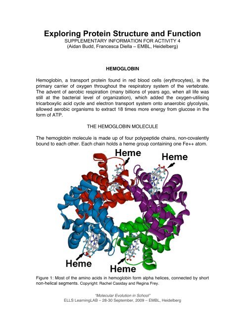
The hemoglobin molecule is made up of four polypeptide chains, non-covalently<br />
bound to each other. Each chain holds a heme group containing one Fe++ atom.<br />
Figure 1: Most of the amino acids in hemoglobin form alpha helices, connected by short<br />
non-helical segments. Copyright: Rachel Casiday <strong>and</strong> Regina Frey.<br />
“Molecular Evolution in School”<br />
ELLS LearningLAB – 28-30 September, 2009 – EMBL, Heidelberg
The four polypeptides chains of the hemoglobin are: two alpha chains (Alpha 1<br />
<strong>and</strong> Alpha 2) of 141 amino acid residues each <strong>and</strong> two beta chains (Beta 1 <strong>and</strong><br />
Beta 2) of 146 amino acid residues each. The alpha <strong>and</strong> beta chains have<br />
different sequences of amino acids, but fold up to form similar three-dimensional<br />
structures (Hemoglobin has no beta str<strong>and</strong>s <strong>and</strong> no disulfide bonds.). The four<br />
chains are held together by non-covalent interactions. There are four binding<br />
sites for oxygen on the hemoglobin molecule, because each chain contains one<br />
heme group. In the alpha chain, the residue 87 is histidine <strong>and</strong> in the beta chain<br />
the residue 92 is histidine . A heme group is attached to each of the four<br />
histidines. The heme consists of an organic part <strong>and</strong> an iron atom. The iron atom<br />
in heme binds to the four nitrogens in the center of the protoporphyrin ring. The<br />
hemoglobin molecule is nearly spherical, with a diameter of 55 angstroms. The<br />
four chains are packed together to form a tetramer. The heme groups are located<br />
in crevices near the exterior of the molecule, one in each subunit. Each alpha<br />
chain is in contact with both beta chains. However, there are few interactions<br />
between the two alpha chains or between the two beta chains.<br />
“Molecular Evolution in School”<br />
ELLS LearningLAB – 28-30 September, 2009 – EMBL, Heidelberg
TRANSPORT OF OXYGEN<br />
When the iron is bound to oxygen, the heme group is red in colour<br />
(oxyhameoglobin), <strong>and</strong> when it lacks oxygen (deoxygenated form) it is blue-red.<br />
As blood passes through the lungs, the hemoglobin picks up oxygen because of<br />
the increased oxygen pressure in the capillaries of the lungs, <strong>and</strong> can then<br />
release this oxygen to body cells where the oxygen pressure in the tissues is<br />
lower. In addition, the red blood cells can pick up the waste product, carbon<br />
dioxide, some of which is carried by the hemoglobin (at a different site from<br />
where it carries the oxygen), while the rest is dissolved in the plasma. An<br />
elemental oxygen molecule binds to the ferrous iron atom in the lungs where<br />
oxygen is abundant, <strong>and</strong> is released later in tissues that need oxygen.<br />
Figure 2: oxygen transport in the human body. Copyright Deborah Maizels, 2003<br />
“Molecular Evolution in School”<br />
ELLS LearningLAB – 28-30 September, 2009 – EMBL, Heidelberg
TYPES OF HEMOGLOBIN<br />
Different types of hemoglobin are classified according to the type of protein<br />
chains they contain. Normal hemoglobin types include:<br />
Hb A - makes up about 95 - 98% of the Hb found in adults; Hb A contains two<br />
alpha (α) protein chains <strong>and</strong> two beta (ß) protein chains.<br />
Hb A 2 - makes up about 2 - 3.5% of Hb; has two alpha (α) <strong>and</strong> two delta (δ)<br />
protein chains.<br />
Hb F - makes up to 2%; has two alpha (α) <strong>and</strong> two gamma (γ) protein chains.<br />
This is the primary hemoglobin produced by the fetus during gestation. Its<br />
production usually falls to a low level within a year after birth.<br />
HEMOGLOBIN AND DISEASE<br />
Defects in the hemoglobin genes can produce abnormal hemoglobins <strong>and</strong><br />
anemia, a condition termed "hemoglobinopathia". Abnormal hemoglobins are<br />
usually caused by a mutation in one of the hemoglobin genes. Two scenarios will<br />
arise: a) structural defects in the hemoglobin molecule <strong>and</strong> b) reduced production<br />
of one of the hemoglobin subunits.<br />
Sickle cell anemia was first described by Linus Pauling. Sickle hemoglobin<br />
differs from normal hemoglobin B by a single amino acid: valine replaces<br />
glutamate at position 6 on the surface of the beta chain (hemoglobin S). This<br />
creates a new hydrophobic spot. When deoxygenated, a small hydrophobic patch<br />
appears on the surface. The hydrophobic spots stick to each other (excluding<br />
water) causing deoxygenated hemoglobin to aggregate into chains. In a sickled<br />
red blood cell, the valine 6 (beta chain) binds to a different hydrophobic patch.<br />
The polymerized hemoglobin distorts red blood cells into an abnormal sickle<br />
shape. Heterozygotes have a mixture of normal hemoglobin A <strong>and</strong> mutant<br />
hemoglobin S. The hemoglobin A stops polymerization, preventing serious<br />
sickling.<br />
Sickle cell anemia was recognized to be the result of a genetic mutation, inherited<br />
according to the Mendelian principle of incomplete dominance <strong>and</strong> it represent a<br />
particularly interesting example of heterozygote superiority among humans. The<br />
HbS allele occurs in some African <strong>and</strong> Asian populations with a high frequency.<br />
This formerly was puzzling because the severity of the anemia, representing a<br />
strong natural selection against homozygotes, should have eliminated the<br />
defective allele. But researchers noticed that the HbS allele occurred at high<br />
frequency precisely in regions of the world where a particularly severe form of<br />
malaria, which is caused by the parasite Plasmodium falciparum, was endemic.<br />
It was hypothesized that the heterozygotes, HbAHbS, were resistant to malaria,<br />
“Molecular Evolution in School”<br />
ELLS LearningLAB – 28-30 September, 2009 – EMBL, Heidelberg
whereas the homozygotes HbAHbA were not. In malaria-infested regions then<br />
the heterozygotes survived better than either of the homozygotes, which were<br />
more likely to die from either malaria (HbAHbA homozygotes) or anemia<br />
(HbSHbS homozygotes). This hypothesis has been confirmed in various ways,<br />
e.g. most hospital patients suffering from severe or fatal forms of malaria are<br />
homozygotes HbAHbA.<br />
<strong>Structure</strong> of Normal Hemoglobin<br />
Molecule (HbA)<br />
<strong>Structure</strong> of Sickle Cell Disease<br />
Molecule<br />
Hemoglobin in Persons with Sickle Cell<br />
Disease<br />
Hemoglobin in Persons with Sickle Cell<br />
Trait<br />
2 alpha <strong>and</strong> 2 beta chains<br />
2 alpha <strong>and</strong> 2 s chains<br />
All hemoglobin molecules consist of 2<br />
alpha <strong>and</strong> 2 s chains<br />
Half of the hemoglobin molecules<br />
consist of 2 alpha <strong>and</strong> 2 beta chains,<br />
<strong>and</strong> half of 2 alpha <strong>and</strong> 2 s chains<br />
Thalassemias are a heterogeneous group of inherited disorders of hemoglobin<br />
synthesis resulting in a reduction in the amount of the particular globin chain<br />
produced. The result of an unbalanced globin-chain synthesis is the production of<br />
an excess of the partner chain. As a consequence of this, unusual forms of<br />
hemoglobin will precipitate <strong>and</strong> accumulate in the red cells. The majority of the<br />
beta thalassemias are characterized by the over-synthesis of the fetal<br />
hemoglobin (HbF). The thalassemias are usually broadly classified by the type of<br />
globin chain (alpha <strong>and</strong> beta thalassemias) whose synthesis is reduced. The<br />
thalassemias are extremely heterogeneous at the molecular level: more than 200<br />
different mutations of the globin genes have been found, <strong>and</strong> the thalassemias<br />
are almost as varied. Every severely-affected population in the world has a few<br />
common mutations unique to a particular region, together with varying numbers<br />
of rare ones.<br />
Porphyrias is not a single disease but a group of inherited (or acquired)<br />
disorders of certain enzymes in the heme biosynthetic (porphyrin) pathway. Each<br />
step of the process is controlled by eight enzymes. If any one of the enzymes is<br />
deficient, the process is disrupted. As a result, porphyrin or its precursors—<br />
chemicals formed at earlier steps of the process—may build up in body tissues<br />
<strong>and</strong> cause illness (mostly it has effects on the nervous system or the skin). The<br />
term "porphyria" is derived from the Greek word "porphyrus" meaning purple.<br />
Urine from some porphyria patients may be reddish in color due to the presence<br />
of excess porphyrins <strong>and</strong> related substances in the urine, <strong>and</strong> the urine may<br />
darken after exposure to light.<br />
“Molecular Evolution in School”<br />
ELLS LearningLAB – 28-30 September, 2009 – EMBL, Heidelberg
HEMOGLOBIN AND EVOLUTION<br />
We usually expect that, after a gene has duplicated, both copies of the gene will<br />
evolve (more or less) independently of each other i.e. changes in one copy of the<br />
gene will not tend to also be acquired by the other copy.<br />
In contrast, "concerted evolution" is a process in which related genes within a<br />
species undergo genetic exchange, causing their sequence evolution to be<br />
concerted over some period of time. This means that each gene locus in the<br />
family comes to have the same genetic variant.<br />
The family of the globin genes of primates, to which the hemoglobin belongs to,<br />
exists almost since the time life originated on earth, nearly four billion years ago<br />
<strong>and</strong> it well illustrates the principle - several copies of the gene lie very close to<br />
each other on the same chromosome, <strong>and</strong> are evolving concertedly.<br />
Figure 3: a phylogeny of the human globin genes. The genes multiplied by gene<br />
duplications, <strong>and</strong> the dates on the figure are the times of the duplications as inferred<br />
from the molecular clock. From Jeffreys et al. (1983).<br />
All primates have two alpha globins; we can therefore assume that the common<br />
ancestor of primates had two alpha globin genes. The sequence of each alpha<br />
globin gene differs between primate species; in the great apes any two species<br />
differ by about 2.5 amino acid substitutions in each gene. If one gene<br />
accumulates about 2.5 amino acid changes in the time between two species,<br />
then two different genes (alpha1 <strong>and</strong> alpha2) which have been separated for<br />
maybe 300 million years should have accumulated many more changes if they<br />
have been evolving independently. The conclusion is that they have not evolved<br />
independently - i.e. they are undergoing concerted evolution.<br />
“Molecular Evolution in School”<br />
ELLS LearningLAB – 28-30 September, 2009 – EMBL, Heidelberg
Note that there are two ways for a gene to have a common ancestor: by gene<br />
duplication <strong>and</strong> by speciation. When two genes share a common ancestor due to<br />
a duplication event we call them paralogous (a-hemoglobin <strong>and</strong> ß-hemoglobin in<br />
human are paralogous). When two genes share a common ancestor due to a<br />
speciation event we call them orthologous (a-hemoglobin in human <strong>and</strong> a-<br />
hemoglobin in chimps).<br />
These globin proteins are widely distributed in many organisms, appearing in the<br />
cells of plants, animals <strong>and</strong> even bacteria. Some globin proteins found in this<br />
family are:<br />
• Myoglobin (used as a reserve supply of oxygen, <strong>and</strong> facilitates the movement of<br />
oxygen within muscles)<br />
• Neuroglobin (involved in oxygen transport in the brain)<br />
• Cytoglobin (involved in intracellular oxygen storage or transfer)<br />
• Leghemoglobin (provides oxygen to bacteroids, which is essential for symbiotic<br />
nitrogen fixation)<br />
• Erythrocruorin (giant hemoglobin of worms)<br />
• Plant hemoglobin (may act as an oxygen sensor)<br />
• Flavohemoglobin (involved in NO detoxification)<br />
Reference:<br />
Jobling, Hurles, Smith “Human evolutionary genetics” Garl<strong>and</strong> Science (2003)<br />
http://www.rcsb.org/pdb/home/home.do<br />
http://en.wikipedia.org/wiki/Hemoglobin<br />
“Molecular Evolution in School”<br />
ELLS LearningLAB – 28-30 September, 2009 – EMBL, Heidelberg