ìì± ë° ì ì± ê°ìì ë³ë³ììì galectin-3, skp2, p27, cyclin D1 ë°í ...
ìì± ë° ì ì± ê°ìì ë³ë³ììì galectin-3, skp2, p27, cyclin D1 ë°í ...
ìì± ë° ì ì± ê°ìì ë³ë³ììì galectin-3, skp2, p27, cyclin D1 ë°í ...

Create successful ePaper yourself
Turn your PDF publications into a flip-book with our unique Google optimized e-Paper software.
The Korean Journal of Pathology<br />
2008; 42: 134-9<br />
양성 및 악성 갑상샘 병변에서의 <strong>galectin</strong>-3, <strong>skp2</strong>, <strong>p27</strong>, <strong>cyclin</strong> <strong>D1</strong><br />
발현의 연관성<br />
홍순억∙홍민의∙권귀영 1 ∙김미경<br />
중앙대학교 의과대학 병리학교실<br />
1<br />
중앙대학교병원 병리과<br />
접 수 : 2008년 2월 13일<br />
게재승인 : 2008년 5월 21일<br />
책임저자 : 권귀영<br />
우 156-755 서울시 동작구 흑석동 224-1<br />
중앙대학교병원 병리과<br />
전화: 02-6299-2754<br />
Fax: 02-6293-5630<br />
E-mail: hsu108@hanmail.net<br />
Correlation of Expression of <strong>galectin</strong>-3, <strong>skp2</strong>, <strong>p27</strong> and <strong>cyclin</strong> <strong>D1</strong><br />
in Benign and Malignant Thyroid Lesions<br />
Soon Auck Hong, Min Eui Hong, Gui Young Kwon 1 and Mi Kyung Kim<br />
Department of Pathology, Chung Ang Uinversity College of Medicine, Seoul; 1 Department of<br />
Pathology, Chung Ang University Hospital, Seoul, Korea<br />
Background : The overexpression of <strong>cyclin</strong> <strong>D1</strong> and <strong>galectin</strong>-3 and the loss of <strong>p27</strong> in thyroid<br />
cancers have recently been reported by many studies. The S-phase kinase associated protein<br />
2 (<strong>skp2</strong>) plays an important role in the degradation of <strong>p27</strong>. We compared the correlation<br />
of the expressions of <strong>galectin</strong>-3, <strong>p27</strong>, <strong>cyclin</strong> <strong>D1</strong> and <strong>skp2</strong> in thyroid lesions. Methods : Sixty<br />
five cases were included in this study and immunohistochemical staining for <strong>galectin</strong>-3, <strong>skp2</strong>,<br />
<strong>p27</strong> and <strong>cyclin</strong> <strong>D1</strong> was performed. Results : The expression of <strong>galectin</strong>-3 increased in the<br />
order of nodular hyperplasia, follicular adenoma, follicular carcinoma and papillary carcinoma<br />
(p
양성 및 악성 갑상샘 병변에서의 <strong>galectin</strong>-3, <strong>skp2</strong>, <strong>p27</strong>, <strong>cyclin</strong> <strong>D1</strong> 발현의 연관성 135<br />
나 비수질성 갑상샘 종양에서는 소포 기원의 악성 종양에서만<br />
국한적으로 발현되고 있다.<br />
또한 다른 종류의 <strong>galectin</strong>이 정상적인 여러 신체 조건에서<br />
발견되는 것과 다르게 <strong>galectin</strong>-3는 정상 또는 태아의 갑상샘<br />
세포에서는 발견되지 않으며 10 증식성 병변이나 양성 갑상샘 종<br />
양에서도 발현되지 않으므로 11,12 수술 전 갑상샘 병변의 진단 또<br />
는 예후 인자로써 그 유용성이 중시되고 있다. 11<br />
한편 유방암 세포에서 <strong>galectin</strong>-3는 세포 주기 G1 시점에서<br />
의 주요한 양성 조절자이며, 인체의 여러 암종에서 잠재적인 종<br />
양 유전자로 작용하는 <strong>cyclin</strong> <strong>D1</strong>의 발현에 중요한 역할을 한다<br />
고 알려져 있다. 12 특히 <strong>cyclin</strong> <strong>D1</strong>의 과발현은 유방암, 간암, 식<br />
도암, 두경부 편평세포암종 그리고 갑상샘 암종 등에서 관찰되<br />
는데, 13-15<br />
정상 또는 양성 갑상샘 종양에서는 거의 발현되지 않<br />
는 반면, 악성 갑상샘 종양에서는 발현이 증가됨으로써 <strong>cyclin</strong><br />
<strong>D1</strong>의 발현이 갑상샘 종양의 진행에 중요한 역할을 한다고 밝혀<br />
졌다. 15 그러나 이러한 갑상샘 종양에서 <strong>galectin</strong>-3와 <strong>cyclin</strong> <strong>D1</strong><br />
과의 연관성에 대한 연구는 아직 많이 부족한 실정이다.<br />
한편 <strong>cyclin</strong>-dependent kinase 활동을 조절하는 세포 주기<br />
조절 단백인 <strong>p27</strong>은 G1-S 세포 주기의 진행을 억제하여 세포의<br />
증식을 조절하는 기능을 하는데, 16,17 최근 여러 정상 내분비계 혹<br />
은 그 외의 장기에서는 발현이 증가되고, 유방암, 대장암 등에서<br />
는 발현이 감소되는 등 이의 감소가 종양의 진행이나 나쁜 예후<br />
와 관련이 있다는 보고가 발표되고 있어 종양 억제 유전자로서<br />
의 가능성이 제기되고 있다. 18-21 특히 종양에서 <strong>p27</strong>의 비활성화<br />
는 S-phase kinase associated protein 2 (<strong>skp2</strong>)에 의해 분해가<br />
촉진되어 일어나거나 <strong>cyclin</strong> D3에 의해 분리되어 일어난다. 22<br />
이에 본 연구에서는 갑상샘 종양에서 유용한 예후 인자로 알<br />
려져 있는 <strong>galectin</strong>-3와 여러 암종에서 중요한 세포 주기 조절<br />
인자로 알려져 있는 <strong>cyclin</strong> <strong>D1</strong> 그리고 <strong>p27</strong>과 <strong>skp2</strong>의 발현을 갑<br />
상샘의 양성 증식성 결절, 양성 갑상샘종, 갑상샘 암종 등 다양<br />
한 병변에서 면역조직화학염색을 통해 상호 비교해보고, 각각<br />
병변에서 이들 단백의 발현 여부와 이들 단백 발현 간의 연관성<br />
을 살펴봄으로써, 갑상샘의 발암 과정에 있어 이들 단백의 역할<br />
에 대해 알아보고자 한다.<br />
재료와 방법<br />
소포샘종이 12예 그리고 소포암종이 9예였다. 한편 이 연구에는<br />
포르말린에 고정된 파라핀 포매 조직을 이용하였다.<br />
방법<br />
면역조직화학 염색<br />
선택된 환자의 파라핀 포매 조직을 4-5 μm 두께로 박절하고,<br />
자일렌으로 탈 파라핀 과정을 거친 후(5분간 3회) 무수 알코올<br />
90%, 75% 그리고 50% 에탄올에 각각 2분씩 처리하여 함수시<br />
켰다. 그 후 내인성 과산화 효소의 활성을 억제하기 위해 0.3%<br />
hydrogen peroxide-methanol에 10분간 처리하고, 증류수로 세<br />
척한 다음 50 mM Tris 완충 용액(TBS, pH 7.5)으로 수세한<br />
후, 비 특이성 반응을 제거하기 위해 30분간 염소 혈청으로 처리<br />
하였다. 그 후 여분의 용액을 제거하고 일차 항체인 <strong>galectin</strong>-3<br />
(Novocastra, Newcatle, UK), <strong>skp2</strong> (Zymed Co., South San<br />
Francisco, CA, USA), <strong>p27</strong> (Dako, Capinteria, CA, USA),<br />
<strong>cyclin</strong> <strong>D1</strong> (Dako)을 실온에서 2시간 작용시켰다.<br />
일차 항체 반응 후 TBS로 5분간 3회 수세한 다음 biotin이<br />
부착된 이차 항체(1:300, Zymed Co.)에 20분간 작용시키고,<br />
통상적인 avidin-biotin complex법으로 염색하였다. 발색제는<br />
AEC를 사용하고 Mayer's hematoxylin으로 대조 염색하였다.<br />
결과 판정<br />
Galectin-3는 전체 종양 세포 중 세포질에서 과립상으로 진하<br />
게 갈색으로 염색된 세포의 수를 세어 백분율로 표시한 뒤, 5%<br />
미만인 경우를 음성, 5% 이상인 경우를 양성으로 판정하였으며,<br />
<strong>cyclin</strong> <strong>D1</strong>과 <strong>skp2</strong>는 전체 종양 세포 중 핵에서 진하게 갈색으로<br />
염색된 세포의 수를 세어 백분율로 표시한 뒤, 5% 미만인 경우<br />
를 음성, 5% 이상인 경우를 양성으로 판정하였다. 한편 <strong>p27</strong>은<br />
전체 종양 세포의 10% 미만에서 관찰된 경우 발현이 저하된 것<br />
으로 판정하였다.<br />
통계학적 분석<br />
Window용 SPSS 12.0 (SPSS, Chicago, IL, USA) 통계프<br />
로그램을 이용하여 카이제곱 검정과 상관분석법을 실시, p값이<br />
0.05 이하일 때를 통계학적으로 유의성이 있는 것으로 하였다.<br />
재료<br />
2003년 9월부터 2004년 12월까지 중앙대학교 부속 필동병원<br />
과 용산병원 그리고 2005년 1월부터 6월까지 중앙대학교 병원<br />
에서 갑상샘 종괴로 수술을 받았던 환자 중 병리적으로 갑상샘<br />
의 결절성 증식증, 갑상샘종, 갑상샘 유두암 그리고 소포암으로<br />
진단받은 예들을 대상으로 조직 보관 상태가 양호한 65예를 선<br />
별하였다. 이 중 결절성 증식증은 14예였으며, 유두암종이 30예,<br />
결 과<br />
결절성 증식증과 유두암종의 비교<br />
Galectin-3는 결절성 증식증의 7.1% (1예) 그리고 유두암종<br />
의 96.7% (29예)에서 발현되어, 결절성 증식증에 비해 유두암종<br />
에서 더 많이 발현되었음(p
136<br />
홍순억∙홍민의∙권귀영 외 1인<br />
결절성 증식증 모두에서 발현되지 않은 반면, 유두암종에서는<br />
20% (6예) 발현되어 두 군 간 통계학적 차이를 보이지 않았다<br />
(p=0.084). 한편 <strong>p27</strong>단백의 발현 감소는 결절성 증식증 21.4%<br />
(3예), 유두암종 96.5% (29예)의 예에 나타남으로써 결절성 증<br />
Table 1. Expression of <strong>galectin</strong>-3, <strong>skp2</strong> and <strong>cyclin</strong> D, and loss of <strong>p27</strong> between nodular hyperplasia and papillary carcinoma<br />
Galectin-3<br />
<strong>skp2</strong><br />
Loss of <strong>p27</strong><br />
Cyclin <strong>D1</strong><br />
(%) (n)<br />
(%) (n)<br />
(%) (n)<br />
(%) (n)<br />
- +<br />
- +<br />
- +<br />
- +<br />
Nodular hyperplasia (n=14) 92.9 (13) 7.1 (1) 100 (14) 0 (0) 78.6 (11) 21.4 (3) 92.9 (13) 7.1 (1)<br />
Papillary carcinoma (n=30) 3.3 (1) 96.7 (29) 80 (24) 20 (6) 3.3 (1) 96.7 (29) 66.6 (20) 33.3 (10)<br />
p value 0.0001 0.084 0.0001 0.062<br />
A B C D<br />
E F G H<br />
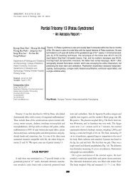
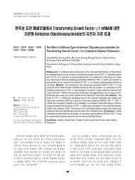
Fig. 1. Immunohistochemical staining of <strong>galectin</strong>-3 (A, B), <strong>skp2</strong> (C, D), <strong>p27</strong> (E, F) and <strong>cyclin</strong> <strong>D1</strong> (G, H) in nodular hyperplasia and papillary<br />
carcinoma; the positve stains of <strong>galectin</strong>-3 (B), <strong>skp2</strong> (D), <strong>cyclin</strong> <strong>D1</strong> (H) are noted in papillary carcinoma. Galectin-3 (A), <strong>skp2</strong> (C),<br />
<strong>cyclin</strong> <strong>D1</strong> (G) show negative stains in nodular hyperplasia. Loss of <strong>p27</strong> (F) in the papillary carcinoma and strong expression of <strong>p27</strong> (E) in<br />
the nodular hyperplasia are shown, respectively.<br />
A B C D<br />
E F G H<br />
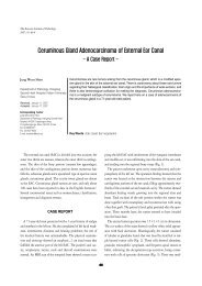
Fig. 2. Immunohistocheimical staining of <strong>galectin</strong>-3 (A, B), <strong>skp2</strong> (C, D), <strong>p27</strong> (E, F) and <strong>cyclin</strong> <strong>D1</strong> (G, H) in follicular adenoma and follicular<br />
carcinoma; increased expression of <strong>galectin</strong>-3 in the follicular carcinoma (B) is compared with the expression of <strong>galectin</strong>-3 in the nodular<br />
hyperplasia (A). Increased expression of <strong>skp2</strong> in the folliulcar adenoma (C) and follicular carcinoma (D) is noted. Loss of <strong>p27</strong> in the follicular<br />
adenoma (E) and follicular carcinoma (F) is present. The strong expression of <strong>cyclin</strong> <strong>D1</strong> in follicular carcinoma (H) is noted. Decreased<br />
expression of <strong>cyclin</strong> <strong>D1</strong> is seen in follicular adenoma (G).
양성 및 악성 갑상샘 병변에서의 <strong>galectin</strong>-3, <strong>skp2</strong>, <strong>p27</strong>, <strong>cyclin</strong> <strong>D1</strong> 발현의 연관성 137<br />
Table 2. Expression of <strong>galectin</strong>-3, <strong>skp2</strong> and <strong>cyclin</strong> <strong>D1</strong>, and loss of <strong>p27</strong> in nodular hyperplasia, follicular adenoma and follicular carcinoma<br />
Galectin-3<br />
<strong>skp2</strong><br />
Loss of <strong>p27</strong><br />
Cyclin <strong>D1</strong><br />
(%) (n)<br />
(%) (n)<br />
(%) (n)<br />
(%) (n)<br />
- +<br />
- +<br />
- +<br />
- +<br />
Nodular hyperplasia (n=14) 92.9 (13) 7.1 (1) 100 (14) 0 (0) 78.6 (11) 21.4 (3) 92.9 (13) 7.1 (1)<br />
Follicular adenoma (n=12) 83.3 (10) 16.7 (2) 83.3 (10) 16.7 (2) 8.3 (1) 91.7 (11) 41.7 (5) 58.3 (7)<br />
Follicular carcinoma (n=9) 44.4 (4) 55.6 (5) 66.7 (6) 33.3 (3) 0 (0) 100.0 (9) 55.6 (5) 44.4 (4)<br />
p value 0.0022 0.080 0.0001 0.018<br />
Table 3. Comparison of loss of <strong>p27</strong> expression and expression<br />
of <strong>galectin</strong>-3 in benign to malignant thyroid lesion<br />
Loss of <strong>p27</strong><br />
(-) (%) (n) (+) (%) (n)<br />
Galectin-3<br />
(-) (n=28) 39.3 (11) 60.7 (17)<br />
(+) (n=28) 5.4 (2) 94.6 (35)<br />
p value 0.001<br />
Table 4. Comparison of expression of <strong>cyclin</strong> <strong>D1</strong> and <strong>skp2</strong> in<br />
benign to malignant thyroid lesion<br />
Cyclin <strong>D1</strong><br />
(-) (%) (n) (+) (%) (n)<br />
<strong>skp2</strong><br />
(-) (n=54) 72.2 (39) 27.8 (15)<br />
(+) (n=11) 36.4 (4) 63.6 (7)<br />
p value 0.022<br />
식증에 비해 유두암종에서 더 많은 예의 발현 감소가 일어남을<br />
알 수 있었으며(p
138<br />
홍순억∙홍민의∙권귀영 외 1인<br />
한편 S-phase kinae 2-cdk2/<strong>cyclin</strong> A와 연관된 단백질인<br />
<strong>skp2</strong>는 세포 주기가 S기에서 G1기로 갈 때 발현되는데, 이때<br />
악성으로 변화된 세포에서 <strong>skp2</strong>의 발현이 증가되었다.<br />
또한 상피 세포의 암종, 림프종과 같은 많은 악성 종양에서<br />
발현 감소되는 <strong>p27</strong>은 23 악성 종양 세포의 조절되지 않는 증식을<br />
일으키는데, 이것은 <strong>p27</strong>이 세포 주기에서 S기에 관여하는 cdk2/<br />
<strong>cyclin</strong> E와 cdk2/<strong>cyclin</strong> A에 대해 억제성 조절을 하기 때문이<br />
다. 24 이러한 <strong>p27</strong>의 감소는 단백 분해를 포함한 세포 기능에 참<br />
여하는 아미노산 화합물(ubiquitination)의 가수 분해 과정이 증<br />
가함으로써 발생하는데, 이때 <strong>p27</strong>의 분해가 항진될 수 있는 첫<br />
번째 기전이 <strong>skp2</strong>의 과발현이다. 22 <strong>skp2</strong>는 최근에 악성 종양성<br />
의 잠재력을 가지고 있으며, 구강 편평 상피 세포 암종과 림프<br />
종 등에서 발현이 증가되었다는 보고가 있어 25 <strong>skp2</strong>의 과발현이<br />
구강 편평 상피 세포 암종과 위암종 그리고 폐암종에서 좋지 않<br />
은 예후를 시사하는 인자라고 추측되고 있다. 22 그러나 본 연구<br />
에서는 양성 결절성 증식증에서 0%, 소포샘종에서 16.7%, 유두<br />
암종에서 20% 그리고 소포암종에서 33.3%의 <strong>skp2</strong>발현이 관찰<br />
됨으로써 각 군 간 유의한 발현의 차이는 없었다.<br />
또한 <strong>p27</strong>은 결절성 증식증의 21.4%, 소포샘종의 91.7%, 유두<br />
암종의 96.7%, 그리고 소포암종의 80%에서 발현이 감소되어 결<br />
절성 증식증에 비해 유두암종에서 발현이 더 감소되는 것을 알<br />
수 있었다(p
양성 및 악성 갑상샘 병변에서의 <strong>galectin</strong>-3, <strong>skp2</strong>, <strong>p27</strong>, <strong>cyclin</strong> <strong>D1</strong> 발현의 연관성 139<br />
참고문헌<br />
1. Haugen BR. Initial treatment of differentiated thyroid carcinoma.<br />
Rev Endocr Metab Disord 2000; 1: 139-45.<br />
2. Mazzaferri EL. NCCN Thyroid carcinoma practice guidelines. NCCN<br />
Proceedings 2000; 391-442.<br />
3. Sherman SI, Brierley JD, Sperling M, et al. Prospective multi center<br />
study of thyroid carcinoma treatment- initial analysis of staging<br />
and outcome. Cancer 1998; 83: 1012-21.<br />
4. Baloch Z, LiVolsi VA, Henricks WH, Sebak BA. Encapsulated follicular<br />
variant of papillary thyroid carcinoma. Am J Clin Pathol<br />
2002; 118: 603-5.<br />
5. Baloch ZW, Fleisher S, LiVolsi VA, Gupta PK. Diagnosis of ‘‘follicular<br />
neoplasm’’: a gray zone in thyroid fine needle aspiration cytology.<br />
Diagn Cytopathol 2002; 26: 41-4.<br />
6. Kini SR, Miller JM, Hamburger JI, Smith-Purslow MJ. Cytopathology<br />
of follicular lesions of thyroid gland. Diagn Cytopathol 1985; 1:<br />
123-32.<br />
7. Orlandi F, Saggiorato E, Pivano G, et al. Galectin-3 is a presurgical<br />
marker of human thyroid carcinoma. Cancer Res 1998; 58: 3015-20.<br />
8. Bartolazzi A, Gasbarri A, Papotti M, et al. Application of an immunodiagnostic<br />
method for improving preoperative diagnosis of nodular<br />
thyroid lesions. Lancet 2001; 357: 1644-50.<br />
9. Papotti M, Volante M, Saggiorato E, et al. Role of <strong>galectin</strong>-3 immunodetectionin<br />
the cytological diagnosis of thyroid cystic papillary carcinoma.<br />
Eur J Endocrinol 2002; 147: 515-21.<br />
10. Savin SB, Cvejic DS, Jankovic MM. Expression of <strong>galectin</strong>-1 and<br />
<strong>galectin</strong>-3 in human fetal thyroid gland. J Histochem Cytochem<br />
2003; 51: 479-83.<br />
11. Volante M, Bozzalla-Cassione F, Orlandi F, et al. Diagnostic role of<br />
<strong>galectin</strong>-3 in follicular thyroid tumors. Virchow Arch 2004; 444:<br />
309-12.<br />
12. Kim KR, Lin HM, Biliran H, Raz A. Cell cycle arrest and inhibition<br />
of anoikis by <strong>galectin</strong>-3 in human breast epithelial cells. Cancer Res<br />
1999; 59: 4148-54.<br />
13. Hall M, Peters G. Genetic alterations of <strong>cyclin</strong>s, <strong>cyclin</strong>-dependent<br />
kinases, and cdk inhibitors in human cancer. Adv Cancer Res 1996;<br />
68: 67-108.<br />
14. Hunter T, Pines J. Cyclins and cancer II: <strong>cyclin</strong> D and CDK inhibitors<br />
come of age. Cell 1994; 79: 573-82.<br />
15. Lazzereschi D, Sambuco L, Carnovale-scalzo C, et al. Cyclin <strong>D1</strong> and<br />
<strong>cyclin</strong> E expression in malignant thyroid cells and in human thyroid<br />
carcinomas. Int J Cancer 1998; 76: 806-11.<br />
16. Sherr CJ, Roberts JM. Inhibitors of mammalian G1 <strong>cyclin</strong>-dependent<br />
kinases. Genes Dev 1995; 9: 1149-63.<br />
17. Reed SI, Baily E, Dulic V, et al. G1 control in mammalian cells. J Cell<br />
Sci 1994; 18(Suppl): 69-73.<br />
18. Carr K, Heffess C, Jin L, et al. Immunohistochemical analysis of thyroid<br />
carcinomas utilizing antibodies to p53 and Ki-67. Appl Immunohistochem<br />
1993; 1: 201-7.<br />
19. Catzavelos C, Bhattachara N, Ung YC, et al. Decreased levels of the<br />
cell-cycle inhibitor <strong>p27</strong> kip1 protein: prognostic implications in primary<br />
breast cancer. Nat Med 1997; 3: 227-30.<br />
20. Porter P, Malone KE, Heagery PJ, et al. Expression of cell-cycle regulator<br />
<strong>p27</strong> kip1 and <strong>cyclin</strong> E, alone and in combination, correlate with<br />
survival in young breast cancer patients. Nat Med 1996; 3: 222-4.<br />
21. Loda M, Cukor B, Tam SW, et al. Increased proteasome-dependent<br />
degradation of the <strong>cyclin</strong>-dependent kinase inhibitor <strong>p27</strong> in aggressive<br />
colorectal carcinomas. Nat Med 1997; 3: 231-4.<br />
22. Masuda TA, Inoue H, Sonoda H, et al. Clinical and biological significance<br />
of S-phase kinase associated protein 2 (<strong>skp2</strong>) gene expression<br />
in gastric carcinoma: modulation of malignant phenotype by<br />
<strong>skp2</strong> overexpression, possibly via <strong>p27</strong> proteolysis. Cancer Res 2002;<br />
62: 3819-25.<br />
23. Slingerland J, Pagano M. Regulation of the cdk inhibitor <strong>p27</strong> and<br />
its deregulation in cancer. J Cell Physiol 2000; 183: 10-7.<br />
24. Nakayama K, Nakahama H, Minamishima YA, et al. Targeted disruption<br />
of <strong>skp2</strong> results in accumulation of <strong>cyclin</strong> E and <strong>p27</strong> kip1 . polyploidy<br />
and centrosome overduplication. EMBO J 2000; 19: 2069-81.<br />
25. Gstaiger M, Jordan R, Lim M, et al. Skp2 is oncogenic and overexpressed<br />
in human cancers. Proct Natl Acad Sci USA 2001; 98: 5043-8.<br />
26. Trocone G, Iaccarino A, Caleo A, et al. P27 kip1 protein expression in<br />
Hashimoto`s thyroiditis. J Clin Pathol 2003; 56: 587-91.<br />
27. Erikson LA, Jin L, Wollan PC, et al. Expression of <strong>p27</strong> kip1 and Ki-67 in<br />
benign and malignant thyroid tumors. Mod Pathol 1998; 11: 169-74.<br />
28. LaBaer J, Garrett MD, Stevenson LF, et al. New funtional activities<br />
for the p21 family of CDK inhibitors. Genes Dev 1997; 11: 847-62.<br />
29. Motti ML, Califano D, Troncone G, et al. Complex regulation of the<br />
<strong>cyclin</strong>-dependent kinase inhibitor <strong>p27</strong> kip1 in thyroid cancer cells by<br />
the PI3K/AKT pathway. Regulation of <strong>p27</strong> kip1 expression and localization.<br />
Am J Pathol 2005; 166: 737-49.<br />
30. Paron I, Scaloni A, Pines A, et al. Nuclear localization of <strong>galectin</strong>-3<br />
in transformed thyroid cells: a role in transcriptional regulation.<br />
Biochem Biophys Res Commun 2003; 302: 545-53.