Printable PDF Version - Gore Medical
Printable PDF Version - Gore Medical
Printable PDF Version - Gore Medical

Create successful ePaper yourself
Turn your PDF publications into a flip-book with our unique Google optimized e-Paper software.
Intracardiac Echocardiography Considerations During<br />
Implantation of the GORE ® HELEX Septal Occluder<br />
Continued from cover...<br />
An eight-year-old girl presented for<br />
transcatheter ASD closure at a weight<br />
of 24.6 kg. A prior transthoracic<br />
echo showed a defect diameter<br />
of approximately 10 mm, and the<br />
posterosuperior (SVC), anterosuperior<br />
(aortic), anteroinferior (tricuspid<br />
valve), posteroinferior (IVC), and<br />
posterosuperior (pulmonary<br />
vein) rims were all adequate.<br />
Using general anesthesia and<br />
heparin anticoagulation, cardiac<br />
catheterization revealed normal right<br />
heart pressures and a Q p<br />
/ Q s<br />
of 1.3.<br />
ICE imaging was performed using an<br />
8F ACUNAV Intracardiac Echo Catheter<br />
through a contralateral venous sheath.<br />
As part of the usual imaging protocol,<br />
a complete ICE evaluation was<br />
completed as follows:<br />
Note: Any control knob adjustments<br />
of the ICE catheter are best made by<br />
moving the locking ring clockwise<br />
to the locked position first and then<br />
moving the A / P or L / R control knob<br />
as needed, thereby maintaining their<br />
position.<br />
1. “Home” view (Figure 1): the ICE<br />
catheter arrow indicator at the control<br />
end corresponds to the transducer<br />
orientation at the catheter tip, and<br />
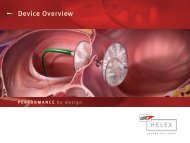
Right Atrial Disk<br />
Left Atrial Disk<br />
Figure 2 Implanted GORE ® HELEX Septal Occluder<br />
in hypothetical heart model.<br />
this should be oriented toward patient<br />
left and slightly anterior; the tricuspid<br />
valve is seen with the right atrium<br />
above and the right ventricle below;<br />
superior is screen right, and inferior is<br />
screen left; occasionally adjusting the<br />
control knob slightly to the Anterior<br />
(A) location is necessary.<br />
2. Using both hands (right hand<br />
at control knob area, left hand on<br />
catheter near introducer sheath entry),<br />
the catheter is rotated clockwise (when<br />
looking from patient’s feet to head) or<br />
away from operator; during this slow<br />
rotation, one may see a prominent<br />
Eustachian valve or Chiari network,<br />
but typically the coronary sinus starts<br />
“rounding” out, and subsequently<br />
the LV and RV outflow tracts are<br />
seen; variations in cardiac anatomy<br />
and position often require gentle<br />
adjustment of the L / R control knob to<br />
optimize the image.<br />
3. With further clockwise rotation,<br />
the mitral valve, left atrial appendage,<br />
and a fully round cross-section of the<br />
coronary sinus are visible (Figures 2<br />
and 3); the atrial septum is well seen;<br />
as clockwise rotation continues,<br />
the left-sided pulmonary veins are<br />
typically seen as an upside down<br />
“Y” connection, with the left superior<br />
vein to screen right and the left<br />
inferior vein to screen left; color flow<br />
is red / orange as the direction of flow<br />
is toward the transducer, which is<br />
situated in the right atrium (Figure 4).<br />
4. As one continues a clockwise<br />
rotation around the back of the heart,<br />
the descending aorta is seen next in<br />
a horizontal orientation.<br />
5. Just to patient right of the<br />
descending aorta are the right-sided<br />
pulmonary veins, again found by<br />
clockwise rotation beyond the<br />
descending aorta. Without any<br />
control knob adjustment, these most<br />
commonly appear as a foreshortened<br />
upside down “Y” connection;<br />
significantly enhanced imaging of the<br />
right-sided veins can be achieved by<br />
locking the L / R control knob toward<br />
Figure 1. Home view.<br />
Figure 3. View of the left atrial appendage<br />
and mitral valve. The atrial is also well seen.<br />
Figure 4. Left superior and inferior pulmonary<br />
veins.