- Page 2:
This page intentionally left blank
- Page 8:
Introduction to Fungi JohnWebster U
- Page 12:
To Philip M. Booth
- Page 18:
viii CONTENTS Chapter 6 Chytridiomy
- Page 22:
x CONTENTS 15.4 Rhytismataceae 440
- Page 28:
Preface to the first edition There
- Page 36:
Preface to the third edition Major
- Page 40:
Acknowledgements We are indebted to
- Page 46:
2 INTRODUCTION With photosynthetic
- Page 50:
4 INTRODUCTION Fig1.3 Transmission
- Page 54:
6 INTRODUCTION Fig1.5 Structural fo
- Page 58:
8 INTRODUCTION of mushroom-type fru
- Page 62:
10 INTRODUCTION Fig1.8 Diagrammatic
- Page 66:
12 INTRODUCTION Fig1.9 Tubular cont
- Page 70:
14 INTRODUCTION Fig1.11 Ion fluxes
- Page 74:
16 INTRODUCTION grow alongside each
- Page 78:
18 INTRODUCTION Fig1.15 Rhizomorphs
- Page 82:
20 INTRODUCTION carbon/nitrogen rat
- Page 86:
22 INTRODUCTION well shown by the X
- Page 90:
24 INTRODUCTION Fig1.17 Zoospore ty
- Page 94:
26 INTRODUCTION genera, e.g. Hypocr
- Page 98:
28 INTRODUCTION creating a momentum
- Page 102:
30 INTRODUCTION Fig1.22 Chlamydospo
- Page 106:
32 INTRODUCTION As mentioned on p.
- Page 110:
34 INTRODUCTION the cell wall (Tabl
- Page 114:
36 INTRODUCTION Fig1.24 The spatial
- Page 118:
38 INTRODUCTION Table1.2. The class
- Page 122:
2 Protozoa: Myxomycota (slime mould
- Page 126:
42 PROTOZOA: MYXOMYCOTA (SLIME MOUL
- Page 130:
44 PROTOZOA: MYXOMYCOTA (SLIME MOUL
- Page 134:
46 PROTOZOA: MYXOMYCOTA (SLIME MOUL
- Page 138:
48 PROTOZOA: MYXOMYCOTA (SLIME MOUL
- Page 142:
50 PROTOZOA: MYXOMYCOTA (SLIME MOUL
- Page 146:
52 PROTOZOA: MYXOMYCOTA (SLIME MOUL
- Page 150:
3 Protozoa: Plasmodiophoromycota 3.
- Page 154:
56 PROTOZOA: PLASMODIOPHOROMYCOTA F
- Page 158:
58 PROTOZOA: PLASMODIOPHOROMYCOTA F
- Page 162:
60 PROTOZOA: PLASMODIOPHOROMYCOTA p
- Page 166:
62 PROTOZOA: PLASMODIOPHOROMYCOTA I
- Page 170:
64 PROTOZOA: PLASMODIOPHOROMYCOTA a
- Page 174:
66 PROTOZOA: PLASMODIOPHOROMYCOTA a
- Page 178:
68 STRAMINIPILA: MINOR FUNGAL PHYLA
- Page 182:
70 STRAMINIPILA: MINOR FUNGAL PHYLA
- Page 186:
72 STRAMINIPILA: MINOR FUNGAL PHYLA
- Page 190:
74 STRAMINIPILA: MINOR FUNGAL PHYLA
- Page 194:
76 STRAMINIPILA: OOMYCOTA Fig 5.1 A
- Page 198:
78 STRAMINIPILA: OOMYCOTA Fig 5.3 L
- Page 202:
80 STRAMINIPILA: OOMYCOTA Table 5.1
- Page 206:
82 STRAMINIPILA: OOMYCOTA strains o
- Page 210:
84 STRAMINIPILA: OOMYCOTA Fig 5.5 S
- Page 214:
86 STRAMINIPILA: OOMYCOTA The oogon
- Page 218:
88 STRAMINIPILA: OOMYCOTA Fig 5.9 A
- Page 222:
90 STRAMINIPILA: OOMYCOTA was the f
- Page 226:
92 STRAMINIPILA: OOMYCOTA Fig 5.12
- Page 230:
94 STRAMINIPILA: OOMYCOTA Fig 5.14
- Page 234:
96 STRAMINIPILA: OOMYCOTA that desc
- Page 238:
98 STRAMINIPILA: OOMYCOTA Fig 5.16
- Page 242:
100 STRAMINIPILA: OOMYCOTA In some
- Page 246:
102 STRAMINIPILA: OOMYCOTA haploid
- Page 250:
104 STRAMINIPILA: OOMYCOTA Fig 5.20
- Page 254:
106 STRAMINIPILA: OOMYCOTA Fig 5.22
- Page 258:
108 STRAMINIPILA: OOMYCOTA Fig 5.24
- Page 262:
110 STRAMINIPILA: OOMYCOTA Fig 5.26
- Page 266:
112 STRAMINIPILA: OOMYCOTA firmly t
- Page 270:
114 STRAMINIPILA: OOMYCOTA present,
- Page 274:
116 STRAMINIPILA: OOMYCOTA convinci
- Page 278:
118 STRAMINIPILA: OOMYCOTA Fig 5.29
- Page 282:
120 STRAMINIPILA: OOMYCOTA and pars
- Page 286:
122 STRAMINIPILA: OOMYCOTA sterigma
- Page 290:
124 STRAMINIPILA: OOMYCOTA The spor
- Page 294:
126 STRAMINIPILA: OOMYCOTA Fig 5.35
- Page 298:
128 CHYTRIDIOMYCOTA N-acetylglucosa
- Page 302:
130 CHY TRIDIOMYCOTA Fig 6.2 Flagel
- Page 306:
132 CHY TRIDIOMYCOTA the reproducti
- Page 310:
134 CHY TRIDIOMYCOTA Table 6.1. Ord
- Page 314:
136 CHY TRIDIOMYCOTA Fig 6.7 Synchy
- Page 318:
138 CHY TRIDIOMYCOTA already been d
- Page 322:
140 CHYTRIDIOMYCOTA Fig 6.9 Synchyt
- Page 326:
142 CHYTRIDIOMYCOTA Canter & Jawors
- Page 330:
144 CHYTRIDIOMYCOTA exit tube which
- Page 334:
146 CHYTRIDIOMYCOTA some of the spe
- Page 338:
148 CHYTRIDIOMYCOTA distinguished v
- Page 342:
150 CHYTRIDIOMYCOTA Fig 6.17 Scanni
- Page 346:
152 CHYTRIDIOMYCOTA Fig 6.18 Neocal
- Page 350:
154 CHYTRIDIOMYCOTA Fig 6.19 Blasto
- Page 354:
156 CHYTRIDIOMYCOTA and if dried sa
- Page 358:
158 CHYTRIDIOMYCOTA Fig 6.21 Life c
- Page 362:
160 CHYTRIDIOMYCOTA both thin-walle
- Page 366:
162 CHYTRIDIOMYCOTA pathways. There
- Page 370:
164 CHYTRIDIOMYCOTA Fig 6.25 Monobl
- Page 374:
166 ZYGOMYCOTA Fig 7.1 Recent phylo
- Page 378:
168 ZYGOMYCOTA Fig 7.3 Mucor rouxii
- Page 382:
170 ZYGOMYCOTA Fig 7.5 Sporangiopho
- Page 386:
172 ZYGOMYCOTA The columella is cur
- Page 390:
174 ZYGOMYCOTA Fig 7.8 Phycomyces b
- Page 394:
176 ZYGOMYCOTA Phycomyces, after ar
- Page 398:
178 ZYGOMYCOTA Fig 7.11 Development
- Page 402:
180 ZYGOMYCOTA Fig 7.12 Life cycle
- Page 406:
182 ZYGOMYCOTA Fig 7.14 Mucor racem
- Page 410:
184 ZYGOMYCOTA that many workers co
- Page 414:
186 ZYGOMYCOTA Fig 7.18 Phycomyces
- Page 418:
188 ZYGOMYCOTA the sporangiophore o
- Page 422:
190 ZYGOMYCOTA makes contact with a
- Page 426:
192 ZYGOMYCOTA Fig 7.24 Thamnidium
- Page 430:
194 ZYGOMYCOTA (i.e. striate) wall
- Page 434:
196 ZYGOMYCOTA sporangiospores, eac
- Page 438:
198 ZYGOMYCOTA Fig 7.30 Mortierella
- Page 442:
200 ZYGOMYCOTA Fig 7.32 Mortierella
- Page 446:
202 ZYGOMYCOTA of germ tubes toward
- Page 450:
204 ZYGOMYCOTA Fig 7.34 Basidiobolu
- Page 454: 206 ZYGOMYCOTA Fig 7.36 Basidiobolu
- Page 458: 208 ZYGOMYCOTA Fig 7.37 The eventfu
- Page 462: 210 ZYGOMYCOTA Fig 7.39 Conidiobolu
- Page 466: 212 ZYGOMYCOTA Fig 7.4 0 Carcass of
- Page 470: 214 ZYGOMYCOTA develop. These are t
- Page 474: 216 ZYGOMYCOTA apparent (Fig. 7.43e
- Page 478: 218 ZYGOMYCOTA mycorrhiza (AM). The
- Page 482: 220 ZYGOMYCOTA Fig 7.4 6 Vesicular
- Page 486: 222 ZYGOMYCOTA interest (Allen, 199
- Page 490: 224 ZYGOMYCOTA Fig 7.47 Harpella me
- Page 494: 8 Ascomycota (ascomycetes) 8.1 Intr
- Page 498: 228 ASCOMYCOTA (ASCOMYCETES) Fig 8.
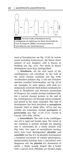
- Page 502: 230 ASCOMYCOTA (ASCOMYCETES) wall a
- Page 508: CONIDIUM PRODUCTION IN ASCOMYCETES
- Page 512: CONIDIUM PRODUCTION IN ASCOMYCETES
- Page 516: DEVELOPMENT OF ASCI 237 These nucle
- Page 520: DEVELOPMENT OF ASCI 239 appendages
- Page 524: DEVELOPMENT OF ASCI 241 may converg
- Page 528: DEVELOPMENT OF ASCI 243 Fig 8.14 As
- Page 532: TYPESOFFRUITBODY 245 the spores may
- Page 536: CLASSIFICATION 247 mutants and the
- Page 540: CLASSIFICATION 249 An outline of cu
- Page 544: TAPHRINALES 251 9.2 Taphrinales The
- Page 548: SCHIZOSACCHAROMYCETALES 253 about 5
- Page 552: SCHIZOSACCHAROMYCETALES 255 importa
- Page 556:
SCHIZOSACCHAROMYCETALES 257 Fig 9.5
- Page 560:
PNEUMOCYSTIS 259 formation of the i
- Page 564:
10 Hemiascomycetes 10.1 Introductio
- Page 568:
SACCHAROMYCES (SACCHAROMYCETACEAE)
- Page 572:
SACCHAROMYCES (SACCHAROMYCETACEAE)
- Page 576:
SACCHAROMYCES (SACCHAROMYCETACEAE)
- Page 580:
SACCHAROMYCES (SACCHAROMYCETACEAE)
- Page 584:
SACCHAROMYCES (SACCHAROMYCETACEAE)
- Page 588:
SACCHAROMYCES (SACCHAROMYCETACEAE)
- Page 592:
SACCHAROMYCES (SACCHAROMYCETACEAE)
- Page 596:
CANDIDA (ANAMORPHIC SACCHAROMYCETAL
- Page 600:
CANDIDA (ANAMORPHIC SACCHAROMYCETAL
- Page 604:
GALACTOMYCES (DIPODASCACEAE) 281 of
- Page 608:
SACCHAROMYCOPSIS (SACCHAROMYCOPSIDA
- Page 612:
11 Plectomycetes 11.1 Introduction
- Page 616:
ASCOSPHAERALES 287 ascocarps, and i
- Page 620:
ONYGENALES 289 infected larvae rath
- Page 624:
ONYGENALES 291 Onygenaceae as human
- Page 628:
ONYGENALES 293 disturb the soil. On
- Page 632:
ONYGENALES 295 Fig11.7 Friesian cat
- Page 636:
EUROTIALES 297 have gymnothecia wit
- Page 640:
EUROTIALES 299 penicilli, a further
- Page 644:
EUROTIALES 301 biseriate species As
- Page 648:
EUROTIALES 303 Cheese production in
- Page 652:
EUROTIALES 305 has similarities to
- Page 656:
EUROTIALES 307 are uncommon, with t
- Page 660:
EUROTIALES 309 Fig11.16 Eurotium re
- Page 664:
EUROTIALES 311 Fig11.18 Conidiophor
- Page 668:
EUROTIALES 313 Fig11.21 Elaphomyces
- Page 672:
12 Hymenoascomycetes: Pyrenomycetes
- Page 676:
SORDARIALES 317 The dark-coloured p
- Page 680:
SORDARIALES 319 vitamins by the sti
- Page 684:
SORDARIALES 321 Fig12.3 Schizotheci
- Page 688:
SORDARIALES 323 degenerates. Cultur
- Page 692:
SORDARIALES 325 Mating type factors
- Page 696:
SORDARIALES 327 kilns and bakeries
- Page 700:
SORDARIALES 329 Fig12.8 Neurospora
- Page 704:
SORDARIALES 331 heterokaryotic (A
- Page 708:
XYLARIALES 333 been placed in sever
- Page 712:
XYLARIALES 335 Daldinia concentrica
- Page 716:
HYPOCREALES 337 Fig12.13 serpens. T
- Page 720:
HYPOCREALES 339 mushroom mycelium.
- Page 724:
HYPOCREALES 341 Fig12.16 Conidiopho
- Page 728:
HYPOCREALES 343 Fig12.18 Canker cau
- Page 732:
HYPOCREALES 345 Fig12.21 Nectria ma
- Page 736:
HYPOCREALES 347 Fig12.22 Fusarium c
- Page 740:
CLAVICIPITALES 349 thick apical cap
- Page 744:
CLAVICIPITALES 351 Fig12.25 The hom
- Page 748:
CLAVICIPITALES 353 and dumb, and, b
- Page 752:
CLAVICIPITALES 355 alkaloid product
- Page 756:
CLAVICIPITALES 357 Fig12.29 Epichlo
- Page 760:
CLAVICIPITALES 359 Fig12.31 Endophy
- Page 764:
CLAVICIPITALES 361 artificially inf
- Page 768:
CLAVICIPITALES 363 conidia. The ass
- Page 772:
OPHIOSTOMATALES 365 Fig12.36 Ophios
- Page 776:
OPHIOSTOMATALES 367 Fig12.37 Asexua
- Page 780:
MICROASCALES 369 Fig12.38 Scopulari
- Page 784:
MICROASCALES 371 Fig12.40 Trichurus
- Page 788:
DIAPORTHALES 373 Aspects of pathoge
- Page 792:
DIAPORTHALES 375 enveloped by hypha
- Page 796:
MAGNAPORTHACEAE 377 cAMP levels wer
- Page 800:
MAGNAPORTHACEAE 379 Fig12.44 The in
- Page 804:
MAGNAPORTHACEAE 381 with a hydropho
- Page 808:
MAGNAPORTHACEAE 383 Fig12.47 (a) Co
- Page 812:
MAGNAPORTHACEAE 385 intracellular s
- Page 816:
GLOMERELLACEAE 387 Fig12.51 Colleto
- Page 820:
GLOMERELLACEAE 389 infection proces
- Page 824:
INTRODUCTION 391 Fig13.1 Summary of
- Page 828:
BLUMERIA GRAMINIS 393 of the Erysip
- Page 832:
BLUMERIA GRAMINIS 395 Fig13.3 An ex
- Page 836:
BLUMERIA GRAMINIS 397 leaf surface.
- Page 840:
BLUMERIA GRAMINIS 399 Fig13.5 Inter
- Page 844:
ERYSIPHE 401 that B. graminis survi
- Page 848:
ERYSIPHE 403 Fig13.9 Summer shoot o
- Page 852:
PHYLLACTINIA AND LEVEILLULA 405 Fig
- Page 856:
PHYLLACTINIA AND LEVEILLULA 407 Fig
- Page 860:
CONTROL OF POWDERY MILDEW DISEASES
- Page 864:
CONTROL OF POWDERY MILDEW DISEASES
- Page 868:
Plate1 Slimemoulds(Myxomycota).(a)W
- Page 872:
Plate 3 Chytridiomycota (a c) and Z
- Page 876:
Plate 5 Pyrenomycetes. (a) Daldinia
- Page 880:
Plate 7 Inoperculate Discomycetes (
- Page 884:
Plate 9 Fruit bodies of Homobasidio
- Page 888:
Plate11 Gasteromycetes (a f) and He
- Page 892:
CONTROL OF POWDERY MILDEW DISEASES
- Page 896:
PYRONEMA (PYRONEMATACEAE) 415 Table
- Page 900:
ALEURIA (PYRONEMATACEAE) 417 simult
- Page 904:
ASCOBOLUS (ASCOBOLACEAE) 419 Aleuri
- Page 908:
ASCOBOLUS (ASCOBOLACEAE) 421 Fig14.
- Page 912:
TUBER (TUBERACEAE) 423 of neighbour
- Page 916:
TUBER (TUBERACEAE) 425 Fig14.7 Tube
- Page 920:
MORCHELLA (MORCHELLACEAE) 427 surro
- Page 924:
15 Hymenoascomycetes: Helotiales (i
- Page 928:
SCLEROTINIACEAE 431 Fig15.1 Sclerot
- Page 932:
SCLEROTINIACEAE 433 Fig15.3 Monilin
- Page 936:
SCLEROTINIACEAE 435 Fig15.4 Non-vol
- Page 940:
SCLEROTINIACEAE 437 Fig15.6 Life cy
- Page 944:
DERMATEACEAE 439 Further, B. cinere
- Page 948:
RHYTISMATACEAE 441 Fig15.8 Infectio
- Page 952:
OTHER REPRESENTATIVES OF THE HELOTI
- Page 956:
OTHER REPRESENTATIVES OF THE HELOTI
- Page 960:
GENERAL ASPECTS OF LICHEN BIOLOGY 4
- Page 964:
Table 16.1. Summary of the most imp
- Page 968:
GENERAL ASPECTS OF LICHEN BIOLOGY 4
- Page 972:
GENERAL ASPECTS OF LICHEN BIOLOGY 4
- Page 976:
LECANORALES 455 listed in Table 16.
- Page 980:
LECANORALES 457 Fig16.8 Cladonia py
- Page 984:
17 Loculoascomycetes 17.1 Introduct
- Page 988:
PLEOSPORALES 461 Pseudoparaphyses a
- Page 992:
PLEOSPORALES 463 Fig17.3 Leptosphae
- Page 996:
PLEOSPORALES 465 Fig17.5 Ascochyta
- Page 1000:
PLEOSPORALES 467 Fig17.8 Epicoccum
- Page 1004:
PLEOSPORALES 469 used (Ellis, 1971b
- Page 1008:
PLEOSPORALES 471 Fig17.11 Alternari
- Page 1012:
PLEOSPORALES 473 Fig17.12 Helmintho
- Page 1016:
PLEOSPORALES 475 Fig17.14 Curvulari
- Page 1020:
PLEOSPORALES 477 1999). Victorin bi
- Page 1024:
PLEOSPORALES 479 Fig17.17 Venturia
- Page 1028:
DOTHIDEALES 481 Table 17.3. Some an
- Page 1032:
DOTHIDEALES 483 Fig17.20 Wheat leaf
- Page 1036:
DOTHIDEALES 485 Fig17.22 Cercospori
- Page 1040:
18 Basidiomycota 18.1 Introduction
- Page 1044:
DEVELOPMENT OF BASIDIA 489 Fig18.2
- Page 1048:
BASIDIOSPORE DEVELOPMENT 491 Fig18.
- Page 1052:
THE MECHANISM OF BASIDIOSPORE DISCH
- Page 1056:
NUMBERS OF BASIDIOSPORES 495 Fig18.
- Page 1060:
497 BASIDIOSPORE GERMINATION AND HY
- Page 1064:
BASIDIOSPORE GERMINATION AND HYPHAL
- Page 1068:
ASEXUAL REPRODUCTION 501 Fig18.12 S
- Page 1072:
ASEXUAL REPRODUCTION 503 Fig18.14 A
- Page 1076:
ASEXUAL REPRODUCTION 505 Fig18.16 S
- Page 1080:
MATING SYSTEMS IN BASIDIOMYCETES 50
- Page 1084:
MATING SYSTEMS IN BASIDIOMYCETES 50
- Page 1088:
RELATIONSHIPS 511 colonized by this
- Page 1092:
CLASSIFICATION 513 These taxonomic
- Page 1096:
INTRODUCTION 515 Fig19.1 Examples o
- Page 1100:
STRUCTURE AND MORPHOGENESIS OF BASI
- Page 1104:
STRUCTURE AND MORPHOGENESIS OF BASI
- Page 1108:
STRUCTURE AND MORPHOGENESIS OF BASI
- Page 1112:
STRUCTURE AND MORPHOGENESIS OF BASI
- Page 1116:
IMPORTANCE OF HOMOBASIDIOMYCETES 52
- Page 1120:
IMPORTANCE OF HOMOBASIDIOMYCETES 52
- Page 1124:
IMPORTANCE OF HOMOBASIDIOMYCETES 52
- Page 1128:
IMPORTANCE OF HOMOBASIDIOMYCETES 53
- Page 1132:
EUAGARICS CLADE 533 Fig19.14 Basidi
- Page 1136:
EUAGARICS CLADE 535 primordium has
- Page 1140:
EUAGARICS CLADE 537 psychromorbidus
- Page 1144:
EUAGARICS CLADE 539 Fig19.15 Second
- Page 1148:
EUAGARICS CLADE 541 (see Figs. 19.1
- Page 1152:
EUAGARICS CLADE 543 Fig19.16 Basidi
- Page 1156:
EUAGARICS CLADE 545 flesh of the fr
- Page 1160:
EUAGARICS CLADE 547 Fig19.18 Basidi
- Page 1164:
EUAGARICS CLADE 549 Fig19.19 Gravit
- Page 1168:
EUAGARICS CLADE 551 Control of witc
- Page 1172:
EUAGARICS CLADE 553 Fig19.20 Basidi
- Page 1176:
BOLETOID CLADE 555 fungus (P. namek
- Page 1180:
BOLETOID CLADE 557 consumption. The
- Page 1184:
BOLETOID CLADE 559 such timbers tha
- Page 1188:
POLYPOROID CLADE 561 Fig19.23 Basid
- Page 1192:
POLYPOROID CLADE 563 Fig19.24 Trame
- Page 1196:
POLYPOROID CLADE 565 distinct compo
- Page 1200:
RUSSULOID CLADE 567 Fig19.26 Basidi
- Page 1204:
RUSSULOID CLADE 569 predominantly b
- Page 1208:
RUSSULOID CLADE 571 Fig19.28 The bo
- Page 1212:
HYMENOCHAETOID CLADE 573 one of a g
- Page 1216:
GOMPHOID PHALLOID CLADE 575 bodies
- Page 1220:
20 Homobasidiomycetes: gasteromycet
- Page 1224:
EVOLUTION AND PHYLOGENY OF GASTEROM
- Page 1228:
GASTEROMYCETES IN THE EUAGARICS CLA
- Page 1232:
GASTEROMYCETES IN THE EUAGARICS CLA
- Page 1236:
GASTEROMYCETES IN THE BOLETOID CLAD
- Page 1240:
GASTEROMYCETES IN THE BOLETOID CLAD
- Page 1244:
GASTEROMYCETES IN THE GOMPHOID PHAL
- Page 1248:
GASTEROMYCETES IN THE GOMPHOID PHAL
- Page 1252:
21 Heterobasidiomycetes 21.1 Introd
- Page 1256:
CERATOBASIDIALES 595 affected (Sneh
- Page 1260:
CERATOBASIDIALES 597 Fig 21.2 Rhizo
- Page 1264:
DACRYMYCETALES 599 Fig 21.4 Dacrymy
- Page 1268:
AURICULARIALES 601 of basidiospores
- Page 1272:
AURICULARIALES 603 Fig 21.7 Life cy
- Page 1276:
TREMELLALES 605 conjugation of comp
- Page 1280:
TREMELLALES 607 Fig 21.12 Life cycl
- Page 1284:
22 Urediniomycetes: Uredinales (rus
- Page 1288:
UREDINALES: THE RUST FUNGI 611 Fig
- Page 1292:
UREDINALES: THE RUST FUNGI 613 Homo
- Page 1296:
UREDINALES: THE RUST FUNGI 615 Fig
- Page 1300:
UREDINALES: THE RUST FUNGI 617 Fig
- Page 1304:
UREDINALES: THE RUST FUNGI 619 form
- Page 1308:
PUCCINIA GRAMINIS, THE CAUSE OF BLA
- Page 1312:
PUCCINIA GRAMINIS, THE CAUSE OF BLA
- Page 1316:
PUCCINIA GRAMINIS, THE CAUSE OF BLA
- Page 1320:
OTHER CEREAL RUSTS 627 race charact
- Page 1324:
PUCCINIA AND UROMYCES 629 Fig 22.13
- Page 1328:
OTHER MEMBERS OF THE PUCCINIACEAE 6
- Page 1332:
OTHER MEMBERS OF THE PUCCINIACEAE 6
- Page 1336:
MELAMPSORACEAE 635 Fig 22.15 Sectio
- Page 1340:
THE ‘TRUE’ SMUT FUNGI (USTILAGI
- Page 1344:
THE ‘TRUE’ SMUT FUNGI (USTILAGI
- Page 1348:
THE ‘TRUE’ SMUT FUNGI (USTILAGI
- Page 1352:
THE ‘TRUE’ SMUT FUNGI (USTILAGI
- Page 1356:
THE ‘TRUE’ SMUT FUNGI (USTILAGI
- Page 1360:
THE ‘TRUE’ SMUT FUNGI (USTILAGI
- Page 1364:
THE ‘TRUE’ SMUT FUNGI (USTILAGI
- Page 1368:
THE ‘TRUE’ SMUT FUNGI (USTILAGI
- Page 1372:
MICROBOTRYALES (UREDINIOMYCETES) 65
- Page 1376:
EXOBASIDIALES (USTILAGINOMYCETES) 6
- Page 1380:
EXOBASIDIALES (USTILAGINOMYCETES) 6
- Page 1384:
INTRODUCTION 659 Fig 24.1 Basidiomy
- Page 1388:
HETEROBASIDIOMYCETE YEASTS 661 Tabl
- Page 1392:
HETEROBASIDIOMYCETE YEASTS 663 Fig
- Page 1396:
HETEROBASIDIOMYCETE YEASTS 665 wide
- Page 1400:
UREDINIOMYCETE YEASTS 667 Fig 24.4
- Page 1404:
UREDINIOMYCETE YEASTS 669 Fig 24.6
- Page 1408:
USTILAGINOMYCETE YEASTS 671 Fig 24.
- Page 1412:
25 Anamorphic fungi (nematophagous
- Page 1416:
NEMATOPHAGOUS FUNGI 675 We owe much
- Page 1420:
NEMATOPHAGOUS FUNGI 677 Fig 25.3 Ar
- Page 1424:
NEMATOPHAGOUS FUNGI 679 Fig 25.5 Da
- Page 1428:
NEMATOPHAGOUS FUNGI 681 Fig 25.7 Ne
- Page 1432:
NEMATOPHAGOUS FUNGI 683 (group 1),
- Page 1436:
AQUATIC HYPHOMYCETES (INGOLDIAN FUN
- Page 1440:
AQUATIC HYPHOMYCETES (INGOLDIAN FUN
- Page 1444:
AQUATIC HYPHOMYCETES (INGOLDIAN FUN
- Page 1448:
AQUATIC HYPHOMYCETES (INGOLDIAN FUN
- Page 1452:
AQUATIC HYPHOMYCETES (INGOLDIAN FUN
- Page 1456:
AQUATIC HYPHOMYCETES (INGOLDIAN FUN
- Page 1460:
AERO-AQUATIC FUNGI 697 Table 25.3.
- Page 1464:
AERO-AQUATIC FUNGI 699 Fig 25.22 Pr
- Page 1468:
AERO-AQUATIC FUNGI 701 means of dis
- Page 1472:
REFERENCES 703 Aist, J. R. & Israel
- Page 1476:
REFERENCES 705 Arnold, D. L., Blake
- Page 1480:
REFERENCES 707 Barr, D. J. S. (1987
- Page 1484:
REFERENCES 709 Beakes, G. W. & Gloc
- Page 1488:
REFERENCES 711 Beuchat, L. R. (1995
- Page 1492:
REFERENCES 713 Bourett, T. M., Czym
- Page 1496:
REFERENCES 715 Brown, J. S., Whan,
- Page 1500:
REFERENCES 717 Callac, P. (1995). B
- Page 1504:
REFERENCES 719 Castlebury, L. A., R
- Page 1508:
REFERENCES 721 Chiu, S. W. & Moore,
- Page 1512:
REFERENCES 723 Cooke, L. R. & Littl
- Page 1516:
REFERENCES 725 Culvenor, C. C., Bec
- Page 1520:
REFERENCES 727 Degousée, N., Gupta
- Page 1524:
REFERENCES 729 Dissing, H. (1986).
- Page 1528:
REFERENCES 731 Edgar, J. A., Frahn,
- Page 1532:
REFERENCES 733 Falk, S. P., Gadoury
- Page 1536:
REFERENCES 735 Foster, S. J. & Fitt
- Page 1540:
REFERENCES 737 Gauger, W. L. (1975)
- Page 1544:
REFERENCES 739 Haptoglossa heteromo
- Page 1548:
REFERENCES 741 Griffith, J. M., Dav
- Page 1552:
REFERENCES 743 Hampson, M. C. (1988
- Page 1556:
REFERENCES 745 Heath, M. C. & Skala
- Page 1560:
REFERENCES 747 Hohl, H. R. & Iselin
- Page 1564:
REFERENCES 749 Huang, B., Li, Z. G.
- Page 1568:
REFERENCES 751 Ingold, C. T. & Zobe
- Page 1572:
REFERENCES 753 Jiang, J., Stephenso
- Page 1576:
REFERENCES 755 Kendrick, B., ed. (1
- Page 1580:
REFERENCES 757 Ko, W. H. (1980). Ho
- Page 1584:
REFERENCES 759 concern: cytological
- Page 1588:
REFERENCES 761 Latunde-Dada, A. O.,
- Page 1592:
REFERENCES 763 Lingappa, B. T. (195
- Page 1596:
REFERENCES 765 Luttrell, E. S. (198
- Page 1600:
REFERENCES 767 Markovich, N. A. & K
- Page 1604:
REFERENCES 769 Menge, J. A. (1984).
- Page 1608:
REFERENCES 771 Molina, R., Trappe,
- Page 1612:
REFERENCES 773 Moss, M. O. & Long,
- Page 1616:
REFERENCES 775 inoperculaten Discom
- Page 1620:
REFERENCES 777 Obermayer, W. & Poel
- Page 1624:
REFERENCES 779 Ott, S., Meier, T. &
- Page 1628:
REFERENCES 781 Percudani, R., Trevi
- Page 1632:
REFERENCES 783 Pommer, E.-H. (1995)
- Page 1636:
REFERENCES 785 Raper, J. R. (1939).
- Page 1640:
REFERENCES 787 Reynolds, D. R. (197
- Page 1644:
REFERENCES 789 Roncal, T., Cordobé
- Page 1648:
REFERENCES 791 Sánchez, C. (2004).
- Page 1652:
REFERENCES 793 K. A. Powell, A. Ren
- Page 1656:
REFERENCES 795 Silliker, M. E., Mon
- Page 1660:
REFERENCES 797 Solla, A. & Gil, L.
- Page 1664:
REFERENCES 799 Stensrud, Ø., Hywel
- Page 1668:
REFERENCES 801 Swann, E. C. & Taylo
- Page 1672:
REFERENCES 803 Thines, E., Weber, R
- Page 1676:
REFERENCES 805 Uchida, W., Matsunag
- Page 1680:
REFERENCES 807 Verstrepen, K. J., D
- Page 1684:
REFERENCES 809 Waters, H., Butler,
- Page 1688:
REFERENCES 811 Weeks, R. J., Padhye
- Page 1692:
REFERENCES 813 Willetts, H. J. & Bu
- Page 1696:
REFERENCES 815 Xu, J.-T. & Mu, C. (
- Page 1700:
Index Page numbers with images are
- Page 1704:
INDEX 819 ascospore-delimiting memb
- Page 1708:
INDEX 821 Candida parapsilosis 227,
- Page 1712:
INDEX 823 Cordyceps capitata 362 Co
- Page 1716:
INDEX 825 Erysiphe polygoni 402 Ery
- Page 1720:
INDEX 827 hemicellulose 528 Hemilei
- Page 1724:
INDEX 829 Leveillula taurica 407 Le
- Page 1728:
INDEX 831 mycosporine-alanine 387 m
- Page 1732:
INDEX 833 Phallus ravenelii 591 Pha
- Page 1736:
INDEX 835 Puccinia coronata 476, 61
- Page 1740:
INDEX 837 scolytid beetles 366 Scop
- Page 1744:
INDEX 839 thallic conidiogenesis 30
- Page 1748:
INDEX 841 yeasts 3; see Archiascomy