How is DNA Analyzed.pdf - Mrs Stovel
How is DNA Analyzed.pdf - Mrs Stovel
How is DNA Analyzed.pdf - Mrs Stovel

You also want an ePaper? Increase the reach of your titles
YUMPU automatically turns print PDFs into web optimized ePapers that Google loves.
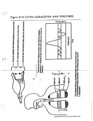
LABORATORY WORKSHEET:<br />
HOW IS <strong>DNA</strong> ANALYZED?<br />
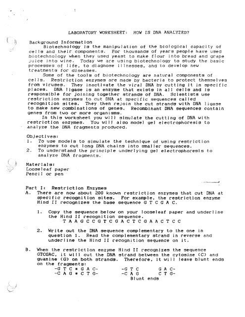
Background Information<br />
Biotechnology Is the manipulation of the biological capacity of<br />
cells and their components. For thousands of years people have used<br />
biotechnology when they used yeast to make flour Into bread and grape<br />
juice into wine. Today we are using biotechnology to study the basic<br />
processes of life, to diagnose Illnesses, and to develop new<br />
treatments for d<strong>is</strong>eases.<br />
Some of the tools of biotechnology are natural components of<br />
cells. Restriction enzymes are made by bacteria to protect themselves<br />
from viruses. They inactivate the viral <strong>DNA</strong> by cutting It In specific<br />
places. <strong>DNA</strong>. I Igase i s an enzyme that ex<strong>is</strong>ts In all cells and Is<br />
responsible for joining together strands of <strong>DNA</strong>. Scient<strong>is</strong>ts use<br />
restriction enzymes to cut <strong>DNA</strong> at specific sequences called<br />
recognition sites. They then rejoin the cut strands with <strong>DNA</strong> ligase<br />
to.make new combinations of genes. Recombinant <strong>DNA</strong> sequences contain<br />
genes from two or more organ<strong>is</strong>ms.<br />
In th<strong>is</strong> worksheet you will simulate the cutting of <strong>DNA</strong> with<br />
restriction enzymes. You will also model gel electrophores<strong>is</strong> to<br />
analyze the <strong>DNA</strong> fragments produced.<br />
Objectives:<br />
1. To use models to simulate the technique of using restriction<br />
enzymes to cut long. <strong>DNA</strong> chains into smaller sequences.<br />
2. To understand the principle underlying gel electrophores<strong>is</strong> to<br />
analyze <strong>DNA</strong> fragments.<br />
Materials:<br />
Looseleaf paper<br />
Pencil or pen<br />
Part I: Restriction Enzymes<br />
A. There are now about 200 known restriction enzymes that cut <strong>DNA</strong> at<br />
specific recognition sites. For example , the restriction enzyme<br />
Hind 11 recognizes the base sequence G T C G A C.<br />
I. Copy the sequence below on your looseleaf paper and<br />
the Hind II recognition sequence.<br />
T A A G C C G T C G A C T C G A A C T C C<br />
underline<br />
2. Write out the <strong>DNA</strong> sequence complementary to the one in<br />
question 1 . Read the complementary strand in reverse and<br />
underline the Hind II recognition sequence on It.<br />
When the restriction enzyme Hind II recognizes the sequence<br />
GTCGAC, it will cut the <strong>DNA</strong> strand between the cytosine (C) and<br />
guanine (G) on both strands. Therefore, It will leave blunt ends<br />
on the fragments:<br />
-G T C a^ G A C- -WG T C G A C-<br />
-C A G* C T G- -C A G C T G--<br />
Blunt ends
The restriction enzyme Eco RI cuts its recognition site at<br />
nonadjacent points on the <strong>DNA</strong> molecule, leaving "sticky" ends.<br />
Eco RI recognizes the base sequence G A A T T C and cuts th<strong>is</strong><br />
sequence between the guanine (G ) and adenine (A) bases:<br />
-G* A A T T C- -G A A T T C-<br />
--C TTAA*G- ---CTTAA\G-<br />
Sticky ends<br />
Sticky ends can bind to similar sticky ends from other Eco<br />
RI-digested fragments. After recombining, the ends are joined by<br />
<strong>DNA</strong> IIgase to form a new pattern of bases. By cutting <strong>DNA</strong> from<br />
two different organ<strong>is</strong>ms with the same enzyme and recombining with<br />
<strong>DNA</strong> ligase, scient<strong>is</strong>ts make recombinant <strong>DNA</strong>.<br />
3. Copy the sequence given below and complete the strand<br />
complementary to It. On both strands Indicate the Eco RI<br />
restriction sites with arrows. Remember to read the<br />
complementary strand in reverse.<br />
G C C T C T A A G A A T T C A G T T C G<br />
4. Once the Eco RI has cut the above <strong>DNA</strong> chain, how many<br />
fragments of <strong>DNA</strong> would there be? Would the ends be blunt or<br />
sticky? <strong>How</strong> many bases would there be In each fragment?<br />
Note. When counting the length of a <strong>DNA</strong> fragment, count only<br />
the number of bases in the upper strand.<br />
C. Below you will see two sequences of <strong>DNA</strong>---<strong>DNA</strong> IA and <strong>DNA</strong> IS. (<strong>DNA</strong><br />
IB <strong>is</strong> a mutant variation of <strong>DNA</strong> IA.)<br />
<strong>DNA</strong> IA<br />
TTG CAA GTC AGA AGA ATT CAA CCT AGG AAT TCT AAG CGC<br />
AAC GTT CAG TCT TCT TAA GTT GGA TCC TTA AGA TTC GCG<br />
<strong>DNA</strong> IB<br />
TTG CAA GTC AGA AGA AGT CAA CCT AGG AAT TCT AAG CGC<br />
AAC GTT CAG TCT TCT TCA GTT GGA TCC TTA AGA TTC GCG<br />
5. What <strong>is</strong> the difference between the two sequences?<br />
6. Copy the sequences onto your paper. Identify the Eco RI<br />
recognition sites on both sequences and mark the sites where<br />
Eco RI would cut with arrows. (You will use these sequences<br />
again In Part II.)<br />
7. <strong>How</strong> many fragments of <strong>DNA</strong> were made from each sequence after<br />
digestion with Eco RI?<br />
8. What are the lengths (in basepairs) of the fragments from the<br />
<strong>DNA</strong> IA and <strong>DNA</strong> IP digestions?<br />
9. Can you recombine any of the "sticky " ends of <strong>DNA</strong> IA and <strong>DNA</strong><br />
IB to make a new sequence of <strong>DNA</strong>? If so, write out the<br />
sequence for one such recombination.
Part II: Gel Electrophores<strong>is</strong><br />
D. Scient<strong>is</strong>ts Identify differences in <strong>DNA</strong> sequences by measuring the<br />
length and number of fragments created by digestion with<br />
restriction enzymes. A technique called gel electrophores<strong>is</strong> Is<br />
used to separate fragments according to length. <strong>DNA</strong> fragments<br />
(cut with an appropriate restriction enzyme) are placed on one end<br />
of a specially-prepared block of agarose called a gel. An<br />
electric current Is applied across the agarose which causes the<br />
strands to migrate through the gel. (Since <strong>DNA</strong> molecules are<br />
negatively charged, they migrate towards the positive electrode.)<br />
The agarose <strong>is</strong> like a sponge with small holes in it. Therefore,<br />
the smaller <strong>DNA</strong> fragments can move through the gel at a faster<br />
rate than larger fragments. Th<strong>is</strong> means that the larger fragments<br />
are found nearer the point of origin. Scient<strong>is</strong>ts then use a<br />
special stain to make the <strong>DNA</strong> fragments v<strong>is</strong>ible as bands. By<br />
counting the number of bands researchers can tell how many<br />
fragments ex<strong>is</strong>t . By observing the d<strong>is</strong>tance each fragment has<br />
migrated, they can determine how big each fragment <strong>is</strong>.<br />
Model el ElectroPhores<strong>is</strong><br />
A. Restriction Enzyme Digestion<br />
Two different strands of <strong>DNA</strong> 1,000 base pairs (bp) long, cut<br />
with the same restriction enzyme (RE).<br />
<strong>DNA</strong> X<br />
<strong>DNA</strong> Y<br />
RE RE RE<br />
10. <strong>How</strong> many fragments are produced by cutting <strong>DNA</strong> X? By cutting<br />
<strong>DNA</strong> Y?<br />
B. Migration D<strong>is</strong>tances of <strong>DNA</strong> In Gels<br />
The size of the <strong>DNA</strong> fragments <strong>is</strong> proportional to the d<strong>is</strong>tance<br />
traveled.<br />
Fragmeent Size (bp)<br />
600 1<br />
400 3<br />
300 4<br />
200: S<br />
too 6
C. Gel Electrophores<strong>is</strong><br />
Appearance of <strong>DNA</strong>s X and Y after digestion and separation by<br />
gel electrophores<strong>is</strong>.<br />
D<strong>is</strong>tance Migrated<br />
X Y Origin<br />
I<br />
1<br />
I<br />
l 1 cm<br />
I<br />
I<br />
1<br />
! 2 cm<br />
1 1<br />
1 3 cm<br />
i<br />
I<br />
1 4 cm<br />
I<br />
1<br />
i<br />
1 5 cm<br />
1 !<br />
1 1 6 cm<br />
11. The fragments generated from <strong>DNA</strong> X and <strong>DNA</strong> Y were then<br />
analyzed by gel electrophores<strong>is</strong>. From the data in figures B<br />
and C, calculate the sizes of the fragments.<br />
12. On your paper draw a simulated gel. Then draw bands<br />
corresponding to the positions to which the Eco RI digests of<br />
<strong>DNA</strong> IA and <strong>DNA</strong> IB would migrate . The fragments of each type<br />
of <strong>DNA</strong> should be in separate lanes and at d<strong>is</strong>tances from the<br />
origin proportional to the numbers of bases In each fragment.<br />
From the following d<strong>is</strong>tances, determine how far your fragments<br />
will migrate : 40 base pairs will migrate 2.5 cm :. 30 bp will<br />
migrate 5 cm; 20 bp will migrate 7.5 cm; and 1 0 bp will<br />
migrate 10 cm. Make sure all the <strong>DNA</strong> <strong>is</strong> accounted for.<br />
1.3. If a mutation causes one of the base pairs in a recognition<br />
site to change, what effect will it have on the ability of the<br />
restriction enzyme to cut at that site?<br />
14. Explain how scient<strong>is</strong>ts use restriction analys<strong>is</strong> to determine<br />
that one sequence of <strong>DNA</strong> Is different from another.