Structure and Function of the Eye: Study Questions 1 ... - Mrs Stovel
Structure and Function of the Eye: Study Questions 1 ... - Mrs Stovel
Structure and Function of the Eye: Study Questions 1 ... - Mrs Stovel

Create successful ePaper yourself
Turn your PDF publications into a flip-book with our unique Google optimized e-Paper software.
Biology 200<br />
<strong>Structure</strong> <strong>and</strong> <strong>Function</strong> <strong>of</strong> <strong>the</strong> <strong>Eye</strong>: <strong>Study</strong> <strong>Questions</strong><br />
1) What stimuli cause <strong>the</strong> eye to blink reflexively?<br />
2) What is <strong>the</strong> purpose <strong>of</strong>: regular blinking?<br />
tears?<br />
3) What causes ambylopia?<br />
4) Which muscles would cause your right eye to look to <strong>the</strong><br />
left?<br />
5) What allows <strong>the</strong> eyeball to retain i ts shape?<br />
6) What causes glaucoma?<br />
7) Describe <strong>the</strong> layers <strong>of</strong> <strong>the</strong> eyeball <strong>and</strong> <strong>the</strong>ir function.<br />
8) Describe <strong>the</strong> function <strong>of</strong> <strong>the</strong> muscles in <strong>the</strong> iris df <strong>the</strong><br />
eye,<br />
9) Describe <strong>the</strong> muscle control <strong>of</strong> <strong>the</strong> lens <strong>of</strong> <strong>the</strong> eye.<br />
10) In what orientation are images projected on <strong>the</strong> retina<br />
<strong>of</strong> <strong>the</strong> eye <strong>and</strong> why do we not perceive <strong>the</strong>m in this way?<br />
11) What are <strong>the</strong> 3 reflex adjustments that are known as<br />
accomodation?<br />
12) What are <strong>the</strong> 2 types <strong>of</strong> receptor cells found in <strong>the</strong><br />
retina <strong>and</strong> what is <strong>the</strong>ir function?<br />
13) Describe <strong>the</strong> adaptation in <strong>the</strong> cells <strong>of</strong> <strong>the</strong> retina that<br />
serves to sharpen <strong>the</strong> visual image <strong>and</strong> fr rove<br />
contrast.<br />
14) What is visual acuity?<br />
15) Describe what <strong>the</strong> fovea centralis is <strong>and</strong> its<br />
implications for vision.<br />
16) What does <strong>the</strong> term 20/20 vision refer to?<br />
17) In terms <strong>of</strong> <strong>the</strong> Snellen test, describe what hyperopic<br />
<strong>and</strong> myopic conditions are.<br />
18) Why is it that <strong>the</strong> rod cells function best in low light<br />
conditions?<br />
19) Explain how <strong>the</strong> cone cells in <strong>the</strong> retina perceive<br />
different colors <strong>of</strong> light,
20) What is <strong>the</strong> cell ular basis for color blindness? What<br />
part <strong>of</strong> <strong>the</strong> population does it affect <strong>the</strong> most?<br />
21) Briefly describe <strong>the</strong> following vision disorders:<br />
a) presbyopia<br />
b) cataracts<br />
c) myopia<br />
d) hyperopia<br />
e) astigmatism<br />
22) What is binocular vision?
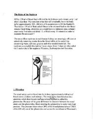
<strong>Structure</strong> <strong>and</strong> <strong>Function</strong> <strong>of</strong> <strong>the</strong><br />
External <strong>Structure</strong><br />
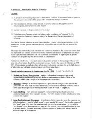
The human eyeball is a slightly battened sphere<br />
about 25 mm (I inch) in diameter. The eyes are<br />
protected from behind by <strong>the</strong> cone-shaped `b ny<br />
eye sockets (orbits) which are padded by layers <strong>of</strong><br />
fatty cushions. Protection <strong>of</strong> <strong>the</strong> exposed surface<br />
(cornea) <strong>of</strong> <strong>the</strong> eye is provided by moveable<br />
eyelids, fringed with protective hairs (eyelashes]<br />
<strong>and</strong> moistened by tears secreted by <strong>the</strong> lacrimal<br />
<strong>and</strong> conjunctival ;l<strong>and</strong>s (Fig. 1).<br />
The eyelids shut re flexively in respoi se t_<br />
-a )ioi v :sporoadhzng objects <strong>and</strong>, certatu<br />
stimuli such as sudden bright ilghts. Ever-, ew<br />
seconds throughout <strong>the</strong> waking hours, <strong>the</strong> eyelids<br />
blink shut for a fraction <strong>of</strong> a second to clear debris<br />
from <strong>the</strong> cornea <strong>and</strong> to spread <strong>the</strong> tears evenly over<br />
it. Tears flow at all times, washing away foreign<br />
particles, lubricating <strong>the</strong> eyelids. <strong>and</strong> keeping <strong>the</strong>.<br />
transparent cornea moist. Excess tears drain into<br />
<strong>the</strong> nasal cavity through <strong>the</strong> nasolocri a l duct.<br />
The flow <strong>of</strong> tears is reflexively increased by irritation<br />
<strong>of</strong> <strong>the</strong> eyes. emotional stress, <strong>and</strong> physical<br />
pain. Tears stream from under <strong>the</strong> lower lids while<br />
o<strong>the</strong>rs drain into <strong>the</strong> nasal cavity, producing <strong>the</strong><br />
familiar "sniffles;" that accompany crying.<br />
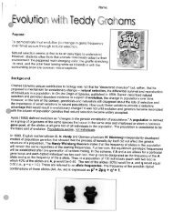
<strong>Eye</strong> Muscles<br />
Obiique<br />
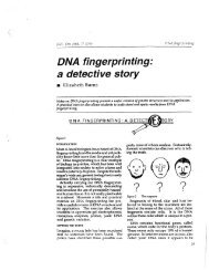
Figure 2 Extrins'c eye muac€ea, Side one too dims <strong>of</strong> <strong>the</strong> a e<br />
<strong>and</strong> its muscular attachments ure shown n <strong>the</strong> upper <strong>and</strong> cwsr<br />
Iiustretions, respectsv fy. ^ontracaon ,t <strong>the</strong> na i! ra, *,us <strong>and</strong><br />
relaxation <strong>of</strong> <strong>the</strong> iaterai rectus cause <strong>the</strong> eye to ro!Pze rtnwarti<br />
(toward <strong>the</strong> nose). 5im:ler actions <strong>of</strong> tt^e o<strong>the</strong>r wpoosingg p3r3 Qt<br />
muscles allow <strong>the</strong> eye to look upward. do°wn rard, or laiefaity,<br />
Six extrinsic (extraocular) muscles cause <strong>the</strong> eve to<br />
move about in its orbit. This makes it possible to<br />
fix <strong>the</strong> gaze on stationary or moving objects, <strong>and</strong> to<br />
coo. dinate <strong>the</strong> two eyes so <strong>the</strong>y ordinarily look at<br />
<strong>the</strong> same thing at <strong>the</strong> same time (Fig. 2). These<br />
muscles originate on <strong>the</strong> bones <strong>of</strong> <strong>the</strong> orbit <strong>and</strong><br />
insert on <strong>the</strong> eyeball itself. The control <strong>and</strong> coerduration<br />
<strong>of</strong> <strong>the</strong>se muscles, which represent some<br />
<strong>of</strong> <strong>the</strong> quickest <strong>and</strong> most precise movements <strong>the</strong><br />
body can make, appears to be a learned process<br />
which develops with <strong>the</strong> maturation <strong>of</strong> v?suci<br />
areas in <strong>the</strong> brain. Malfunctions cause a variety <strong>of</strong><br />
defects in visual alignment. including "crosseves"<br />
<strong>and</strong> amblvopia, which is w<strong>and</strong>ering <strong>of</strong> <strong>the</strong><br />
eye from <strong>the</strong> intended point <strong>of</strong> focus.<br />
Three pairs <strong>of</strong> striated extrinsic muscles are responsible<br />
for each eye's tall range <strong>of</strong> movement.<br />
Four <strong>of</strong> <strong>the</strong> muscles are attached to <strong>the</strong> superior,<br />
inferior, medial, <strong>and</strong> lateral surfaces <strong>of</strong> <strong>the</strong> eyeball<br />
<strong>and</strong>, are named accordingly: superior rectus, to<br />
ter c::r medial rectus, <strong>and</strong> lateral r,-,ctus.<br />
T hey cause <strong>the</strong> eyeball to turn up, down, medially<br />
(inward], <strong>and</strong> laterally (outward), respectively<br />
(F g. 3The two re raining muscles are tart=<br />
sta er c,r am fi `e:;:: r ohiiques which, acting alone,<br />
rotate <strong>the</strong> eyeball.<br />
The <strong>Eye</strong>ball<br />
Eac:h eye consists <strong>of</strong> a three-layered membranous<br />
sac <strong>and</strong> its',ontents. <strong>the</strong>ciqueous humci, vitrcut s
Inferior reCtUS Superior oblique inferior oblique<br />
Figure 3 <strong>Eye</strong> movements caused by <strong>the</strong> extrinsic eye muscles.<br />
The eye at <strong>the</strong> top is focused straight ahead.<br />
body, <strong>and</strong> lens (Fig. 4). Although <strong>the</strong> eye is basically<br />
a saclike structure. its shape is maintained as<br />
a fairly rigid sphere by <strong>the</strong> presence <strong>of</strong> sern.i-fluid<br />
material within distinct anterior <strong>and</strong> posterior<br />
compartments. The anterior chamber, located between<br />
<strong>the</strong> cornea <strong>and</strong> <strong>the</strong> lens, is filled with semiliquid<br />
aqueous humor produced by <strong>the</strong> ciliary<br />
Jody. The aqueous humor drains into tiny canals,<br />
<strong>the</strong> canals <strong>of</strong> Schlemm, near <strong>the</strong> junction <strong>of</strong> <strong>the</strong><br />
cornea <strong>and</strong> iris, at a rate sufficient to maintain <strong>the</strong><br />
shape <strong>of</strong> <strong>the</strong> eyeball. Insufficient drainage <strong>of</strong> aqueous<br />
humor may result in an elevated intrancular<br />
pressure, a painful condition (glaucoma) that can<br />
seriously damage <strong>the</strong> eye.<br />
The large posterior cavity <strong>of</strong> <strong>the</strong> eye, bordered<br />
by <strong>the</strong> retina, ciliary body, suspensory ligaments,<br />
<strong>and</strong> lens, is called <strong>the</strong> vitreous body. It is filled<br />
with a transparent jelly which, like <strong>the</strong> aqueous<br />
humor, maintains <strong>the</strong> spherical shape <strong>of</strong> <strong>the</strong><br />
eyeball.<br />
The outermost layer is a tough, fibrous, graywhite<br />
membrane in <strong>the</strong> posterior part <strong>of</strong> <strong>the</strong> eye<br />
called <strong>the</strong> sclera or sclerotic coat, This layer is<br />
continuous with <strong>the</strong> transparent cornea at <strong>the</strong><br />
front <strong>of</strong> <strong>the</strong> eye <strong>and</strong> appears at each margin <strong>of</strong> <strong>the</strong><br />
cornea as <strong>the</strong> white <strong>of</strong> <strong>the</strong> eye.<br />
The middle laver <strong>of</strong> <strong>the</strong> eve wall is <strong>the</strong> choroid<br />
layer which contains many blood vessels that<br />
bring oxygen <strong>and</strong> nourishment to <strong>the</strong> cells <strong>of</strong> this<br />
structure <strong>and</strong> to <strong>the</strong> o<strong>the</strong>r coats as well. Many<br />
?,igment granules are found in <strong>the</strong> chcroid laver,<br />
,`ausing it to have a deep reddish-purple color.<br />
This prevents te:lection <strong>of</strong> light inside <strong>the</strong> eye in<br />
much <strong>the</strong> sane way as a coating <strong>of</strong> black paint<br />
inside .3 c_ur era el,minates stray rays <strong>of</strong> light irsat<br />
might o<strong>the</strong>rwise ruin <strong>the</strong> film.<br />
Around <strong>the</strong> edge <strong>of</strong> <strong>the</strong> cornea <strong>the</strong> choroid<br />
coat forms <strong>the</strong> ciliary, body, a thickened structure<br />
containing smooth muscle. Circling <strong>the</strong> anterior<br />
margin <strong>of</strong> <strong>the</strong> ciliary body is a thin muscular diaphragm<br />
with an opening in <strong>the</strong> center called <strong>the</strong><br />
iris. The iris may be blue, brown, green, gray, or<br />
black depending on <strong>the</strong> amount <strong>of</strong> pigment (c elanin)<br />
contained in its tissue. If no pigment is present,<br />
<strong>the</strong> iris appears to be blue. Small amounts <strong>of</strong><br />
pigment cause <strong>the</strong> eve to have a gray or green color<br />
<strong>and</strong>, as <strong>the</strong> amount <strong>of</strong> pigment increases, <strong>the</strong> depth<br />
<strong>of</strong> color approaches black. <strong>Eye</strong> color is a hereditary<br />
trait, meaning that <strong>the</strong> distribution <strong>of</strong> melanin<br />
pigment in <strong>the</strong> eye is controlled by genes.<br />
Two sets <strong>of</strong> smooth muscles are found Within<br />
<strong>the</strong> iris <strong>of</strong> <strong>the</strong> eye. The inner set is circular <strong>and</strong><br />
serves to constrict <strong>the</strong> inner edge <strong>of</strong> <strong>the</strong> iris. The<br />
o<strong>the</strong>r set is perpendicular to <strong>the</strong> circular muscles.<br />
When this set <strong>of</strong> muscles contracts, <strong>the</strong> inner edge<br />
<strong>of</strong> <strong>the</strong>...iri is dt w-n open. Contraction <strong>of</strong> <strong>the</strong> iris<br />
muscles is regulated by <strong>the</strong> autonomic nervous<br />
system through <strong>the</strong> oculomotor (third cranial)<br />
nerve.<br />
The opening in <strong>the</strong> center <strong>of</strong> <strong>the</strong> iris is <strong>the</strong><br />
pupil, through which light enters <strong>the</strong> eye. The<br />
diameter <strong>of</strong> <strong>the</strong> pupil varies continually <strong>and</strong> is<br />
regulated by <strong>the</strong> amount <strong>of</strong> contraction <strong>of</strong> <strong>the</strong> two<br />
sets <strong>of</strong> iris muscles. Thus, <strong>the</strong> amount <strong>of</strong> light<br />
allowed to enter <strong>the</strong> eye is determined by <strong>the</strong> size<br />
<strong>of</strong> <strong>the</strong> pupil, If a bright light is flashed into <strong>the</strong> eye,<br />
<strong>the</strong> circular muscles contract very rapidly <strong>and</strong> <strong>the</strong><br />
pupil becomes smaller. In dim light <strong>the</strong> o<strong>the</strong>r set <strong>of</strong><br />
iris muscles contracts <strong>and</strong> <strong>the</strong> pupil becomes<br />
larger.<br />
The Lens<br />
Directly behind <strong>the</strong> pupil is <strong>the</strong> lens, a transparent,<br />
semi-elastic structure shaped somewhat like a<br />
biconvex disk. As show in Figure 5. <strong>the</strong> lens is<br />
held in place by suspensory ligaments attached to<br />
<strong>the</strong> ciliary body <strong>of</strong> <strong>the</strong> choroid laver. The suspensory'<br />
ligaments control <strong>the</strong> shape <strong>of</strong> <strong>the</strong> lens by<br />
exerting varying amounts <strong>of</strong> tension on it. An increase<br />
in tension causes <strong>the</strong> lens to flatten slightly:,<br />
while a decrease in tension allows <strong>the</strong> lens to<br />
return to a thicker configuration by virtue <strong>of</strong> its<br />
elasticity. The amount <strong>of</strong> tension imposed on <strong>the</strong><br />
6
Oistart<br />
okjoct<br />
.'7'uSCiSrr3t3F`;F<br />
tiyga?t9i't<br />
Latera l recfu3 _<br />
Figure pole <strong>of</strong> th lens it acorn nodatic n fordatant arts n r<br />
vision. 7co- Raiaxation <strong>of</strong> <strong>the</strong> c:ilarv musi.ls causes f' sac:: r:<br />
sory ligam ents todraw <strong>the</strong> iens.nto a flattened configurati or: so<br />
provides ::ha minimum amount <strong>of</strong> focusing power for cistart<br />
vision. Bcc rr: Contraction <strong>of</strong> <strong>the</strong> ciliary muscle rsi Uva,3 rtj.<br />
tension on <strong>the</strong> suspensory ligamen ts <strong>and</strong> allays <strong>the</strong> era to<br />
thicken ' r greater focusing power i, near vision.<br />
refroc: on, 4"Vh ' a parallel light rains pass 1 ough<br />
<strong>the</strong> biconvex lens <strong>of</strong>'.:ie eye, <strong>the</strong> y are bent so as to<br />
converge bt a single point to form a bright pot on<br />
<strong>the</strong>retin ;:., The point at which a clear focus c:c: nt<br />
is <strong>the</strong> `+, 3F point or principal rocus <strong>and</strong> its -, i -<br />
ance from <strong>the</strong> Jens is <strong>the</strong> focal length (Fig. 61. he<br />
focal l ength decreases as <strong>the</strong> curvature o :cr s<br />
becomess more pronounced <strong>and</strong> increases as <strong>the</strong><br />
Flguru 4 Top: Horizontal cross section through <strong>the</strong> right eye<br />
lens becomes flatter. Thusv when distant obji is<br />
showing interior structures. The bloo d vessels run a,ong <strong>the</strong> hack<br />
<strong>of</strong> <strong>the</strong> eye between <strong>the</strong> retina <strong>and</strong> <strong>the</strong> vitreous body. Bottom: are viewed. <strong>the</strong> lens is pulled into a slighs_iy flatter<br />
Vert #cat cross section <strong>of</strong> <strong>the</strong> antarior portion <strong>of</strong> <strong>the</strong> eye showing shape by increased tensiat:t in <strong>the</strong> suspensor,, hg<br />
<strong>the</strong> lens ;n relation to <strong>the</strong> suspensory ligaments e,td chary muscle.<br />
aments, A decrease in tension, accomplished by<br />
contraction <strong>of</strong> <strong>the</strong> ciliary muscle, allows t`e ien. to<br />
thicken slightly, which provides additional headli<br />
%ng <strong>of</strong> lig h t rays for near vsitr:°s.<br />
t r^ lens by <strong>the</strong> <strong>the</strong> suspensor ligaments light is re ulated bysource is an abject instead <strong>of</strong><br />
<strong>the</strong> ciliary muscle, a smooth -muscle in <strong>the</strong> viii .rv point. many light rays from. each part <strong>of</strong> <strong>the</strong> object<br />
body.<br />
come into focus at a partic ular distance from <strong>the</strong><br />
ens. That Light rays, way, which normally an travel inverter straight image <strong>of</strong> <strong>the</strong> obh tct is<br />
lines, slow down when passing through transparent<br />
materials such as water, glass, or <strong>the</strong> '.ens <strong>of</strong> <strong>the</strong> prltted '-jy <strong>the</strong> brain, it is flipped over <strong>and</strong> app4, 3As<br />
projected onto <strong>the</strong> retina. When <strong>the</strong> image is i`:ter--<br />
eye. "'he path <strong>of</strong> <strong>the</strong> light beam, i s bent as it slows so right side= flp.<br />
it travels at a different angle. an event caked Since light rays are bent whenever <strong>the</strong>:, ; : s
-'.._"'-.....^ Point Source<br />
<strong>of</strong> light<br />
- C<br />
Figure 6 Refrectwon (bending) <strong>of</strong> 4fight rays by <strong>the</strong> lens sv9tc' <strong>of</strong> -,e eye. i',e kiyht soi.°r
erabie degree before it reaches <strong>the</strong> Ae o-lying .onto<br />
<strong>and</strong> cones, so several photoreceptors may be<br />
stimulated by a tiny point <strong>of</strong> light. If ail <strong>of</strong> <strong>the</strong><br />
receptors responded, you would perceive only<br />
fuzzy blob <strong>of</strong> light surrounded by a halo. However.<br />
nerve impulses that arise from a light-stimulated<br />
retinal receptor inhibit <strong>the</strong> adjacent receptors so<br />
<strong>the</strong>y fail to respond to <strong>the</strong> illumination. This inhibition<br />
sharpens <strong>the</strong> visual image <strong>and</strong> improves its<br />
contrast. The mutual inhibition <strong>of</strong> retinal cells is<br />
demonstrated in Figure B.<br />
When an evenly illuminated surface is<br />
3<br />
:'tO%'=' t;d. all <strong>of</strong> 11-e retinal rece ptors arc partly infiic)<br />
ited: as brightness increases, so does inhibition.<br />
with <strong>the</strong> result that <strong>the</strong> visual field does not seem<br />
as bright as it o<strong>the</strong>rwise would. Therefore, a<br />
brightly sunlit day appears only a few times brighter<br />
than a cloudy one. although <strong>the</strong> difference in<br />
illumination may be as great as a hundredfold.<br />
'visual Acuity<br />
x tte amp c at at detail <strong>the</strong> eye can distinguish ;s<br />
known as vi sual acuity <strong>and</strong> depends upon. <strong>the</strong><br />
retina a s well as <strong>the</strong> refractive system <strong>of</strong> <strong>the</strong> eve.<br />
For two distant objects to be perceived as separate<br />
objects, <strong>the</strong> light rays from those objects must be<br />
focused on separate retinal receptors with at least<br />
are unstimulated receptor in between. in <strong>the</strong><br />
center <strong>of</strong> <strong>the</strong> retina is a tiny depression called <strong>the</strong><br />
fovea centralis. This area contains only cones.<br />
which are packed toge<strong>the</strong>r more densely than in<br />
any o<strong>the</strong>r portion <strong>of</strong> <strong>the</strong> retina. Therefore, <strong>the</strong> fovea<br />
centralis is <strong>the</strong> region <strong>of</strong> greatest visual acuity.<br />
When one looks directly at an object, most <strong>of</strong> <strong>the</strong><br />
light rays from that object fall upon <strong>the</strong> fovea.<br />
The angle formed by light rays entering <strong>the</strong><br />
eye from two separate points determines <strong>the</strong> ability<br />
to distinguish <strong>the</strong> points as definite o bjects.<br />
Ught<br />
Figure; Dlagrarnmatic cross section <strong>of</strong> <strong>the</strong> human retina. incoming light must pass through <strong>the</strong> fusers <strong>and</strong> Cella <strong>of</strong><br />
<strong>the</strong> various retinal layers before reaching <strong>the</strong> Sensitive tips <strong>of</strong> <strong>the</strong> rocs <strong>and</strong> cones. Nerve mpulses from <strong>the</strong> rods <strong>and</strong><br />
cones travel' toward <strong>the</strong> brain via <strong>the</strong> optic nerve fibers.<br />
9
Figure S The grillwork Wusion is an exa.-mpie <strong>of</strong> optical inriibitior, Ei<strong>the</strong>r set <strong>of</strong> diagonal bars !!eft <strong>and</strong> center) can by<br />
viewed as a pattern <strong>of</strong> alternating black-<strong>and</strong>-white stripes. However, when <strong>the</strong> patterns are superimposed to form a<br />
gridwork, as in <strong>the</strong> right h<strong>and</strong> illustration, nonexistent gray spots seem to appear at <strong>the</strong> intersections <strong>of</strong> <strong>the</strong> white<br />
Stripes.<br />
n<br />
Visual acuity can be measured with <strong>the</strong> aid <strong>of</strong> a<br />
Snellen eye chart (Fig. 0 <strong>and</strong> 10).<br />
At a distance <strong>of</strong> 20 feet from <strong>the</strong> chart, a normal<br />
eye can read <strong>the</strong> lines down through <strong>the</strong><br />
2{}-foot ILne, but none smaller. The visual acuity <strong>of</strong><br />
this eye is said to be 200/20 because at 20 feet <strong>the</strong><br />
individual can read <strong>the</strong> 20-foot line. This is normal<br />
vision, which also requires a normal refractive<br />
system because visual acuity is reduced by defective<br />
refraction. A farsighted (hyperopic) person<br />
may be able to read <strong>the</strong> 15-foot line <strong>and</strong> have 20115<br />
vision, meaning that he can distinguish letters at<br />
20 feet that a person with normal vision would be<br />
able to read only at 15 feet. Conversely, if <strong>the</strong><br />
smallest line that a nearsighted (myopic) individual<br />
can read is that for 100 feet, his visual acuity<br />
is 201100, considerably less than normal according<br />
to <strong>the</strong> Snellen test.<br />
Chemistry <strong>of</strong> Vision<br />
Rhodopsin, a chemical compound found in <strong>the</strong><br />
rod cells, <strong>and</strong> a series <strong>of</strong> closely related chemicals<br />
located in <strong>the</strong> cones are <strong>the</strong> substances ultimately<br />
responsible for <strong>the</strong> ability to see. These chemicals<br />
are broken down by <strong>the</strong> action <strong>of</strong> light, stimulating<br />
<strong>the</strong> photoreceptor to discharge a small electrical<br />
impulse that is transmitted to o<strong>the</strong>r neurons in <strong>the</strong><br />
retina <strong>and</strong>, by way <strong>of</strong> tho optic nerve, to <strong>the</strong> brain.<br />
Rhodopsin <strong>and</strong> <strong>the</strong> o<strong>the</strong>r visual chemicals are <strong>the</strong>n<br />
resyn<strong>the</strong>sized by <strong>the</strong> photoreceptors.<br />
Rhodopsin consists <strong>of</strong> a form <strong>of</strong> vitamin A<br />
10<br />
combined with a large protein molecule cailc&J.<br />
opsin, Strong daylight or artificial 'tight causes<br />
rhodopsin to be broken clown to retiner.e <strong>and</strong><br />
scotopsin. Since this breakdown occurs faster than<br />
<strong>the</strong> rhodops n can be resy°nt.hesized (Fig. 11), <strong>the</strong><br />
rod cells do not function ore] under strong illumination,<br />
althhough <strong>the</strong>y participate to some decree In<br />
daylight vision. The rods function mainly under<br />
conditions <strong>of</strong> low light intensity, providing us<br />
with <strong>the</strong> ability to see at night (night vision). Vision<br />
in dim light has poor detail <strong>and</strong> little or no color.<br />
Objects can be seen more clearly in dim light if<br />
viewed from <strong>the</strong> side <strong>of</strong> <strong>the</strong> eye, because rods are<br />
lacking in <strong>the</strong> central portion <strong>of</strong> <strong>the</strong> posterior retinal<br />
lining: (fovea centralis) where <strong>the</strong> majority <strong>of</strong><br />
<strong>the</strong> incoming light is normally focused.<br />
Lack <strong>of</strong> dietary vitamin A leads to rhodop,-;ire<br />
deficiency, which results in night blindness<br />
(rnyctalopia). Long-term vitamin A deficiency may<br />
lead to permanent loss <strong>of</strong> rod cells which, like<br />
o<strong>the</strong>r types <strong>of</strong> nerve tissues. are incapable <strong>of</strong> .regeneration.<br />
Color Vision<br />
Visible light can be separated into six distinct c^ for<br />
components-violet, blue, green, yellow, orange,<br />
<strong>and</strong> red-by passing <strong>the</strong> light through a prism (Fig.<br />
12). The wavelengths <strong>of</strong> <strong>the</strong>se light rays go from<br />
00 0 A (violet) up to 7000 A (red), <strong>the</strong> limits <strong>of</strong><br />
4<br />
F 'S Y<br />
h1rnan colot reception.<br />
Any visible color can be produced by corr^biu
I-1 N<br />
D F 9^I<br />
1-0 T<br />
T F<br />
0 F N P T H<br />
P H U N T D Z<br />
Figure 10 At <strong>the</strong> prescribed viewing distance. ail ie*lters -„- i<br />
visual acuity chart subtend <strong>the</strong> same visual angle <strong>and</strong> for ni rettr<br />
images <strong>of</strong> <strong>the</strong> same silo.<br />
It is a common experience to look directly fit L<br />
bright light for an instant <strong>and</strong> Find thati a similar<br />
ima ge o <strong>the</strong> glowing bulb remains in ihe<br />
f.e:id for some time afterward. The phenomi::no n<br />
railed a positive afterimage because t r u C:f<br />
<strong>the</strong> image are both about <strong>the</strong> same col Dr. `yc<br />
prolonged exposure <strong>of</strong> <strong>the</strong> eye to <strong>the</strong> same bri<br />
light may result in an afterimage called a negat;ve<br />
afterimage. Viewing <strong>the</strong> light for a few se o^:_<br />
through a piece <strong>of</strong> red cellophane will produce<br />
negative afterimage that appears to be <strong>the</strong> complementary<br />
color, green. Repeating <strong>the</strong> experimeu`<br />
with green cellophane will yield a red negative.<br />
afterimage.<br />
N P X 'r z F, H<br />
1<br />
Figure 9 The st<strong>and</strong>ard Snatlen visual acuity chart. The numbers<br />
along <strong>the</strong> right border indicate <strong>the</strong> distance from whicn a person<br />
with "normal" visual acuity can perceive <strong>the</strong> lertara in each line.<br />
The numbers along <strong>the</strong> left border give <strong>the</strong> visual acuity index for<br />
each line <strong>of</strong> print when <strong>the</strong> chart is viewed from a distance <strong>of</strong> 2C<br />
Net,<br />
Rhodopsin<br />
Lmi-rhodpsin<br />
i<br />
ing lights <strong>of</strong> three optical primary coicrs-red,<br />
blue, <strong>and</strong> green-in varying proportions (Fig. 13).<br />
The retina contains three types <strong>of</strong> cones, each sensitive<br />
to a wavelength <strong>of</strong> light that corresponds to<br />
one <strong>of</strong> <strong>the</strong> primary colors. The sensitivities <strong>of</strong> <strong>the</strong><br />
retinal cone cells are not exactly to red, blue, <strong>and</strong><br />
green light, so considerable overlap exists (Fig. 14).<br />
Any <strong>of</strong> <strong>the</strong> hundreds <strong>of</strong> visible colors can be<br />
achieved by stimulating <strong>the</strong> various cones in <strong>the</strong><br />
proper proportions, just as a painter can obtain any<br />
color by mixing certain amounts <strong>of</strong> <strong>the</strong> three primary<br />
hues. Equal stimulation <strong>of</strong> <strong>the</strong> three types <strong>of</strong><br />
cones is perceived as white light.<br />
Ffgu;e #t A schematic summary <strong>of</strong> <strong>the</strong> chemical changes .n t=a<br />
rhodopsin cycle during exposure to light <strong>and</strong> dark cwe^oitior^..<br />
Rhodopsin is converted to scotopsin <strong>and</strong> retinene in <strong>the</strong> pr, w.<br />
ence <strong>of</strong> strong light. iesyn<strong>the</strong>sis <strong>of</strong> rhodopsin, which orc.::: r.-<br />
in deiknes$, reguirss a certain amount <strong>of</strong> vitamin A.<br />
I<br />
,,Jal r<br />
Scotopsin<br />
Retinerle<br />
4 t<br />
Vitamin A
A<br />
Figure 12 A Prism seParates white !igttt into The colors <strong>of</strong> <strong>the</strong> spectru<br />
wavelengths <strong>of</strong> visible light are shown in Angstrom nos.<br />
The occurrence <strong>of</strong> visual afterimages is<br />
thought to be dependent upon <strong>the</strong> way incoming<br />
light rays are processed by <strong>the</strong> retina. As explained<br />
earlier, light reaching <strong>the</strong> eye is focused by <strong>the</strong> lens<br />
to form an image on <strong>the</strong> reti na. -Brief exposure to an<br />
ntense light source apparently stimulates <strong>the</strong><br />
cone cells to convey signals to <strong>the</strong> visual cortex in<br />
<strong>the</strong> brain for a few moments after <strong>the</strong> eye has been<br />
directed away from <strong>the</strong> light, resulting in a posit; ve<br />
afterimage. Prolonged exposure to <strong>the</strong> light<br />
stimulus causes <strong>the</strong> cones responding to those particular<br />
wavelengths to become temporarily<br />
fatigued or aadapted. The negative afterimage, produced<br />
by continued activity <strong>of</strong> cones that are less<br />
intensely stimulated., is darker than <strong>the</strong> bright light<br />
that caused it: The appearance <strong>of</strong> afterimages in<br />
complementary colors (red following green:. o„<br />
example) can be explained in <strong>the</strong> same way.<br />
Afteric-oages are Illusions, serving to remind<br />
us that <strong>the</strong> senses are sometimes imperfect<br />
mediators between <strong>the</strong> external world <strong>and</strong> our<br />
perception <strong>of</strong> it. O<strong>the</strong>r types <strong>of</strong> optical illusions<br />
include those involving <strong>the</strong> iaterprotatiori <strong>of</strong> spatial<br />
relationships (Fig. 16),<br />
1<br />
,figure 13 V arious =ntermediate colors are produced when <strong>the</strong><br />
?rrmary co lors <strong>of</strong> light are superimposed. Wrritu light results<br />
when equal amounts <strong>of</strong> <strong>the</strong> three primary coicrs (red. uue :in,d<br />
aree„' are mixed.<br />
Color Deficiency<br />
inadequate function <strong>of</strong> one or more types <strong>of</strong> cones<br />
results in co'or deficiency, <strong>the</strong> inability to perceive<br />
correctly one or more <strong>of</strong> <strong>the</strong> primary colors. This<br />
term is synonymous with "color blindness,'<br />
which is no longer used because <strong>of</strong> <strong>the</strong> negative<br />
feelings generated by <strong>the</strong> word "blindness." The<br />
degree <strong>of</strong> deficiency is variable, ranging fro °rl<br />
minor confusion <strong>of</strong> color shades to a total lack <strong>of</strong><br />
color inerce.ption. The :nest common fn m <strong>of</strong> color<br />
deficiency is red-green color deficiency, which is<br />
due to s l ro e:m with tie red receptors. Lorafu u.u<br />
12
4000 bow<br />
An"<br />
wfY^'en'h IA)<br />
Figure to Sensitivity ranges <strong>of</strong> <strong>the</strong> three retinal pigments,<br />
between certain shades <strong>of</strong> red <strong>and</strong> green occurs,<br />
since light rays <strong>of</strong> <strong>the</strong>se wavelengths stimulate <strong>the</strong><br />
same combination <strong>of</strong> cones. This defect is a sex -<br />
linked genetic trait which affects 8 to 9 percent <strong>of</strong><br />
<strong>the</strong> male population.<br />
Figure 15 Demonstration <strong>of</strong> negat.ve<br />
afterimages. Stare fixedly at <strong>the</strong> center<br />
<strong>of</strong> <strong>the</strong> cross for 20 to 30 seconds, <strong>the</strong>n<br />
close your eyes or look at a •,srh €te scar..<br />
face. You was see a cross with <strong>the</strong> col<br />
ors reversed (i is., <strong>the</strong> vertical limb will=.<br />
be red <strong>and</strong> <strong>the</strong> hor €zontal limb green) if<br />
<strong>the</strong> image 3 s produced on a white surface.<br />
i ts size will depend upon <strong>the</strong> d„-<br />
tance <strong>of</strong> <strong>the</strong> surface from <strong>the</strong> eyes<br />
fl<br />
I<br />
Figure 1fi Optical illusions. Left: Two identical ractanrjies super imposed on a photograph <strong>of</strong> receding railroad<br />
tracks. Although <strong>the</strong> rectangles are exactly <strong>the</strong> same size, <strong>the</strong> uoDer one seems to be longer. We know that <strong>the</strong> distant<br />
railroad ties are as large as those in <strong>the</strong> foreground, but any object lying between <strong>the</strong> rails in <strong>the</strong> middle distance fin<br />
this case. <strong>the</strong> top rectangle) is unc snsoiousty enlarged. -enter An i;ptiCal rIiusion in which <strong>the</strong> vertical lines in bath<br />
figures are exactly <strong>the</strong> same length, although <strong>the</strong> one on ins +e`t appears to be apprec,abiy longer Extending <strong>the</strong><br />
diagonal lines outward in <strong>the</strong> left illustration ano inward in <strong>the</strong> rigryt ilustration causes <strong>the</strong> viewer to meite an incorrect<br />
comparison <strong>of</strong> <strong>the</strong> lengths <strong>of</strong> <strong>the</strong> two vertical lines. Rrght An ambiguous "gore redrawn from an illustration by<br />
cartoonist W. E- HO demonstrates <strong>the</strong> importance <strong>of</strong> interpretation in <strong>the</strong> process <strong>of</strong> vi,ual perception. Ei<strong>the</strong>ra young<br />
g€ri or an old woman can be seen, depending upon <strong>the</strong> viewer s perspective. The Chln at <strong>the</strong> young girl s also <strong>the</strong> nose<br />
<strong>of</strong> <strong>the</strong>y old woman.
Common Disorders <strong>of</strong> Vision<br />
Presbyopia<br />
Thickening <strong>of</strong> <strong>the</strong> lens for near vision is permitted<br />
by contraction <strong>of</strong> <strong>the</strong> ciliary muscle, which lessens<br />
<strong>the</strong> tension imposed upon <strong>the</strong> lens by <strong>the</strong> suspensory<br />
, ,gaments. However, it is <strong>the</strong> elasUeftye#<br />
lens thatactually causes <strong>the</strong> thickening because<br />
<strong>the</strong> lens automatically assumes a fairly round<br />
shape as external tension is removed. An estimation<br />
<strong>of</strong> <strong>the</strong> degree <strong>of</strong> lens elasticity can be obtained<br />
by measuring <strong>the</strong> near point, which is <strong>the</strong> shortest<br />
distance from <strong>the</strong> eye that an object can be viewed<br />
clearly.<br />
One <strong>of</strong> <strong>the</strong> normal consequences <strong>of</strong> aging is a<br />
gradual loss <strong>of</strong> lens elasticity, which begins at an<br />
early age but proceeds more rapidly during later<br />
years. As <strong>the</strong> elasticity <strong>of</strong> <strong>the</strong> lens decreases, <strong>the</strong><br />
near point recedes <strong>and</strong> accommodation becomes<br />
diminished to <strong>the</strong> point where reading becomes<br />
difficult (Fig. 17). This lack <strong>of</strong> near vision, known<br />
as presbyopia, is <strong>of</strong>ten corrected with convex<br />
lenses or bifocals (a convex insert in regular eyeglasses).<br />
100<br />
90a<br />
7<br />
Cataract<br />
The lens <strong>of</strong> <strong>the</strong> eye occasionally becomes opaque<br />
<strong>and</strong> light rays are prevented from reaching <strong>the</strong><br />
tw 20 30 i0 50 00 70<br />
Age to Y"ra<br />
Figure 17 The near point recedes with advancing ag:e.oecauss<br />
<strong>the</strong> lens <strong>of</strong> <strong>the</strong> eye gradually loses its ability to accomr: ;date `,,r<br />
near vision. The most dramatic changes are usually no,eo in<br />
humans beyond 40 years <strong>of</strong> age.<br />
Figure 18 Left: A normal lens transrnits it -°n ng iinh' so it fFilis on toe r t;na `Ovesa.. Ighr: C^pactty <strong>of</strong> ire lens, a<br />
condition known as cataract, prevents <strong>the</strong> incoming Sight rays from oass?ng through to <strong>the</strong> retina.<br />
14
ens<br />
Norm,<br />
eye<br />
MacuO .- _^^._ # tit<br />
r_ _.-<br />
lutes L<br />
Astigmat"u.'n<br />
Figure 1 S ton: Nesrsigfltecf ess. or myos ,a, s a c onc;itioa in n?sr. ;ncc^rnang ri:fit t r2ys oc spa to focus in ira t 2" °ne<br />
retina. The ur biem is Cnrrecre by placing a biconcave lens in !r:nt <strong>of</strong> <strong>the</strong> eye, i'enrer: The focal coint for,ncoro:rr,g<br />
light rays is behind <strong>the</strong> retina lr, <strong>the</strong> :ar dition known as farsighteortess or hyperopia. A biconvex lens is used to correct<br />
this problem Bottom: Astigmatism its condition in which uneven focus^nq <strong>of</strong> fns visual image results from distort,Q",<br />
<strong>of</strong> Me curvature <strong>of</strong> <strong>the</strong> lens or Cornea. The problem is corrected with an optical lens that has <strong>the</strong> same degree u<br />
astigmatic n: but at right angles 'o <strong>the</strong> otarie <strong>of</strong> astigmatisrn In ti^e eye.<br />
retinal photoreceptors. This condition. .:own as<br />
cataract, is treated by S irgirc l re.Tr va <strong>of</strong> th e lens<br />
<strong>and</strong> strong eyegleisses to conip risate or its absence<br />
(Fig. 18). A new treatment, which involves<br />
repiacement <strong>of</strong> <strong>the</strong> clouded lens with a plastic one<br />
F<br />
lacking <strong>the</strong> power <strong>of</strong> accommodation, is stil l<br />
in <strong>the</strong><br />
experimental stage.<br />
Nearsightedness<br />
Nearsightedness or myopia occurs rshert Nle<br />
eyeball is abnormally long. This causes <strong>the</strong> visual<br />
imeg€ in come i.ito focus .',?7 front <strong>of</strong> <strong>the</strong> -tiny (A '<br />
t 91.<br />
myopic 'individual Can See i ell at slhoct<br />
.%xr F:t`sa, but tf e lens cannot flatten -n, g i E<br />
focus distant obiects on <strong>the</strong> retina. The, problem IS<br />
ct . cectesd with a conc .ve lens, which causes light<br />
m vs from a distant obiect to diverse li gfitly as t .€;i<br />
ente.1 <strong>the</strong> eye o <strong>the</strong> focal point falls or, ti e: retina.<br />
Farsightedness<br />
TN opposite <strong>of</strong> nears'gtitfrlness is t € t dies:,<br />
or iIvperr^i^irz, wahich occurs ^vh n <strong>the</strong> y==ball i,,<br />
kJ
abnormally short. This Ca^nses <strong>the</strong> visua<br />
to be focused behind <strong>the</strong> retina (Fig. %91. The eye<br />
inctions normally when distant objects are<br />
sewed, but <strong>the</strong> refractive system, lacks adequate<br />
strength for accommodation in near vision. The<br />
condition is treated by placing a convex lens in<br />
front <strong>of</strong> <strong>the</strong> eye so light rays from near objects<br />
converge slightly before <strong>the</strong>y enter <strong>the</strong> eye, This<br />
causes <strong>the</strong> visual image to, be focused properly on<br />
<strong>the</strong> retina.<br />
Astigmatism<br />
unfortunately, <strong>the</strong> elements <strong>of</strong> <strong>the</strong> refractive systern<br />
are not always perfectly symmetrical. imperfections<br />
in <strong>the</strong> shape <strong>of</strong> <strong>the</strong> cornea or lens cause<br />
incoming light rays to converge unevenly so <strong>the</strong>y<br />
do not focus at a specific point on <strong>the</strong> retina. This<br />
condition is called astigmatism (Fig. 1`'i <strong>and</strong> results<br />
in blurred or distorted visual images. Using<br />
an astigmatism testing chart (Fig. 20), even minor<br />
amounts <strong>of</strong> astigmatism can be detected. To an<br />
individual with an asymmetrical cornea, lines in<br />
one plane appear sharp <strong>and</strong> clear while those at<br />
right angles are fuzzy <strong>and</strong> poorly focused. The<br />
tuation is corrected by a lens with <strong>the</strong> same de-<br />
,;#ee <strong>of</strong> astigmatism. but at right angles to that <strong>of</strong> <strong>the</strong><br />
cornea so <strong>the</strong> effects cancel each o<strong>the</strong>r,<br />
Figure 20 An asi^gmatism testing chart. € he radiating lines we<br />
appear to be darker aro bolder in t he planes <strong>of</strong> <strong>the</strong> eye which are<br />
astigmatic. Since Eight rays are bent to a greater degree where <strong>the</strong><br />
cornea has <strong>the</strong> most pronounced curvature, light passing<br />
through that area wilt come to a tacos in front <strong>of</strong> <strong>the</strong> retina,<br />
resulting in a blur. The eye tends to focus light from Me flattened<br />
(astigmatic) area; that from She normally curved surfaces will be<br />
refracted too much.<br />
16
Visual Fields <strong>and</strong> Binocular Vision<br />
We have seen how rays <strong>of</strong> light are focused by <strong>the</strong><br />
lens to fall in a concentrated pattern on <strong>the</strong> sensitive<br />
photoreceptors <strong>of</strong> <strong>the</strong> retina. thus stimulating<br />
<strong>the</strong> formation <strong>of</strong> visual images. The portion <strong>of</strong> <strong>the</strong><br />
external world t be seen. Brit t mmoving,<br />
by one eye is called <strong>the</strong> visual field. The extent <strong>of</strong><br />
<strong>the</strong> visual field is limited chiefly by <strong>the</strong> bony structure<br />
<strong>of</strong> <strong>the</strong> eye socket <strong>and</strong> by <strong>the</strong> bridge <strong>of</strong> <strong>the</strong> nose,<br />
in humans <strong>the</strong> two eyes are only a few centimeters<br />
apart so <strong>the</strong>y register similar views <strong>of</strong> <strong>the</strong> external<br />
world. In fact. <strong>the</strong> visual fields actually overlap,<br />
which makes binocular vision possible. Binocular<br />
vision is <strong>the</strong> reception <strong>of</strong> slightly different visual<br />
images by each <strong>of</strong> <strong>the</strong> two eyes. Light rays from<br />
different parts <strong>of</strong> <strong>the</strong> visual field come to a focus at<br />
a particular point on each retina. The light rays<br />
from <strong>the</strong> object <strong>of</strong> focus (i,e.. <strong>the</strong> object that is being<br />
looked at) fail upon <strong>the</strong> fovea centralis. while<br />
those from surrounding areas come into focus at<br />
nearby retinal sites.<br />
When a single object such as a pencil tip is<br />
viewed with both eyes, <strong>the</strong> light rays from it fall<br />
upon <strong>the</strong> fovea <strong>of</strong> each retina, yet <strong>the</strong> visual image<br />
formed is <strong>of</strong> a single object. The retinal sites stimulated<br />
in <strong>the</strong> two eyes are said to be corresponding<br />
points. All sites in <strong>the</strong> binocular field <strong>of</strong> vision<br />
have corresponding points in <strong>the</strong> two retinas, <strong>and</strong><br />
when <strong>the</strong>se points are stimulated, a single visual<br />
image is seen. If <strong>the</strong> extrinsic eye muscles do not<br />
move one <strong>of</strong> <strong>the</strong> eyes properly, <strong>the</strong> visual image<br />
may not fall on corresponding points <strong>and</strong>, as a<br />
result, two images are perceived. This condition <strong>of</strong><br />
double vision is called diplopia, The individual<br />
<strong>of</strong>ten learns to ignore <strong>the</strong> aberrant image <strong>and</strong>, if <strong>the</strong><br />
situation is not corrected, <strong>the</strong> ignored eye may<br />
eventually become functionally blind.<br />
--------- Visual F.eid <strong>of</strong> Rignt <strong>Eye</strong> ----------+.<br />
po'l,o r <strong>of</strong> v'Sclal<br />
fisid pro €actao onto w...__<br />
lets Yasuai cortex<br />
T*W "W-01 We"<br />
xl<br />
Depth Perception<br />
Depth perception is <strong>the</strong> ability to judge <strong>the</strong> relative<br />
distances between far <strong>and</strong> near objects. This ability<br />
is required in many situations, such as judging <strong>the</strong><br />
speed <strong>of</strong> oncoming cars to avoid a collision. The<br />
basis <strong>of</strong> meaningful depth perception is <strong>the</strong> fact<br />
that <strong>the</strong> two eyes do not see exactly <strong>the</strong> same visual<br />
field. With only one eye <strong>the</strong> field <strong>of</strong> view appears<br />
to be two-dimensional; a person with only one eye<br />
must rely upon learned cues to judge distances,<br />
Flgurn 21 The visual fields overlap to permit birtocu'ar vi ,<br />
Ugt't reflected from a single obtect causes nerve it puaos to<br />
transmitted from woth retinas. but <strong>the</strong>y travel to only orte cere,)F ai<br />
ham.sphere, Nerve impulses from <strong>the</strong> left sides <strong>of</strong> both ret:ras a,'a<br />
Conducted via <strong>the</strong> optic nerve to <strong>the</strong> optic Chlasrn, 'cvhc e t^,a<br />
fibers from <strong>the</strong> medial portion <strong>of</strong> <strong>the</strong> right eye cross over to ;ci7<br />
<strong>the</strong> eft optic tract. This crossover occurs in <strong>the</strong> opposite direction<br />
rt <strong>the</strong> case <strong>of</strong> optic ne-ve tibers from <strong>the</strong> medial s e E ` t:=<br />
eft retina <strong>and</strong> <strong>the</strong> lateral portion <strong>of</strong> <strong>the</strong> right retina. After syrap•s,<br />
ing <strong>the</strong> 'lateral geniculate bodies, <strong>the</strong> nerve imt+ul :as proceec<br />
to <strong>the</strong> r•,mary visual areas rn <strong>the</strong> ocC;pltaf lobes <strong>of</strong> <strong>the</strong> person-n
such as <strong>the</strong> fact that objects appear to become<br />
smaller as <strong>the</strong>y move away. Two eyes result in<br />
greater perception, because <strong>the</strong> two eyes see<br />
slightly different views <strong>of</strong> <strong>the</strong> same objects. These<br />
images are interpreted in <strong>the</strong> visual centers <strong>of</strong> <strong>the</strong><br />
brain as one three-dimensional image.<br />
Central Visual Pathways<br />
he translation <strong>of</strong> light rays into detailed visual<br />
images is a complicated process involving <strong>the</strong><br />
brain as well as <strong>the</strong> eye (Fig. 21). Light rays from<br />
objects in <strong>the</strong> left portion <strong>of</strong> <strong>the</strong> visual field fall on<br />
<strong>the</strong> right side <strong>of</strong> <strong>the</strong> retina, <strong>and</strong> those from objects<br />
in <strong>the</strong> right portion <strong>of</strong> <strong>the</strong> visual field come into<br />
focus on <strong>the</strong> left side <strong>of</strong> <strong>the</strong> retina. Nerve fibers<br />
from all <strong>of</strong> <strong>the</strong> retinal receptors come toge<strong>the</strong>r to<br />
form <strong>the</strong> optic nerve, which conveys impulses<br />
from <strong>the</strong> light-stimulated receptors to <strong>the</strong> brain,<br />
Some <strong>of</strong> <strong>the</strong> nerve fibers cross over at <strong>the</strong> optic<br />
chiasm, so all fibers arising from <strong>the</strong> nasal (medial)<br />
half <strong>of</strong> each retina -,ross to <strong>the</strong> opposite side. As a<br />
result <strong>the</strong> left optic tract carries fibers from <strong>the</strong> left<br />
half <strong>of</strong> each retina, <strong>and</strong> this represents <strong>the</strong> right<br />
half <strong>of</strong> <strong>the</strong> visual field for both eyes. The optic tract<br />
continues to <strong>the</strong> lateral geniculate body, a part <strong>of</strong><br />
<strong>the</strong> thalamus, <strong>and</strong> onward to <strong>the</strong> visual cortex in<br />
<strong>the</strong> occipital lobes <strong>of</strong> <strong>the</strong> cerebrum. The primary<br />
visual receiving area is localized mainly in <strong>the</strong><br />
medial portion <strong>of</strong> <strong>the</strong> occipital lobes, which is in<br />
<strong>the</strong> hind part <strong>of</strong> <strong>the</strong> cerebrum. Each occipital lobe<br />
receives visual stimuli from one half <strong>of</strong> each retina<br />
(Fig. 21).<br />
Anterior to <strong>the</strong> visual : e.eiving area are <strong>the</strong><br />
visual association areas where integration, interpretation,<br />
<strong>and</strong> storage <strong>of</strong> visual information<br />
occur. This is <strong>the</strong> final point in <strong>the</strong> complex<br />
neurochemical pathway along which light rays<br />
reflected from objects in our external world are<br />
perceived in terms <strong>of</strong> form, color, <strong>and</strong> detail.<br />
18
Suggested Activities<br />
Visual Acuity<br />
Visual acuity, <strong>the</strong> degree <strong>of</strong> detail <strong>the</strong> eye can<br />
distinguish. is measured with a Snellen visual<br />
acuity chart (Fig. 9). The chart should be attached<br />
to a wall or door so it can be viewed from,* distance<strong>of</strong><br />
exactly 20 feet.<br />
The subject should perform <strong>the</strong> test initially<br />
without eyeglasses or contact lenses. The test can<br />
<strong>the</strong>n be repeated with corrective eyewear. St<strong>and</strong>ing<br />
20 feet from <strong>the</strong> Snelle:n chart, <strong>the</strong> subject<br />
covers one eye with a slip <strong>of</strong> paper <strong>and</strong> reads <strong>the</strong><br />
successively smaller lines <strong>of</strong> print until <strong>the</strong> letters<br />
can no longer be distinguished correctly. The last<br />
line that can be read without mistakes identifies<br />
<strong>the</strong> limit <strong>of</strong> visual acuity for that eye. The test is<br />
<strong>the</strong>n repeated for <strong>the</strong> o<strong>the</strong>r eye.<br />
If <strong>the</strong> smallest letters that can be read are on<br />
<strong>the</strong> 20-foot line (marked on <strong>the</strong> border <strong>of</strong> <strong>the</strong> Snelten<br />
chart), <strong>the</strong> subject has 20120 vision in that eye.<br />
This is considered to be normal visual acuity. If <strong>the</strong><br />
letters below <strong>the</strong> 100-foot line cannot be distinguished,<br />
<strong>the</strong> subject's visual acuity is 20/100, considerably<br />
less than normal. On <strong>the</strong> o<strong>the</strong>r b<strong>and</strong>,<br />
visual acuity <strong>of</strong> 20115 is better than average according<br />
to <strong>the</strong> Sneilen test, but a farsighted (hyperopic)<br />
individual who achieves this score may not be able<br />
to focus well on closer objects.<br />
Astig matism<br />
Astigmatism is blurring or distortion <strong>of</strong> <strong>the</strong> visual<br />
image by improper convergence <strong>of</strong> light rays as<br />
<strong>the</strong>y enter <strong>the</strong> eye through imperfections in <strong>the</strong><br />
cornea. Many people have varying degrees <strong>of</strong> astigmatism<br />
without being aware <strong>of</strong> <strong>the</strong> condition.<br />
The test for astigmatism should be performed<br />
without corrective lenses because astigmatic compensation<br />
is usually included in eyeglass prescriptions.<br />
The astigmatism test chart (Fig. 20) resembles<br />
a wheel with <strong>the</strong> spokes radiating outward from<br />
<strong>the</strong> center. When viewed from a distance <strong>of</strong> a to iC<br />
feet, <strong>the</strong> lines in certain planes will seem exceptionally<br />
thick <strong>and</strong> dark to <strong>the</strong> astigmatic eye. The<br />
chart should be evenly lighted <strong>and</strong> viewed<br />
monocularly (one eye at a time) to test for astigmatism<br />
in each eye individually. Repeat <strong>the</strong> test<br />
with corrective lenses, if usually worn, to determine<br />
whe<strong>the</strong>r total astigmatic compensation is<br />
provided.<br />
Visual Fields<br />
The portion <strong>of</strong> <strong>the</strong> external world that can be<br />
viewed by one eye while <strong>the</strong> attention is focused<br />
on a stationary point is <strong>the</strong> visual field <strong>of</strong> that eye _<br />
\.isu t fields are measured with a perimeter,<br />
which is a device shaped like a large half circle<br />
marked with a white fixation point in <strong>the</strong> center <strong>of</strong><br />
<strong>the</strong> arc, The subject is positioned behind <strong>the</strong><br />
perimeter so <strong>the</strong> curvature <strong>of</strong> <strong>the</strong> arc comes toward<br />
his or her head in <strong>the</strong> horizontal plane. WVith one<br />
eye occluded, <strong>the</strong> subject directs <strong>the</strong> focus <strong>of</strong> <strong>the</strong><br />
o<strong>the</strong>r ;ye upon <strong>the</strong> fixation point. The person conducting<br />
<strong>the</strong> test places a movable marker, similar<br />
to <strong>the</strong> fixation point, beside <strong>the</strong> fixation point <strong>and</strong><br />
slowly moves <strong>the</strong> marker around <strong>the</strong> arc t: ware<br />
<strong>the</strong> side <strong>of</strong> <strong>the</strong> subject's head until <strong>the</strong> subject c n<br />
no longer see it.<br />
t is important for <strong>the</strong> subject to refrain fm m<br />
following <strong>the</strong> marker with his or her eye. T. he angle<br />
marked on <strong>the</strong> perimeter at <strong>the</strong> point whexe <strong>the</strong><br />
moving target is no longer visible defines <strong>the</strong> lateral<br />
limit <strong>of</strong> <strong>the</strong> visual field horizontally. The same<br />
procedure is followed to establish <strong>the</strong> limit in <strong>the</strong><br />
opposite direction (toward <strong>the</strong> nose), These points<br />
are recorded on a perimetey record chart. The process<br />
is repeated with <strong>the</strong> perimeter set venicady<br />
<strong>and</strong> at 45' angles in both directions. The o<strong>the</strong>r eye<br />
is <strong>the</strong>n tested in <strong>the</strong> same manner.<br />
Color Vision<br />
Deficiencies in color perception are ascertained by<br />
a variety <strong>of</strong> testing methods. One <strong>of</strong> <strong>the</strong> best is <strong>the</strong><br />
Ichikawa color vision test. This consists <strong>of</strong> a set <strong>of</strong><br />
printed forms containing numbers which cane<br />
recognized only by individuals whose perception<br />
<strong>of</strong> certain colors is normal. The subject examines<br />
all plates in <strong>the</strong> booklet <strong>and</strong> records <strong>the</strong> number<br />
that appears in each- Correct interpretations <strong>of</strong> <strong>the</strong><br />
various colored plates are included with <strong>the</strong> testing<br />
materials.<br />
The i-Loirngren color vision test is ano<strong>the</strong>r c-...-<br />
cellent method for testing color perception in thou<br />
classroom. A set <strong>of</strong> wool str<strong>and</strong>s, each dyed a<br />
specific color, is mounted in a folder. The subject<br />
attempts to match each str<strong>and</strong> with an identic.ai<br />
strip <strong>of</strong> yarn. Mismatching or hesitation in placing<br />
<strong>the</strong> str<strong>and</strong>s correctly is probable evidence <strong>of</strong> color<br />
perception deficienccy.<br />
5
Near Point <strong>and</strong> Accommodation<br />
Near point <strong>and</strong> accommodation exercises are performed<br />
by teams <strong>of</strong> two students. The near point is<br />
determined by holding a ruler near <strong>the</strong> outer (lateral)<br />
edge <strong>of</strong> <strong>the</strong> subject's eye while <strong>the</strong> subject<br />
moves a pencil or similar object toward <strong>the</strong> eye<br />
until <strong>the</strong> visual image becomes unclear. Be careful<br />
to avoid actually touching <strong>the</strong> eye. The near point<br />
is <strong>the</strong> distance <strong>of</strong> <strong>the</strong> object from <strong>the</strong> eye (read<br />
directly from <strong>the</strong> ruler). Record <strong>the</strong> near point <strong>and</strong><br />
repeat <strong>the</strong> test for <strong>the</strong> o<strong>the</strong>r eve, Compare <strong>the</strong> two<br />
readings to see if <strong>the</strong> near point is <strong>the</strong> same for both<br />
eyes. Correlate <strong>the</strong>se values with Figure 17 to determine<br />
whe<strong>the</strong>r <strong>the</strong> near point is normal for <strong>the</strong><br />
subject's age.<br />
Make a pinhole in a slip <strong>of</strong> paper with <strong>the</strong> tip<br />
<strong>of</strong> a pencil. Repeat <strong>the</strong> near point test while <strong>the</strong><br />
subject views <strong>the</strong> object through <strong>the</strong> pinhole. The<br />
pinhole causes incoming light rays to be directed<br />
through <strong>the</strong> central portion <strong>of</strong> <strong>the</strong> lens so a clear<br />
visual image is formed, even at distances shorter<br />
than <strong>the</strong> near point. The pinhole in this case serves<br />
<strong>the</strong> same purpose as pupillary constriction in accommodation<br />
for near vision, although <strong>the</strong> iris <strong>of</strong><br />
<strong>the</strong> eye cannot produce such a small aperture.<br />
There are three simultaneous reflex adjustrents<br />
in accommodation: bulging <strong>of</strong> <strong>the</strong> lens, constriction<br />
<strong>of</strong> <strong>the</strong> pupils, <strong>and</strong> convergence <strong>of</strong> <strong>the</strong><br />
eyeballs. Of <strong>the</strong> three, only <strong>the</strong> latter two can be<br />
demonstrated in <strong>the</strong> classroom. Hold a pencil<br />
about 60 cm (2 ft) in front <strong>of</strong> <strong>the</strong> subject's eyes <strong>and</strong><br />
move it slowly toward his or her face. Watch for<br />
convergence <strong>and</strong> pupillary constriction as <strong>the</strong> object<br />
approaches <strong>the</strong> near point. List <strong>the</strong> muscles<br />
involved in <strong>the</strong> process <strong>of</strong> accommodation,<br />
face through <strong>the</strong> pinhole card used in <strong>the</strong> last<br />
exercise. Close <strong>the</strong> o<strong>the</strong>r eye, <strong>and</strong> note <strong>the</strong> apparent<br />
increase in size <strong>of</strong> <strong>the</strong> illuminated field. Reopening<br />
<strong>the</strong> eve causes <strong>the</strong> field to become smaller. The<br />
changes in size <strong>of</strong> <strong>the</strong> illuminated field are <strong>the</strong><br />
result <strong>of</strong> alternate dilation <strong>and</strong> constriction <strong>of</strong> <strong>the</strong><br />
pupil <strong>of</strong> <strong>the</strong> eye that remained open. although <strong>the</strong><br />
light <strong>and</strong> dark stimuli were presented to <strong>the</strong> o<strong>the</strong>r<br />
eye.<br />
Afterimages<br />
Flash <strong>the</strong> beam <strong>of</strong> a penlight into one eve for an<br />
instant an <strong>the</strong>n close your eyes. T he light bulb<br />
will be seen for a short time as a positive afterimage<br />
(<strong>the</strong> same color as <strong>the</strong> light). Repeat <strong>the</strong> experiment<br />
two more times with sheets <strong>of</strong> transparent red <strong>and</strong><br />
green plastic in front <strong>of</strong> your eye. What color <strong>the</strong><br />
afterimage produced by <strong>the</strong> red plastic? The green<br />
plastic? if <strong>the</strong> flash <strong>of</strong> light is brief (one second or<br />
less), <strong>the</strong> afterimage will be positive (red for <strong>the</strong> red<br />
plastic <strong>and</strong> green for <strong>the</strong> o<strong>the</strong>r).<br />
Shine <strong>the</strong> penlight into your eve for about 20<br />
seconds <strong>and</strong> quickly look at <strong>the</strong> piece <strong>of</strong> white<br />
paper. Blinking your eye will make <strong>the</strong> afterimage<br />
more apparent. Repeat <strong>the</strong> procedure using <strong>the</strong> red<br />
<strong>and</strong> green plastic sheets. The afterimages produced<br />
are opposite <strong>the</strong> colors <strong>of</strong> <strong>the</strong> light stimuli<br />
(dark for <strong>the</strong> yellow light, red for green light, an 6<br />
green for red light). These are negative afterimages<br />
caused by <strong>the</strong> bleaching or fatiguing <strong>of</strong> certain<br />
visual pigments by prolonged contact with colored<br />
light. Saturation <strong>of</strong> <strong>the</strong> pigments responsive to red<br />
light gives a negative afterimage in <strong>the</strong> complementary<br />
color (green) <strong>and</strong> vice versa.<br />
Consensual Light Reflex<br />
The amount <strong>of</strong> light entering <strong>the</strong> eye is determined<br />
by <strong>the</strong> size <strong>of</strong> <strong>the</strong> pupil. Direct <strong>the</strong> beam <strong>of</strong> a penlight<br />
into one eye <strong>of</strong> <strong>the</strong> subject <strong>and</strong> observe what<br />
happens to <strong>the</strong> pupils <strong>of</strong> both eyes. W,Vhen a bright<br />
light is flashed into one eye, <strong>the</strong> circular iris muscles<br />
<strong>of</strong>both eyes contract. constricting both pupils,<br />
This phenomenon is <strong>the</strong> consensual light reflex.<br />
Darken one eye while observing <strong>the</strong> pupil <strong>of</strong> <strong>the</strong><br />
o<strong>the</strong>r eve. Do <strong>the</strong> pupils dilate in both eyes when<br />
oniv one eve is darkened?<br />
The consensual light reflex can be observed in<br />
yet ano<strong>the</strong>r way. Look at a swell-illuminated sur-<br />
The Blind Spot<br />
We are ordinarily uneware <strong>of</strong> <strong>the</strong> blind spot in <strong>the</strong><br />
visual field produced by <strong>the</strong> optic disc. its existence<br />
can he demonstrated by closing <strong>the</strong> left eye<br />
while focusing <strong>the</strong> right eye on <strong>the</strong> cross in Figure<br />
22. hold <strong>the</strong> page about 50 cm {20 inches) fro dm<br />
20
your race with <strong>the</strong> figure directly in front <strong>of</strong> your<br />
right eye. At that location. <strong>the</strong> cross <strong>and</strong> <strong>the</strong> dot<br />
should both be visible, While keeping your left eye<br />
closed. slowly move <strong>the</strong> page toward your face<br />
until <strong>the</strong> dot seems to disappear. At this point, <strong>the</strong><br />
image <strong>of</strong> <strong>the</strong> dot fails an <strong>the</strong> optic disc or blind<br />
spot, which contains nei<strong>the</strong>r rods nor cones.<br />
Measure <strong>the</strong> distance at which <strong>the</strong> dot disappears<br />
<strong>and</strong> compare this value with results obtained by<br />
o<strong>the</strong>r members <strong>of</strong> <strong>the</strong> class. Plot <strong>the</strong>se data on a<br />
graph with distance as <strong>the</strong> X-axis <strong>and</strong> <strong>the</strong> number<br />
<strong>of</strong> students on <strong>the</strong> Y-axis.<br />
The size <strong>of</strong> <strong>the</strong> blind field can be determined<br />
quite easily. Hang a sheet <strong>of</strong> plain white paper an a<br />
wall <strong>and</strong> mark a dark "X" on <strong>the</strong> left h<strong>and</strong> side,<br />
The subject should close <strong>the</strong> left eye <strong>and</strong> st<strong>and</strong> 3C+<br />
cm (12 inches) from <strong>the</strong> wall, staring intently at <strong>the</strong><br />
"X" with <strong>the</strong> right eye. It is easy to maintain <strong>the</strong><br />
30-cm distance by placing one end <strong>of</strong> a ruler<br />
against <strong>the</strong> wall <strong>and</strong> <strong>the</strong> o<strong>the</strong>r end against <strong>the</strong><br />
subject's forehead. The subject's partner moves a<br />
pencil, covered with white paper except for <strong>the</strong><br />
tip, slowly across <strong>the</strong> sheet <strong>of</strong> paper. When <strong>the</strong><br />
pencil point seems to disappear, a mark is made on<br />
<strong>the</strong> paper. When it reappears, ano<strong>the</strong>r mark is<br />
made. This procedure is repeated in various directions<br />
until an area is mapped on <strong>the</strong> paper which<br />
cannot be seen while staring at <strong>the</strong> "X." Repeat <strong>the</strong><br />
optic di sc i s located on <strong>the</strong> med°iai portion <strong>of</strong> .,. .<br />
retina.<br />
The optics <strong>of</strong> this experiment are shown diagraammatically<br />
in Figure 23. From <strong>the</strong> results <strong>of</strong><br />
your experiments <strong>and</strong> <strong>the</strong> following formula, ccl<br />
culate <strong>the</strong> diameter <strong>of</strong> your own retinal blind s pot:<br />
Ala CD<br />
A • Dimenian on paper ifmm experiment)<br />
s = Dimension on retina (unknown)<br />
C = Distance from paper to eye (300 mm)<br />
D = Focal length <strong>of</strong> eye (17 mm)<br />
if <strong>the</strong> diameter <strong>of</strong> <strong>the</strong> blind field an ' ; € r==<br />
is -40 :mn. <strong>the</strong> retinal blind spot can he eterm.zr<br />
by applying; <strong>the</strong> formula as follows:<br />
40 .m = „ Gi,<br />
a 17mm<br />
300 B aao rpm<br />
a 2.,3 mm (diameter)<br />
The distance from <strong>the</strong> center <strong>of</strong> th e bii= ;,f s,-J,.-<br />
to <strong>the</strong>. fixation point in <strong>the</strong> center <strong>of</strong> <strong>the</strong> tos rs r a<br />
be determined by <strong>the</strong> same formula, usi r: ft c<br />
distance from <strong>the</strong> center <strong>of</strong> <strong>the</strong> blind field to t<br />
"X" as value A.<br />
Dominant <strong>Eye</strong> Determination,<br />
Most experiment individuals for <strong>the</strong> left eye, using a sheet <strong>of</strong> paper do not make equal use <strong>of</strong> e<br />
with an "X" on <strong>the</strong> right h<strong>and</strong> side. The area mapped<br />
for each eye is <strong>the</strong> blind field. which is located than <strong>the</strong> o<strong>the</strong>r, making it <strong>the</strong> dominant ey,. T_<br />
eyes. One eye is usually relied upon more heed<br />
an <strong>the</strong> lateral side <strong>of</strong> <strong>the</strong> visual field because <strong>the</strong> determine which eye is dominan t, roll a sheer<br />
300 T<br />
Targ4<
paper into a tube about 4 cm (1'° inches) in diameter.<br />
Using both eyes simultaneously, view some<br />
object across <strong>the</strong> room through <strong>the</strong> tube. Holding<br />
<strong>the</strong> tube steady, close <strong>and</strong> reopen first one eye <strong>and</strong><br />
<strong>the</strong>n <strong>the</strong> o<strong>the</strong>r. The eye that is most closely aligned<br />
with <strong>the</strong> object being viewed through <strong>the</strong> tube is<br />
<strong>the</strong> dominant eye.<br />
Retinal Blood Vessels<br />
The layers <strong>of</strong> <strong>the</strong> human retina (Fig. 7) are arranged<br />
so incoming light rays must pass through <strong>the</strong> entire<br />
retina, including <strong>the</strong> vascular bed, before reaching<br />
<strong>the</strong> rods <strong>and</strong> cones. The blood vessels, <strong>the</strong>refore,<br />
cast a continuous shadow on <strong>the</strong> visual field. We<br />
are not usually aware <strong>of</strong> <strong>the</strong>se shadows, because<br />
receptor adaptation <strong>and</strong> fatigue make it impossible<br />
Fur<strong>the</strong>r Reading<br />
Favreau, O.E. <strong>and</strong> Corballis, ,4.C., Negative aftereffects<br />
in visual perception, Scientific American,<br />
1976, 235(6), 42-48.<br />
Guyton, A.C., Basic Human Physiology: Normal<br />
<strong>Function</strong> <strong>and</strong> Mechanisms <strong>of</strong> Disease, W.B.<br />
Saunders Co., Philadelphia, 1977.<br />
)acob. S.;'., Francone, C.A., <strong>and</strong> Lassow, L't',).,<br />
<strong>Structure</strong> <strong>and</strong> <strong>Function</strong> in Man,<br />
Saunders Co., Philadelphia, 1978.<br />
L<strong>and</strong>au, B,R., Essential Human Anatomy <strong>and</strong> FhvsioloW,f.<br />
Scott, Foresman <strong>and</strong> Co., Glenvie^^ ,<br />
ill., 1976,<br />
Luciano. '7.S.. V<strong>and</strong>er, Al,, .<strong>and</strong> Sherman. J.H,,<br />
Human <strong>Function</strong> <strong>and</strong> <strong>Structure</strong>, McGraw-<br />
Hill, Inc., New York, 1973.<br />
artin, to view indefinitely R., What any image fixed it means in position on to be color deficicn"',<br />
<strong>the</strong> retinal surface, The retinal blood vessels become<br />
apparent, however, when we make <strong>the</strong>ir )ulesz, B., Cooperative phenomena in binocular<br />
innov io s in Sight, May, 1979.<br />
shadows move.<br />
depth perception, American Scientist, 19"4.<br />
Using <strong>the</strong> pinhole card made previously to 62, 32-43.<br />
determine near point <strong>and</strong> accommodation. look at McClintic, 1.R., Physiology <strong>of</strong> <strong>the</strong> Human Body,<br />
a brightly illuminated white surface while moving John Wiley <strong>and</strong> Sons, New York, 1978.<br />
<strong>the</strong> card rapidly from side-to-side, Within a short Michael, --,R.. ,Retinal processing <strong>of</strong> visual images,<br />
time you should become aware <strong>of</strong> a delicate, lacy Scientific American, 1969, 220(5), 104-114.<br />
network with an open space in <strong>the</strong> center. The lacy Morrison, T.F., Cornett. F.]., Te<strong>the</strong>r, l.E., <strong>and</strong> Gratz.<br />
filaments represent <strong>the</strong> shadows <strong>of</strong> <strong>the</strong> retinal P., Human Physiology, Holt, Rinehart <strong>and</strong><br />
capillaries <strong>and</strong> <strong>the</strong> clear opening is <strong>the</strong> fovea centrails,<br />
from which most <strong>of</strong> <strong>the</strong>se vessels are lack-<br />
Soloman, E.P, <strong>and</strong> Davis, P:W., Underst<strong>and</strong>ing<br />
Winston, New York, 1977.<br />
ing.<br />
Human Anatomy <strong>and</strong> Physiology, McGraw-<br />
Hill, New York, 1978.<br />
22