Cold Spring Harb Symp Quant Biol-1953-Watson-SQB ... - Biology
Cold Spring Harb Symp Quant Biol-1953-Watson-SQB ... - Biology
Cold Spring Harb Symp Quant Biol-1953-Watson-SQB ... - Biology

You also want an ePaper? Increase the reach of your titles
YUMPU automatically turns print PDFs into web optimized ePapers that Google loves.
Downloaded from symposium.cshlp.org on March 26, 2009 - Published by <strong>Cold</strong> <strong>Spring</strong> <strong>Harb</strong>or Laboratory Press<br />
STRUCTURE OF DNA 125<br />
structure is a well-defined one and all bond distances<br />
and angles, including van der Waal distances,<br />
are stereochemicaUy acceptable.<br />
The essential element of the structure is the manner<br />
in which the two chains are held together by<br />
hydrogen bonds between the bases. The bases are<br />
perpendicular to the fiber axis and joined together<br />
in pairs. The pairing arrangement is very specific,<br />
and only certain pairs of bases will fit into the<br />
structure. The basic reason for this is that we have<br />
assumed that the backbone of each polynucleotide<br />
chain is in the form of a regular helix. Thus, irrespective<br />
of which bases are present, the glucosidic<br />
bonds (which join sugar and base) are arranged<br />
in a regular manner in space. In particular,<br />
any two glucosidic bonds (one from each chain)<br />
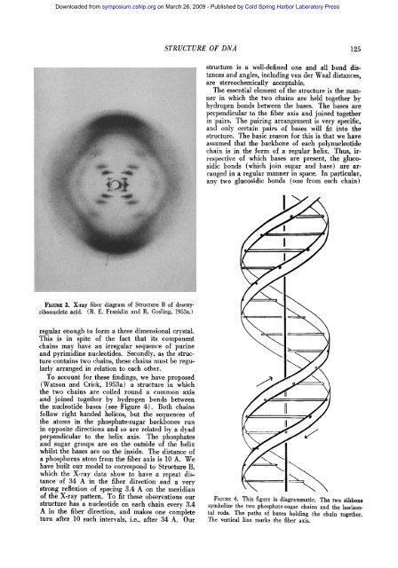
FiGnag 3. X-ray fiber diagram of Structure B of desoxyribonudeie<br />
acid. (R. E. Franklin and R. Gosling, <strong>1953</strong>a.)<br />
regular enough to form a three dimensional crystal.<br />
This is in spite of the fact that its component<br />
chains may have an irregular sequence of purine<br />
and pyrimidine nucleotides. Secondly, as the structure<br />
contains two chains, these chains must be regularly<br />
arranged in relation to each other.<br />
To account for these findings, we have proposed<br />
(<strong>Watson</strong> and Crick, <strong>1953</strong>a) a structure in which<br />
the two chains are coiled round a common axis<br />
and joined together by hydrogen bonds between<br />
the nucleotide bases (see Figure 4). Both chains<br />
follow right handed helices, but the sequences of<br />
the atoms in the phosphate-sugar backbones run<br />
in opposite directions and so are related by a dyad<br />
perpendicular to the helix axis. The phosphates<br />
and sugar groups are on the outside of the helix<br />
whilst the bases are on the inside. The distance of<br />
a phosphorus atom from the fiber axis is 10 A. We<br />
have built our model to correspond to Structure B,<br />
which the X-ray data show to have a repeat distance<br />
of 34 A in the fiber direction and a very<br />
strong reflexion of spacing 3.4 A on the meridian<br />
of the X-ray pattern. To fit these observations our<br />
structure has a nucleotide on each chain every 3.4<br />
A in the fiber direction, and makes one complete<br />
turn after 10 such intervals, i.e., after 34 A. Our<br />
FIGURE 4. This figure is diagrammatic. The two ribbons<br />
symbolize the two phosphate-sugar chains and the horizontal<br />
rods. The paths of bases holding the chain together.<br />
The vertical line marks the fiber axis.