Neuromuscular Junction: Development
Neuromuscular Junction: Development
Neuromuscular Junction: Development

Create successful ePaper yourself
Turn your PDF publications into a flip-book with our unique Google optimized e-Paper software.
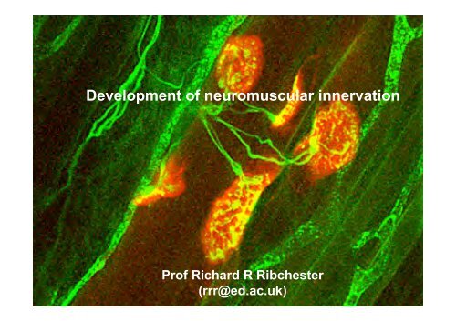
<strong>Development</strong> of neuromuscular innervation<br />
Prof Richard R Ribchester<br />
(rrr@ed.ac.uk)
Main topics for this lecture<br />
• The neurobiological context..<br />
• Motor Neurone birth<br />
• <strong>Neuromuscular</strong> synapse formation<br />
• Motor Neurone death<br />
• <strong>Neuromuscular</strong> synapse elimination<br />
• Molecular changes in ACh Receptors<br />
• Neonatal disease of motor units: Spinal Muscular Atrophy (SMA)<br />
• <strong>Development</strong> of myelin sheaths<br />
• Motor nerve sprouting and regeneration: recapitulation of<br />
development
The neurobiological context….
Induction<br />
Proliferation<br />
Progressive<br />
Migration<br />
Aggregation<br />
Synapse<br />
formation<br />
Axon<br />
outgrowth<br />
Cell Death<br />
Regressive<br />
Synapse<br />
Elimination
Remodelling of connections is a feature of development<br />
Levay, Stryker & Shatz. J. comp. Neurol. 179,223-244.(1978)
Remodelling of connections is activity-dependent
Activity-dependent remodelling may be based on<br />
competition for neurotrophic factors<br />
From<br />
Purves,D.(1988) Body &<br />
Brain: a trophic theory of<br />
neural connections. Harvard<br />
University Press.<br />
“When an axon of cell A is near enough to excite a cell B and repeatedly and persistently<br />
takes part in firing it, some growth process or metabolic change takes place in one or both<br />
cells such that Aʼs efficiency as one of the cells firing B is increased.”<br />
D.O.Hebb (1949) The Organization of Behaviour.
“The neuromuscular<br />
junction... [is] an<br />
experimentally<br />
favourable object<br />
whose study could<br />
throw considerable<br />
light on synaptic<br />
mechanisms<br />
elsewhere”<br />
Sir Bernard Katz, Fenn Lecture,<br />
IUPS Glasgow, 1993
Overview of motor units and their development…
The “Final Common Path”….
Motor neurone cell bodies occupy the ventral horn of grey matter<br />
http://www.tmin.ac.jp/english/dept/07/neurology2.jpg<br />
http://www.shef.ac.uk/content/1/c6/02/25/50/ps2.jpg
<strong>Neuromuscular</strong> connections frequently occupy a tight band<br />
in skeletal muscle<br />
1 mm
Mature NMJ’s are mononeuronally innervated<br />
500 µm
Confocal microscopy of the NMJ<br />
30 µm
Four cell types at the NMJ<br />
tSC<br />
KC<br />
NT<br />
MF<br />
1 µm
Motor units are expanded and muscle fibres hyperinnervated in neonates<br />
50 µm<br />
P5 DL<br />
Teriakidis
Neonatal muscle fibres are polyneuronally innervated (π)<br />
Court<br />
Teriakidis
Remodelling <strong>Neuromuscular</strong> Synapses
Summary of key stages in the development of rodent NMJ’s<br />
NMJ Expand<br />
NMJ Reshape<br />
AChR γ->ε<br />
NMJ Elim<br />
Myelin Form<br />
NMJ Form<br />
MF Form<br />
MN Die<br />
Progressive<br />
Regressive<br />
Remodel<br />
MN Form<br />
-20 days Birth<br />
+30 days
Rodent NMJ’s are stable in form but grow throughout life<br />
Balice-Gordon & Lichtman (1990) J Neurosci 10, 894
Quantal Content (variance<br />
method) at NMJ of rat HD<br />
100<br />
First EPP<br />
Plateau EPP (10 Hz)<br />
50<br />
0<br />
0 100 200 300 400<br />
Age<br />
(Based on Kelly & Roberts, 1977 and Kelly, 1978)
Formation of motor neurone pools…
The neural tube contains gradients of<br />
transcription factors<br />
BMP<br />
Shh<br />
The progenitors of motor neurons<br />
and interneurons are formed within<br />
distinct regionally-restricted domains<br />
of the ventral neural tube. The<br />
p0–p3 domains give rise to various<br />
interneuron subtypes, whereas the<br />
pMN domain is the source of motor<br />
neurons. The progenitor domains<br />
are identified by segmental<br />
expression of sets of transcription<br />
factors that are activated or<br />
repressed by different threshold<br />
concentrations of sonic hedgehog<br />
(Shh). The Shh gradient is denoted<br />
by pink dots. Shh signalling is<br />
thought to regulate the initial<br />
expression of transcription factors in<br />
the ventral neural tube (for example,<br />
Nkx2.2, Olig2, Pax6 and Irx3), which<br />
subsequently engage in crossregulatory<br />
interactions to sharpen<br />
and maintain the domain<br />
boundaries. Finally, combinatorial<br />
interactions between transcription<br />
factors expressed in each domain<br />
regulate downstream genes that<br />
determine progenitor identity. The<br />
positions of the floorplate (FP), an<br />
important source of Shh proteins,<br />
and the roofplate (RP), a source of<br />
bone morphogenetic proteins<br />
(BMPs), are shown.
Other transcription factors specify medial-lateral<br />
(intrasegmental) and rostro-caudal motor neurone identity<br />
Dasen et al (2005) Cell
Selective outgrowth of axons…
S. Ramon y Cajal, ca 1900, identifies neuronal growth cones
http://www.bio.miami.edu/ktosney/
Growth cones both extend and retract<br />
http://growthcones.neuroscience.umn.edu/Videos.html
Growth cones are semi-autonomous<br />
Remove<br />
Cell body…<br />
…a few<br />
minutes<br />
later…
Growth cones respond to adhesive and chemical<br />
gradients<br />
http://growthcones.neuroscience.umn.edu/Videos.html
In immature spinal cord in vivo, netrins attract and<br />
semaphorins repel motor neurone growth cones
Growth cone<br />
properties may<br />
underly the<br />
specificity of<br />
connections<br />
Lance-Jones & Landmesser (1980) J Physiol
Topographic projections are determined by<br />
neural identity, not location<br />
Lance-Jones & Landmesser (1980) J Physiol
Formation of muscle fibres and NMJ’s…
Myogenesis<br />
Myoblasts<br />
Myotubes<br />
Muscle Fibres
<strong>Neuromuscular</strong> connections frequently occupy a tight band<br />
in skeletal muscle<br />
1 mm
Two types of synapse formation: FaSyn & DeSyn
Acetylcholine receptors cluster under the<br />
influence of Agrin<br />
http://faculty.washington.edu/afolch/images/Concept_Synaptogen.jpg
ACh Receptor maturation…
“Plaque”<br />
“Pretzel”<br />
Slater (1982)
Agrin clusters ACh receptors via Muscle-Specific Kinase<br />
Neuregulin modulates AChR synthesis via ErbB receptors<br />
Musk<br />
AChR<br />
NRG receptor (ErbB)<br />
http://www.mun.ca/biology/desmid/brian/BIOL3530/DB_Ch11/fig11_36.jpg
The nicotinic ACh Receptor at NMJ<br />
8 nm<br />
/ε
Neonate: AChR - γ<br />
Adult AChR - ε
Fetal: AChR - γ<br />
Adult AChR - ε
AChR-ε knockout mice continue to make AChR-γ<br />
and initially NMJ develop normally<br />
Witzemann et al. (1996) PNAS 93, 13286<br />
Missias et al. (1997) <strong>Development</strong> 124, 5075
But AChR-ε are required for long-term maintenance of NMJ and survival<br />
Witzemann et al. (1996) PNAS 93, 13286<br />
Age (days)<br />
Missias et al. (1997) <strong>Development</strong> 124, 5075
Natural Motor Neurone Death…
MN death is a normal part of PRENATAL development
Target size regulates the number of motor neurones
Growth Factors possibly implicated in activitydependent,<br />
negative-feedback control of<br />
motor neurone survival:<br />
BDNF<br />
CNTF<br />
GDNF<br />
TGFβ<br />
CT-1<br />
HGF<br />
VEGF<br />
Reg-2<br />
Fas
Muscle paralysis inhibits embryonic motor neurone death<br />
Normal<br />
Paralysed<br />
dTC - paralysed<br />
Control<br />
Pittman & Oppenheim (1978) Nature
The mitochondrial Bcl-2/Bax System regulates motor neurone apoptosis
Natural Synapse Elimination…
Muscle fibres are initially “polyneuronally” innervated<br />
J.F. Tello<br />
Polyneuronal innervation in<br />
fetal human muscle<br />
(1917)
Physiological methods of measuring PI<br />
AB<br />
B<br />
A<br />
PI= [(A+B)-AB]/A<br />
A B A B<br />
PI= AB/A
Synapse elimination during postnatal development establishes<br />
mononeuronal innervation of motor endplates<br />
Walsh & Lichtman (2003). Neuron
Time lapse imaging of synapse elimination<br />
π<br />
µ<br />
π<br />
µ<br />
Walsh & Lichtman (2003) Neuron 37,67-73
Animation courtesy of Jean Livet & Mark Terasaki
Loss of motor neurones?<br />
Or elimination of connections?
Motor unit size can be estimated from their<br />
isometric forces
Motor unit size decreases postnatally<br />
Elimination Loss of motor of connections neurones..X✓
Synapse elimination during postnatal development establishes<br />
mononeuronal innervation of motor endplates<br />
Keller-Peck, C. et al.(2001) Neuron 31,381-394<br />
Walsh & Lichtman (2003) Neuron 37,67-73
Neurones retract some of their synapses while stabilising others<br />
Keller-Peck, C. et al.(2001) Neuron 31,381-394
Is Synapse Elimination globally “programmed”<br />
or due to local “competition”…
Neuronal/synaptic competition:<br />
“The negative effects that one neurone has on<br />
others by consuming, or controlling access to,<br />
resources at synapses that are limited in<br />
availability”<br />
(Based on a definition by Keddy, P.(1989) Competition. Chapman &<br />
Hall)
“Intrinsic” withdrawal ?<br />
Or “competitive” take-over ?
The rate of synapse elimination is activity-dependent<br />
100<br />
80<br />
Stimulation<br />
Paralysis<br />
Normal<br />
60<br />
40<br />
20<br />
0<br />
0 10 20 30<br />
Age (days)
“When an axon of cell A is near enough to excite a cell B and repeatedly and persistently<br />
takes part in firing it, some growth process or metabolic change takes place in one or both<br />
cells such that Aʼs efficiency as one of the cells firing B is increased.”<br />
D.O.Hebb (1949) The Organization of Behaviour.<br />
From<br />
Purves,D.(1988) Body &<br />
Brain: a trophic theory of<br />
neural connections. Harvard<br />
University Press.
Transgenic expression of a growth factor, GDNF, delays elimination<br />
Nguyen QT, Parsadanian AS, Snider WD, Lichtman JW (1998) Science 279:1725–1729.
X<br />
Evidence for<br />
competiton: no<br />
change in size of<br />
surviving motor units<br />
after partial<br />
denervation at birth<br />
Betz et al. (1980)<br />
Evidence against<br />
competiton: surviving<br />
motor unit size<br />
continues to decline<br />
after partial<br />
denervation at birth<br />
Fladby & Jansen, 1988
We can determine motor unit size in thy1.2-YFP mice by<br />
counting the number of muscle fibres that are innervated<br />
Adrianna Teriakidis
Neonate<br />
~ 1 month after<br />
partial denervation<br />
Adrianna Teriakidis
Motor unit sizes 2 days after partial denervation<br />
are larger than motor unit sizes 4-6 weeks later.<br />
Adrianna Teriakidis
Is every one of a dominant motor<br />
neurone’s synapses a winner?<br />
Kasthuri & Lichtman (2003) Nature
Motor units may compete in a “dominance hierarchy”
Disease correlate : Spinal Muscular Atrophy<br />
- SMA Type I (Werdnig Hoffman disease)<br />
- Neonatal, “floppy baby”<br />
- Fatal, no cure<br />
- Incidence 1:15,000 births<br />
- 95% cases have mutations in SMN1 gene<br />
- Compensation by SMN2 gene in other SMA types<br />
- Mouse KO/transgenic models show NMJ defects
Impaired NMJ form and function in neonatal NMJ’s in smn knockout mouse models of SMA<br />
Murray et al (2008) Human Mol Genet<br />
Cifuentes-Diaz et al (2002) Human Mol Genet<br />
Kong et al (2009) J Neuroscience
Summary of key stages in the development of rodent NMJ’s<br />
NMJ Expand<br />
NMJ Reshape<br />
AChR γ->ε<br />
NMJ Elim<br />
Myelin Form<br />
NMJ Form<br />
MF Form<br />
MN Die<br />
Progressive<br />
Regressive<br />
Remodel<br />
MN Form<br />
-20 days Birth<br />
+30 days
Myelin formation…
Schwann cells arise from the neural crest<br />
Carmeliet (2003) Nature Reviews Genetics
Myelin forms from compacted Schwann cell membranes
Myelin sheaths form postnatally<br />
Slater, C.R. (1982)
Regeneration…
Remodelling <strong>Neuromuscular</strong> Synapses
MPN LPN SN<br />
<br />
4DL
Partial denervation triggers axonal sprouting
Axonal sprouting is preceded by Schwann cell sprouting<br />
Son et al (1996) TINS 19,280
Regeneration reverses axonal sprouting
Polyneuronal innervation (π) in reinnervated adult muscles<br />
resembles development<br />
Neonatal<br />
Reinnervated adult
Polyneuronal innervation in reinnervated muscle<br />
is also activity-dependent …<br />
J.A.Barry & R.R. Ribchester<br />
J.Neurosci. 15,6327-6339(1995)<br />
E.M. Costanzo, J.A. Barry & R.R. Ribchester<br />
J.Physiol. 521.2,365-374 (1999)
…but some polyinnervation persists at some NMJ after activity resumes<br />
60<br />
%PI<br />
40<br />
20<br />
0<br />
0 2 4 6 8<br />
Weeks Recovery
SUMMARY<br />
• <strong>Neuromuscular</strong> <strong>Junction</strong>s (NMJ’s) are an excellent<br />
model system for studying synaptic development<br />
• Motor neurone (MN) birth, muscle fibre growth, myelin formation<br />
and endplate expansion are progressive features of motor unit<br />
development<br />
• MN death and synapse elimination at NMJ’s are regressive<br />
features of motor unit development<br />
• Endplates additionally undergo structural remodelling and<br />
adjustments in AChR expression<br />
• Some aspects of MN/NMJ development are competitive,<br />
establishing hierarchies of motor units<br />
• Motor unit development goes awry in diseases such as Spinal<br />
Muscular Atrophy, due to mutations in SMN genes<br />
• Axonal and synaptic regeneration after nerve injury partially<br />
recapitulates neuromuscular development