Muscle fatigue and muscle weakness: what we know and ... - Frontiers
Muscle fatigue and muscle weakness: what we know and ... - Frontiers
Muscle fatigue and muscle weakness: what we know and ... - Frontiers

You also want an ePaper? Increase the reach of your titles
YUMPU automatically turns print PDFs into web optimized ePapers that Google loves.
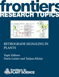
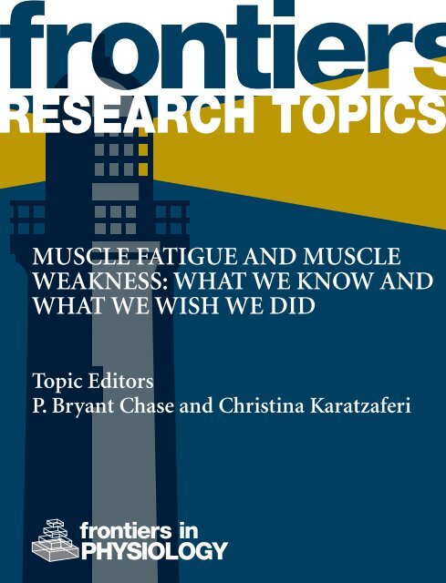
MUSCLE FATIGUE AND MUSCLE<br />
WEAKNESS: WHAT WE KNOW AND<br />
WHAT WE WISH WE DID<br />
Topic Editors<br />
P. Bryant Chase <strong>and</strong> Christina Karatzaferi<br />
PHYSIOLOGY
FRONTIERS COPYRIGHT<br />
STATEMENT<br />
© Copyright 2007-2013<br />
<strong>Frontiers</strong> Media SA.<br />
All rights reserved.<br />
All content included on this site,<br />
such as text, graphics, logos, button<br />
icons, images, video/audio clips,<br />
downloads, data compilations <strong>and</strong><br />
software, is the property of or is<br />
licensed to <strong>Frontiers</strong> Media SA<br />
(“<strong>Frontiers</strong>”) or its licensees <strong>and</strong>/or<br />
subcontractors. The copyright in the<br />
text of individual articles is the<br />
property of their respective authors,<br />
subject to a license granted to<br />
<strong>Frontiers</strong>.<br />
The compilation of articles<br />
constituting this e-book, as <strong>we</strong>ll as<br />
all content on this site is the<br />
exclusive property of <strong>Frontiers</strong>.<br />
Images <strong>and</strong> graphics not forming<br />
part of user-contributed materials<br />
may not be downloaded or copied<br />
without permission.<br />
Articles <strong>and</strong> other user-contributed<br />
materials may be downloaded <strong>and</strong><br />
reproduced subject to any copyright<br />
or other notices. No financial<br />
payment or reward may be given for<br />
any such reproduction except to the<br />
author(s) of the article concerned.<br />
As author or other contributor you<br />
grant permission to others to<br />
reproduce your articles, including<br />
any graphics <strong>and</strong> third-party<br />
materials supplied by you, in<br />
accordance with the Conditions for<br />
Website Use <strong>and</strong> subject to any<br />
copyright notices which you include<br />
in connection with your articles <strong>and</strong><br />
materials.<br />
All copyright, <strong>and</strong> all rights therein,<br />
are protected by national <strong>and</strong><br />
international copyright laws.<br />
The above represents a summary<br />
only. For the full conditions see the<br />
Conditions for Authors <strong>and</strong> the<br />
Conditions for Website Use.<br />
Cover image provided by Ibbl sarl,<br />
Lausanne CH<br />
ISSN 1664-8714<br />
ISBN 978-2-88919-163-5<br />
DOI 10.3389/978-2-88919-163-5<br />
ABOUT FRONTIERS<br />
<strong>Frontiers</strong> is more than just an open-access publisher of scholarly articles: it is a pioneering<br />
approach to the world of academia, radically improving the way scholarly research is managed.<br />
The gr<strong>and</strong> vision of <strong>Frontiers</strong> is a world where all people have an equal opportunity to seek, share<br />
<strong>and</strong> generate <strong>know</strong>ledge. <strong>Frontiers</strong> provides immediate <strong>and</strong> permanent online open access to all<br />
its publications, but this alone is not enough to realize our gr<strong>and</strong> goals.<br />
FRONTIERS JOURNAL SERIES<br />
The <strong>Frontiers</strong> Journal Series is a multi-tier <strong>and</strong> interdisciplinary set of open-access, online<br />
journals, promising a paradigm shift from the current review, selection <strong>and</strong> dissemination<br />
processes in academic publishing.<br />
All <strong>Frontiers</strong> journals are driven by researchers for researchers; therefore, they constitute a service<br />
to the scholarly community. At the same time, the <strong>Frontiers</strong> Journal Series operates on a revolutionary<br />
invention, the tiered publishing system, initially addressing specific communities of<br />
scholars, <strong>and</strong> gradually climbing up to broader public underst<strong>and</strong>ing, thus serving the interests<br />
of the lay society, too.<br />
DEDICATION TO QUALITY<br />
Each <strong>Frontiers</strong> article is a l<strong>and</strong>mark of the highest quality, thanks to genuinely collaborative interactions<br />
bet<strong>we</strong>en authors <strong>and</strong> review editors, who include some of the world’s best academicians.<br />
Research must be certified by peers before entering a stream of <strong>know</strong>ledge that may eventually<br />
reach the public - <strong>and</strong> shape society; therefore, <strong>Frontiers</strong> only applies the most rigorous <strong>and</strong><br />
unbiased reviews.<br />
<strong>Frontiers</strong> revolutionizes research publishing by freely delivering the most outst<strong>and</strong>ing research,<br />
evaluated with no bias from both the academic <strong>and</strong> social point of view.<br />
By applying the most advanced information technologies, <strong>Frontiers</strong> is catapulting scholarly<br />
publishing into a new generation.<br />
WHAT ARE FRONTIERS RESEARCH TOPICS?<br />
<strong>Frontiers</strong> Research Topics are very popular trademarks of the <strong>Frontiers</strong> Journals Series: they are<br />
collections of at least ten articles, all centered on a particular subject. With their unique mix<br />
of varied contributions from Original Research to Review Articles, <strong>Frontiers</strong> Research Topics<br />
unify the most influential researchers, the latest key findings <strong>and</strong> historical advances in a hot<br />
research area!<br />
Find out more on how to host your own <strong>Frontiers</strong> Research Topic or contribute to one as an<br />
author by contacting the <strong>Frontiers</strong> Editorial Office: researchtopics@frontiersin.org<br />
<strong>Frontiers</strong> in Physiology August 2013 | <strong>Muscle</strong> <strong>fatigue</strong> <strong>and</strong> <strong>muscle</strong> <strong><strong>we</strong>akness</strong>: <strong>what</strong> <strong>we</strong> <strong>know</strong> <strong>and</strong> <strong>what</strong> <strong>we</strong> wish <strong>we</strong> did | 1
MUSCLE FATIGUE AND MUSCLE<br />
WEAKNESS: WHAT WE KNOW AND<br />
WHAT WE WISH WE DID<br />
Topic Editors:<br />
P. Bryant Chase, Florida State University, USA<br />
Christina Karatzaferi, University of Thessaly, Greece<br />
Image by P. Bryant Chase <strong>and</strong> Christina<br />
Karatzaferi.<br />
The purpose of this Research Topic is to<br />
discuss evidence coming from macroscopic<br />
observations on the whole human/animal<br />
to investigations at the molecular level in an<br />
effort to identify critical factors in <strong>muscle</strong><br />
<strong>fatigue</strong> <strong>and</strong> <strong>muscle</strong> <strong><strong>we</strong>akness</strong>, in health <strong>and</strong><br />
disease.<br />
Why is this important?<br />
Skeletal <strong>muscle</strong>s confer movement to the<br />
human body using vast amounts of energy<br />
provided through complex metabolic<br />
pathways. Thus, whole body mobility <strong>and</strong><br />
energy balance are dictated by <strong>muscle</strong><br />
contraction. On the other side, <strong>muscle</strong><br />
function reflects overall health status, as<br />
chronic conditions <strong>and</strong>/or ageing affect either<br />
or both of <strong>muscle</strong> quality (protein <strong>and</strong> fat content) <strong>and</strong> quantity (mass). In health, <strong>muscle</strong><br />
<strong>fatigue</strong> is temporary <strong>and</strong> recovery occurs rapidly. In ageing or chronic disease ho<strong>we</strong>ver,<br />
<strong>muscle</strong> <strong>fatigue</strong> may occur prematurely <strong>and</strong> be persistent, endangering thus a person’s safety<br />
(i.e. <strong><strong>we</strong>akness</strong> leads to falls), <strong>and</strong> leading the sufferer in a self-perpetuating vicious cycle<br />
of inactivity -> disuse <strong>muscle</strong> atrophy/metabolic disturbance <strong>and</strong> so on, that compounds<br />
morbidity (i.e. causing metabolic syndrome, fatness, hypertension, <strong>muscle</strong> cachexia) <strong>and</strong> that<br />
eventually leads to premature death.<br />
<strong>Frontiers</strong> in Physiology August 2013 | <strong>Muscle</strong> <strong>fatigue</strong> <strong>and</strong> <strong>muscle</strong> <strong><strong>we</strong>akness</strong>: <strong>what</strong> <strong>we</strong> <strong>know</strong> <strong>and</strong> <strong>what</strong> <strong>we</strong> wish <strong>we</strong> did | 2
Research in skeletal <strong>muscle</strong> physiology <strong>and</strong> biophysics is at an exciting phase. The advent of<br />
new methodologies <strong>and</strong> technological advancements have allo<strong>we</strong>d researchers to advance our<br />
<strong>know</strong>ledge on the “how’s” <strong>and</strong> “why’s” of <strong>muscle</strong> contraction – ho<strong>we</strong>ver there are still many<br />
unans<strong>we</strong>red questions especially when disease states are implicated.<br />
Such questions need addressing if as a scientific community <strong>we</strong> aspire to secure better<br />
designed interventions to improve <strong>muscle</strong> function, <strong>and</strong> thus improve quality of life <strong>and</strong> life<br />
prognosis for the ageing population <strong>and</strong> chronic disease patients.<br />
<strong>Frontiers</strong> in Physiology August 2013 | <strong>Muscle</strong> <strong>fatigue</strong> <strong>and</strong> <strong>muscle</strong> <strong><strong>we</strong>akness</strong>: <strong>what</strong> <strong>we</strong> <strong>know</strong> <strong>and</strong> <strong>what</strong> <strong>we</strong> wish <strong>we</strong> did | 3
Table of Contents<br />
05 <strong>Muscle</strong> Fatigue <strong>and</strong> <strong>Muscle</strong> Weakness: What We Know <strong>and</strong> What We<br />
Wish We Did<br />
Christina Karatzaferi <strong>and</strong> P. Bryant Chase<br />
Mechanisms of <strong>Muscle</strong> Function at the Sarcomeric Level<br />
08 Recent Insights Into <strong>Muscle</strong> Fatigue at the Cross-Bridge Level<br />
Edward P. Debold<br />
22 The Multiple Roles of Phosphate in <strong>Muscle</strong> Fatigue<br />
David G. Allen <strong>and</strong> Sofie Trajanovska<br />
30 The Sarcomeric Protein Nebulin: Another Multifunctional Giant in Charge of<br />
<strong>Muscle</strong> Strength Optimization<br />
Coen A. C. Ottenheijm, Henk Granzier <strong>and</strong> Siegfried Labeit<br />
Modeling <strong>Muscle</strong> Function<br />
39 A Physiologically Based, Multi-Scale Model of Skeletal <strong>Muscle</strong> Structure <strong>and</strong><br />
Function<br />
O. Röhrle, J. B. Davidson <strong>and</strong> A. J. Pullan<br />
Physical Activity <strong>and</strong> Exercise Performance<br />
53 Effects of Physical Activity <strong>and</strong> Inactivity on <strong>Muscle</strong> Fatigue<br />
Gregory C. Bogdanis<br />
68 Antioxidants <strong>and</strong> Skeletal <strong>Muscle</strong> Performance: “Common Knowledge” vs.<br />
Experimental Evidence<br />
Andrés Hernández, Arthur Cheng <strong>and</strong> Håkan Westerblad<br />
The <strong>Muscle</strong>-Brain Connection of Fatigue in Health <strong>and</strong> Disease<br />
74 <strong>Muscle</strong> Fatigue <strong>and</strong> Cognition: What is the Link?<br />
Tali Kobilo <strong>and</strong> Henriette van Praag<br />
77 Hemodialysis Fatigue: Just “Simple” Fatigue or A Syndrome on Its Own Right?<br />
Giorgos K. Sakkas <strong>and</strong> Christina Karatzaferi<br />
81 Fatigue is a Brain-Derived Emotion That Regulates the Exercise Behavior to<br />
Ensure the Protection of Whole Body Homeostasis<br />
Timothy David Noakes<br />
<strong>Frontiers</strong> in Physiology August 2013 | <strong>Muscle</strong> <strong>fatigue</strong> <strong>and</strong> <strong>muscle</strong> <strong><strong>we</strong>akness</strong>: <strong>what</strong> <strong>we</strong> <strong>know</strong> <strong>and</strong> <strong>what</strong> <strong>we</strong> wish <strong>we</strong> did | 4
EDITORIAL<br />
published: 30 May 2013<br />
doi: 10.3389/fphys.2013.00125<br />
<strong>Muscle</strong> <strong>fatigue</strong> <strong>and</strong> <strong>muscle</strong> <strong><strong>we</strong>akness</strong>: <strong>what</strong> <strong>we</strong> <strong>know</strong> <strong>and</strong><br />
<strong>what</strong> <strong>we</strong> wish <strong>we</strong> did<br />
Christina Karatzaferi 1,2 * <strong>and</strong> P. Bryant Chase 3<br />
1<br />
Department of Physical Education <strong>and</strong> Sports Science, University of Thessaly, Trikala, Greece<br />
2<br />
Department of Kinesiology, Center for Research <strong>and</strong> Technology Thessaly, Trikala, Greece<br />
3<br />
Department of Biological Science, The Florida State University, Tallahassee, FL, USA<br />
*Correspondence: ck@pe.uth.gr<br />
Edited by:<br />
Peter J. Reiser, Ohio State University, USA<br />
Revie<strong>we</strong>d by:<br />
Peter J. Reiser, Ohio State University, USA<br />
This Research Topic on <strong>muscle</strong> <strong>fatigue</strong> <strong>and</strong> <strong>muscle</strong> <strong><strong>we</strong>akness</strong><br />
presents the latest ideas, arguments, <strong>and</strong> evidence from investigations<br />
at the molecular level to macroscopic observations on<br />
whole animals including humans, in an effort to identify critical<br />
factors underlying <strong>fatigue</strong> <strong>and</strong> <strong><strong>we</strong>akness</strong> in health <strong>and</strong> disease.<br />
Skeletal <strong>muscle</strong>s confer movement to the human body using<br />
vast amounts of energy provided through complex metabolic<br />
pathways such that whole body mobility <strong>and</strong> energy balance<br />
are largely dictated by <strong>muscle</strong> activity. Conversely, <strong>muscle</strong> function<br />
reflects overall health status as exercise history <strong>and</strong> chronic<br />
conditions affect either or both <strong>muscle</strong> quality, including protein<br />
<strong>and</strong> fat content, <strong>and</strong> <strong>muscle</strong> mass. In health, <strong>muscle</strong> <strong>fatigue</strong><br />
is temporary <strong>and</strong> recovery occurs rapidly, <strong>and</strong> recreational or<br />
competitive athletes are always pursuing the next best <strong>fatigue</strong><br />
“fix.” Ho<strong>we</strong>ver, after inactivity—whether due to lifestyle choices,<br />
injury or chronic disease—<strong>muscle</strong> <strong>fatigue</strong> may occur prematurely<br />
<strong>and</strong> persist, endangering a person’s safety because <strong><strong>we</strong>akness</strong><br />
can lead to falls that may result in loss of independence.<br />
Individuals are then trapped in a self-perpetuating, vicious cycle<br />
of inactivity, disuse <strong>muscle</strong> atrophy/<strong><strong>we</strong>akness</strong>, <strong>and</strong> metabolic<br />
disturbance that compounds morbidity (i.e., causing metabolic<br />
syndrome, obesity, hypertension, cachexia) <strong>and</strong> eventually premature<br />
death. Such issues transcend many scientific disciplines<br />
<strong>and</strong> it becomes evident that not only recognizing fundamental<br />
factors in <strong>muscle</strong> <strong>fatigue</strong> <strong>and</strong> <strong>muscle</strong> <strong><strong>we</strong>akness</strong> is necessary,<br />
but also evaluating their interaction with factors outside of the<br />
<strong>muscle</strong> is essential if <strong>we</strong> aspire to design better interventions<br />
that improve <strong>muscle</strong> function <strong>and</strong> thus improve quality of life<br />
<strong>and</strong> life prognosis for the ageing population <strong>and</strong> chronic disease<br />
patients.<br />
Fatigue <strong>and</strong> <strong><strong>we</strong>akness</strong> may stem from changes within myocytes<br />
that affect cross-bridge function or Ca 2+ activation, to changes<br />
within the circulation or function of the nervous system. Within<br />
myocytes, metabolic products of ATP hydrolysis in the cytoplasm<br />
such as inorganic phosphate (Pi), protons (H + or pH), <strong>and</strong> ADP<br />
have often been considered as agents that could disrupt force<br />
generation at the sarcomere level (Fabiato <strong>and</strong> Fabiato, 1978;<br />
Cooke <strong>and</strong> Pate, 1985; Metzger <strong>and</strong> Moss, 1987; Nosek et al.,<br />
1987, 1990; Chase <strong>and</strong> Kushmerick, 1988, 1995; Cooke et al.,<br />
1988; Godt <strong>and</strong> Nosek, 1989; Pate <strong>and</strong> Cooke, 1989; Metzger<br />
<strong>and</strong> Moss, 1990a,b; Pate et al., 1995, 1998; Wiseman et al.,<br />
1996; Karatzaferi et al., 2003, 2008). These effects may be due<br />
to direct binding to proteins, or due to a more global alteration<br />
of cellular energetics (G ATP )inthemyocyte(Karatzaferi et al.,<br />
2004).<br />
In this Research Topic, Debold (2012) consolidates the most<br />
recent information, including single molecule assays <strong>and</strong> molecular<br />
biological approaches, about the mechanisms by which Pi,<br />
H + , <strong>and</strong> ADP inhibit actomyosin cross-bridge cycling <strong>and</strong> thin<br />
filament Ca 2+ -activation. Allen <strong>and</strong> Trajanovska (2012) provide<br />
a synthesis on the multiple roles of Pi in <strong>fatigue</strong>, including<br />
novel results from their group, showing that Pi is even more<br />
detrimental when its effects on Ca 2+ release are combined with<br />
inhibition of actomyosin force generation <strong>and</strong> Ca 2+ activation.<br />
In addition to activity-driven changes in metabolites <strong>and</strong> cellular<br />
energetics, mutations in sarcomeric proteins have been associated<br />
with prolonged <strong>muscle</strong> <strong><strong>we</strong>akness</strong> in myopathies. Moving<br />
away from actomyosin events, Ottenheijm et al. (2012) consider<br />
the role of nebulin in sarcomere function, <strong>and</strong> how transgenic<br />
mouse models can inform us about mutations in the giant filamentous<br />
protein nebulin, <strong>and</strong> mutations in other thin filament<br />
<strong>and</strong> closely related proteins that are associated with nemaline<br />
myopathy.<br />
To fully test our underst<strong>and</strong>ing of <strong>muscle</strong> <strong>fatigue</strong>, appropriately<br />
detailed models of <strong>muscle</strong> function will be necessary. Röhrle<br />
et al. (2012) make major advances in that arena by presenting a<br />
multi-scale, finite element model of the human tibialis anterior.<br />
Their model has the advantage of allowing simulation of <strong>fatigue</strong><br />
at the cellular <strong>and</strong> motor unit levels, <strong>and</strong> can incorporate altered<br />
recruitment patterns of motor units due to central components of<br />
<strong>fatigue</strong>. Thus their model can serve an invaluable role as <strong>we</strong> bridge<br />
our underst<strong>and</strong>ing bet<strong>we</strong>en the cellular <strong>and</strong> tissue levels.<br />
<strong>Muscle</strong>’s plasticity is most readily evident in its adaptation<br />
to repeated exercise, <strong>and</strong> conversely to inactivity that may be<br />
associated with various injuries <strong>and</strong> disease states. Bogdanis<br />
(2012) reviews the long-term changes in <strong>muscle</strong> at the molecular,<br />
cellular, <strong>and</strong> tissue levels, as <strong>we</strong>ll as the corresponding<br />
functional changes that are associated with these adaptations to<br />
activity level history. Fatigability is a key functional characteristic<br />
of different <strong>muscle</strong> fiber types, <strong>and</strong> can vary greatly with<br />
activity, or inactivity, <strong>and</strong> Bogdanis evaluates the utility of highintensity<br />
bouts of exercise for modulating fatigability by training,<br />
www.frontiersin.org May 2013 | Volume 4 | Article 125 | 5
Karatzaferi <strong>and</strong> Chase<br />
Research topic: <strong>muscle</strong> <strong>fatigue</strong><br />
or as a component in therapy. Bogdanis’ section on effects of reactive<br />
oxygen species (ROS) sets the stage for the succinct review<br />
on antioxidants by Hernández et al. (2012). Despitethepopularity<br />
of antioxidants as nutritional supplements, Hernández<br />
et al. report that their utility for either minimizing or speeding<br />
recovery from <strong>fatigue</strong> appears to be limited to specific <strong>muscle</strong><br />
types. Moreover, Bogdanis’ (2012) section on neural factors<br />
opens the discussion on the role of non-<strong>muscle</strong> factors in<br />
<strong>fatigue</strong> <strong>and</strong> serves as a bridge to the articles by Kobilo <strong>and</strong><br />
van Praag (2012), Sakkas <strong>and</strong> Karatzaferi (2012), <strong>and</strong>Noakes<br />
(2012).<br />
What is the extent to which <strong>muscle</strong> activity <strong>and</strong> <strong>fatigue</strong><br />
influence the function of other physiological systems of the<br />
body, particularly the nervous system upon which skeletal <strong>muscle</strong><br />
depends for activation, <strong>and</strong> how much of fatigability is<br />
determined centrally? In the commentary by Kobilo <strong>and</strong> van<br />
Praag (2012), pharmacological activation of AMP-activated protein<br />
kinase (AMPK)—a metabolic regulator that is activated<br />
during exercise—is shown to alter performance in a test of<br />
spatial memory <strong>and</strong> hippocampal neurogenesis in mice in a<br />
time-dependent manner. How can diseases <strong>and</strong> treatments modify<br />
the experience <strong>and</strong> presentation of <strong>fatigue</strong>? In their opinion<br />
article, Sakkas <strong>and</strong> Karatzaferi (2012) consider available<br />
evidence on the complex symptomatology of <strong>fatigue</strong> in renal<br />
patients on hemodialysis treatment. By drawing analogies to<br />
Chronic Fatigue Syndrome, Sakkas <strong>and</strong> Karatzaferi (2012) present<br />
the view that <strong>fatigue</strong>, as experienced by patients undergoing<br />
routine hemodialysis, might be better addressed by caregivers<br />
as a syndrome <strong>and</strong> not with isolated measures since<br />
its apparent complexity requires a cross-disciplinary therapeutic<br />
approach. While hemodialysis <strong>and</strong> some other patients may<br />
be afflicted with specific syndromes, the rest of us have all<br />
heard the expression “mind over matter.” Does it apply to<br />
<strong>muscle</strong>? Noakes (2012) concludes the series with a challenging<br />
review, partly historical in nature, arguing that the key component<br />
in <strong>fatigue</strong> is central. The author discusses the accepted<br />
models on the limits of human exercise performance, <strong>and</strong><br />
presents his central governor model of exercise regulation, arguing<br />
that <strong>fatigue</strong> is brain-derived, being an important homeostatic<br />
mechanism that protects an organism from catastrophic<br />
overexertion.<br />
It is our sincere hope that this Research Topic will not<br />
only provide readers with new insights <strong>and</strong> viewpoints on<br />
the issue of <strong>muscle</strong> <strong>fatigue</strong> <strong>and</strong> <strong><strong>we</strong>akness</strong>, but will also stimulate<br />
novel ideas, experiments, <strong>and</strong> further advances in this<br />
research field.<br />
ACKNOWLEDGMENTS<br />
We thank all authors <strong>and</strong> revie<strong>we</strong>rs for their invaluable contributions<br />
in this Research Topic. Christina Karatzaferi is supported by<br />
NSRF 2007–2013, European Social Fund (University of Thessaly-<br />
4525, MIS 377260).<br />
REFERENCES<br />
Allen, D. G., <strong>and</strong> Trajanovska, S. (2012).<br />
The multiple roles of phosphate<br />
in <strong>muscle</strong> <strong>fatigue</strong>. Front. Physiol.<br />
3:463. doi: 10.3389/fphys.2012.<br />
00463<br />
Bogdanis, G. C. (2012). Effects of<br />
physical activity <strong>and</strong> inactivity<br />
on <strong>muscle</strong> <strong>fatigue</strong>. Front. Physiol.<br />
3:142. doi: 10.3389/fphys.2012.<br />
00142<br />
Chase, P. B., <strong>and</strong> Kushmerick, M. J.<br />
(1988). Effects of pH on contraction<br />
of rabbit fast <strong>and</strong> slow skeletal<br />
<strong>muscle</strong> fibers. Biophys. J. 53,<br />
935–946. doi: 10.1016/S0006-3495<br />
(88)83174-6<br />
Chase, P. B., <strong>and</strong> Kushmerick, M.<br />
J. (1995). Effect of physiological<br />
ADP levels on contraction of single<br />
skinned fibers from rabbit fast <strong>and</strong><br />
slow <strong>muscle</strong>s. Am. J. Physiol. 268,<br />
C480–C489.<br />
Cooke, R., Franks, K., Luciani, G.<br />
B., <strong>and</strong> Pate, E. (1988). The inhibition<br />
of rabbit skeletal <strong>muscle</strong><br />
contraction by hydrogen ions<br />
<strong>and</strong> phosphate. J. Physiol. 395,<br />
77–97.<br />
Cooke, R., <strong>and</strong> Pate, E. (1985).<br />
The effects of ADP <strong>and</strong> phosphate<br />
on the contraction of<br />
<strong>muscle</strong> fibers. Biophys. J. 48,<br />
789–798. doi: 10.1016/S0006-3495<br />
(85)83837-6<br />
Debold, E. P. (2012). Recent insights<br />
into <strong>muscle</strong> <strong>fatigue</strong> at the crossbridge<br />
level. Front. Physiol.<br />
3:151. doi: 10.3389/fphys.2012.<br />
00151<br />
Fabiato, A., <strong>and</strong> Fabiato, F. (1978).<br />
Effects of pH on the myofilaments<br />
<strong>and</strong> the sarcoplasmic reticulum<br />
of skinned cells from cardiac<br />
<strong>and</strong> skeletal <strong>muscle</strong>s. J. Physiol. 276,<br />
233–255.<br />
Godt, R. E., <strong>and</strong> Nosek, T. M. (1989).<br />
Changes in intracellular milieu with<br />
<strong>fatigue</strong> or hypoxia depress contraction<br />
of skinned rabbit skeletal<br />
<strong>and</strong> cardiac <strong>muscle</strong>. J. Physiol. 412,<br />
155–180.<br />
Hernández, A., Cheng, A., <strong>and</strong><br />
Westerblad, H. (2012). Antioxidants<br />
<strong>and</strong> skeletal <strong>muscle</strong> performance:<br />
“common <strong>know</strong>ledge” vs. experimental<br />
evidence. Front. Physiol.<br />
3:46. doi: 10.3389/fphys.2012.<br />
00046<br />
Karatzaferi, C., Chinn, M. K., <strong>and</strong><br />
Cooke, R. (2004). The force exerted<br />
by a <strong>muscle</strong> cross-bridge depends<br />
directly on the strength of the<br />
actomyosin bond. Biophys. J. 87,<br />
2532–2544. doi: 10.1529/biophysj.<br />
104.039909<br />
Karatzaferi, C., Franks-Skiba, K., <strong>and</strong><br />
Cooke, R. (2008). Inhibition of<br />
shortening velocity of skinned skeletal<br />
<strong>muscle</strong> fibers in conditions<br />
that mimic <strong>fatigue</strong>. Am. J. Physiol.<br />
Regul. Integr. Comp. Physiol. 294,<br />
R948–R955. doi: 10.1152/ajpregu.<br />
00541.2007<br />
Karatzaferi, C., Myburgh, K. H., Chinn,<br />
M. K., Franks-Skiba, K., <strong>and</strong> Cooke,<br />
R. (2003). Effect of an ADP analog<br />
on isometric force <strong>and</strong> ATPase<br />
activity of active <strong>muscle</strong> fibers.<br />
Am. J. Physiol. Cell Physiol. 284,<br />
C816–C825. doi: 10.1152/ajpcell.<br />
00291.2002<br />
Kobilo, T., <strong>and</strong> van Praag, H. (2012).<br />
<strong>Muscle</strong> <strong>fatigue</strong> <strong>and</strong> cognition:<br />
<strong>what</strong> is the link? Front. Physiol.<br />
3:14. doi: 10.3389/fphys.2012.<br />
00014<br />
Metzger, J. M., <strong>and</strong> Moss, R. L. (1987).<br />
Greater hydrogen ion-incuced<br />
depression of tension <strong>and</strong> velocity<br />
in skinned single fibres of rat fast<br />
than slow <strong>muscle</strong>s. J. Physiol. 393,<br />
727–742.<br />
Metzger, J. M., <strong>and</strong> Moss, R. L. (1990a).<br />
Effects on tension <strong>and</strong> stiffness<br />
due to reduced pH in mammalian<br />
fast- <strong>and</strong> slow-twitch skinned skeletal<br />
<strong>muscle</strong> fibres. J. Physiol. 428,<br />
737–750.<br />
Metzger, J. M., <strong>and</strong> Moss, R. L. (1990b).<br />
pH modulation of the kinetics of<br />
aCa 2+ -sensitive cross-bridge state<br />
transition in mammalian single<br />
skeletal <strong>muscle</strong> fibres. J. Physiol. 428,<br />
751–764.<br />
Noakes, T. D. (2012). Fatigue is a<br />
brain-derived emotion that regulates<br />
the exercise behavior to<br />
ensure the protection of whole<br />
body homeostasis. Front. Physiol.<br />
3:82. doi: 10.3389/fphys.2012.<br />
00082<br />
Nosek, T. M., Fender, K. Y., <strong>and</strong><br />
Godt, R. E. (1987). It is diprotonated<br />
inorganic phosphate that<br />
depresses force in skinned skeletal<br />
<strong>muscle</strong> fibers. Science 236,<br />
191–193. doi: 10.1126/science.<br />
3563496<br />
Nosek, T. M., Leal-Cardoso, J. H.,<br />
McLaughlin, M., <strong>and</strong> Godt, R.<br />
E. (1990). Inhibitory influence of<br />
phosphate <strong>and</strong> arsenate on contraction<br />
of skinned skeletal <strong>and</strong> cardiac<br />
<strong>muscle</strong>. Am. J. Physiol. 259,<br />
C933–C939.<br />
Ottenheijm, C. A. C., Granzier, H.,<br />
<strong>and</strong> Labeit, S. (2012). The sarcomeric<br />
protein nebulin: another multifunctional<br />
giant in charge of <strong>muscle</strong><br />
strength optimization. Front.<br />
Physiol. 3:37. doi: 10.3389/fphys.<br />
2012.00037<br />
Pate, E., Bhimani, M., Franks-Skiba,<br />
K., <strong>and</strong> Cooke, R. (1995). Reduced<br />
effect of pH on skinned rabbit<br />
psoas <strong>muscle</strong> mechanics<br />
at high temperatures: implications<br />
for <strong>fatigue</strong>. J. Physiol. 486,<br />
689–694.<br />
<strong>Frontiers</strong> in Physiology | Striated <strong>Muscle</strong> Physiology May2013|Volume4|Article125| 6
Karatzaferi <strong>and</strong> Chase<br />
Research topic: <strong>muscle</strong> <strong>fatigue</strong><br />
Pate, E., <strong>and</strong> Cooke, R. (1989). Addition<br />
of phosphate to active <strong>muscle</strong><br />
fibers probes actomyosin states<br />
within the po<strong>we</strong>rstroke. Pflügers<br />
Arch. 414, 73–81. doi: 10.1007/<br />
BF00585629<br />
Pate, E., Franks-Skiba, K., <strong>and</strong> Cooke,<br />
R. (1998). Depletion of phosphate<br />
in active <strong>muscle</strong> fibers probes<br />
actomyosin states within the po<strong>we</strong>rstroke.<br />
Biophys. J. 74, 369–380.<br />
doi: 10.1016/S0006-3495(98)<br />
77794-X<br />
Röhrle, O., Davidson, J. B., <strong>and</strong> Pullan,<br />
A. J. (2012). A physiologically based,<br />
multi-scale model of skeletal <strong>muscle</strong><br />
structure <strong>and</strong> function. Front.<br />
Physiol. 3:358. doi: 10.3389/fphys.<br />
2012.00358<br />
Sakkas, G. K., <strong>and</strong> Karatzaferi, C.<br />
(2012). Hemodialysis <strong>fatigue</strong>: just<br />
“simple” <strong>fatigue</strong> or a syndrome on<br />
its own right? Front. Physiol. 3:306.<br />
doi: 10.3389/fphys.2012.00306<br />
Wiseman, R. W., Beck, T. W., <strong>and</strong><br />
Chase, P. B. (1996). Effect of<br />
intracellular pH on force development<br />
depends on temperature in<br />
intact skeletal <strong>muscle</strong> from mouse.<br />
Am. J. Physiol. Cell Physiol. 271,<br />
C878–C886.<br />
Received: 04 May 2013; accepted: 13 May<br />
2013; published online: 30 May 2013.<br />
Citation: Karatzaferi C <strong>and</strong> Chase PB<br />
(2013) <strong>Muscle</strong> <strong>fatigue</strong> <strong>and</strong> <strong>muscle</strong> <strong><strong>we</strong>akness</strong>:<br />
<strong>what</strong> <strong>we</strong> <strong>know</strong> <strong>and</strong> <strong>what</strong> <strong>we</strong> wish <strong>we</strong><br />
did. Front. Physiol. 4:125. doi: 10.3389/<br />
fphys.2013.00125<br />
This article was submitted to <strong>Frontiers</strong> in<br />
Striated <strong>Muscle</strong> Physiology, a specialty of<br />
<strong>Frontiers</strong> in Physiology.<br />
Copyright © 2013 Karatzaferi <strong>and</strong><br />
Chase. This is an open-access article<br />
distributed under the terms of the<br />
Creative Commons Attribution License,<br />
which permits use, distribution <strong>and</strong><br />
reproduction in other forums, provided<br />
the original authors <strong>and</strong> source are<br />
credited <strong>and</strong> subject to any copyright<br />
notices concerning any third-party<br />
graphics etc.<br />
www.frontiersin.org May 2013 | Volume 4 | Article 125 | 7
REVIEW ARTICLE<br />
published: 01 June 2012<br />
doi: 10.3389/fphys.2012.00151<br />
Recent insights into <strong>muscle</strong> <strong>fatigue</strong> at the cross-bridge<br />
level<br />
Edward P. Debold*<br />
Department of Kinesiology, University of Massachusetts, Amherst, MA, USA<br />
Edited by:<br />
Christina Karatzaferi, University of<br />
Thessaly, Greece<br />
Revie<strong>we</strong>d by:<br />
Marieke Johanna Bloemink,<br />
University of Kent, UK<br />
Roger Cooke, University of California<br />
San Francisco, USA<br />
*Correspondence:<br />
Edward P. Debold, Department of<br />
Kinesiology, University of<br />
Massachusetts, 158 Totman Building,<br />
Amherst, MA 01003, USA.<br />
e-mail: edebold@kin.umass.edu<br />
The depression in force <strong>and</strong>/or velocity associated with muscular <strong>fatigue</strong> can be the result of<br />
a failure at any level, from the initial events in the motor cortex of the brain to the formation<br />
of an actomyosin cross-bridge in the <strong>muscle</strong> cell. Since all the force <strong>and</strong> motion generated<br />
by <strong>muscle</strong> ultimately derives from the cyclical interaction of actin <strong>and</strong> myosin, researchers<br />
have focused heavily on the impact of the accumulation of intracellular metabolites [e.g.,<br />
P i ,H + <strong>and</strong> adenosine diphoshphate (ADP)] on the function these contractile proteins. At<br />
saturating Ca ++ levels, elevated P i appears to be the primary cause for the loss in maximal<br />
isometric force, while increased [H + ] <strong>and</strong> possibly ADP act to slow unloaded shortening<br />
velocity in single <strong>muscle</strong> fibers, suggesting a causative role in muscular <strong>fatigue</strong>. Ho<strong>we</strong>ver<br />
the precise mechanisms through which these metabolites might affect the individual function<br />
of the contractile proteins remain unclear because intact <strong>muscle</strong> is a highly complex<br />
structure. To simplify problem isolated actin <strong>and</strong> myosin have been studied in the in vitro<br />
motility assay <strong>and</strong> more recently the single molecule laser trap assay with the findings<br />
showing that both P i <strong>and</strong> H + alter single actomyosin function in unique ways. In addition<br />
to these new insights, <strong>we</strong> are also gaining important information about the roles played by<br />
the <strong>muscle</strong> regulatory proteins troponin (Tn) <strong>and</strong> tropomyosin (Tm) in the <strong>fatigue</strong> process.<br />
In vitro studies, suggest that both the acidosis <strong>and</strong> elevated levels of P i can inhibit velocity<br />
<strong>and</strong> force at sub-saturating levels of Ca ++ in the presence of Tn <strong>and</strong> Tm <strong>and</strong> that this inhibition<br />
can be greater than that observed in the absence of regulation. To underst<strong>and</strong> the<br />
molecular basis of the role of regulatory proteins in the <strong>fatigue</strong> process researchers are<br />
taking advantage of modern molecular biological techniques to manipulate the structure<br />
<strong>and</strong> function of Tn/Tm. These efforts are beginning to reveal the relevant structures <strong>and</strong><br />
how their functions might be altered during <strong>fatigue</strong>. Thus, it is a very exciting time to study<br />
<strong>muscle</strong> <strong>fatigue</strong> because the technological advances occurring in the fields of biophysics<br />
<strong>and</strong> molecular biology are providing researchers with the ability to directly test long held<br />
hypotheses <strong>and</strong> consequently reshaping our underst<strong>and</strong>ing of this age-old question.<br />
Keywords: <strong>muscle</strong>, <strong>fatigue</strong>, myosin, actin, phosphate, acidosis, troponin, tropomyosin<br />
INTRODUCTION<br />
The quest to identify the cause(s) of <strong>muscle</strong> <strong>fatigue</strong> has been a<br />
quintessential question in the field of physiology for more than<br />
100 years. In that time our underst<strong>and</strong>ing of the etiology of <strong>fatigue</strong><br />
has evolved greatly <strong>and</strong> <strong>we</strong> have a greater appreciation for the<br />
complexity of the phenomenon, recognizing that there are many<br />
potential factors that may contribute to <strong>fatigue</strong>. For example,<br />
the rate <strong>and</strong> extent of <strong>fatigue</strong> is highly dependent of the mode<br />
<strong>and</strong> intensity of contractility activity (Fitts, 1994). Based on these<br />
observations it is now clear that, in general, the factors that cause<br />
a <strong>muscle</strong> to <strong>fatigue</strong> from low intensity stimulation are distinctly<br />
different from the factors that elicit <strong>fatigue</strong> from high intensity<br />
stimulation (Fitts, 1994). This review focuses on the recent revelations<br />
with regard to <strong>fatigue</strong> resulting from short <strong>and</strong> intense bouts<br />
of contractile activity (i.e., high intensity <strong>fatigue</strong>) with a particular<br />
focus on the role of the cross-bridge cycle. This type of <strong>muscle</strong><br />
<strong>fatigue</strong> was classically defined by a decrease in force in response<br />
to repeated intense contractile activity, ho<strong>we</strong>ver this definition has<br />
been broadened to a decrease in the expected or required po<strong>we</strong>r<br />
output (Fitts, 1994). This revision has two important implications;<br />
firstly it indicates that <strong>fatigue</strong> can occur at submaximal, as<br />
<strong>we</strong>ll as maximal, contractile intensities; <strong>and</strong> secondly that <strong>fatigue</strong><br />
can result from a drop in force <strong>and</strong>/or the velocity of contraction.<br />
This more widely accepted definition of <strong>fatigue</strong> now defines for<br />
researchers in the field the parameters on which to focus on or<br />
identifying the underlying causes of this kind of transient loss of<br />
<strong>muscle</strong> function.<br />
Work in the late 1970s <strong>and</strong> early 1980s used NMR spectroscopy<br />
on in vivo <strong>muscle</strong> to establish that the accumulation of metabolites,<br />
principally hydrogen ions (H + , i.e., acidosis), inorganic phosphate<br />
(P i ), <strong>and</strong> adenosine diphosphate (ADP), <strong>we</strong>re correlated with the<br />
development of <strong>fatigue</strong> in response to intense bouts of contractile<br />
activity (Dawson et al., 1978). Parallel efforts using chemically<br />
skinned single <strong>muscle</strong> fibers demonstrated that elevated levels of<br />
these ions directly inhibit <strong>muscle</strong>’s ability to generate maximal<br />
isometric force <strong>and</strong> unloaded shortening velocity (Cooke et al.,<br />
www.frontiersin.org June 2012 | Volume 3 | Article 151 | 8
Debold<br />
<strong>Muscle</strong> <strong>fatigue</strong> at the cross-bridge<br />
1988), providing strong evidence for a causative role in <strong>fatigue</strong>.<br />
While it is clear from the skinned single <strong>muscle</strong> fiber studies<br />
that elevated levels of metabolites directly affect the force <strong>and</strong><br />
motion generating capacity of <strong>muscle</strong>, it is still not clear how<br />
this occurs at a molecular level. More sophisticated experiments<br />
in single fibers led to hypotheses about how these ions might<br />
inhibit force <strong>and</strong> velocity at the level of a single cross-bridge<br />
including how P i might rebind to myosin <strong>and</strong> reverse the <strong>we</strong>ak<br />
to strong-binding transition (Hibberd et al., 1985; Dantzig et al.,<br />
1992). Our current underst<strong>and</strong>ing of the role of the cross-bridge<br />
cycle in <strong>fatigue</strong> based on <strong>muscle</strong> fiber experiments has recently<br />
been revie<strong>we</strong>d (Fitts, 2008). In the present review <strong>we</strong> examine<br />
the research at the molecular level largely incorporating in vitro<br />
findings that provide more detailed insight into the underlying<br />
mechanisms of putative agents of <strong>fatigue</strong> on actomyosin function.<br />
The big advantage of using these methods is that the behavior<br />
of a single cross-bridge can be directly observed rather than<br />
inferred from the properties of a whole <strong>muscle</strong> or even single <strong>muscle</strong><br />
fiber where the parameters measured represent the collective<br />
action of more than a billion individual cross-bridges. Furthermore,<br />
intact <strong>muscle</strong> contains a host of proteins in addition to<br />
actin <strong>and</strong> myosin that act to modulate <strong>and</strong> regulate contractile<br />
function, making it difficult to isolate which proteins are mediating<br />
the effects of <strong>fatigue</strong>. For these reasons researchers have<br />
resorted to in vitro approaches to underst<strong>and</strong> both which proteins<br />
are involved <strong>and</strong> how the function of a single cross-bridge is<br />
affected.<br />
Great technological advances in the fields of biophysics <strong>and</strong><br />
molecular biology are now enabling researchers to gain unprecedented<br />
insight into some of the most fundamental mechanisms<br />
underlying the loss of the force <strong>and</strong> motion generating capacities<br />
of <strong>muscle</strong> during <strong>fatigue</strong>. It is these recent efforts that will be the<br />
focus of this review. It is important to note that at the molecular<br />
level the efforts to underst<strong>and</strong> <strong>muscle</strong> <strong>fatigue</strong> are often confluent<br />
with the efforts to underst<strong>and</strong> the basic molecular mechanism<br />
of contraction <strong>and</strong> thus this review incorporates some literature<br />
focused on the basic mechanism of contraction as it pertains to<br />
underst<strong>and</strong> <strong>fatigue</strong>.<br />
THE CROSS-BRIDGE CYCLE<br />
The force <strong>and</strong> motion generated by <strong>muscle</strong> are ultimately the<br />
result of the cyclical interaction of myosin <strong>and</strong> actin in a process<br />
coupled to the hydrolysis of ATP. This process, referred to as the<br />
cross-bridge cycle, links myosin’s ATPase cycle with the mechanical<br />
events that drive force <strong>and</strong> motion. Although many of the<br />
specific details remain controversial, extensive study over many<br />
years has provided a basic model for how a myosin molecule<br />
converts the energy from ATP hydrolysis into force <strong>and</strong> motion<br />
(Holmes <strong>and</strong> Geeves, 2000). This simple working model of the<br />
cross-bridge cycle that incorporates the salient features of the cycle<br />
can be used to explore the molecular basis of <strong>fatigue</strong> (Figure 1).<br />
In this basic model, P i -release is closely associated with myosin’s<br />
lever arm rotation, the key molecular event responsible for force<br />
<strong>and</strong> motion in <strong>muscle</strong>. This event is follo<strong>we</strong>d by two kinetic transitions<br />
that occur while myosin is strongly bound to actin. In the<br />
first step myosin, now strongly bound to actin, releases ADP from<br />
the active site putting actomyosin in a rigor state; subsequently,<br />
in the second step, the cross-bridge waits in a rigor state until a<br />
new ATP molecule rebinds to myosin’s active site <strong>and</strong> facilitates<br />
the dissociation from actin. While myosin is detached (or more<br />
precisely <strong>we</strong>akly bound) from actin, ATP is hydrolyzed, a biochemical<br />
event coupled to resetting of the lever arm, ensuring that<br />
the next binding event causes another productive displacement. It<br />
is important to point out that more complex models have been<br />
proposed based on recent in vitro findings including additional<br />
AM.ADP states that could be coupled to a structural change in<br />
the position of the lever arm (Capitanio et al., 2006) <strong>and</strong> strongly<br />
strongly bound pre-po<strong>we</strong>rstroke states (Takagi et al., 2004) but<br />
this simple model is consistent with much of the data from in vitro<br />
experiments (Palmiter et al., 1999; Baker et al., 2002; Debold et al.,<br />
2008, 2010) <strong>and</strong> provides the best starting point for underst<strong>and</strong>ing<br />
theeffectsofelevatedlevelsofP i ,H + , <strong>and</strong> ADP on actomyosin.<br />
In fact, years of investigations using skinned <strong>muscle</strong> fibers have<br />
led to several hypotheses regarding how elevated levels of P i ,H +<br />
<strong>and</strong> ADP during <strong>fatigue</strong> could directly inhibit specific steps in the<br />
cross-bridge cycle (Cooke, 2007). More recent in vitro approaches<br />
using isolated proteins have attempted to address these hypotheses<br />
FIGURE 1 | A model of the cross-bridge cycle depicting the biochemical<br />
mechanical events. d indicates the po<strong>we</strong>rstroke that causes the<br />
displacement. t on refers to the duration of strong actomyosin strong-binding.<br />
t on is made up of t ADP corresponding to the ADP-lifetime <strong>and</strong> the rigor lifetime<br />
t rigor . Figure is modified from Debold et al. (2011), reprinted with permission<br />
from the American Physiological Society.<br />
<strong>Frontiers</strong> in Physiology | Striated <strong>Muscle</strong> Physiology June 2012 | Volume 3 | Article 151 | 9
Debold<br />
<strong>Muscle</strong> <strong>fatigue</strong> at the cross-bridge<br />
more directly by taking advantage of the advances in the field of<br />
biophysics (Debold et al., 2008, 2011).<br />
EFFECTS OF ACIDOSIS<br />
As far back as 1880 acidosis was <strong>know</strong>n to increase in contracting<br />
<strong>muscle</strong> <strong>and</strong> was postulated to be directly involved in the loss<br />
of force during periods of stress (Gaskell, 1880). The idea that<br />
the build-up of acidosis was causative in <strong>fatigue</strong> gained support<br />
from observations that the decrease in <strong>muscle</strong> performance could<br />
be temporally correlated with a decrease in the intracellular pH<br />
(Dawson et al., 1978). Consistent with this hypothesis early work<br />
on isolated <strong>muscle</strong> suggested that fatiguing levels of acidosis inhibit<br />
force production (Donaldson et al., 1978; Fabiato <strong>and</strong> Fabiato,<br />
1978; Edman <strong>and</strong> Lou, 1990; Kentish, 1991; Ricciardi et al., 1994).<br />
Ho<strong>we</strong>ver, more recent work in isolated single fibers demonstrates<br />
that the effect of acidosis on force is highly temperature-dependent<br />
(Pate et al., 1995). In fact, at physiological temperatures most studies<br />
now indicate that there is little, if any, effect of acidosis on<br />
maximal isometric force (Pate et al., 1995; Knuth et al., 2006).<br />
Some authors have even suggested that acidosis may actually prevent,<br />
rather than contribute to, the loss in force during <strong>fatigue</strong><br />
(Pedersen et al., 2004), but this remains controversial (Kristensen<br />
et al., 2005). Thus at the very least it appears that acidosis contributes<br />
little if at all to the decrease in force during <strong>fatigue</strong>, at least<br />
at saturating levels of intracellular Ca ++ (see Effects of Acidosis<br />
on <strong>Muscle</strong> Activation).<br />
In contrast to its minimal effects on force,there is good evidence<br />
from <strong>muscle</strong> fibers that it may play a role in slowing the velocity<br />
of contraction during <strong>fatigue</strong> (Knuth et al., 2006; Karatzaferi et al.,<br />
2008). Single fiber studies demonstrate that decreasing pH from a<br />
resting value (∼7.0) to a value reached during <strong>fatigue</strong> (∼6.2) can<br />
slow unloaded shortening velocity by over 30% even near physiological<br />
temperatures (30˚C). These findings suggest acidosis may<br />
have other effects on the actomyosin interaction, specifically that it<br />
might slow the step in the cross-bridge cycle that limits shortening<br />
velocity. Ho<strong>we</strong>ver, since these studies <strong>we</strong>re done in <strong>muscle</strong> fibers<br />
it is not clear exactly which step in the cross-bridge cycle might be<br />
affected or which of the contractile proteins are helping to mediate<br />
the effect.<br />
Confirmation that acidosis can directly affect the actomyosin<br />
interaction was demonstrated using an in vitro motility assay<br />
(a measure analogous to unload shortening velocity), where the<br />
velocity at which isolated <strong>muscle</strong> myosin translocates fluorescently<br />
labeled actin filaments can be quantified (Debold et al.,<br />
2008). Increasing acidosis from a resting level (7.4) to fatiguing<br />
levels (6.4), near physiological temperatures (30˚C), in this assay<br />
decreases actin filament velocity (V actin )byover65%(Debold<br />
et al., 2008), a finding qualitatively consistent with observations in<br />
single <strong>muscle</strong> fibers (Knuth et al., 2006). Interestingly, the acidosisinduced<br />
decrease in V actin is greater than that observed in fibers,<br />
suggesting that the structure of the sacromere <strong>and</strong>/or the presence<br />
of additional contractile proteins present in intact <strong>muscle</strong><br />
may attenuate some of the loss in unloaded shortening velocity.<br />
For example, the highly ordered arraignment of the thick filaments<br />
in <strong>muscle</strong> fibers absent in in vitro assays where myosin is<br />
r<strong>and</strong>omly coated on the surface. And there is evidence that the<br />
orientation of myosin to actin affects the single molecule mechanics<br />
(Tanaka et al., 1998). Alternatively, the difference in magnitude<br />
may be related to the absence of the regulatory proteins in the<br />
in vitro assays discussed above. Here there is strong evidence for<br />
the involvement of the troponin <strong>and</strong> tropomyosin in the depressive<br />
effects of acidosis (as detailed below) <strong>and</strong> these proteins may<br />
act to attenuate the magnitude of the acidosis (Fujita <strong>and</strong> Ishiwata,<br />
1999). It will be important <strong>and</strong> informative to reconcile this difference<br />
as <strong>we</strong> attempt to underst<strong>and</strong> the potential role of acidosis<br />
in slowing velocity during <strong>fatigue</strong>.<br />
In the simplest model (Huxley, 1990), V actin is proportional<br />
to myosin’s unitary step size (d) <strong>and</strong> the duration of the actomyosin<br />
strong-binding (t on ; i.e., V actin = d/t on ). Using this paradigm<br />
the acidosis-induced decrease in V actin could result from<br />
either a decrease in d <strong>and</strong>/or an increase in t on . The advent of the<br />
three-bead laser trap assay (Finer et al., 1994) now provides an<br />
unprecedented means of directly determining which single molecule<br />
parameter is affected by acidosis (Figure 2). Using this assay<br />
Debold et al. (2008) found that while d was largely unaffected by<br />
acidosis at 30˚C, but that t on was increased by almost threefold at<br />
low pH, which could quantitatively account for the 65% decrease<br />
in V actin that was observed in the in vitro motility assay (Debold<br />
et al., 2008).<br />
As illustrated in Figure 1, t on is composed of two biochemical<br />
states of myosin; the ADP bound state (AM.ADP) <strong>and</strong> the rigor<br />
state (AM). By manipulating the ATP concentration in the single<br />
molecule laser trap assay one can further delineate whether<br />
the prolongation of t on results from a slowing of the rate of ADP<br />
release or the rate of ATP-induced dissociation from rigor. The<br />
results of this series of experiments suggested that acidosis has<br />
little effect on the rate of ATP-induced dissociation of actin <strong>and</strong><br />
myosin, but increases the duration of the ADP-bound state by over<br />
threefold (Debold et al., 2008). The latter effect can fully explain<br />
the acidosis-induced increase in t on <strong>and</strong> therefore the slowing<br />
of V actin at a fatiguing level acidosis. Thus these findings provided<br />
some of the first direct evidence of the effects of a fatiguing<br />
level of acidosis on a single cross-bridge, suggesting that the effect<br />
can be attributed to a single biochemical transition in myosin’s<br />
cross-bridge cycle.<br />
FIGURE 2 | Three-bead laser trap assay. A schematic representation of<br />
the three-bead laser trap assay showing a single myosin molecule<br />
interacting with a single actin filament. A quadrant photodiode detector<br />
tracks the motion a trapped bead connected to the actin filament.<br />
www.frontiersin.org June 2012 | Volume 3 | Article 151 | 10
Debold<br />
<strong>Muscle</strong> <strong>fatigue</strong> at the cross-bridge<br />
Now that this information is <strong>know</strong>n <strong>what</strong> <strong>we</strong> wish <strong>we</strong> knew is<br />
the specific structures <strong>and</strong> mechanisms responsible for mediating<br />
this effect. Given the <strong>know</strong>ledge gained from revelation of both the<br />
full amino acid sequence <strong>and</strong> myosin’s atomic structure (Rayment<br />
et al., 1993) this task may now be a less daunting one. An interesting<br />
starting point to identify the important structures would be<br />
to take advantage of the differential sensitivity to acidosis of fast<br />
type II <strong>and</strong> slow type I <strong>muscle</strong> fibers (Metzger <strong>and</strong> Moss, 1990).<br />
Fibers expressing the fast type II, IIx, or IIb myosin heavy chain<br />
(MHC) seem to be more sensitive to a decrease in acidosis than<br />
fibers with slow type I MHC. A Comparison of all the different<br />
residues of each protein might be inefficient, ho<strong>we</strong>ver since velocity<br />
is strongly governed by the rate of ADP-release (Siemankowski<br />
et al., 1985) the structures affecting this rate present an attractive<br />
area to target in myosin’s heavy chain. The sequences for type I<br />
<strong>and</strong> each type II isoform (IIa, IIx, <strong>and</strong> IIb) are highly homologous<br />
except for the surface binding loops (Chikuni et al., 2004).<br />
And there is evidence that these binding loops can influence the<br />
overall ATPase rate, shortening velocity, <strong>and</strong> the ADP-release rate<br />
(Kurzawa-Goertz et al., 1998) therefore this may be a key area to<br />
probe the differential sensitivity to acidosis.<br />
EFFECTS OF ACIDOSIS ON MUSCLE ACTIVATION<br />
In addition to direct effects on the actomyosin cross-bridge cycle,<br />
acidosis is also thought to indirectly affect the actomyosin interaction<br />
by altering the Ca ++ sensitivity of <strong>muscle</strong>. A decreased<br />
Ca ++ -sensitivity means that less force will be produced at the<br />
same level of activation, thus unlike the reduced effect of acidosis<br />
observed at saturating Ca ++ levels (Pate et al., 1995) if<br />
acidosis disrupts <strong>muscle</strong> activation force will be compromised at<br />
sub-saturating Ca ++ levels. This may play a particularly important<br />
role during the latter stages of <strong>fatigue</strong>, when the intracellular<br />
[Ca ++ ] is thought to compromised, due to decreased release from<br />
the sarcoplasmic reticulum (Lee et al., 1991).<br />
<strong>Muscle</strong> activation starts when a complex series of molecular<br />
motions following the binding of Ca ++ to TnC ultimately lead<br />
to the movement of tropomyosin away from a position where it<br />
blocks the myosin-binding sites on actin. Improvements in biophysical<br />
techniques combined with new high resolution structures<br />
of the contractile proteins have revealed new details of this process<br />
(Galinska-Rakoczy et al., 2008). These findings are leading to more<br />
sophisticated hypotheses about how contraction is regulated at<br />
the molecular level <strong>and</strong> will be crucial to underst<strong>and</strong>ing how this<br />
process might be disrupted during <strong>fatigue</strong>. Much of these new<br />
data support a model that posits that Tm oscillates bet<strong>we</strong>en three<br />
distinct positions on actin (McKillop <strong>and</strong> Geeves, 1993); the first<br />
being a “Blocked-state” where the myosin binding sites on actin<br />
are completely unavailable; a second,“Closed-state”where Tm only<br />
reveals the <strong>we</strong>ak binding sites on actin; <strong>and</strong> a third,“Open-state”in<br />
which the sites for strong myosin-binding are available <strong>and</strong> myosin<br />
can therefore generate force <strong>and</strong> motion (Gordon et al., 2000). In<br />
this model the binding of Ca ++ to TnC increases occupancy of<br />
the Closed-state, but only after myosin strongly binds to actin in<br />
the Open-state is the filament fully activated. Thus full activation<br />
of the thin filament requires both Ca ++ dependent process <strong>and</strong><br />
myosin being strongly bound to actin. We also <strong>know</strong> that this<br />
process is highly cooperative, meaning that the binding of one<br />
myosin increases the probability that neighboring myosin molecules<br />
will bind to actin. And there is evidence that the putative<br />
agents of <strong>fatigue</strong> might affect this cooperative behavior (Debold<br />
et al., 2006), which would likely show the greatest alterations under<br />
conditions where Ca ++ release is compromised.<br />
Support for the above model of activation <strong>and</strong> its cooperative<br />
properties have been provided from structural evidence showing<br />
that Tm can exist in three distinct positions on actin (Galinska-<br />
Rakoczy et al., 2008). In addition, recent single molecule laser<br />
trap experiments using actin filaments reconstituted with TnTm<br />
demonstrate that in the absence of Ca ++ TnTm decrease the probability<br />
of myosin strong-binding by 100-fold, but that the binding<br />
of one myosin increases the probability of a neighboring myosin<br />
binding by more than 10-fold (Kad et al., 2005). The later result<br />
is consistent both with this “three-state model” of thin filament<br />
activation <strong>and</strong> nicely demonstrates the cooperative aspect at the<br />
single molecule level. This later technique could provide interesting<br />
insight into how acidosis might exerts its effects on during<br />
<strong>fatigue</strong>.<br />
In addition to these studies which highlight the recent work<br />
characterizing Tm dynamics, the revelation of the crystal structure<br />
of the core domain of Tn has provided exciting details of<br />
the intra-molecular dynamics of Tn resulting from Ca ++ -binding<br />
to TnC (Takeda et al., 2003), <strong>and</strong> the movements that might ultimately<br />
couple to the positions of Tm on actin. The most significant<br />
finding from these structural investigations suggests that in the<br />
absence of Ca ++ actin <strong>and</strong> myosin are prevented from interacting<br />
because the C-terminal portion of the inhibitory subunit of Tn<br />
(TnI) is tightly bound to actin. This constrains Tm in a position<br />
that blocks the myosin binding sites on actin. Binding of Ca ++<br />
to TnC opens up a hydrophobic patch the N-terminal lobe TnC<br />
that has an affinity for a specific helix in the C-terminal portion<br />
of TnI causing it to dissociate from actin <strong>and</strong> bind to N-terminal<br />
lobe of TnC. This dissociation of TnI from actin frees Tm to move<br />
out of way of the myosin binding sites on actin, allowing actin,<br />
<strong>and</strong> myosin to bind. These new molecular insights are providing<br />
unprecedented detail of the molecular basis of <strong>muscle</strong> activation<br />
<strong>and</strong> will therefore help us to pinpoint where activation process<br />
acidosis might exert its depressive effects.<br />
Early work, using skinned single <strong>muscle</strong> fibers, established that<br />
acidosis decreases Ca ++ sensitivity (Fabiato <strong>and</strong> Fabiato, 1978)<br />
<strong>and</strong> more recent evidence indicates this effect can be recapitulated<br />
in the in vitro motility assay where acidosis can slow the V actin of<br />
actin filaments reconstituted with TnTm (Sata et al., 1995; VanBuren<br />
et al., 2002). To better underst<strong>and</strong> the molecular basis of this<br />
effect <strong>and</strong> isolate the specific structures involved researchers have<br />
used advances in molecular biology <strong>and</strong> biophysical techniques to<br />
both manipulate the structural of the regulatory proteins <strong>and</strong> then<br />
directly assess the impact on function in vitro.<br />
The interest in identifying the structural elements responsible<br />
for the acidosis-induced decrease in Ca ++ -sensitivity originally<br />
stemmed from an interest in underst<strong>and</strong>ing the effects of acute<br />
ischemia on cardiac function (Blanchard <strong>and</strong> Solaro, 1984; Solaro<br />
et al., 1988), a condition that shares with <strong>fatigue</strong> the rapid accumulation<br />
of metabolites. It was readily apparent that cardiac <strong>muscle</strong><br />
was much more strongly affected by acidosis than skeletal <strong>muscle</strong>,<br />
particularly at sub-maximal levels of activation. In fact after<br />
<strong>Frontiers</strong> in Physiology | Striated <strong>Muscle</strong> Physiology June 2012 | Volume 3 | Article 151 | 11
Debold<br />
<strong>Muscle</strong> <strong>fatigue</strong> at the cross-bridge<br />
cardiac <strong>muscle</strong>, fast skeletal <strong>muscle</strong> is the next most sensitive<br />
follo<strong>we</strong>d by slow skeletal, which shows the smallest decrease in<br />
Ca ++ -sensitivity under acidic conditions (Morimoto et al., 1999).<br />
The structural differences bet<strong>we</strong>en skeletal isoforms may provide<br />
avenues to pinpoint the key structures <strong>and</strong> molecular motions<br />
which gives rise to these effects. Fortuitously, researchers have<br />
focused heavily on underst<strong>and</strong>ing the effects acidosis, often using<br />
pH levels experienced during <strong>fatigue</strong> (Fabiato <strong>and</strong> Fabiato, 1978;<br />
Solaro et al., 1988; Ball et al., 1994). Thus the findings provide<br />
important insight into the role of acidosis in the <strong>fatigue</strong> process<br />
as <strong>we</strong>ll.<br />
Some of the initial work suggested that the differential sensitivity<br />
bet<strong>we</strong>en cardiac <strong>and</strong> skeletal <strong>muscle</strong> might be attributable to<br />
the subtle structural differences in the isoforms of troponin (Blanchard<br />
<strong>and</strong> Solaro, 1984). A subsequent comparison of neonatal<br />
cardiac <strong>muscle</strong>, which expresses a slow skeletal isoform of TnI<br />
(ssTnI) with adult cardiac <strong>muscle</strong>, which expresses cTnI, revealed<br />
that the neonatal isoform of TnI was much less sensitive to acidosis<br />
(Solaro et al., 1988). This suggested that the differential<br />
response might be attributed to the specific regions in TnI that differ<br />
bet<strong>we</strong>en ssTnI <strong>and</strong> cTnI. Ho<strong>we</strong>ver, it is important to point out<br />
that in this experiment the isoforms of TnC <strong>and</strong> the tropomyosin<br />
binding subunit of TnT <strong>we</strong>re also slightly different, preventing the<br />
authors from attributing the effect exclusively to TnI. To attempt<br />
to resolve this issue Ball et al. (1994) extracted Tn from fast skeletal<br />
<strong>muscle</strong> fibers (fsTn) <strong>and</strong> replaced the fsTnI with the cardiac isoform<br />
of TnI isoform <strong>and</strong> then incorporated the full Tn complex<br />
back into the skeletal <strong>muscle</strong> fibers. These manipulated skeletal<br />
fibers displayed the mild acidosis sensitivity characteristic of<br />
skeletal <strong>muscle</strong> fibers despite containing cardiac TnI, suggesting<br />
that TnI alone does not govern the differential response to acidosis.<br />
In a subsequent set of experiments both the fsTnI <strong>and</strong> fsTnC<br />
<strong>we</strong>re replaced with cTnI <strong>and</strong> cTnC <strong>and</strong> under these conditions the<br />
fibers demonstrated this increased sensitivity to acidosis characteristic<br />
of cardiac <strong>muscle</strong>. This led the authors to conclude that<br />
the interaction bet<strong>we</strong>en TnC <strong>and</strong> TnI is crucial for mediating<br />
the acidosis-induced depression in Ca ++ -sensitivity. This finding<br />
may not be surprising in light of the recent structural evidence<br />
that the interaction of these two subunits is crucial for activation<br />
(Takeda et al., 2003) but clearly more precise manipulation of<br />
the structures was required to underst<strong>and</strong> this more effect more<br />
fully.<br />
Based on the above evidence researchers have focused on<br />
indentifying the crucial structural regions within the TnI subunit<br />
that might be responsible for the differential response of<br />
cardiac <strong>and</strong> skeletal <strong>muscle</strong>. Initial experiments in this process<br />
used chimeras of ssTnI <strong>and</strong> cTnI <strong>and</strong> measured the effect on<br />
the force-pCa relationship (Day et al., 2007). By systematically<br />
reducing the number of amino acids that differed bet<strong>we</strong>en the<br />
cardiac <strong>and</strong> skeletal isoforms researchers <strong>we</strong>re able to assign a<br />
good portion of the differential sensitivity to acidosis to the<br />
carboxy-terminus region of TnI. In fact the same research group<br />
used point mutations to suggest that the differential response<br />
to acidosis was largely attributable to a difference of one amino<br />
acid at residue 164 (Day et al., 2006). In an elegant followup<br />
study they directly tested this hypothesis by substituting the<br />
alanine at residue 164 in cTnI for the histadine, present in<br />
ssTnI, made the cardiac <strong>muscle</strong> respond nearly identical to slow<br />
skeletal <strong>muscle</strong>, essentially protecting it against the depressive<br />
effects of acidosis (Day et al., 2006). The authors postulated<br />
that the since pKa of histadine is in the physiological range,<br />
its presence makes ssTnI better able to buffer the free protons<br />
<strong>and</strong> therefore maintain its Ca ++ -sensitivity under acidic<br />
conditions (Westfall <strong>and</strong> Metzger, 2007). This of course has<br />
important potential applications for the treatment of myocardial<br />
ischemia but also highlights nicely how functional biophysical<br />
assays paired with molecular biology provide exceptional insight<br />
into the molecular basis of the depressive effects of acidosis during<br />
<strong>fatigue</strong>.<br />
Based on these experiments it is clear that the depressive effects<br />
of acidosis involve TnI, particularly in cardiac <strong>muscle</strong>, ho<strong>we</strong>ver<br />
there is also evidence that the other subunits of Tn may be involved<br />
in this decreased Ca ++ -sensitivity, particularly in skeletal <strong>muscle</strong>.<br />
In an effort to delineate a possible role for TnC in the acidosisinduced<br />
depression of Ca ++ -sensitivity, Metzger et al. (1993)<br />
replaced the TnC in skeletal <strong>muscle</strong> fibers (sTnC) with the cardiac<br />
isoform (cTnC) <strong>and</strong> found that this increased the sensitivity of<br />
the skeletal fibers to acidosis suggesting it may be more important<br />
to <strong>fatigue</strong> than TnI (Metzger et al., 1993). Based on this evidence<br />
<strong>and</strong> findings that indicate that acidosis directly reduces the affinity<br />
of Ca ++ binding sites on TnC (Parsons et al., 1997) authors have<br />
suggested that high levels of H + may temporally alter the structure<br />
of the Ca ++ binding sites on TnC. In addition, the C-terminal<br />
region of TnC makes important contacts with TnI during activation<br />
(Takeda et al., 2003) <strong>and</strong> this could help explain why both TnI<br />
<strong>and</strong> TnC are implicated in mediating this depressive effect of acidosis<br />
(Ball et al., 1994). Since the effects of acidosis on TnC’s Ca ++<br />
binding affinity are significant in both cardiac <strong>and</strong> fast skeletal isoforms,but<br />
the effects mediated through TnI are more prominent in<br />
cardiac <strong>muscle</strong>,this suggests that the effects mediated through TnC<br />
may be more important for skeletal <strong>muscle</strong> <strong>fatigue</strong>. The greater<br />
potential involvement of TnC in the <strong>fatigue</strong> process could stem<br />
from the fact that the fsTnC has two low-affinity Ca ++ -binding<br />
sites while cTnC has just one (Gordon et al., 2000), although this<br />
is an idea that needs further exploration.<br />
There is also evidence that Tm-binding subunit of Tn (TnT)<br />
is involved in the acidosis-induced decrease in Ca ++ sensitivity<br />
in skeletal <strong>muscle</strong> (Ogut et al., 1999). In support of this notion<br />
Nosek et al. (2004) demonstrated in mice, genetically engineered<br />
to express the fast skeletal isoform of TnT in the myocardium, that<br />
there was an increased sensitivity to acidosis compared to those<br />
with the normal those with the normal cTnT. This is particularly<br />
interesting for skeletal <strong>muscle</strong> <strong>fatigue</strong> because it suggests that<br />
while fsTnI may attenuate the Ca ++ -sensitivity, fsTnT may actually<br />
accentuate the decrease in Ca ++ -sensitivity in skeletal <strong>muscle</strong>.<br />
This would imply a greater role for TnT than TnI in skeletal <strong>muscle</strong><br />
<strong>fatigue</strong>. Since the structural differences bet<strong>we</strong>en the cardiac<br />
<strong>and</strong> skeletal isoforms of TnT are now <strong>we</strong>ll <strong>know</strong>n the authors<br />
concluded that the subtle charge differences in the NH-terminal<br />
domain bet<strong>we</strong>en cardiac <strong>and</strong> fast skeletal may account for the differential<br />
response to acidosis (Nosek et al., 2004). Here a systemic<br />
approach, similar to the one for taken for TnI, could be employed<br />
to identify the structures <strong>and</strong> mechanisms responsible TnT’s role<br />
in the acidosis-sensitivity of skeletal <strong>muscle</strong>.<br />
www.frontiersin.org June 2012 | Volume 3 | Article 151 | 12
Debold<br />
<strong>Muscle</strong> <strong>fatigue</strong> at the cross-bridge<br />
These investigations into the structure <strong>and</strong> function of Tn<br />
have been very important for underst<strong>and</strong>ing how the loss of<br />
contractility during ischemic heart disease <strong>and</strong> provide an important<br />
starting point for underst<strong>and</strong>ing the role in of acidosis in<br />
the decreased Ca ++ -sensitivity during <strong>fatigue</strong>. Interestingly, while<br />
much of the effect in cardiac <strong>muscle</strong> can be localized to a specific<br />
region in TnI the evidence from skeletal <strong>muscle</strong> suggests a greater<br />
involvement of TnC <strong>and</strong> TnT. Future work in which systematic<br />
series of structural perturbations of each Tn subunit <strong>and</strong> Tm are<br />
paired with functional assessments in an in vitro setting will more<br />
clearly delineate the role that the regulatory proteins play in the<br />
acidosis-induced decrease in Ca ++ -sensitivity.<br />
EFFECTS OF P i<br />
One of the strongest correlates with the loss of force during <strong>fatigue</strong><br />
is the accumulation of P i in <strong>muscle</strong>, as demonstrated in NMR<br />
studies using in vivo <strong>muscle</strong> (Dawson et al., 1978). The associated<br />
decrease in force is believed to be due to a direct effect in<br />
the contractile proteins since in non-<strong>fatigue</strong>d <strong>muscle</strong> fibers it is<br />
routinely observed that high levels of P i reduce maximal isometric<br />
force (Cooke <strong>and</strong> Pate, 1985; Hibberd et al., 1985; Nosek et al.,<br />
1987; Cooke et al., 1988; Pate <strong>and</strong> Cooke, 1989; Debold et al., 2004,<br />
2006).<br />
Muscular force, at the molecular level, is dependent on the<br />
product of the unitary force of a single cross-bridge (F uni ) <strong>and</strong><br />
the duty cycle (the percentage of the ATPase cycle spent strongly<br />
bound to actin; VanBuren et al., 1995). Therefore P i could affect<br />
either one or both of these parameters to cause a reduction in<br />
force. The prevailing model suggests that the duty cycle is reduced<br />
because the rebinding of P i rapidly reverses the lever arm rotation<br />
<strong>and</strong> leads to detachment of myosin from actin reducing the number<br />
of strongly bound cross-bridges (Takagi et al., 2004). Strong<br />
support for this model comes from the observation that elevated<br />
levels of P i reduce maximal isometric force in skinned <strong>muscle</strong><br />
fibers in a concentration-dependent manner (Pate <strong>and</strong> Cooke,<br />
1989). Furthermore,elevating P i in a single molecule assay suggests<br />
that P i release <strong>and</strong> myosin’s lever arm rotation are closely coupled<br />
(Baker et al., 2002). Therefore the presence of high levels of P i<br />
would act to decrease myosin’s duty cycle with little or no effect on<br />
F uni . Ho<strong>we</strong>ver, this mechanism of force production still remains<br />
controversial because isolated <strong>muscle</strong> preparations that incorporate<br />
spin-labeled probes to sensitive to myosin’s lever arm rotation<br />
do not detect a reverse shift the distribution of myosin heads from<br />
the post-po<strong>we</strong>rstroke to pre-po<strong>we</strong>rstroke state following exposure<br />
tohighlevelsofP i . This finding suggests that rather than reversing<br />
lever arm rotation <strong>and</strong> inducing detachment elevated P i may<br />
decrease the force per cross-bridge (Baker et al., 1999). Findings<br />
such as this have led to the proposal of thermodynamic models of<br />
muscular contraction (Baker et al., 1999; Karatzaferi et al., 2004)<br />
as opposed to the more conventional molecular models (Huxley,<br />
1957). In support of these types of models maximal isometric<br />
force is linearly decreases with the logarithm of the [Pi] (Pate<br />
<strong>and</strong> Cooke, 1989). This suggests that elevated levels of P i during<br />
<strong>fatigue</strong> decrease force by decreasing the available energy from the<br />
hydrolysis of ATP.<br />
In addition, to the thermodynamic information <strong>we</strong> are also<br />
gaining important atomic level insight into the internal motions<br />
of myosin that involve P i . In fact, the effects of P i on the crossbridge<br />
cycle are important to underst<strong>and</strong>ing the molecular basis<br />
of contraction because the release of P i from myosin is believed<br />
to be the step most closely associated to the rotation of myosin’s<br />
lever arm, the key molecular event underlying the generation of<br />
force <strong>and</strong> motion (Holmes <strong>and</strong> Geeves, 2000). In a conventional<br />
molecular model of the cross-bridge cycle P i is thought to reduce<br />
force by rebinding to myosin in the AM.ADP state <strong>and</strong> rapidly,<br />
causing both reversal of myosin’s lever arm rotation <strong>and</strong> the dissociation<br />
from actin (Takagi et al.,2004). In isometrically contracting<br />
<strong>muscle</strong> this molecular process would lead to a decrease in the<br />
number of strongly bound cross-bridges <strong>and</strong> therefore force. In<br />
the 1980s this may have provided a complete mechanistic explanation<br />
for the effect of P i on force, ho<strong>we</strong>ver advances in structural<br />
biology leading to the revelation of the 3-D atomic structure of<br />
myosin (Rayment et al., 1993; Figure 3) means that <strong>we</strong> can now<br />
gather much more detail about how this process might occur.<br />
For example, <strong>what</strong> internal molecular motions within myosin’s<br />
active site lead to the P i -induced reversal of the lever arm rotation<br />
that ultimately lead to the reversal of the lever arm rotation <strong>and</strong><br />
detachment from actin? Atomic resolution structures of myosin<br />
in different nucleotide states have provided important clues into<br />
these motions, at least from the perspective of P i -release <strong>and</strong> lever<br />
are rotation(Holmes <strong>and</strong> Geeves, 2000). In fact, with all this new<br />
structural information, the suggestion that P i reduces force during<br />
<strong>fatigue</strong> by simply reversing force generating step is analogous to<br />
suggesting that a clock tells time by the moving its h<strong>and</strong>s. In the<br />
case of the clock you would be ignoring the intricate motions of<br />
gears <strong>and</strong> springs behind the face of the clock that precisely govern<br />
the rotation of the h<strong>and</strong>s. Similarly, <strong>we</strong> now <strong>know</strong> that the reversal<br />
of force-generating step by myosin likely involves the coordinated<br />
movement of several regions within the myosin molecule.<br />
FIGURE 3 | Atomic structure of a myosin molecule A ribbon<br />
representation of the head portion myosin (S1). The heavy chain is<br />
colored in blue, gray, green, <strong>and</strong> red, denoting different regions of the<br />
protein. The light chains are colored in yellow (Essential) <strong>and</strong> pink<br />
(Regulatory). The upper portion of the motor domain is separated by a large<br />
cleft (actin-binding cleft), the opening <strong>and</strong> closing of which modulates actin<br />
affinity. Subtle changes in the nucleotide-binding site are amplified in the<br />
converter region resulting in a large rotation of the lever arm. Figure from<br />
Geeves <strong>and</strong> Holmes (2005), reprinted with permission from the publisher.<br />
<strong>Frontiers</strong> in Physiology | Striated <strong>Muscle</strong> Physiology June 2012 | Volume 3 | Article 151 | 13
Debold<br />
<strong>Muscle</strong> <strong>fatigue</strong> at the cross-bridge<br />
In recent years systematic <strong>and</strong> careful experimental designs<br />
have providing important insights into the basic intra-molecular<br />
motions deep within myosin that link subtle changes in the ATP<br />
binding site to large conformational changes in the converter<br />
region <strong>and</strong> lever arm that are associated with force generation<br />
(Holmes <strong>and</strong> Geeves, 2000). Since myosin is quite a large protein,<br />
there must be pathways of communication that allow distant<br />
regions of the protein coordinate the biochemical <strong>and</strong> structural<br />
to ensure energy is not wasted. Most importantly, strong-binding<br />
of myosin to actin must be precisely timed <strong>and</strong> coordinated with<br />
rotation of the lever arm to effectively displace an actin filament<br />
<strong>and</strong>/or generate force. Both of these events are also coupled to<br />
the release of P i from myosin’s active site, this mechanochemical<br />
coupling is thought to ensure efficient use of the energy from the<br />
hydrolysis of ATP (Holmes <strong>and</strong> Geeves, 2000). Coordination of<br />
these events is believed to be initiated through the opening <strong>and</strong><br />
closing of a large cleft in the motor domain, involving a strut that<br />
spans the cleft (Figure 3). This large cleft, which reaches from the<br />
actin-binding domain deep into active site, closes upon strongbinding<br />
to actin, a movement coupled to subtle rearrangements<br />
within the nucleotide binding pocket <strong>and</strong> to the release of P i .<br />
Through a series of linkages <strong>and</strong> switches these subtle motions in<br />
the active site are communicated <strong>and</strong> amplified by the converter<br />
domain to cause the lever arm to rotate <strong>and</strong> generate the ∼10 nm<br />
displacement of an actin filament (Holmes, 2005). The events of<br />
rebinding are not yet <strong>know</strong>n in the same intricate detail, but it is<br />
generally assumed that the reaction is reversible <strong>and</strong> the rebinding<br />
of P i during <strong>fatigue</strong> reverses this sequence of events to reduce force.<br />
Thus these basic findings have the potential to provide unprecedented<br />
molecular detail of the <strong>fatigue</strong> within <strong>muscle</strong>’s molecular<br />
motor <strong>and</strong> it will be interesting to follow the developments as the<br />
number of structures <strong>and</strong> the resolution increases.<br />
It is clear these <strong>and</strong> similar structural data are providing important<br />
insights into the molecular basis of <strong>muscle</strong> contraction <strong>and</strong><br />
indirectly insight into the process of <strong>fatigue</strong>, ho<strong>we</strong>ver it is important<br />
to point out that structures are only “snapshots in time.”<br />
Therefore they do not provide a full characterization of all the<br />
motions within the protein through time. What <strong>we</strong> wish <strong>we</strong> knew,<br />
<strong>and</strong> could see directly, is how P i rebinds to the active site of myosin<br />
<strong>and</strong> how this event is linked to conformational changes in the<br />
actin-binding site, converter, <strong>and</strong> lever-arm in real-time. Given the<br />
time scales of these events this will be difficult but recent developments<br />
in molecular dynamics simulations may soon provide<br />
important insights into these events in the near future (Cecchini<br />
et al., 2010).<br />
Despite extensive work on the effects of P i on force <strong>and</strong> the<br />
<strong>we</strong>alth of <strong>know</strong>ledge of structural states there are still effects of<br />
P i on contractile function that are not easily explained with existing<br />
models. For example, the effects of P i on shortening velocity<br />
are much more equivocal, <strong>and</strong> seemingly more complex, than the<br />
effects on force. In skinned single <strong>muscle</strong> fibers high levels of<br />
P i most often have little effect on unloaded shortening velocity<br />
(Cooke et al., 1988), ho<strong>we</strong>ver altering the conditions slightly can<br />
cause P i to elicit a decrease (Pate <strong>and</strong> Cooke, 1989) or even a minor<br />
increase in velocity (Pate <strong>and</strong> Cooke, 1989). Similar observations<br />
have been made in in vitro motility experiments where P i can<br />
either increase or decrease V actin depending on the experimental<br />
conditions (Debold et al., 2011). In both preparations the effects<br />
of P i are highly dependent on the ATP concentration, at high ATP<br />
P i can cause a slight increases in V actin (Homsher et al., 1992)<br />
while at μM ATP levels it slows V actin (Warshaw et al., 1991). This<br />
issue has recently been re-examined <strong>and</strong> the data suggest that the<br />
opposing effects result from the ability of P i to rebind to the either<br />
AM.ADP or rigor states (see Figure 1) of myosin (Debold et al.,<br />
2011). In this variation of the simple detachment-limited model<br />
(Figure 1), P i rebinding to the AM.ADP state facilitates dissociation<br />
from a post-po<strong>we</strong>rstroke state <strong>and</strong> therefore limits duration<br />
of strong-actin binding <strong>and</strong> increases velocity by ∼5%. Ho<strong>we</strong>ver<br />
the lifetime of the AM.ADP state is very short in skeletal <strong>muscle</strong><br />
myosin <strong>and</strong> the affinity rather low so this sequence is not readily<br />
observed under normal conditions. By contrast the when P i<br />
rebinds to the rigor state it prevents ATP from binding, prolonging<br />
the strongly bound state (i.e., t on ) <strong>and</strong> therefore slows velocity.<br />
Simulations governed by this mechanism <strong>we</strong>re able to accurately<br />
capture both the P i -induced increase in velocity at ATP <strong>and</strong> the<br />
depression in velocity at low ATP (Debold et al., 2011). These findings<br />
<strong>we</strong>re then extended to more fully underst<strong>and</strong> the effect during<br />
<strong>fatigue</strong>-like conditions by manipulating the pH as <strong>we</strong>ll as the P i<br />
levels (Debold et al., 2011). In this experiment it was demonstrated<br />
that the P i -induced increase in V actin was significantly enhanced at<br />
low pH. For example, at pH 6.5, the addition of 30 mM P i caused<br />
V actin to almost double (Figure 4). The effect was still significant<br />
but smaller in magnitude in a less acidic pH of 6.8 <strong>and</strong> recently<br />
FIGURE 4 | The effect of P i at low pH on V actin . In vitro motility actin<br />
filament velocities from chicken skeletal myosin <strong>and</strong> actin under various<br />
levels of pH <strong>and</strong> P i . Values represent mean ± SEM for V actin at each pH <strong>and</strong><br />
P i level. Data <strong>we</strong>re analyzed using a two-way ANOVA, * indicates<br />
significantly (p < 0.05) different from 0 mM P i <strong>and</strong> # indicates a significant<br />
interaction, meaning that the increase in V actin in response to P i was greater<br />
at both pH 6.8 <strong>and</strong> 6.5 than at 7.4. Figure from Debold et al. (2011),<br />
reprinted with permission from the American Physiological Society.<br />
www.frontiersin.org June 2012 | Volume 3 | Article 151 | 14
Debold<br />
<strong>Muscle</strong> <strong>fatigue</strong> at the cross-bridge<br />
a similar effect was reported under even more severe <strong>fatigue</strong>-like<br />
conditions (Greenberg et al., 2010). Taken together these findings<br />
strongly suggest the effects of Pi on velocity are dependent on the<br />
pH. Debold et al. (2011) interpreted the findings using the same<br />
modified simple model described above. The simulations using<br />
the model suggested that the changes in t on <strong>we</strong>re due to either<br />
acidosis prolonging the AM.ADP state or by P i shortening the lifetime<br />
of the AM.ADP state. Since our earlier work demonstrated<br />
that pH prolongs the AM.ADP state this suggests that there is more<br />
time for P i to rebind to actomyosin, making the increase in V actin<br />
more pronounced at low pH, providing a kinetic basis for the pH<br />
dependence of the effect (Debold et al., 2011).<br />
These findings have relevance to <strong>fatigue</strong> because they suggest<br />
that the depressive effects of acidosis on V actin may be offset by the<br />
effects of P i . Ho<strong>we</strong>ver, since pH also slows myosin’s overall ATPase<br />
rate (Stone <strong>and</strong> Prevost, 1973) these ions would likely act additively<br />
to reduce force in <strong>muscle</strong> <strong>and</strong> thus po<strong>we</strong>r. This hypothesis<br />
is supported by the observation that in a loaded in vitro motility<br />
assay a <strong>fatigue</strong>-like milieu (pH 6.2, 30 mM P i , 0.3 mM ADP)<br />
reduced myosin’s force generating capacity at a given velocity <strong>and</strong><br />
translated into profound reduction in the peak po<strong>we</strong>r generating<br />
capacity of myosin (Figure 5). Therefore a P i -induced increase in<br />
shortening velocity may come at the expense of force generation<br />
<strong>and</strong> ultimately po<strong>we</strong>r at the molecular level. Here <strong>we</strong> wish <strong>we</strong> knew<br />
how these ionic changes might affect force <strong>and</strong> po<strong>we</strong>r in vitro <strong>and</strong><br />
with recent advances in the laser trap assay (Debold et al., 2005)<br />
this is something that could be ans<strong>we</strong>red in the near future.<br />
EFFECTS OF P i ON MUSCLE ACTIVATION<br />
The effects of fatiguing levels of P i , like acidosis, typically decrease<br />
the Ca ++ -sensitivity of force in single <strong>muscle</strong> fibers (Millar <strong>and</strong><br />
Homsher, 1990, 1992; Walker et al., 1992; Palmer <strong>and</strong> Kentish,<br />
1994; Metzger, 1996). Furthermore, recent work suggests that<br />
this effect this is more pronounced near physiological temperatures<br />
vs lo<strong>we</strong>r temperatures (Debold et al., 2006; Figure 6). This<br />
effect may be particularly relevant for <strong>fatigue</strong> because it means<br />
that during the later stages of <strong>fatigue</strong>, when intracellular Ca ++<br />
is reduced (Westerblad et al., 1991), increases in P i would have<br />
a more dramatic depressive effect on isometric force. Despite<br />
being a <strong>we</strong>ll-established phenomenon, the molecular mechanisms<br />
underlying this P i -induced depression of Ca ++ -sensitivity effect<br />
remain poorly understood.<br />
In contrast to the putative effects of acidosis on <strong>muscle</strong> activation,<br />
most evidence indicates that P i does not disrupt the function<br />
of Tn, including Ca ++ binding to TnC (Palmer <strong>and</strong> Kentish,<br />
1994). This suggests that the effect is mediated through a distinctly<br />
different mechanism than the acidosis-induced depression<br />
in Ca ++ -sensitivity (Fabiato <strong>and</strong> Fabiato, 1978). Rather most evidence<br />
suggests that P i ’s strong depressive effect on the force–pCa<br />
relationship is likely mediated through P i ’s effect on a strongly<br />
bound actomyosin cross-bridge (Millar <strong>and</strong> Homsher, 1990, 1992;<br />
Walker et al., 1992; Palmer <strong>and</strong> Kentish, 1994; Metzger, 1996;<br />
Debold et al., 2006). Strong-binding of myosin to actin is required<br />
for full activation of the thin filament (McKillop <strong>and</strong> Geeves,<br />
1993) therefore at high concentrations, P i rebinding to myosin<br />
is thought to cause the reversal of the force generating step (Takagi<br />
et al., 2004). This is believed to have several effects on the level of<br />
thin filament activation; first the stabilization of Tm in the Open<br />
state (i.e., full activation) requires myosin strong binding (Gordon<br />
et al., 2000) thus this effect would be reversed by elevated<br />
P i . Secondly, strong-binding also contributes to the cooperative<br />
binding of myosin through propagation of the motions of Tm<br />
on actin (Gordon et al., 2000) from one regulatory unit (7Actin<br />
monomers, 1Tn, 1Tm) to its nearest neighbors, here a P i -induced<br />
decrease in strong-binding would be expected to reduce this effect<br />
<strong>and</strong> thus activation. Another source of cooperative activation that<br />
is also dependent on strong-binding is the force-induced increase<br />
in TnC’s affinity for Ca ++ (Gordon et al., 2000), an increase in P i<br />
would reduce this effect, leading to a <strong>muscle</strong> that is less sensitive<br />
to Ca ++ all of these contributing to a reduced sensitivity to Ca ++<br />
in a force–pCa relationship.<br />
While the above findings provide plausible mechanisms for the<br />
rightward shift in the force–pCa relationship there are some findings<br />
that suggest our underst<strong>and</strong>ing is incomplete. For example,<br />
elevated levels of P i can increase the steepness of the force–<br />
pCa curve suggesting an increase rather than a decrease in<br />
FIGURE 5 | In vitro force–velocity (A) <strong>and</strong> force–po<strong>we</strong>r (B) relationships.<br />
Fatigue was simulated by exposing the myosin pH 6.2, 30 mM P i <strong>and</strong> 0.3 mM<br />
ADP <strong>and</strong> the effects on skeletal <strong>muscle</strong> myosin’s force–velocity relationship<br />
determined. Load was applied by adding increasing amounts of an<br />
actin-binding protein (alpha-actinin). Data <strong>we</strong>re fit with the Hill force–velocity<br />
equation (Hill, 1938) with the black line normal conditions <strong>and</strong> the gray line<br />
<strong>fatigue</strong>-like conditions. The force–po<strong>we</strong>r relationships (B) <strong>we</strong>re derived by<br />
calculating the product of force <strong>and</strong> velocity <strong>and</strong> fit to the Hill force–po<strong>we</strong>r<br />
equation (Hill, 1938). Figure reprinted from Greenberg <strong>and</strong> Moore (2010) with<br />
permission from John Wiley <strong>and</strong> Sons.<br />
<strong>Frontiers</strong> in Physiology | Striated <strong>Muscle</strong> Physiology June 2012 | Volume 3 | Article 151 | 15
Debold<br />
<strong>Muscle</strong> <strong>fatigue</strong> at the cross-bridge<br />
FIGURE 6|EffectofP i on the force–pCa relationship. Skinned single<br />
fiber <strong>muscle</strong> forces measured at increasing concentrations of free<br />
Ca ++ (displayed in pCa, −log units) in the presence <strong>and</strong> absence of<br />
30 mM P i (A). The forces are normalized to the maximum reached at<br />
saturating pCa (4.5) <strong>and</strong> fit with the Hill plot (B). The leftward shift in the<br />
curve indicates that elevated P i makes the fibers less sensitive to Ca ++ .<br />
Figure is modified from Debold et al. (2006), reprinted with permission<br />
from the American Physiological Society.<br />
cooperativity, even in the face of decreasing Ca ++ -sensitivity as<br />
evidenced by a decrease in the pCa50 (Millar <strong>and</strong> Homsher, 1990).<br />
The increased cooperativity seems inconsistent with the mechanism<br />
outlined above <strong>and</strong> suggests that a cross-bridge to which P i<br />
rebinds behaves differently than a myosin molecule in a state prior<br />
to P i -release. In addition, the effects of the velocity–pCa relationship<br />
have been equivocal with the earliest work demonstrating that<br />
P i decreases Ca ++ sensitivity of V actin (Sata et al., 1995), yet more<br />
recent work suggests that elevated P i seems to have little if any<br />
impact on the velocity–pCa relationship (Gorga et al., 2003). This<br />
resolution of these issues will be crucial to underst<strong>and</strong>ing how<br />
this important ion mediates its potent effects on <strong>fatigue</strong>. Here it<br />
might be informative to directly observe the effects on P i on the<br />
behavior of a single actomyosin cross-bridge by using a regulated<br />
thin filament in the laser trap assay (Kad et al., 2005) both under<br />
unloaded <strong>and</strong> loaded conditions.<br />
EFFECTS OF ADP<br />
At rest, ADP is typically maintained quite low (∼10 μM) inside<br />
the cell due to the creatine kinase reaction strongly favoring the<br />
formation of ATP. Ho<strong>we</strong>ver, with intense contractile activity the<br />
concentration of creatine phosphate rapidly decreases <strong>and</strong> creatine<br />
reciprocally increases. These changes shift the equilibrium of the<br />
reaction so that when creatine phosphate is completely depleted<br />
intracellular levels of ADP can be as high as 1 mM (Nagesser et al.,<br />
1993). Ho<strong>we</strong>ver others have argued that this might significantly<br />
overestimate the values reached during <strong>fatigue</strong> in in vivo <strong>muscle</strong><br />
(Cooke, 2007). This controversy aside it is clear that even 0.5 mM<br />
ADP can significantly affect both the force <strong>and</strong> velocity generating<br />
capacity of myosin in vitro (Greenberg et al., 2010).<br />
Observations from skinned single <strong>muscle</strong> fibers consistently<br />
reveal that mM levels of ADP slow unloaded shortening velocity<br />
(Cooke <strong>and</strong> Pate, 1985; Metzger, 1996). ADP has a similar effect<br />
on V actin measured in a motility assay, suggesting that ADP slows<br />
velocity by directly inhibiting myosin function (Greenberg et al.,<br />
2010). In contrast high levels of ADP have the opposite effect<br />
on fiber force, consistently causing small but significant increases<br />
maximal isometric force (Cooke <strong>and</strong> Pate, 1985; Hoar et al., 1987;<br />
Metzger, 1996; Fukuda et al., 2000).<br />
The opposing effects on force <strong>and</strong> velocity can be explained<br />
at the molecular level by assuming that ADP competes with ATP<br />
for binding to myosin’s active site. In a simple detachment model<br />
(Figure 1), when levels are high ADP can readily rebind to myosin’s<br />
empty nucleotide-binding site in the rigor state resulting in the<br />
reformation of the AM.ADP state. This state is a strongly bound<br />
post-po<strong>we</strong>rstroke state, therefore high ADP acts to prolong t on ,<br />
while having no effect on d, <strong>and</strong> therefore slows velocity (according<br />
to V = d/t on ). This molecular concept is supported by observations<br />
from the single molecule laser trap assay where mM ADP<br />
levels have been shown to prolong the strongly bound lifetime of<br />
a single actomyosin bond without affecting d (Baker et al., 2002).<br />
Prolongation of t on , while slowing velocity, would also increase the<br />
number of strongly bound cross-bridges at any given moment.<br />
Therefore, since force is directly dependent on the number of<br />
attached cross-bridges this would act to increase maximal isometric<br />
force in <strong>muscle</strong>. These data <strong>and</strong> accompanying interpretations<br />
provide a simple, molecular level explanation for the slowing of<br />
velocity but increased force observed with increasing ADP levels.<br />
While these data provide important information, maximal isometric<br />
force <strong>and</strong> unloaded shortening velocity only represent two<br />
www.frontiersin.org June 2012 | Volume 3 | Article 151 | 16
Debold<br />
<strong>Muscle</strong> <strong>fatigue</strong> at the cross-bridge<br />
endpoints on the force–velocity relationship continuum. Therefore<br />
to fully underst<strong>and</strong> if ADP contributes to the loss of muscular<br />
po<strong>we</strong>r during <strong>fatigue</strong> it may be more informative to characterize<br />
its effects on myosin’s load-dependent velocity. This was recently<br />
accomplished using a modified motility assay where myosin<br />
worked against the load of an actin-binding protein (Greenberg<br />
et al.,2010). The results revealed that elevated levels of ADP attenuated<br />
load-induced decrease in V actin that characterizes <strong>muscle</strong> <strong>and</strong><br />
myosin. This implies that elevated ADP levels may have little effect<br />
on myosin’s po<strong>we</strong>r-generating capacity <strong>and</strong> thus might play only a<br />
small or even protective role in in vivo <strong>fatigue</strong>. Here, examining the<br />
effect of ADP the load dependence of a single actomyosin interaction,<br />
a technique recently used to characterize the load dependence<br />
of smooth <strong>muscle</strong> <strong>muscle</strong> (Veigel et al., 2003). These are related<br />
techniques may provide even more crucial insight into both the<br />
mechanism of <strong>fatigue</strong> <strong>and</strong> the role of ADP-release in contraction.<br />
EFFECTS OF ADP ON Ca ++ -SENSITIVITY<br />
As detailed in the previous section the potential role of ADP in<br />
<strong>fatigue</strong> has been strongly questioned due to the debate over the<br />
concentrations achieved during high intensity exercise (Cooke,<br />
2007). Ho<strong>we</strong>ver the highest estimates suggest the ADP can reach<br />
1 mM <strong>and</strong> this level of ADP can have a significant impact on the<br />
Ca ++ -sensitivity both in <strong>muscle</strong> fibers <strong>and</strong> in vitro (Greenberg<br />
et al., 2010). In skinned single <strong>muscle</strong> fibers elevated levels of ADP<br />
slow unloaded shortening velocity but enhance maximal isometric<br />
force <strong>and</strong> it also increases the sensitivity to Ca ++ evidenced by<br />
a leftward-shift in the force–pCa relationship (Hoar et al., 1987).<br />
This might seem beneficial during <strong>fatigue</strong> since it could potentially<br />
offset a loss in force caused by other metabolites, ho<strong>we</strong>ver ADP<br />
has the opposite effect on contraction velocity, slowing unloaded<br />
shortening velocity at both maximal <strong>and</strong> submaximal Ca ++ concentrations<br />
(Metzger, 1996). Thus the ADP-induced slowing of<br />
velocity might offset an increase in force <strong>and</strong> as a result po<strong>we</strong>r may<br />
ultimately be compromised.<br />
The ADP-induced slowing of V actin is thought to be mediated<br />
by a direct effect on myosin since the observations from<br />
the in vitro motility assay using reconstituted thin filaments<br />
(actin + Tn + Tm) also show ADP slows actin filament velocity<br />
(Gorga et al., 2003). In these in vitro investigations, ADP levels<br />
of only 0.5 mM can significantly decreases V actin at both maximal<br />
<strong>and</strong> submaximal Ca ++ levels, <strong>and</strong> this effect is concentration<br />
dependent with 5 mM ADP causing V actin to decrease by ∼80%<br />
at maximal Ca ++ (Gorga et al., 2003; Figure 7).<br />
In the model of the cross-bridge cycle, described above<br />
(Figure 1), elevated ADP is thought to rebind to myosin in the<br />
rigor state, pushing the cross-bridge back into the AM.ADP state.<br />
This scenario presumably prolongs the duration of the strongly<br />
bound state causing velocity to slow, but it would increase activation<br />
of the thin filament via strong-binding activation. This model<br />
nicely accounts for the increase in force <strong>and</strong> Ca ++ -sensitivity via<br />
an increased number of strongly bound cross-bridges <strong>and</strong> also<br />
the decrease in velocity due to the same increase in t on . Therefore<br />
while the increase in force due to elevated ADP might attenuate the<br />
effects of other metabolites (Hoar et al., 1987), any benefit to <strong>muscle</strong><br />
performance would be offset by the much more pronounced<br />
decrease in velocity (Gorga et al., 2003). Thus this evidence implies<br />
FIGURE 7|EffectofADPonthevelocity–pCa relationship in vitro.<br />
Regulated actin filament velocities plotted as a function of increasing ADP<br />
concentrations. The ADP levels <strong>we</strong>re increased from 0.5 mM ADP (squares,<br />
dotted line), to 2 mM (triangles up, dashed regression), <strong>and</strong> 5 mM (triangles<br />
down, dash-dot regression) <strong>and</strong> compared to control experiments with no<br />
added ADP (circles, solid regression). Elevated ADP significantly decreased<br />
maximal velocity but increased the pCa 50 of the thin filament in a<br />
concentration-dependent manner. Figure modified from Gorga et al., 2003,<br />
reprinted with permission from Cell Press.<br />
a small but potentially significant role for elevated levels of ADP<br />
in <strong>fatigue</strong>.<br />
EFFECTS OF REACTIVE OXYGEN SPECIES AND OTHER STRUCTURAL<br />
CHANGES<br />
Reactive oxygen species (ROS) <strong>and</strong> nitric oxide (NO) are some<strong>what</strong><br />
ne<strong>we</strong>r putative agents of <strong>fatigue</strong>, but an accumulating body<br />
of literature is beginning to indicate that they may play a significant<br />
role in the loss of muscular force <strong>and</strong> velocity during<br />
<strong>fatigue</strong> (Smith <strong>and</strong> Reid, 2006). ROS are oxygen containing molecules,<br />
such as superoxide (O2 − ), that have unpaired electrons<br />
making them highly reactive <strong>and</strong> thus capable of damaging membranes<br />
<strong>and</strong> structurally modifying various contractile proteins.<br />
The potential structural modifications include the formation of<br />
disulfide bridges bet<strong>we</strong>en neighboring cysteine residues as <strong>we</strong>ll as<br />
modifications of methionine residues. Many species of ROS <strong>and</strong><br />
NO have now been demonstrated to accumulate inside contracting<br />
<strong>muscle</strong> cells during intense activity. In addition, there is also<br />
evidence that they may be playing a causative role in the <strong>fatigue</strong><br />
process (Smith <strong>and</strong> Reid, 2006). Ho<strong>we</strong>ver, the effects are concentration<br />
dependent in a complex manner. When levels of ROS are<br />
low, as might occur at rest or even in the early stages of <strong>fatigue</strong>,<br />
they appear to enhance force production in isolated <strong>muscle</strong> (Reid<br />
et al., 1993). In contrast, at higher concentrations (as might occur<br />
in the later stages of <strong>fatigue</strong>) certain species, such as H 2 O 2 ,begin<br />
to inhibit contractile function (Andrade et al., 1998). Thus while<br />
small amounts of ROS may actually improve contractile function,<br />
the negative impact of higher concentrations suggests they can still<br />
play a causative role in the <strong>fatigue</strong> process.<br />
Several groups are now beginning to explore the molecular<br />
basis of the effects of ROS <strong>and</strong> NO species on contractile function<br />
<strong>Frontiers</strong> in Physiology | Striated <strong>Muscle</strong> Physiology June 2012 | Volume 3 | Article 151 | 17
Debold<br />
<strong>Muscle</strong> <strong>fatigue</strong> at the cross-bridge<br />
in vitro. Snook et al. (2008), recently set out to the probe the<br />
molecular basis of the effects of the ROS peroxynitrite (ONOO − )<br />
using an in vitro motility assay, exposing both myosin <strong>and</strong> actin<br />
to increasing concentrations of ONOO − <strong>and</strong> measuring its effects<br />
myosin’s ability to move actin <strong>and</strong> generate force. At concentrations<br />
greater than 10 μM, ONOO − caused significant reductions<br />
in V actin , suggesting that this species <strong>and</strong> concentration negatively<br />
can negatively affect actomyosin function. They complemented<br />
these functional assessments with biochemical experiments to<br />
determine the nature of these structural changes, <strong>and</strong> found that<br />
ONOO − caused an increase in tyrosine nitration of both actin <strong>and</strong><br />
myosin, with myosin showing the strongest relationship bet<strong>we</strong>en<br />
tyrosine nitration <strong>and</strong> the decrease in V actin (Snook et al., 2008).<br />
Interestingly, exposing myosin to the same level of ONOO − actually<br />
increased its force-generating capacity, an observation that<br />
agrees with findings from intact <strong>muscle</strong> <strong>and</strong> skinned fibers (Allen<br />
et al., 2008). This suggests that this species might attenuate the loss<br />
of force during <strong>fatigue</strong>. It is interesting that the opposite effects on<br />
velocity <strong>and</strong> force are similar to the effects of ADP on myosin,<br />
suggesting that ONOO − may slow the same step in the crossbridge<br />
cycle, ADP-release. In terms of <strong>fatigue</strong>, ho<strong>we</strong>ver, this makes<br />
interpretation of the role of ONOO − in the <strong>fatigue</strong> process more<br />
difficult because it has opposite effects on force <strong>and</strong> velocity. The<br />
implications for its role in the <strong>fatigue</strong> process may depend on the<br />
relative magnitude of the effect force vs velocity <strong>and</strong> ultimately on<br />
po<strong>we</strong>r generation. This will have to be delineated by assessing the<br />
effect of ONOO − on the force–velocity <strong>and</strong> force–po<strong>we</strong>r relationships<br />
of myosin in vitro. It should also be noted that the concentrations<br />
used in this <strong>and</strong> similar in vitro studies <strong>we</strong>re <strong>we</strong>ll above<br />
the levels believed to be reached in vivo during <strong>fatigue</strong>, further<br />
complicating the interpretation of such studies (Allen et al., 2008).<br />
Some of the strongest evidence for the role of ROS in <strong>fatigue</strong><br />
comes from the observations that the progression of <strong>fatigue</strong> can<br />
be greatly slo<strong>we</strong>d (Moopanar <strong>and</strong> Allen, 2005). In addition, in<br />
<strong>muscle</strong> that has been severely <strong>fatigue</strong>d exposure to a ROS scavenger<br />
can readily reverse much of the loss in force generating<br />
capacity (Moopanar <strong>and</strong> Allen, 2006). The reversibility of these<br />
changes suggests they are only transient structural modifications<br />
but the specific nature of these modifications, such as the proteins<br />
modified <strong>and</strong> the nature of the structural changes are not yet clear.<br />
Moopanar <strong>and</strong> Allen studied the effects of fatiguing levels of<br />
ROS on single fiber contractile properties <strong>and</strong> observed that this<br />
level caused a significant depression in force. Measures of intracellular<br />
Ca ++ enabled them to attribute much of the depressive<br />
effect of ROS to an alteration in the Ca ++ -sensitivity, suggesting<br />
the involvement of TnTm. In support of this notion, Snook<br />
et al. (2008) recently presented evidence that ROS can directly<br />
affect Tn/Tm function slowing velocity in an in vitro motility assay<br />
(Snook et al., 2008). In this experiment when both the myosin <strong>and</strong><br />
the regulated filaments (Actin + Tn + Tm) <strong>we</strong>re pretreated with<br />
peroxynitrite ONOO − the effects onV actin <strong>we</strong>re more pronounced<br />
than that observed when myosin alone was treated, suggesting in<br />
addition to myosin Tn/Tm are modified <strong>and</strong> functional altered by<br />
ONOO − . Interestingly, the depressive effects using reconstituted<br />
thin filaments (Snook et al., 2008) <strong>we</strong>re less pronounced than in<br />
<strong>muscle</strong> fibers (Moopanar <strong>and</strong> Allen, 2005) suggesting that other<br />
as yet unidentified factors may be contributing in in vivo <strong>muscle</strong>.<br />
Thus the role of ROS in <strong>fatigue</strong> is beginning to be explored<br />
but future efforts will have to determine whether the ultimate<br />
effects on muscular performance in an in vivo setting are beneficial<br />
or detrimental. Other key issues to resolve related to ROS<br />
<strong>and</strong> <strong>fatigue</strong> include the determination of the actual concentration<br />
of ROS reached during <strong>fatigue</strong> <strong>and</strong> whether the structural alterations<br />
induced by ROS are readily reversed in a live cell where<br />
antioxidants <strong>and</strong> other natural reducing agents exist to reverse the<br />
changes to structure <strong>and</strong> function.<br />
ADDITIONAL STRUCTURAL MODIFICATIONS<br />
In addition, to the transient changes induced by ROS <strong>and</strong> NO<br />
species there may also be longer lasting structural modifications to<br />
the contractile proteins that occur during <strong>fatigue</strong> that in some cases<br />
may be secondary to ROS damage. For example, when diaphragm<br />
strips are repeatedly contracted under hypoxic conditions there<br />
is a loss of both TnI <strong>and</strong> TnC from the <strong>muscle</strong> (de Paula et al.,<br />
2001). The authors speculated that the loss might be due to cleavage<br />
<strong>and</strong> degradation of TnI <strong>and</strong> TnC during the <strong>fatigue</strong> <strong>and</strong> that<br />
these structural changes contributed to the loss in force. Furthermore,<br />
much of the loss in force was reversed when full-length<br />
exogenous TnI <strong>and</strong> TnC was added back into the <strong>fatigue</strong>d fibers<br />
(de Paula et al., 2001), adding further support that the cleavage of<br />
these proteins is responsible for the reduction in force. While they<br />
could not identify <strong>what</strong> caused the cleavage the authors speculated<br />
that it may stem from increased ROS production directly damaging<br />
the proteins. This hypothesis could be tested by repeating the<br />
experiments in the presence of a reducing agent to determine if it<br />
prevents the degradation of TnTm <strong>and</strong> the associated loss in force.<br />
This kind of cleavage <strong>and</strong> degradation of the regulatory proteins<br />
is similar to the response observed in cardiac <strong>muscle</strong> following a<br />
transient ischemia/reperfusion injury (a.k.a. myocardial stunning;<br />
Gao et al., 1996). This very <strong>we</strong>ll-characterized phenomenon results<br />
in a progressive cleavage of TnI which is dependent on the severity<br />
of the injury, <strong>and</strong> is linked to a loss in Ca ++ -sensitivity (McDonough<br />
et al., 1999). Thus a similar phenomenon may be occurring<br />
during hypoxic <strong>fatigue</strong>,although more investigation will be needed<br />
to confirm that this effect occurs during <strong>fatigue</strong> in vivo.<br />
Phosphorylation of contractile proteins can also be classed as<br />
a transient structural modification, as it is a covalent modification<br />
of a protein. One particular phosphorylation event related<br />
to <strong>fatigue</strong> that has been the focus of considerable effort is the<br />
contraction-induced phosphorylation of myosin’s regulatory light<br />
chain (RLC). Phosphorylation of myosin’s RLC increases with<br />
repeated contractions <strong>and</strong> thus is high during <strong>fatigue</strong> from intense<br />
contractile activity. These observations led Karatzaferi et al. (2008)<br />
to examine the effects of P i <strong>and</strong> H + on single fiber contractile<br />
properties at high <strong>and</strong> low levels of RLC phosphorylation. Interestingly,<br />
they found that at physiological temperatures, elevated<br />
levels of P i <strong>and</strong> H + had their greatest depressive effect on single<br />
fiber force <strong>and</strong> velocity when the RLC was highly phosphorylated<br />
as they suggested would be the case during <strong>fatigue</strong> (Karatzaferi<br />
et al., 2008). Ho<strong>we</strong>ver knocking out the light-chain phosphorylating<br />
kinase (MLCK) in a mouse model does not seem to prevent<br />
or even attenuate the decrease in tetanic force with <strong>fatigue</strong>, suggesting<br />
little or no role for this phosphorylation event in <strong>fatigue</strong>.<br />
Although the temperatures of the two studies <strong>we</strong>re different (30 vs<br />
www.frontiersin.org June 2012 | Volume 3 | Article 151 | 18
Debold<br />
<strong>Muscle</strong> <strong>fatigue</strong> at the cross-bridge<br />
25˚C) could help explain a portion of the discrepancy, the po<strong>we</strong>r<br />
of the later in vivo work strongly suggests a limited or non-existent<br />
role for MLC phosphorylation in <strong>fatigue</strong>. A similar conclusion was<br />
reached by Greenberg et al. (2010) based findings from an in vitro<br />
motility assay where elevated levels of H + ,P i <strong>and</strong> ADP had a similar<br />
depressive effect on the phosphorylated <strong>and</strong> dephosphorylated<br />
myosin. Therefore while it is clear phosphorylation of MLC influences<br />
tetanic force it is not clear that it has a significant role in<br />
<strong>fatigue</strong>.<br />
In addition to the RLC of myosin there are a many other potential<br />
phosphorylation sites on other contractile proteins, thus this<br />
may not be the only phosphorylation of contractile proteins associated<br />
with <strong>fatigue</strong>. One particularly interesting phosphorylation<br />
linked to oxidative stress in skeletal <strong>muscle</strong>, <strong>and</strong> thus possibly<br />
<strong>fatigue</strong>, is the phosphorylation of Tm (Houle et al., 2003). In<br />
response to elevated levels of H 2 O 2 Houle et al. (2003) <strong>we</strong>re able<br />
to detect an increase in the phosphorylation of Tm. In subsequent<br />
experiments it was demonstrated that in vitro phosphorylation of<br />
Tm can greatly increase the force <strong>and</strong> velocity of actin reconstituted<br />
with Tm in a laser trap assay (Rao et al., 2009). Interestingly, the<br />
results suggested that the effect was due to an increase in the size of<br />
the cooperative unit of the thin filament, implying that phosphorylation<br />
modifies the behavior of Tm on actin to promote strong<br />
actomyosin binding <strong>and</strong> ultimately activation of the thin filament.<br />
While such a change in Tm phosphorylation might occur during<br />
<strong>fatigue</strong> the increase force <strong>and</strong> velocity observed in vitro suggests<br />
this effect might help to prevent the loss of <strong>muscle</strong> function during<br />
<strong>fatigue</strong> rather that exacerbate it. Ho<strong>we</strong>ver, more work will need to<br />
be done to definitely determine the role of this <strong>and</strong> other contraction<br />
dependent changes in phosphorylation in <strong>fatigue</strong> in vivo. In<br />
related work in the future it will be crucial to be able to link the<br />
structural modifications to the function changes. This will likely<br />
take a systemic approach to both locate each modification <strong>and</strong><br />
assess its impact on the function of various contractile proteins in<br />
an in vitro setting.<br />
CONCLUSIONS AND FUTURE DIRECTIONS<br />
The cause of <strong>fatigue</strong> has been a fundamental question of science<br />
for many years <strong>and</strong> the cause of <strong>muscle</strong> <strong>fatigue</strong> specifically has been<br />
studied for more than 100 years. While underst<strong>and</strong>ing the limits<br />
of human performance drove much of the early efforts in this<br />
field it is now clear that identifying the molecular causes will have<br />
important clinical relevance to conditions such as chronic heart<br />
failure <strong>and</strong> ischemic heart disease where the effects of <strong>fatigue</strong> can<br />
be severe enough to limit the daily tasks required for independent<br />
living (Poole-Wilson <strong>and</strong> Buller, 1988). Many of the originally<br />
proposed mechanisms, established decades ago, are now able to<br />
be directly tested <strong>and</strong> challenged with the recent technological<br />
developments in molecular <strong>and</strong> structural biology as <strong>we</strong>ll as single<br />
molecule biophysics. These recent findings have confirmed<br />
some long-held theories but others have been sharply challenged<br />
by the first direct evidence. And maybe more importantly, the<br />
new findings are presenting the field with a new set of questions<br />
about the nature of the mechanism of <strong>fatigue</strong>, forcing us<br />
to think at an even more basic level about the process of <strong>fatigue</strong>.<br />
Thus it is a particularly exciting time to be studying this question<br />
as the rapid pace of technological advances will now allow<br />
the field to push our <strong>know</strong>ledge even further in the very near<br />
future.<br />
ACKNOWLEDGMENTS<br />
Preparation of this manuscript was supported, in part, by a grant<br />
from the American Heart Association to Edward P. Debold (AHA<br />
Grant No. 09SDG2100039).<br />
REFERENCES<br />
Allen, D. G., Lamb, G. D., <strong>and</strong> Westerblad,<br />
H. (2008). Skeletal <strong>muscle</strong><br />
<strong>fatigue</strong>: cellular mechanisms. Physiol.<br />
Rev. 88, 287–332.<br />
Andrade, F. H., Reid, M. B., Allen, D.<br />
G., <strong>and</strong> Westerblad, H. (1998). Effect<br />
of hydrogen peroxide <strong>and</strong> dithiothreitol<br />
on contractile function of<br />
single skeletal <strong>muscle</strong> fibres from<br />
the mouse. J. Physiol. 509(Pt 2),<br />
565–575.<br />
Baker, J. E., Brosseau, C., Joel, P. B., <strong>and</strong><br />
Warshaw, D. M. (2002). The biochemical<br />
kinetics underlying actin<br />
movement generated by one <strong>and</strong><br />
many skeletal <strong>muscle</strong> myosin molecules.<br />
Biophys. J. 82, 2134–2147.<br />
Baker, J. E., LaConte, L. E., Brust-<br />
Mascher, I., <strong>and</strong> Thomas, D. D.<br />
(1999). Mechanochemical coupling<br />
in spin-labeled, active, isometric<br />
<strong>muscle</strong>. Biophys. J. 77, 2657–2664.<br />
Ball, K. L., Johnson, M. D., <strong>and</strong> Solaro,<br />
R. J. (1994). Isoform specific interactions<br />
of troponin I <strong>and</strong> troponin C<br />
determine pH sensitivity of myofibrillar<br />
Ca2+ activation. Biochemistry<br />
33, 8464–8471.<br />
Blanchard, E. M., <strong>and</strong> Solaro, R. J.<br />
(1984). Inhibition of the activation<br />
<strong>and</strong> troponin calcium binding of<br />
dog cardiac myofibrils by acidic pH.<br />
Circ. Res. 55, 382–391.<br />
Capitanio, M., Canepari, M., Cacciafesta,<br />
P., Lombardi, V., Cicchi, R.,<br />
Maffei, M., Pavone, F. S., <strong>and</strong> Bottinelli,<br />
R. (2006). Two independent<br />
mechanical events in the interaction<br />
cycle of skeletal <strong>muscle</strong> myosin with<br />
actin. Proc. Natl. Acad. Sci. U.S.A.<br />
103, 87–92.<br />
Cecchini, M., Alexeev, Y., <strong>and</strong> Karplus,<br />
M. (2010). Pi release from myosin: a<br />
simulation analysis of possible pathways.<br />
Structure 18, 458–470.<br />
Chikuni, K., Muroya, S., <strong>and</strong> Nakajima,<br />
I. (2004). Myosin heavy chain isoforms<br />
expressed in bovine skeletal<br />
<strong>muscle</strong>s. Meat Sci. 67, 87–94.<br />
Cooke, R. (2007). Modulation of<br />
the actomyosin interaction during<br />
<strong>fatigue</strong> of skeletal <strong>muscle</strong>. <strong>Muscle</strong><br />
Nerve 36, 756–777.<br />
Cooke, R., Franks, K., Luciani, G. B.,<br />
<strong>and</strong> Pate, E. (1988). The inhibition<br />
of rabbit skeletal <strong>muscle</strong> contraction<br />
by hydrogen ions <strong>and</strong> phosphate. J.<br />
Physiol. 395, 77–97.<br />
Cooke, R., <strong>and</strong> Pate, E. (1985). The<br />
effects of ADP <strong>and</strong> phosphate on<br />
the contraction of <strong>muscle</strong> fibers.<br />
Biophys. J. 48, 789–798.<br />
Dantzig, J. A., Goldman, Y. E., Millar,<br />
N. C., Lacktis, J., <strong>and</strong> Homsher, E.<br />
(1992). Reversal of the cross-bridge<br />
force-generating transition by photogeneration<br />
of phosphate in rabbit<br />
psoas <strong>muscle</strong> fibres. J. Physiol. 451,<br />
247–278.<br />
Dawson, M. J., Gadian, D. G., <strong>and</strong><br />
Wilkie, D. R. (1978). Muscular<br />
<strong>fatigue</strong> investigated by phosphorus<br />
nuclear magnetic resonance. Nature<br />
274, 861–866.<br />
Day, S. M., Westfall, M. V., Fomicheva,<br />
E. V., Hoyer, K., Yasuda, S., La Cross,<br />
N. C., D’Alecy, L. G., Ingwall, J.<br />
S., <strong>and</strong> Metzger, J. M. (2006). Histidine<br />
button engineered into cardiac<br />
troponin I protects the ischemic<br />
<strong>and</strong> failing heart. Nat. Med. 12,<br />
181–189.<br />
Day, S. M., Westfall, M. V., <strong>and</strong> Metzger,<br />
J. M. (2007). Tuning cardiac performance<br />
in ischemic heart disease<br />
<strong>and</strong> failure by modulating myofilament<br />
function. J. Mol. Med. 85,<br />
911–921.<br />
Debold, E. P., Beck, S. E., <strong>and</strong> Warshaw,<br />
D. M. (2008). The effect of<br />
low pH on single skeletal <strong>muscle</strong><br />
myosin mechanics <strong>and</strong> kinetics.<br />
Am. J. Physiol. Cell Physiol. 295,<br />
C173–C179.<br />
Debold, E. P., Dave, H., <strong>and</strong> Fitts, R. H.<br />
(2004). Fiber type <strong>and</strong> temperature<br />
dependence of inorganic phosphate:<br />
implications for <strong>fatigue</strong>. Am.J.Physiol.<br />
Cell Physiol. 287, C673–C681.<br />
Debold, E. P., Patlak, J. B., <strong>and</strong> Warshaw,<br />
D. M. (2005). Slip sliding<br />
away: Load-dependence of velocity<br />
generated by skeletal <strong>muscle</strong> myosin<br />
<strong>Frontiers</strong> in Physiology | Striated <strong>Muscle</strong> Physiology June 2012 | Volume 3 | Article 151 | 19
Debold<br />
<strong>Muscle</strong> <strong>fatigue</strong> at the cross-bridge<br />
molecules in the laser trap. Biophys.<br />
J. 89, L34–L36.<br />
Debold, E. P., Romatowski, J., <strong>and</strong> Fitts,<br />
R. H. (2006). The depressive effect of<br />
P-i on the force-pCa relationship in<br />
skinned single <strong>muscle</strong> fibers is temperature<br />
dependent. Am. J. Physiol.<br />
290, C1041–C1050.<br />
Debold, E. P., Saber, W., Cheema, Y.,<br />
Bookwalter, C. S., Trybus, K. M.,<br />
Warshaw, D. M. <strong>and</strong> VanBuren,<br />
P. (2010). Human actin mutations<br />
associated with hypertrophic <strong>and</strong><br />
dilated cardiomyopathies demonstrate<br />
distinct thin filament regulatory<br />
properties in vitro. J. Mol. Cell.<br />
Cardiol. 48, 286–292.<br />
Debold, E. P., Turner, M., Stout, J. C.,<br />
<strong>and</strong> Walcott, S. (2011). Phosphate<br />
enhances myosin-po<strong>we</strong>red actin filament<br />
velocity under acidic conditions<br />
in a motility assay. Am.J.Physiol.<br />
Regul. Integr. Comp. Physiol. 300,<br />
R1401–R1408.<br />
de Paula, B. M., van Leyen, S. A.,<br />
Brotto, L. S., Jin, J. P., Nosek,<br />
C. M., <strong>and</strong> Nosek, T. M. (2001).<br />
Hypoxia/<strong>fatigue</strong>-induced degradation<br />
of troponin I <strong>and</strong> troponin C:<br />
new insights into physiologic <strong>muscle</strong><br />
<strong>fatigue</strong>. Pflugers Arch. 442, 738–744.<br />
Donaldson, S. K., Hermansen, L., <strong>and</strong><br />
Bolles, L. (1978). Differential, direct<br />
effects of H+ on Ca2+-activated<br />
force of skinned fibers from the<br />
soleus, cardiac <strong>and</strong> adductor magnus<br />
<strong>muscle</strong>s of rabbits. Pflugers Arch.<br />
376, 55–65.<br />
Edman, K. A., <strong>and</strong> Lou, F. (1990).<br />
Changes in force <strong>and</strong> stiffness<br />
induced by <strong>fatigue</strong> <strong>and</strong> intracellular<br />
acidification in frog <strong>muscle</strong> fibres. J.<br />
Physiol. 424, 133–149.<br />
Fabiato, A., <strong>and</strong> Fabiato, F. (1978).<br />
Effects of pH on the myofilaments<br />
<strong>and</strong> the sarcoplasmic reticulum of<br />
skinned cells from cardiace <strong>and</strong><br />
skeletal <strong>muscle</strong>s. J. Physiol. 276,<br />
233–255.<br />
Finer, J. T., Simmons, R. M., <strong>and</strong> Spudich,J.A.<br />
(1994). Single myosin molecule<br />
mechanics: piconewton forces<br />
<strong>and</strong> nanometre steps. Nature 368,<br />
113–119.<br />
Fitts, R. H. (1994). Cellular mechanisms<br />
of <strong>muscle</strong> <strong>fatigue</strong>. Physiol. Rev. 74,<br />
49–94.<br />
Fitts, R. H. (2008). The cross-bridge<br />
cycle <strong>and</strong> skeletal <strong>muscle</strong> <strong>fatigue</strong>. J.<br />
Appl. Physiol. 104, 551–558.<br />
Fujita, H., <strong>and</strong> Ishiwata, S. (1999).<br />
Tropomyosin modulates pH dependence<br />
of isometric tension. Biophys.<br />
J. 77, 1540–1546.<br />
Fukuda, N., Kajiwara, H., Ishiwata,<br />
S., <strong>and</strong> Kurihara, S. (2000). Effects<br />
of MgADP on length dependence<br />
of tension generation in skinned<br />
rat cardiac <strong>muscle</strong>. Circ. Res. 86,<br />
E1–E6.<br />
Galinska-Rakoczy, A., Engel, P., Xu,<br />
C., Jung, H., Craig, R., Tobacman,<br />
L. S., <strong>and</strong> Lehman, W. (2008).<br />
Structural basis for the regulation<br />
of <strong>muscle</strong> contraction by troponin<br />
<strong>and</strong> tropomyosin. J. Mol. Biol. 379,<br />
929–935.<br />
Gao, W. D., Liu, Y., Mellgren, R., <strong>and</strong><br />
Marban, E. (1996). Intrinsic myofilament<br />
alterations underlying the<br />
decreased contractility of stunned<br />
myocardium. A consequence of<br />
Ca2+-dependent proteolysis? Circ.<br />
Res. 78, 455–465.<br />
Gaskell, W. H. (1880). On the tonicity<br />
of the heart <strong>and</strong> blood vessels. J.<br />
Physiol. 3, 48–92.<br />
Geeves,M. A.,<strong>and</strong> Holmes,K. C. (2005).<br />
The molecular mechanism of <strong>muscle</strong><br />
contraction. Adv. Protein Chem.<br />
71, 161–193.<br />
Gordon, A. M., Homsher, E., <strong>and</strong> Regnier,<br />
M. (2000). Regulation of contraction<br />
in striated <strong>muscle</strong>. Physiol.<br />
Rev. 80, 853–924.<br />
Gorga,J.A.,Fishbaugher,D. E.,<strong>and</strong>Van-<br />
Buren, P. (2003). Activation of the<br />
calcium-regulated thin filament by<br />
myosin strong binding. Biophys. J.<br />
85, 2484–2491.<br />
Greenberg, M. J., Mealy, T. R., Jones, M.,<br />
Szczesna-Cordary, D., <strong>and</strong> Moore,<br />
J. R. (2010). The direct molecular<br />
effects of <strong>fatigue</strong> <strong>and</strong> myosin regulatory<br />
light chain phosphorylation<br />
on the actomyosin contractile apparatus.<br />
Am. J. Physiol. Regul. Integr.<br />
Comp. Physiol. 298, R989–R996.<br />
Greenberg, M. J., <strong>and</strong> Moore, J. R.<br />
(2010). The molecular basis of frictional<br />
loads in the in vitro motility<br />
assay with applications to the study<br />
of the loaded mechanochemistry of<br />
molecular motors. Cytoskeleton 67,<br />
273–285.<br />
Hibberd, M. G., Dantzig, J. A., Trentham,<br />
D. R., <strong>and</strong> Goldman, Y. E.<br />
(1985). Phosphate release <strong>and</strong> force<br />
generation in skeletal <strong>muscle</strong> fibers.<br />
Science 228, 1317–1319.<br />
Hill,A.V. (1938). The heat of shortening<br />
<strong>and</strong> the dynamic constants of <strong>muscle</strong>.<br />
Proc. R. Soc. Lond. B Biol. Sci. 126,<br />
136–195.<br />
Hoar, P. E., Mahoney, C. W., <strong>and</strong> Kerrick,W.<br />
G. (1987). MgADP-increases<br />
maximum tension <strong>and</strong> Ca2+ sensitivity<br />
in skinned rabbit soleus fibers.<br />
Pflugers Arch. 410, 30–36.<br />
Holmes, K. C. (2005). The molecular<br />
basis of cross-bridge function. Adv.<br />
Exp. Med. Biol. 565, 13–22.<br />
Holmes,K. C.,<strong>and</strong> Geeves,M. A. (2000).<br />
The structural basis of <strong>muscle</strong> contraction.<br />
Philos. Trans. R. Soc. Lond.<br />
B Biol. Sci. 355, 419–431.<br />
Homsher, E., Wang, F., <strong>and</strong> Sellers, J. R.<br />
(1992). Factors affecting movement<br />
of F-actin filaments propelled by<br />
skeletal <strong>muscle</strong> heavy meromyosin.<br />
Am. J. Physiol. 262, C714–C723.<br />
Houle, F., Rousseau, S., Morrice, N.,<br />
Luc, M., Mongrain, S., Turner, C.<br />
E., Tanaka, S., Moreau, P., <strong>and</strong><br />
Huot, J. (2003). Extracellular signalregulated<br />
kinase mediates phosphorylation<br />
of tropomyosin-1 to promote<br />
cytoskeleton remodeling in<br />
response to oxidative stress: impact<br />
on membrane blebbing. Mol. Biol.<br />
Cell 14, 1418–1432.<br />
Huxley, A. F. (1957). <strong>Muscle</strong> structure<br />
<strong>and</strong> theories of contraction. Prog.<br />
Biophys. Biophys. Chem. 7, 255–318.<br />
Huxley, H. E. (1990). Sliding filaments<br />
<strong>and</strong> molecular motile systems. J.<br />
Biol. Chem. 265, 8347–8350.<br />
Kad, N. M., Kim, S., Warshaw, D.<br />
M., VanBuren, P., <strong>and</strong> Baker, J. E.<br />
(2005). Single-myosin crossbridge<br />
interactions with actin filaments<br />
regulated by troponin-tropomyosin.<br />
Proc. Natl. Acad. Sci. U.S.A. 102,<br />
16990–16995.<br />
Karatzaferi, C., Chinn, M. K., <strong>and</strong><br />
Cooke, R. (2004). The force exerted<br />
by a <strong>muscle</strong> cross-bridge depends<br />
directly on the strength of the<br />
actomyosin bond. Biophys. J. 87,<br />
2532–2544.<br />
Karatzaferi, C., Franks-Skiba, K., <strong>and</strong><br />
Cooke, R. (2008). The inhibition<br />
of shortening velocity of skinned<br />
skeletal <strong>muscle</strong> fibers in conditions<br />
that mimic <strong>fatigue</strong>. Am. J. Physiol.<br />
Regul. Integr. Comp. Physiol. 294,<br />
R948–R955.<br />
Kentish, J. C. (1991). Combined<br />
inhibitory actions of acidosis <strong>and</strong><br />
phosphate on maximum force production<br />
in rat skinned cardiac <strong>muscle</strong>.<br />
Pflugers Arch. 419, 310–318.<br />
Knuth, S. T., Dave, H., Peters, J. R.,<br />
<strong>and</strong> Fitts, R. H. (2006). Low cell pH<br />
depresses peak po<strong>we</strong>r in rat skeletal<br />
<strong>muscle</strong> fibres at both 30 degrees<br />
C <strong>and</strong> 15 degrees C: implications<br />
for <strong>muscle</strong> <strong>fatigue</strong>. J. Physiol. 575,<br />
887–899.<br />
Kristensen, M., Albertsen, J., Rentsch,<br />
M., <strong>and</strong> Juel, C. (2005). Lactate <strong>and</strong><br />
force production in skeletal <strong>muscle</strong>.<br />
J. Physiol. 562, 521–526.<br />
Kurzawa-Goertz, S. E., Perreault-Micale,<br />
C. L., Trybus, K. M., Szent-<br />
Gyorgyi, A. G., <strong>and</strong> Geeves, M. A.<br />
(1998). Loop I can modulate ADP<br />
affinity, ATPase activity, <strong>and</strong> motility<br />
of different scallop myosins. Transient<br />
kinetic analysis of S1 isoforms.<br />
Biochemistry 37, 7517–7525.<br />
Lee, J. A., Westerblad, H., <strong>and</strong> Allen,<br />
D. G. (1991). Changes in tetanic<br />
<strong>and</strong> resting [Ca2+]i during <strong>fatigue</strong><br />
<strong>and</strong> recovery of single <strong>muscle</strong> fibres<br />
from Xenopus laevis. J. Physiol. 433,<br />
307–326.<br />
McDonough, J. L., Arrell, D. K., <strong>and</strong><br />
Van Eyk, J. E. (1999). Troponin I<br />
degradation <strong>and</strong> covalent complex<br />
formation accompanies myocardial<br />
ischemia/reperfusion injury. Circ.<br />
Res. 84, 9–20.<br />
McKillop, D. F., <strong>and</strong> Geeves, M. A.<br />
(1993). Regulation of the interaction<br />
bet<strong>we</strong>en actin <strong>and</strong> myosin subfragment<br />
1: evidence for three states<br />
of the thin filament. Biophys. J. 65,<br />
693–701.<br />
Metzger, J. M. (1996). Effects of phosphate<br />
<strong>and</strong> ADP on shortening velocity<br />
during maximal <strong>and</strong> submaximal<br />
calcium activation of the thin<br />
filament in skeletal <strong>muscle</strong> fibers.<br />
Biophys. J. 70, 409–417.<br />
Metzger, J. M., <strong>and</strong> Moss, R. L. (1990).<br />
Effects of tension <strong>and</strong> stiffness<br />
due to reduced pH in mammalian<br />
fast- <strong>and</strong> slow-twitch skinned skeletal<br />
<strong>muscle</strong> fibres. J. Physiol. 428,<br />
737–750.<br />
Metzger, J. M., Parmacek, M. S., Barr,<br />
E., Pasyk, K., Lin, W. I., Cochrane,<br />
K. L., Field, L. J., <strong>and</strong> Leiden, J. M.<br />
(1993). Skeletal troponin C reduces<br />
contractile sensitivity to acidosis in<br />
cardiac myocytes from transgenic<br />
mice. Proc. Natl. Acad. Sci. U.S.A. 90,<br />
9036–9040.<br />
Millar, N. C., <strong>and</strong> Homsher, E.<br />
(1990). The effect of phosphate<br />
<strong>and</strong> calcium on force generation in<br />
glycerinated rabbit skeletal <strong>muscle</strong><br />
fibers. A steady-state <strong>and</strong> transient<br />
kinetic study. J. Biol. Chem. 265,<br />
20234–20240.<br />
Millar, N. C., <strong>and</strong> Homsher, E. (1992).<br />
Kinetics of force generation <strong>and</strong><br />
phosphate release in skinned rabbit<br />
soleus <strong>muscle</strong> fibers. Am. J. Physiol.<br />
262, C1239–C1245.<br />
Moopanar, T. R., <strong>and</strong> Allen, D. G.<br />
(2005). Reactive oxygen species<br />
reduce myofibrillar Ca2 + sensitivity<br />
in fatiguing mouse skeletal <strong>muscle</strong>at37degreesC.J.<br />
Physiol. 564,<br />
189–199.<br />
Moopanar, T. R., <strong>and</strong> Allen, D. G.<br />
(2006). The activity-induced reduction<br />
of myofibrillar Ca2 + sensitivity<br />
in mouse skeletal <strong>muscle</strong> is<br />
reversed by dithiothreitol. J. Physiol.<br />
571, 191–200.<br />
Morimoto, S., Harada, K., <strong>and</strong> Ohtsuki,<br />
I. (1999). Roles of troponin isoforms<br />
in pH dependence of contraction<br />
in rabbit fast <strong>and</strong> slow skeletal<br />
<strong>and</strong> cardiac <strong>muscle</strong>s. J. Biochem. 126,<br />
121–129.<br />
Nagesser, A. S., van der Laarse, W. J., <strong>and</strong><br />
Elzinga, G. (1993). ATP formation<br />
<strong>and</strong> ATP hydrolysis during fatiguing,<br />
www.frontiersin.org June 2012 | Volume 3 | Article 151 | 20
Debold<br />
<strong>Muscle</strong> <strong>fatigue</strong> at the cross-bridge<br />
intermittent stimulation of different<br />
types of single <strong>muscle</strong> fibres from<br />
Xenopus laevis. J. <strong>Muscle</strong> Res. Cell.<br />
Motil. 14, 608–618.<br />
Nosek, T. M., Brotto, M. A., <strong>and</strong> Jin, J.<br />
P. (2004). Troponin T isoforms alter<br />
the tolerance of transgenic mouse<br />
cardiac <strong>muscle</strong> to acidosis. Arch.<br />
Biochem. Biophys. 430, 178–184.<br />
Nosek, T. M., Fender, K. Y., <strong>and</strong> Godt,<br />
R. E. (1987). It is diprotonated inorganic<br />
phosphate that depresses force<br />
in skinned skeletal <strong>muscle</strong> fibers.<br />
Science 236, 191–193.<br />
Ogut, O., Granzier, H., <strong>and</strong> Jin, J. P.<br />
(1999). Acidic <strong>and</strong> basic troponin T<br />
isoforms in mature fast-twitch skeletal<br />
<strong>muscle</strong> <strong>and</strong> effect on contractility.<br />
Am. J. Physiol. 276, C1162–<br />
C1170.<br />
Palmer, S., <strong>and</strong> Kentish, J. C. (1994). The<br />
role of troponin C in modulating<br />
the Ca2+ sensitivity of mammalian<br />
skinned cardiac <strong>and</strong> skeletal <strong>muscle</strong><br />
fibres. J. Physiol. 480(Pt 1), 45–60.<br />
Palmiter, K. A., Tyska, M. J., Dupuis, D.<br />
E., Alpert, N. R., <strong>and</strong> Warshaw, D.<br />
M. (1999). Kinetic differences at the<br />
single molecule level account for the<br />
functional diversity of rabbit cardiac<br />
myosin isoforms. J. Physiol. 519(Pt<br />
3), 669–678.<br />
Parsons, B., Szczesna, D., Zhao, J., Van,<br />
S. G., Kerrick, W. G., Putkey, J. A.,<br />
<strong>and</strong> Potter, J. D. (1997). The effect of<br />
pH on the Ca2+ affinity of the Ca2+<br />
regulatory sites of skeletal <strong>and</strong> cardiac<br />
troponin C in skinned <strong>muscle</strong><br />
fibres. J. <strong>Muscle</strong> Res. Cell. Motil. 18,<br />
599–609.<br />
Pate, E., Bhimani, M., Franks-Skiba, K.,<br />
<strong>and</strong> Cooke,R. (1995). Reduced effect<br />
of pH on skinned rabbit psoas <strong>muscle</strong><br />
mechanics at high temperatures:<br />
implications for <strong>fatigue</strong>. J. Physiol.<br />
486(Pt 3), 689–694.<br />
Pate, E., <strong>and</strong> Cooke, R. (1989). Addition<br />
of phosphate to active <strong>muscle</strong><br />
fibers probes actomyosin states<br />
within the po<strong>we</strong>rstroke. Pflugers<br />
Arch. 414, 73–81.<br />
Pedersen, T. H., Nielsen, O. B., Lamb,<br />
G. D., <strong>and</strong> Stephenson, D. G. (2004).<br />
Intracellular acidosis enhances the<br />
excitability of working <strong>muscle</strong>. Science<br />
305, 1144–1147.<br />
Poole-Wilson, P. A., <strong>and</strong> Buller, N.<br />
P. (1988). Causes of symptoms in<br />
chronic congestive heart failure <strong>and</strong><br />
implications for treatment. Am. J.<br />
Cardiol. 62, 31A–34A.<br />
Rao, V. S., Marongelli, E. N., <strong>and</strong> Guilford,<br />
W. H. (2009). Phosphorylation<br />
of tropomyosin extends cooperative<br />
binding of myosin beyond<br />
a single regulatory unit. Cell Motil.<br />
Cytoskeleton 66, 10–23.<br />
Rayment, I., Rypniewski, W. R.,<br />
Schmidt-Base, K., Smith, R., Tomchick,<br />
D. R., Benning, M. M.,<br />
Winkelmann, D. A., Wesenberg,<br />
G., <strong>and</strong> Holden, H. M. (1993).<br />
Three-dimensional structure of<br />
myosin subfragment-1: a molecular<br />
motor. Science 261, 50–58.<br />
Reid,M. B.,Khawli,F.A.,<strong>and</strong> Moody,M.<br />
R. (1993). Reactive oxygen in skeletal<br />
<strong>muscle</strong>. III. Contractility of un<strong>fatigue</strong>d<br />
<strong>muscle</strong>. J. Appl. Physiol. 75,<br />
1081–1087.<br />
Ricciardi, L., Bottinelli, R., Canepari,<br />
M., <strong>and</strong> Reggiani, C. (1994). Effects<br />
of acidosis on maximum shortening<br />
velocity <strong>and</strong> force-velocity relation<br />
of skinned rat cardiac <strong>muscle</strong>. J. Mol.<br />
Cell. Cardiol. 26, 601–607.<br />
Sata, M., Sugiura, S., Yamashita, H.,<br />
Fujita, H., Momomura, S., <strong>and</strong> Serizawa,<br />
T. (1995). MCI-154 increases<br />
Ca2+ sensitivity of reconstituted<br />
thin filament. A study using a novel<br />
in vitro motility assay technique.<br />
Circ. Res. 76, 626–633.<br />
Siemankowski, R. F., Wiseman, M. O.,<br />
<strong>and</strong> White, H. D. (1985). ADP dissociation<br />
from actomyosin subfragment<br />
1 is sufficiently slow to limit<br />
the unloaded shortening velocity in<br />
vertebrate <strong>muscle</strong>. Proc. Natl. Acad.<br />
Sci. U.S.A. 82, 658–662.<br />
Smith, M. A., <strong>and</strong> Reid, M. B. (2006).<br />
Redox modulation of contractile<br />
function in respiratory <strong>and</strong> limb<br />
skeletal <strong>muscle</strong>. Respir. Physiol. Neurobiol.<br />
151, 229–241.<br />
Snook, J. H., Li, J., Helmke, B. P., <strong>and</strong><br />
Guilford,W. H. (2008). Peroxynitrite<br />
inhibits myofibrillar protein function<br />
in an in vitro assay of motility.<br />
Free Radic. Biol. Med. 44, 14–23.<br />
Solaro, R. J., Lee, J. A., Kentish, J. C.,<br />
<strong>and</strong> Allen, D. G. (1988). Effects of<br />
acidosis on ventricular <strong>muscle</strong> from<br />
adult <strong>and</strong> neonatal rats. Circ. Res. 63,<br />
779–787.<br />
Stone, D., <strong>and</strong> Prevost, S. (1973). Characterization<br />
of modified myosin at<br />
low ionic strength. Enzymatic <strong>and</strong><br />
spin-label studies. Biochemistry 12,<br />
4206–4211.<br />
Takagi, Y., Shuman, H., <strong>and</strong> Goldman,Y.<br />
E. (2004). Coupling bet<strong>we</strong>en<br />
phosphate release <strong>and</strong> force generation<br />
in <strong>muscle</strong> actomyosin. Philos.<br />
Trans. R. Soc. Lond. B Biol. Sci. 359,<br />
1913–1920.<br />
Takeda, S.,Yamashita,A., Maeda, K., <strong>and</strong><br />
Maeda, Y. (2003). Structure of the<br />
core domain of human cardiac troponin<br />
in the Ca2+-saturated form.<br />
Nature 424, 35–41.<br />
Tanaka, H., Ishijima, A., Honda, M.,<br />
Saito, K., <strong>and</strong> Yanagida, T. (1998).<br />
Orientation dependence of displacements<br />
by a single one-headed<br />
myosin relative to the actin filament.<br />
Biophys. J. 75, 1886–1894.<br />
VanBuren, P., Alix, S. L., Gorga, J. A.,<br />
Begin, K. J., LeWinter, M. M., <strong>and</strong><br />
Alpert, N. R. (2002). Cardiac troponin<br />
T isoforms demonstrate similar<br />
effects on mechanical performance<br />
in a regulated contractile system.<br />
Am. J. Physiol. Heart Circ. Physiol.<br />
282, H1665–H1671.<br />
VanBuren, P., Guilford, W. H., Kennedy,<br />
G., Wu, J., <strong>and</strong> Warshaw, D. M.<br />
(1995). Smooth <strong>muscle</strong> myosin:<br />
a high force-generating molecular<br />
motor. Biophys. J. 68, 256S–258S.<br />
Veigel, C., Molloy, J. E., Schmitz, S.,<br />
<strong>and</strong> Kendrick-Jones, J. (2003).<br />
Load-dependent kinetics of force<br />
production by smooth <strong>muscle</strong><br />
myosin measured with optical<br />
t<strong>we</strong>ezers. Nat. Cell Biol. 5, 980–986.<br />
Walker, J. W., Lu, Z., <strong>and</strong> Moss, R.<br />
L. (1992). Effects of Ca2+ on the<br />
kinetics of phosphate release in<br />
skeletal <strong>muscle</strong>. J. Biol. Chem. 267,<br />
2459–2466.<br />
Warshaw, D. M., Desrosiers, J. M., Work,<br />
S. S., <strong>and</strong> Trybus, K. M. (1991).<br />
Effects of MgATP, MgADP, <strong>and</strong> Pi<br />
on actin movement by smooth <strong>muscle</strong><br />
myosin. J. Biol. Chem. 266,<br />
24339–24343.<br />
Westerblad, H., Lee, J. A., Lannergren,<br />
J., <strong>and</strong> Allen, D. G. (1991). Cellular<br />
mechanisms of <strong>fatigue</strong> in skeletal<br />
<strong>muscle</strong>. Am. J. Physiol. 261, C195–<br />
C209.<br />
Westfall, M. V., <strong>and</strong> Metzger, J. M.<br />
(2007). Single amino acid substitutions<br />
define isoform-specific effects<br />
of troponin I on myofilament Ca2+<br />
<strong>and</strong> pH sensitivity. J. Mol. Cell. Cardiol.<br />
43, 107–118.<br />
Conflict of Interest Statement: The<br />
author declares that the research was<br />
conducted in the absence of any commercial<br />
or financial relationships that<br />
could be construed as a potential conflict<br />
of interest.<br />
Received: 18 January 2012; accepted: 02<br />
May 2012; published online: 01 June<br />
2012.<br />
Citation: Debold EP (2012) Recent<br />
insights into <strong>muscle</strong> <strong>fatigue</strong> at the crossbridge<br />
level. Front. Physio. 3:151. doi:<br />
10.3389/fphys.2012.00151<br />
This article was submitted to <strong>Frontiers</strong> in<br />
Striated <strong>Muscle</strong> Physiology, a specialty of<br />
<strong>Frontiers</strong> in Physiology.<br />
Copyright © 2012 Debold. This is an<br />
open-access article distributed under the<br />
terms of the Creative Commons Attribution<br />
Non Commercial License, which permits<br />
non-commercial use, distribution,<br />
<strong>and</strong> reproduction in other forums, provided<br />
the original authors <strong>and</strong> source are<br />
credited.<br />
<strong>Frontiers</strong> in Physiology | Striated <strong>Muscle</strong> Physiology June 2012 | Volume 3 | Article 151 | 21
REVIEW ARTICLE<br />
published: 11 December 2012<br />
doi: 10.3389/fphys.2012.00463<br />
The multiple roles of phosphate in <strong>muscle</strong> <strong>fatigue</strong><br />
David G. Allen* <strong>and</strong> Sofie Trajanovska<br />
School of Medical Sciences, Bosch Institute <strong>and</strong> Sydney School of Medicine, University of Sydney, Sydney, NSW, Australia<br />
Edited by:<br />
Christina Karatzaferi, University of<br />
Thessaly, Greece<br />
Revie<strong>we</strong>d by:<br />
Edward Debold, University of<br />
Massachusetts, Amherst, USA<br />
Roger Cooke, UCSF, USA<br />
*Correspondence:<br />
David G. Allen, School of Medical<br />
Sciences, Bosch Institute <strong>and</strong><br />
Sydney School of Medicine,<br />
University of Sydney, Sydney,<br />
NSW 2006, Australia.<br />
e-mail: davida@physiol.usyd.edu.au<br />
<strong>Muscle</strong> <strong>fatigue</strong> is the decline in performance of <strong>muscle</strong>s observed during periods of<br />
intense activity. ATP consumption exceeds production during intense activity <strong>and</strong> there<br />
are multiple changes in intracellular metabolites which may contribute to the changes<br />
in crossbridge activity. It is also <strong>we</strong>ll-established that a reduction in activation, either<br />
through action potential changes or reduction in Ca 2+ release from the sarcoplasmic<br />
reticulum (SR), makes an additional contribution to <strong>fatigue</strong>. In this review <strong>we</strong> focus on<br />
the role of intracellular inorganic phosphate (P i ) whose concentration can increase rapidly<br />
from around 5–30 mM during intense <strong>fatigue</strong>. Studies from skinned <strong>muscle</strong> fibers show<br />
that these changes substantially impair myofibrillar performance although the effects are<br />
strongly temperature dependent. Increased P<br />
2<br />
i can also cause reduced Ca + release from<br />
the SR <strong>and</strong> may therefore contribute to the reduced activation. In a recent study, <strong>we</strong> have<br />
measured both P i <strong>and</strong> Ca 2+ release in a blood-perfused mammalian preparation <strong>and</strong> the<br />
results from this preparation allows us to test the extent to which the combined effects of<br />
Pi<br />
<strong>and</strong> Ca 2+<br />
changes may contribute to <strong>fatigue</strong>.<br />
Keywords: skeletal <strong>muscle</strong>, <strong>fatigue</strong>, intracellular calcium, inorganic phosphate, myofibrillar performance<br />
INTRODUCTION<br />
<strong>Muscle</strong> <strong>fatigue</strong> can be defined as the decline of <strong>muscle</strong> performance<br />
associated with intense <strong>muscle</strong> activity. Many mechanisms<br />
contribute to this decline, from psychological factors within the<br />
cortex down to the state of individual proteins inside the <strong>muscle</strong>.<br />
To study mechanisms in detail it is usually necessary to use<br />
a simplified preparation <strong>and</strong> a <strong>we</strong>ll-defined pattern of stimulation.<br />
One approach has been to use isolated small preparations<br />
of <strong>muscle</strong> <strong>and</strong> stimulate with intermittent, maximal, isometric<br />
tetani; obviously central (cortical) mechanisms do not occur in<br />
this preparation <strong>and</strong> the effects of changes in blood flow are also<br />
absent. Another approach is to use human subjects <strong>and</strong> induce<br />
<strong>fatigue</strong> by maximal or near-maximal contractions repeated regularly.<br />
In suitable <strong>muscle</strong>s both force <strong>and</strong> metabolite changes can<br />
be measured simultaneously <strong>and</strong> repeatedly. Two limitations to<br />
this approach are that both the force <strong>and</strong> the metabolites are usually<br />
averaged across a <strong>muscle</strong> which consists of various fiber types<br />
with different properties. In addition the degree of central vs.<br />
peripheral <strong>fatigue</strong> is often uncertain in human subjects. Given<br />
the interpretative difficulties of these two approaches, a third<br />
approach is of great importance; application of <strong>know</strong>n metabolite<br />
changes to skinned <strong>muscle</strong> preparations. In this preparation, the<br />
metabolites are defined <strong>and</strong> can be changed at will <strong>and</strong> the fiber<br />
type can be determined. Extensive data is available documenting<br />
how the performance of the contractile proteins is affected<br />
by individual metabolite changes.<br />
In this short review <strong>we</strong> will (1) discuss the evidence that inorganic<br />
phosphate (P i ) contributes to the changes in crossbridge<br />
behavior, (2) consider the evidence that phosphate contributes to<br />
the reduced Ca 2+ release, (3) examine the correlation bet<strong>we</strong>en<br />
force <strong>and</strong> P i in fatiguing <strong>muscle</strong>s, <strong>and</strong> (4) describe a new model<br />
of <strong>fatigue</strong> in which both P i <strong>and</strong> intracellular tetanic Ca 2+ concentration<br />
([Ca 2+ ] i ) can be measured <strong>and</strong> analyse how these two<br />
factors in combination contribute to the force decline. This type<br />
of analysis involves many assumptions <strong>and</strong> can only give a general<br />
indication of the magnitude of the contribution that might<br />
arise from these sources. It is accepted that many other factors,<br />
with both positive <strong>and</strong> negative effects, contributed to the overall<br />
effect.<br />
Many metabolites change in <strong>fatigue</strong> <strong>and</strong> a number of them<br />
have been shown in skinned fibers to affect the performance of the<br />
contractile proteins or Ca 2+ release. Historically pH change has<br />
been widely considered a major cause of <strong>fatigue</strong> acting through<br />
a reduction in Ca 2+ sensitivity (Fabiato <strong>and</strong> Fabiato, 1978).<br />
Ho<strong>we</strong>ver, over the last two decades its contribution has been<br />
reassessed mainly because the effects of pH on isometric force<br />
are much reduced at mammalian body temperatures (Pate et al.,<br />
1995). In contrast it seems that acidosis reduces shortening velocity<strong>and</strong><strong>muscle</strong>po<strong>we</strong>ratbodytemperature(Fitts,<br />
2008) sothat<br />
acidosis may still be of importance in many kinds of <strong>fatigue</strong>.<br />
This reassessment has been documented extensively elsewhere<br />
(for review see Westerblad et al., 2002; Cairns, 2006; Cooke,<br />
2007). Other metabolites with probable roles in <strong>fatigue</strong> include<br />
ATP, ADP, PCr, Mg, <strong>and</strong> reactive oxygen <strong>and</strong> nitrogen species<br />
(Allen et al., 2008b). In addition many proteins display posttranslational<br />
changes which affect their function, for instance<br />
phosphorylation, nitrosylation, <strong>and</strong> oxidation <strong>and</strong> the importance<br />
of these are starting to emerge (Bellinger et al., 2008; Mollica<br />
et al., 2012).<br />
<strong>Muscle</strong>s use ATP as the immediate source of energy but the<br />
ADP <strong>and</strong> P i produced are regenerated by a series of metabolic<br />
pathways which utilize stores of PCr, glycogen, <strong>and</strong> lipid within<br />
the cell. Glycogen within the cell can be utilized by anaerobic glycolysis<br />
whose net products are lactate <strong>and</strong> protons. The metabolic<br />
pathways that regenerate ATP are turned on rapidly <strong>and</strong> tightly<br />
regulated so that the net effect is that ATP concentration is only<br />
www.frontiersin.org December 2012 | Volume 3 | Article 463 | 22
Allen <strong>and</strong> Trajanovska<br />
Phosphate <strong>and</strong> <strong>muscle</strong> <strong>fatigue</strong><br />
marginally reduced by moderate exercise but there are major<br />
increases in P i , ADP, creatine, <strong>and</strong> protons while PCr <strong>and</strong> glycogen<br />
decrease. The changes in concentration of ATP, PCr, P i ,<strong>and</strong><br />
ADP occur in a relatively stereotyped fashion which has been<br />
documented in numerous studies (Dawson et al., 1978; Cady<br />
et al., 1989; Lanza et al., 2006). Conversely the degree of acidosis<br />
observed is more variable because the rate <strong>and</strong> extent to<br />
which anaerobic glycolysis is turned on is dependent on the<br />
fiber type <strong>and</strong> the nature of the activity. Furthermore, skeletal<br />
<strong>muscle</strong> fibers contain variable amounts of the lactate transporter<br />
which reduce any intracellular accumulation of lactate <strong>and</strong><br />
protons.<br />
PHOSPHATE CONTRIBUTES TO THE DECLINE IN<br />
CROSSBRIDGE PERFORMANCE<br />
Ruegg et al. (1971), working with skinned insect flight <strong>muscle</strong>,<strong>we</strong>rethefirsttonotethatincreasedP<br />
i inhibited contractile<br />
force. This observation was later repeated <strong>and</strong> extended by many<br />
groups working on skinned skeletal <strong>muscle</strong> (Cooke <strong>and</strong> Pate,<br />
1985; Millar <strong>and</strong> Homsher, 1990). These groups demonstrated<br />
that, in skinned skeletal <strong>muscle</strong> at low temperatures (10–20 ◦ C),<br />
an increase in P i produced a substantial reduction in both maximum<br />
Ca 2+ -activated force <strong>and</strong> in Ca 2+ sensitivity e.g., Figure 1A<br />
from Millar <strong>and</strong> Homsher (1990). It is generally accepted that<br />
this reduction occurs because P i is released from crossbridges<br />
at a stage closely associated with force production so that elevation<br />
of phosphate accelerates the backward rate of this step<br />
<strong>and</strong> thus reduces force (Takagi et al., 2004; Cooke, 2007; Fitts,<br />
2008). Cooke <strong>and</strong> Pate <strong>and</strong> Millar <strong>and</strong> Homsher sho<strong>we</strong>d that<br />
both the reduction of force <strong>and</strong> the shift of Ca 2+ sensitivity <strong>we</strong>re<br />
logarithmic functions of P i . Thus, a 10-fold increase in P i produced<br />
a reduction in maximum Ca 2+ -activated force to 63%<br />
<strong>and</strong> a reduction of Ca 2+ sensitivity of 0.2 pCa units (Millar <strong>and</strong><br />
Homsher, 1990). In order to compare data from skinned fibers<br />
A<br />
B<br />
C<br />
D<br />
FIGURE 1 | Effect of P i on relation bet<strong>we</strong>en pCa <strong>and</strong> force for skinned<br />
skeletal <strong>muscle</strong>. (A) Data from rabbit psoas <strong>muscle</strong> at 10 ◦ C(Millar <strong>and</strong><br />
Homsher, 1990). Ca 2+ concentrations have been adjusted for the excessively<br />
high Ca 2+ EGTA binding constant used in this study. P i concentrations (mM)<br />
as indicated on Panel. Note large fall in maximum Ca 2+ -activated force under<br />
these conditions. (B) Data from Millar <strong>and</strong> Homsher replotted to show how<br />
force falls as a function of P i at various pCa levels. (C) Data from fast<br />
gastrocnemius <strong>muscle</strong> at 30 ◦ C(Debold et al., 2006). Published data <strong>we</strong>re at<br />
0.7 mM (no added P i ) <strong>and</strong> 30 mM P i ; <strong>we</strong> have interpolated curves on the basis<br />
that pCa50 is a logarithmic function of P i . Note the absence of significant<br />
effect of P i on maximum Ca 2+ -activated force under these conditions <strong>and</strong> that<br />
the Debold et al. data is much steeper; Hill coefficients ∼8 whereas for Millar<br />
<strong>and</strong> Homsher (1990) Hill coefficients <strong>we</strong>re 2–6. (D) Data from Debold et al.<br />
(2006) redraw to show force as a function of P i at various pCa levels.<br />
<strong>Frontiers</strong> in Physiology | Striated <strong>Muscle</strong> Physiology December 2012 | Volume 3 | Article 463 | 23
Allen <strong>and</strong> Trajanovska<br />
Phosphate <strong>and</strong> <strong>muscle</strong> <strong>fatigue</strong><br />
with data from intact <strong>muscle</strong>s, <strong>we</strong> have converted the data in<br />
Figure 1A to the relationship bet<strong>we</strong>en force <strong>and</strong> P i (Figure 1B). In<br />
mammalian <strong>muscle</strong>s the resting myoplasmic [P i ]isaround5mM<br />
(Kemp et al., 2007; Fitts, 2008) so<strong>we</strong>havenormalizedtheforce<br />
to 100% at this [P i ] <strong>and</strong> compared the relationship at three different<br />
pCa (5.8, 5.9, <strong>and</strong> 6.0) to represent the levels that may occur<br />
in near-maximally activated tetani. Even at high Ca 2+ ,therelation<br />
bet<strong>we</strong>en force <strong>and</strong> P i declines because of the effect of P i on<br />
maximum Ca 2+ -activated force <strong>and</strong> this effect is largest at low<br />
P i <strong>and</strong> decreases as P i increases. At lo<strong>we</strong>r Ca 2+ levels the relationship<br />
falls more steeply as the effect of changes in pCa50 (pCa<br />
required to give half maximal force) add to the reduced maximum<br />
Ca 2+ -activated force.<br />
The early work on skinned <strong>muscle</strong> cited above was performed<br />
at 10–20 ◦ C <strong>and</strong> a major recent development was the discovery<br />
that the effects of both pH <strong>and</strong> P i on skinned fibers are<br />
quantitatively <strong>and</strong> qualitatively different when repeated at near<br />
physiological temperatures (Pate et al., 1995; Coupl<strong>and</strong> et al.,<br />
2001; Debold et al., 2006). Data from Debold et al. (2006) are<br />
shown in Figure 1C who studied fast <strong>and</strong> slow fibers at both<br />
15 ◦ C<strong>and</strong>30 ◦ C. Over the range of 0.7–30 mM P i ,maximum<br />
Ca 2+ -activated force was reduced by 46% at 15 ◦ Cbutbyonly<br />
2% at 30 ◦ C. In contrast, over the same range of P i ,thepCa50<br />
was reduced by 0.28 log units at 15 ◦ C but was reduced by 0.63<br />
log units at 30 ◦ C. Assuming the relation bet<strong>we</strong>en P i <strong>and</strong> Ca 2+<br />
sensitivity is logarithmic (Millar <strong>and</strong> Homsher, 1990), <strong>we</strong> have<br />
interpolated the data of Debold et al. (2006)at30 ◦ Cintoaseries<br />
of force/pCa curves at various P i (Figure 1C). We have then converted<br />
this data into the relationship bet<strong>we</strong>en force <strong>and</strong> P i for<br />
various pCa levels (Figure 1D). Note that in contrast to the data<br />
at low temperatures (Figures 1A,B), there is little effect of P i on<br />
maximum Ca 2+ -activated force <strong>and</strong> consequently, at the highest<br />
Ca 2+ (pCa 5.8) P i has relatively little effect on force (Figure 1D).<br />
Only when the Ca 2+ is lo<strong>we</strong>r (pCa 5.9 <strong>and</strong> 6.0) does the force<br />
fall substantially <strong>and</strong> the effect is least at low P i <strong>and</strong> greater as P i<br />
increases.<br />
At low temperatures, increases in both P i <strong>and</strong> H + depress <strong>muscle</strong><br />
force <strong>and</strong> when applied simultaneously the effect is larger<br />
<strong>and</strong> generally assumed to be multiplicative. Nosek et al. (1987)<br />
factored out the effect of pH <strong>and</strong> noted that, after this procedure,<br />
the effect on force of a fixed concentration of P i was<br />
very variable. Ho<strong>we</strong>ver, when the total P i was converted to the<br />
H 2 PO − 4 <strong>and</strong> HPO2− 4 forms, there was a good correlation with<br />
the diprotonated form <strong>and</strong> a poor correlation with the monoprotonated<br />
form. On this basis they suggested that it was the<br />
diprotonated form of P i which inhibited force <strong>and</strong> that pH exerted<br />
two effects on force; a direct effect of protons <strong>and</strong> an indirect<br />
effect by converting more of the total P i to the diprotonated<br />
form. Ho<strong>we</strong>ver, some other studies have been unable to reproduce<br />
this effect (e.g., Chase <strong>and</strong> Kushmerick, 1988) <strong>and</strong> it is generally<br />
agreed that this effect is only seen in fast skeletal <strong>muscle</strong><br />
but not in slow or cardiac <strong>muscle</strong>. It is also worth noting that<br />
the experiments on this topic <strong>we</strong>re at 22 ◦ C <strong>and</strong> it is not <strong>know</strong>n<br />
whether the effect persists at mammalian temperatures. Thus,<br />
the issue remains unresolved <strong>and</strong> some experimenters chose to<br />
display force data against total P i while others compare force<br />
with H 2 PO − 4 .<br />
THE EFFECT OF P i ON Ca 2+ RELEASE<br />
Fatigue is <strong>know</strong>n to involve both reduced crossbridge<br />
performance <strong>and</strong> reduced activation. There are many possible<br />
mechanisms by which activation might decline during <strong>fatigue</strong><br />
(for review see Allen et al., 2008a) but in the present article <strong>we</strong><br />
focus on the possibility that P i mayhavesomedirecteffecton<br />
activation. This idea was first raised by Fryer et al. (1995) ina<br />
study on skinned fibers with intact sarcoplasmic reticulum (SR).<br />
Ca 2+ release was triggered by caffeine <strong>and</strong> was found to decrease<br />
substantially after the preparation had been bathed in a high P i<br />
solution. They suggested that during <strong>fatigue</strong> P i entered the SR<br />
reaching concentrations at which it was capable of precipitating<br />
with the high levels of Ca 2+ found in the SR. This Ca 2+ P i<br />
precipitate would thus reduce the free Ca 2+ available in the SR<br />
for release (for review see Allen <strong>and</strong> Westerblad, 2001). This<br />
theory was supported by a study in which P i was microinjected<br />
into single <strong>muscle</strong> fibers (Westerblad <strong>and</strong> Allen, 1996). There was<br />
asmallreductioninCa 2+ sensitivity but a substantial reduction<br />
in tetanic Ca 2+ present after 4 min. These data suggest that much<br />
of the P i entered the SR where it precipitated <strong>and</strong> subsequently<br />
reduced Ca 2+ release. Further support was provided by Laver<br />
et al. (2001) who discovered a phosphate permeable channel<br />
in the SR which could provide the route of entry for P i .This<br />
mechanism was strengthened by the observation that the decline<br />
in tetanic [Ca 2+ ] i which normally occurs during <strong>fatigue</strong> is<br />
reduced or delayed in creatine kinase knockout mice, in which<br />
the normal <strong>fatigue</strong>-induced rise in P i is reduced (Dahlstedt <strong>and</strong><br />
Westerblad, 2001).<br />
More recently Dutka et al. (2005) have extended earlier work<br />
on skinned fibers by showing that the P i -induced reduction in<br />
Ca release initially observed with caffeine triggered release is also<br />
present when Ca release is triggered by T-tubular action potentials.<br />
They also sho<strong>we</strong>d that the total SR Ca 2+ was unaffected by<br />
P i , supporting the idea that Ca 2+ had precipitated as opposed to<br />
leaking out of the SR. In their experiments a 2 min exposure to<br />
30 mM P i was sufficient to reduce Ca 2+ release to a steady level<br />
suggesting that this mechanism would have time to operate during<br />
a <strong>fatigue</strong> protocol of this or greater length. This mechanism<br />
appears to be substantially smaller in slow fibers compared to<br />
fast, which may contribute to the <strong>fatigue</strong>-resistance of slow fibers<br />
(Posterino <strong>and</strong> Dunn, 2008).<br />
In summary, a substantial body of evidence points to the<br />
possibility that elevated P i contributes to reduced Ca 2+ release<br />
during <strong>fatigue</strong>. Definitive evidence probably would require measurements<br />
of Ca 2+ P i precipitates in the SR <strong>and</strong> the timecourse of<br />
their change during <strong>fatigue</strong> <strong>and</strong> recovery. Ho<strong>we</strong>ver, the small size<br />
of the SR makes this a challenging procedure.<br />
THE CORRELATION BETWEEN FATIGUE AND [P i ]<br />
Many studies have correlated the changes in myoplasmic [P i ]<br />
with the changes in force during fatiguing activities. While the<br />
measurements of metabolites from isolated <strong>muscle</strong>s <strong>and</strong> from<br />
biopsies of human <strong>muscle</strong>s have been possible for many years,<br />
these methods are not easily scaled up to obtaining multiple<br />
samples. Thus, a rapid expansion in <strong>know</strong>ledge occurred with<br />
the development of nuclear magnetic resonance which allows<br />
non-invasive sampling of metabolites to be repeated frequently.<br />
www.frontiersin.org December 2012 | Volume 3 | Article 463 | 24
Allen <strong>and</strong> Trajanovska<br />
Phosphate <strong>and</strong> <strong>muscle</strong> <strong>fatigue</strong><br />
Dawson et al. (1978) pioneered this technique in <strong>muscle</strong> <strong>and</strong> <strong>we</strong>re<br />
able to plot PCr, Cr, P i ,ADP,ATP,<strong>and</strong>H + as functions of the<br />
declining force in tetanically stimulated ischaemic frog <strong>muscle</strong>.<br />
H + sho<strong>we</strong>d a strong linear correlation with declining force <strong>and</strong><br />
was therefore a good c<strong>and</strong>idate. P i also correlated <strong>we</strong>ll with force<br />
declinethoughathighP i there was some deviation from linearity.<br />
This work was performed on frog <strong>muscle</strong>s at 4 ◦ C; given the<br />
importance of temperature <strong>we</strong> focus in this section on subsequent<br />
studies in humans or mammals at or near body temperature.<br />
Lanza et al. (2006) examined force <strong>and</strong> metabolite change<br />
in the human tibialis anterior in response to six maximal 12 s<br />
contractions separated by 12 s rest periods. In this protocol force<br />
declined to 74% control <strong>and</strong> over this period H 2 PO − 4<br />
increased<br />
from 3.5 to 15 mM <strong>and</strong> sho<strong>we</strong>d a near linear relation with force<br />
(Figure 2A). We have calculated total P i from their data <strong>and</strong><br />
this also correlates <strong>we</strong>ll with force (Figure 2A). These authors<br />
repeated the protocol under ischaemic conditions with similar<br />
results. Jones et al. (2009) also studied the human tibialis anterior<br />
under ischaemic conditions but using electrical stimulation of the<br />
<strong>muscle</strong> <strong>and</strong> their data show a comparable relationship bet<strong>we</strong>en P i<br />
<strong>and</strong> force decline (Figure 2B). ATP consumption was calculated<br />
<strong>and</strong>, after correction for the decline of force, the economy of<br />
A<br />
B<br />
C<br />
D<br />
FIGURE 2 | Correlation bet<strong>we</strong>en P i <strong>and</strong> force in intact, fatiguing <strong>muscle</strong>.<br />
(A) Human subjects performed six 12 s maximal contractions involving<br />
dorsiflexion of the ankle <strong>and</strong> NMR spectra <strong>we</strong>re collected from the tibialis<br />
anterior <strong>muscle</strong> in 12 s periods bet<strong>we</strong>en contractions (Lanza et al., 2006). In<br />
the original publication only H 2 PO − 4 was plotted; <strong>we</strong> have calculated P i on the<br />
basis P i = H 2 PO − 4 (1 + 10pH−6.75 ). (B) Stimulated maximal tetani of the<br />
human tibialis anterior <strong>muscle</strong> under ischaemic conditions. 1.6 s tetani with<br />
1.6 s intervals bet<strong>we</strong>en tetani continued for 32 s (Jones et al., 2009).<br />
(C) Human subjects performed 60 maximal voluntary contractions of the<br />
quadriceps group, each contraction 3 s in duration with 2 s rest bet<strong>we</strong>en<br />
(Burnley et al., 2010). P i <strong>and</strong> H 2 PO − 4 <strong>we</strong>re recorded bet<strong>we</strong>en each<br />
contraction. Resting P i assumed to be 5 mM. (D) Exteriorized mouse tibialis<br />
anterior stimulated with maximal tetani of 0.4 s duration repeated every 4 s.<br />
<strong>Muscle</strong>s <strong>we</strong>re sampled under control conditions <strong>and</strong> when <strong>fatigue</strong> had<br />
reached a steady state (12 min). A second protocol involved 0.4 s tetani<br />
repeated every 2 s (double duty cycle or DDC <strong>fatigue</strong>). Some <strong>fatigue</strong><br />
protocols <strong>we</strong>re recorded after death when circulation had ceased<br />
(ischaemic <strong>fatigue</strong>). Data from Allen et al. (2011).<br />
<strong>Frontiers</strong> in Physiology | Striated <strong>Muscle</strong> Physiology December 2012 | Volume 3 | Article 463 | 25
Allen <strong>and</strong> Trajanovska<br />
Phosphate <strong>and</strong> <strong>muscle</strong> <strong>fatigue</strong><br />
contraction appeared unchanged during the period of <strong>fatigue</strong>. A<br />
different approach was used by Burnley et al. (2010)whostudied<br />
the quadriceps group during repeated maximal voluntary contractions,<br />
3 s on 2 s off, continued until the force was stable. P i<br />
<strong>and</strong> H 2 PO − 4 <strong>we</strong>re measured at 5 s intervals throughout, so that<br />
the timecourse of P i <strong>and</strong> force could be follo<strong>we</strong>d throughout the<br />
procedure. The results show that P i initially changed more quickly<br />
than force (Figure 2C) so that the plot has a region where force<br />
<strong>and</strong> P i change reasonably linearly but, because force continued to<br />
fall while P i was constant, this gives a near vertical line on the plot.<br />
Because pH continued to decline slowly throughout <strong>fatigue</strong>, the<br />
plot of H 2 PO − 4<br />
is more nearly linear over the whole range. Thus,<br />
this data supports the idea that pH is making some contribution<br />
to <strong>fatigue</strong>, either directly or through the changing proportions of<br />
H 2 PO − 4 /total P i; alternatively the authors suggest that a central<br />
component to <strong>fatigue</strong> may become more prominent during the<br />
later part of the protocol. Another possibility is that Ca 2+ release<br />
is reduced with a delay in this model as observed in many isolated<br />
<strong>muscle</strong> <strong>fatigue</strong> protocols (Westerblad <strong>and</strong> Allen, 1991) <strong>and</strong>in<br />
the exteriorized mouse <strong>muscle</strong> (Figure 3B). The final example is<br />
from Allen et al. (2011) who studied the exteriorized tibialis anterior<br />
of mouse (Figure 2D). Blood flow was intact <strong>and</strong> the <strong>muscle</strong><br />
temperature was 30 ◦ C. The protocol involved direct <strong>muscle</strong> stimulation<br />
with 400 ms tetani repeated every 4 s (duty cycle = 0.1).<br />
Force reached a plateau at ∼50% after 6–10 min <strong>and</strong> P i was determined<br />
in whole <strong>muscle</strong> extracts at the beginning <strong>and</strong> end of the<br />
protocol <strong>and</strong> after recovery (a typical <strong>fatigue</strong> protocol for this<br />
preparation is shown in Figure 3C). The three upper most points<br />
are control, recovery <strong>and</strong> the st<strong>and</strong>ard <strong>fatigue</strong> protocol. The two<br />
lo<strong>we</strong>st points are a more intense stimulation protocol (duty cycle<br />
0.2) <strong>and</strong> a protocol in which the <strong>muscle</strong> was ischaemic. There is a<br />
reasonable correlation over the whole range bet<strong>we</strong>en force decline<br />
<strong>and</strong> P i in this preparation.<br />
All the results cited demonstrate a negative correlation<br />
bet<strong>we</strong>en P i <strong>and</strong> force during fatiguing stimulation. Typically the<br />
effect of P i appears smaller early in <strong>fatigue</strong> <strong>and</strong> the effect appears<br />
to be enhanced in the steady state. A simple explanation of this<br />
would be if P i also decreases Ca 2+ release but after a delay.<br />
MEASUREMENTS OF P i AND Ca 2+ IN MOUSE MUSCLE<br />
DURING FATIGUE<br />
The data in Figure 1 make it clear that the expected reduction<br />
in force as a function of P i is very dependent on the tetanic<br />
[Ca 2+ ] i . Thus, to analyse the consequences of P i during <strong>fatigue</strong><br />
it is necessary to have measurements of force, tetanic [Ca 2+ ] i<br />
<strong>and</strong> P i . We have recently developed a new model of <strong>fatigue</strong> in<br />
which, potentially, all these data can be determined (Allen et al.,<br />
2011). The approach uses the tibialis anterior <strong>muscle</strong> of the anaesthetized<br />
mouse. The distal tendon of the <strong>muscle</strong> is detached <strong>and</strong><br />
tied to a force transducer while the proximal end is undisturbed<br />
so that normal blood supply occurs. Ca 2+ is measured using<br />
genetically-encoded cameleons (Miyawaki et al., 1997) transfected<br />
into the <strong>muscle</strong> 5–10 days before the experiment. The<br />
indicator is expressed in a subset of fibers <strong>and</strong> those within one<br />
or two fibers diameters of the outer surface of the <strong>muscle</strong> can be<br />
examined. Figure 3A shows a fluorescent XY image of the <strong>muscle</strong><br />
surface obtained with a 2-photon microscope. In this image<br />
A<br />
B<br />
C<br />
FIGURE 3 | Fatigue in the exteriorized mouse tibialis anterior <strong>muscle</strong>.<br />
(A) Fluorescence image of tibialis anterior <strong>muscle</strong> 10 days after transfection<br />
with cameleon (YC2). Image was scanned from L to R over 750 ms, <strong>and</strong> the<br />
<strong>muscle</strong> stimulated after 400 ms (indicated by movement artifact). Thus, in<br />
the L part of the image the fibers are resting whereas in the R part of the<br />
image the fibers are tetanized. (B) Records of the ratio of fluorescence of<br />
YFP/CFP obtained from images such as (A) during a <strong>fatigue</strong> protocol. Black<br />
bar indicates period of high duty cycle stimulation. Empty circles indicate<br />
ratios determined during tetani (R h<strong>and</strong> images in Panel A), filled circles<br />
show ratios from the resting <strong>muscle</strong> just prior to tetani (L h<strong>and</strong> images in<br />
Panel A). (C) Record of force during the stimulation protocol. Initially the<br />
<strong>muscle</strong> is stimulated every 2 min <strong>and</strong> force is stable. When duty cycle is<br />
increased to one tetanus every 4 s, force declines exponentially <strong>and</strong><br />
approaches a steady state. Stimuli are then given every 2 min <strong>and</strong> force<br />
recovers to ∼90% of the initial level.<br />
the scanning occurred from left to right so that at the left of the<br />
image a group of five fibers showing variable degrees of expression<br />
are visible. Approximately half way through the image acquisition<br />
(total time 730 ms) the <strong>muscle</strong> was stimulated tetanically,<br />
www.frontiersin.org December 2012 | Volume 3 | Article 463 | 26
Allen <strong>and</strong> Trajanovska<br />
Phosphate <strong>and</strong> <strong>muscle</strong> <strong>fatigue</strong><br />
indicated by the movement artifact, <strong>and</strong> the right h<strong>and</strong> part<br />
of the image shows the same group of fibers during a tetanus.<br />
Cameleons have two fluorophores (YFP <strong>and</strong> CFP) attached to the<br />
ends of a calmodulin molecule plus a linker segment. When Ca 2+<br />
binds to calmodulin, it undergoes a shape change <strong>and</strong> the CFP<br />
<strong>and</strong> YFP become sufficiently close that fluorescence resonance<br />
energy transfer (FRET) occurs bet<strong>we</strong>en them. The molecule is<br />
illuminated with a wavelength that excites CFP which then emits<br />
at its characteristic wavelength. In the presence of Ca 2+ ,FRET<br />
occurs bet<strong>we</strong>en CFP <strong>and</strong> YFP so that some fraction of the emitted<br />
light is now at the YFP emission wavelength. Thus, the ratio of<br />
YFP/CFP emitted fluorescence is Ca 2+ sensitive.<br />
Figure 3C illustrates our st<strong>and</strong>ard <strong>fatigue</strong> protocol. In the control<br />
period the <strong>muscle</strong> is stimulated with maximal tetani at 2 min<br />
intervals leading to stable tetanic force. During the fatiguing protocol<br />
the <strong>muscle</strong> is stimulated every 4 s (duty cycle = 0.1) for<br />
12 min <strong>and</strong> force approaches a steady state by the end of this<br />
period. Subsequently the tetanic frequency is reduced so that at<br />
2 min intervals tetani recover to around 90% of the initial force.<br />
Figure 3B shows the YFP/CFP ratios at rest <strong>and</strong> during selected<br />
tetani during the protocol. Resting [Ca 2+ ] i typically shows no significant<br />
change during this <strong>fatigue</strong> protocol. Tetanic [Ca 2+ ] i falls<br />
during <strong>fatigue</strong> <strong>and</strong> recovers afterwards. If the difference bet<strong>we</strong>en<br />
tetanic <strong>and</strong> resting [Ca 2+ ] i is defined as 100%, then the average<br />
tetanic ratio during <strong>fatigue</strong> declined to 67%. The ratio then<br />
recovers with a timecourse similar to that of force. We have not<br />
calibrated our YFP/CFP ratios but data from Miyawaki et al.<br />
(1999) suggest that the relationship bet<strong>we</strong>en YFP/CFP ratio <strong>and</strong><br />
Ca 2+ should be linear for our indicator (YC2) over the lo<strong>we</strong>r part<br />
of its range.<br />
Metabolic measurements <strong>we</strong>re made by rapidly freezing the<br />
<strong>muscle</strong> in liquid N 2 <strong>and</strong> extraction with perchloric acid. P i was<br />
measured in the supernatant using 31P-NMR spectroscopy. The<br />
relation bet<strong>we</strong>en force <strong>and</strong> P i in control (upper leftmost point)<br />
<strong>and</strong> various types of <strong>fatigue</strong> is depicted in Figure 2D <strong>and</strong> shows<br />
a robust linear relationship bet<strong>we</strong>en P i <strong>and</strong> force over the whole<br />
range of force outputs. In order to decide to <strong>what</strong> extent changes<br />
in P i can explain the reduction in force one needs to compare<br />
data in Figure 2D with the data in Figure 1D. There are several<br />
problems involved in such a comparison. (1) One needs to <strong>know</strong><br />
the appropriate level of Ca 2+ in order to make the comparison.<br />
(2) Both the force <strong>and</strong> the metabolic measurements are averaged<br />
from the whole <strong>muscle</strong> which will consist of a mixture of fiber<br />
types. Percival et al. (2010) have determined the fiber type composition<br />
of mouse tibialis anterior <strong>and</strong> the main fiber types are<br />
Type IIB (54%) <strong>and</strong> Type IIA (34%) with smaller amounts of various<br />
intermediate fiber types. The Type IIB fibers predominate<br />
onthesuperficialhalfofthe<strong>muscle</strong>whiletheTypeIIAfibersare<br />
mainly located in the deeper portion. As a consequence, our Ca 2+<br />
measurements, which are made from superficial fibers, are likely<br />
to be from Type IIB fibers. Larsson et al. (1991) have determined<br />
the fatiguability of these fiber types in rat tibialis anterior using<br />
the single motor unit stimulation protocol. Using a duty cycle<br />
of 0.4, compared to our 0.1, Type IIB fibers <strong>fatigue</strong>d to 2% control<br />
in 4 min whereas Type IIA fibers <strong>fatigue</strong>d to 93% over 4 min.<br />
Assuming rat <strong>and</strong> mouse fibers are similar in fatiguability, then<br />
it would seem that Type IIA fibers should show very little <strong>fatigue</strong><br />
at our duty cycle <strong>and</strong>, conversely, most of the observed <strong>fatigue</strong> in<br />
our preparation is likely to be in the Type IIB fibers.<br />
To underst<strong>and</strong> how P i <strong>and</strong> Ca 2+ interact during <strong>fatigue</strong>, in<br />
Figure 4 <strong>we</strong> plot skinned fiber data at 30 ◦ C (Debold et al.,<br />
2006) showing pCa/force curves at the resting P i (7 mM) <strong>and</strong><br />
the <strong>fatigue</strong>d P i (15 mM). Our Ca 2+ measurements are not calibrated<br />
but, since <strong>we</strong> stimulate at a frequency that gives maximal<br />
force, <strong>we</strong> can assume that Ca 2+ is at least supramaximal on the<br />
appropriate P i curve (for recent review of calcium measurements<br />
in skeletal <strong>muscle</strong> see Baylor <strong>and</strong> Hollingworth, 2011). Thus, for<br />
the un<strong>fatigue</strong>d <strong>muscle</strong> (P i = 7 mM) reading from Figure 4, <strong>we</strong><br />
can see that the tetanic [Ca 2+ ] i should be ∼pCa = 5.9 giving a<br />
tetanic [Ca 2+ ] i = 1.26 µM. This point is marked on Figure 4 as a<br />
black circle. From our Ca 2+ measurements <strong>we</strong> found that tetanic<br />
[Ca 2+ ] i reduced to 67% in the <strong>fatigue</strong>d <strong>muscle</strong> giving a tetanic<br />
[Ca 2+ ] i of 0.83 µM (pCa 6.08). Simultaneously during <strong>fatigue</strong><br />
P i rises to 15 mM so the expected force for these conditions is<br />
given by the empty circle (22% control force). Thus, under these<br />
assumptions, the combination of Pi <strong>and</strong> [Ca 2+ ] i predict a force of<br />
22% in our <strong>fatigue</strong> protocol whereas the measured <strong>fatigue</strong> force<br />
was 49%. This analysis assumes that both the fast <strong>and</strong> slow fibers<br />
had similar changes in Ca 2+ <strong>and</strong> P i <strong>and</strong> that the properties of the<br />
slow <strong>and</strong> fast fibers are similar with respect to P i .Onthefirst<br />
point, Karatzaferi et al. (2001) measured metabolic changes in<br />
both Type IIA fibers <strong>and</strong> the human equivalent of Type IIB (Type<br />
IIXa). Although they did not measure P i ,thedeclineinPCrwas<br />
substantially greater in the IIXa fibers (103 mmol/kg dry <strong>we</strong>ight)<br />
compared to the Type IIA fibers (71 mmol/kg dry <strong>we</strong>ight).<br />
FIGURE 4 | Prediction of the force during <strong>fatigue</strong> based on the P i <strong>and</strong><br />
Ca 2+ data from the mouse tibialis anterior. Skinned fiber data<br />
(continuous lines) is from Debold et al. (2006) <strong>and</strong>showspCaforcedataat<br />
7 mM, the resting P i in mouse tibialis anterior, <strong>and</strong> 15 mM, the P i reached<br />
after the st<strong>and</strong>ard <strong>fatigue</strong> protocol shown in Figure 3. Black circle indicates<br />
the resting un<strong>fatigue</strong>d <strong>muscle</strong> with P i 7mM<strong>and</strong>Ca 2+ assumed just<br />
sufficient to fully saturate the contractile machinery (pCa 5.9). Open circle<br />
shows the predicted force when Ca 2+ falls by 33%, as measured during<br />
<strong>fatigue</strong>, <strong>and</strong> P i rises to that observed in <strong>fatigue</strong>. The skinned fiber data<br />
predict a force of 22% under these conditions. The closed <strong>and</strong> open square<br />
show another estimated with the Ca 2+ higher by 25%. Also shown are<br />
parts of the pCa force curves for P i 12 <strong>and</strong> 18 mM (see text for details).<br />
<strong>Frontiers</strong> in Physiology | Striated <strong>Muscle</strong> Physiology December 2012 | Volume 3 | Article 463 | 27
Allen <strong>and</strong> Trajanovska<br />
Phosphate <strong>and</strong> <strong>muscle</strong> <strong>fatigue</strong><br />
This suggests that P i would be higher than the average in Type IIB<br />
fibers <strong>and</strong> correspondingly lo<strong>we</strong>r in the Type IIA fibers. On the<br />
second point Debold et al. (2006) state that Type IIB <strong>and</strong> IIA fibers<br />
had similar properties with respect to Ca 2+ <strong>and</strong> P i sensitivity. To<br />
further investigate this point <strong>we</strong> have recalculated the expected<br />
force assuming the <strong>muscle</strong> consists of 60% Type IIB <strong>and</strong> 40%<br />
Type IIA. We further assume that P i is 20% greater in the Type<br />
IIB fibers (18 mM) <strong>and</strong> 20% less in the Type IIA fibers (12 mM).<br />
The half shaded circles (Figure 4) show that on these assumptions,<br />
the Type IIB fibers would generate 15% force while the Type<br />
IIA fibers would generate 34%. The whole <strong>muscle</strong> would then<br />
generate 23% (0.4 × 0.34 + 0.6 × 0.15). These new assumptions<br />
only change our estimated force from 22 to 23%. Thus, these two<br />
areas of uncertainty probably would not make a very substantial<br />
change in the estimated force under the <strong>fatigue</strong> conditions. On<br />
the other h<strong>and</strong> the estimated tetanic [Ca 2+ ] i makes a large contribution.<br />
For instance if <strong>we</strong> assume the tetanic [Ca 2+ ] i is 25%<br />
higher than required to saturate (black square in Figure 4), then<br />
the estimated force during <strong>fatigue</strong> after the appropriate reduction<br />
in tetanic [Ca 2+ ] i (open square) becomes 64%. Thus, a 25%<br />
change in our estimated Ca 2+ leads to large (64 vs. 21%) estimate<br />
in the <strong>fatigue</strong>d force. Furthermore, <strong>we</strong> have no information on<br />
the tetanic Ca 2+ in Type IIA fibers or how it might change during<br />
<strong>fatigue</strong>. Our conclusion as present is that the combination of<br />
declining Ca 2+ <strong>and</strong> rising P i during <strong>fatigue</strong> are probably capableofexplainingmuchoftheobservedforcereduction.Tomake<br />
this estimate more precise <strong>we</strong> would need calibrated Ca 2+ measurements<br />
in both fiber types <strong>and</strong> more details on the metabolite<br />
changes in different fiber types.<br />
An unresolved difficulty with these calculations is to explain<br />
the observation of Larsson et al. (1991) showing that Type IIB<br />
fibers would be expected to <strong>fatigue</strong> to near 0 whereas Type IIA<br />
fibers show very little <strong>fatigue</strong>. In contrast our modeling suggests<br />
Type IIB <strong>fatigue</strong> to 15% while Type IIA <strong>fatigue</strong> to 34%. There are<br />
many possible causes of this discrepancy; one is that factors other<br />
than P i <strong>and</strong> Ca 2+ are important in <strong>fatigue</strong>. A second possibility<br />
is that the difference in P i is greater bet<strong>we</strong>en the two fiber types.<br />
A further possibility is that the Ca 2+ changes occurring during<br />
<strong>fatigue</strong> are smaller in the Type IIA fibers <strong>and</strong> larger in the Type IIB<br />
fibers which would be expected if the Ca 2+ changes are a function<br />
of the P i changes as discussed earlier.<br />
CONCLUSIONS<br />
Current data on the changes in P i in <strong>muscle</strong> during <strong>fatigue</strong> suggest<br />
that the combination of the increase in P i <strong>and</strong> the decrease<br />
in tetanic Ca 2+ are capable of explaining much of the observed<br />
decline in force. This analysis is highly dependent on skinned fiber<br />
data obtained at 30 ◦ C<strong>and</strong>moredataat37 ◦ Cwouldbedesirable.<br />
Another area of uncertainty is the level of tetanic Ca 2+ in<br />
the various fibers types <strong>and</strong> the extent to which this changes during<br />
<strong>fatigue</strong> protocols. While the mechanism of change in tetanic<br />
Ca 2+ remains uncertain, a strong possibility is that the change in<br />
P i is instrumental in the reduction of tetanic Ca 2+ suggesting that<br />
evolution has utilized one metabolite to reduce both the myofibrillar<br />
performance <strong>and</strong> the Ca 2+ release. The many other factors<br />
that modulate <strong>fatigue</strong> would appear to do so by fine-tuning the<br />
substantial reduction in force caused by the interactions bet<strong>we</strong>en<br />
P i <strong>and</strong> myofibrillar proteins <strong>and</strong> the Ca 2+ release mechanism.<br />
ACKNOWLEDGMENTS<br />
This work has been supported by a Discovery Grant from the<br />
Australian Research Council.<br />
REFERENCES<br />
Allen, D. G., Clugston, E., Petersen,<br />
Y., Roder, I. V., Chapman, B.,<br />
<strong>and</strong> Rudolf, R. (2011). Interactions<br />
bet<strong>we</strong>en intracellular calcium <strong>and</strong><br />
phosphate in intact mouse <strong>muscle</strong><br />
during <strong>fatigue</strong>. J. Appl. Physiol. 111,<br />
358–366.<br />
Allen, D. G., Lamb, G. D., <strong>and</strong><br />
Westerblad, H. (2008a). Impaired<br />
calcium release during <strong>fatigue</strong>.<br />
J. Appl. Physiol. 104, 296–305.<br />
Allen, D. G., Lamb, G. D., <strong>and</strong><br />
Westerblad, H. (2008b). Skeletal<br />
<strong>muscle</strong> <strong>fatigue</strong>: cellular mechanisms.<br />
Physiol. Rev. 88,<br />
287–332.<br />
Allen, D. G., <strong>and</strong> Westerblad, H. (2001).<br />
Role of phosphate <strong>and</strong> calcium<br />
stores in <strong>muscle</strong> <strong>fatigue</strong>. J. Physiol.<br />
536, 657–665.<br />
Baylor, S. M., <strong>and</strong> Hollingworth, S.<br />
(2011). Calcium indicators <strong>and</strong> calcium<br />
signalling in skeletal <strong>muscle</strong><br />
fibres during excitation-contraction<br />
coupling. Prog. Biophys. Mol. Biol.<br />
105, 162–179.<br />
Bellinger, A. M., Reiken, S., Dura, M.,<br />
Murphy,P.W.,Deng,S.X.,L<strong>and</strong>ry,<br />
D. W., et al. (2008). Remodeling of<br />
ryanodine receptor complex causes<br />
“leaky” channels: a molecular<br />
mechanism for decreased exercise<br />
capacity. Proc. Natl. Acad. Sci.<br />
U.S.A. 105, 2198–2202.<br />
Burnley, M., Vanhatalo, A., Fulford, J.,<br />
<strong>and</strong> Jones, A. M. (2010). Similar<br />
metabolic perturbations during allout<br />
<strong>and</strong> constant force exhaustive<br />
exercise in humans: a (31)P magnetic<br />
resonance spectroscopy study.<br />
Exp. Physiol. 95, 798–807.<br />
Cady, E. B., Jones, D. A., Lynn, J., <strong>and</strong><br />
Newham, D. J. (1989). Changes in<br />
force <strong>and</strong> intracellular metabolites<br />
during <strong>fatigue</strong> of human skeletal<br />
<strong>muscle</strong>. J. Physiol. 418, 311–325.<br />
Cairns, S. P. (2006). Lactic acid <strong>and</strong><br />
exercise performance: culprit or<br />
friend? Sports Med. 36, 279–291.<br />
Chase, P. B., <strong>and</strong> Kushmerick, M. J.<br />
(1988). Effects of pH on contraction<br />
of rabbit fast <strong>and</strong> slow skeletal <strong>muscle</strong><br />
fibers. Biophys. J. 53, 935–946.<br />
Cooke, R. (2007). Modulation of the<br />
actomyosin interaction during<br />
<strong>fatigue</strong> of skeletal <strong>muscle</strong>. <strong>Muscle</strong><br />
Nerve 36, 756–777.<br />
Cooke, R., <strong>and</strong> Pate, E. (1985). The<br />
effects of ADP <strong>and</strong> phosphate on<br />
the contraction of <strong>muscle</strong> fibers.<br />
Biophys. J. 48, 789–798.<br />
Coupl<strong>and</strong>, M. E., Puchert, E.,<br />
<strong>and</strong> Ranatunga, K. W. (2001).<br />
Temperature dependence of active<br />
tension in mammalian (rabbit<br />
psoas) <strong>muscle</strong> fibres: effect of<br />
inorganic phosphate. J. Physiol. 536,<br />
879–891.<br />
Dahlstedt, A. J., <strong>and</strong> Westerblad,<br />
H. (2001). Inhibition of creatine<br />
kinase reduces the <strong>fatigue</strong>-induced<br />
decrease of tetanic [Ca 2+ ] i in<br />
mouse skeletal <strong>muscle</strong>. J. Physiol.<br />
533, 639–649.<br />
Dawson, M. J., Gadian, D. G., <strong>and</strong><br />
Wilkie, D. R. (1978). <strong>Muscle</strong> <strong>fatigue</strong><br />
investigated by phopshorus nuclear<br />
magnetic resonance. Nature 274,<br />
861–866.<br />
Debold, E. P., Romatowski, J., <strong>and</strong><br />
Fitts, R. H. (2006). The depressive<br />
effect of Pi on the force-pCa<br />
relationship in skinned single <strong>muscle</strong><br />
fibers is temperature dependent.<br />
Am. J. Physiol. Cell Physiol. 290,<br />
C1041–C1050.<br />
Dutka, T. L., Cole, L., <strong>and</strong> Lamb, G.<br />
D. (2005). Calcium phosphate<br />
precipitation in the sarcoplasmic<br />
reticulum reduces action<br />
potential-mediated Ca 2+ release<br />
in mammalian skeletal <strong>muscle</strong>.<br />
Am. J. Physiol. Cell Physiol. 289,<br />
C1502–C1512.<br />
Fabiato, A., <strong>and</strong> Fabiato, F. (1978).<br />
Effects of pH on the myofilaments<br />
<strong>and</strong> the sarcoplasmic reticulum<br />
of skinned cells from cardiac<br />
<strong>and</strong> skeletal <strong>muscle</strong>s. J. Physiol. 276,<br />
233–255.<br />
Fitts, R. H. (2008). The cross-bridge<br />
cycle <strong>and</strong> skeletal <strong>muscle</strong> <strong>fatigue</strong>.<br />
J. Appl. Physiol. 104, 551–558.<br />
Fryer,M.W.,O<strong>we</strong>n,V.J.,Lamb,G.<br />
D., <strong>and</strong> Stephenson, D. G. (1995).<br />
Effects of creatine phosphate <strong>and</strong><br />
P i on Ca 2+ movements <strong>and</strong> tension<br />
development in rat skinned skeletal<br />
<strong>muscle</strong> fibres. J. Physiol. 482,<br />
123–140.<br />
Jones, D. A., Turner, D. L., McIntyre,<br />
D. B., <strong>and</strong> Newham, D. J. (2009).<br />
Energy turnover in relation to slowing<br />
of contractile properties during<br />
fatiguing contractions of the human<br />
www.frontiersin.org December 2012 | Volume 3 | Article 463 | 28
Allen <strong>and</strong> Trajanovska<br />
Phosphate <strong>and</strong> <strong>muscle</strong> <strong>fatigue</strong><br />
anterior tibialis <strong>muscle</strong>. J. Physiol.<br />
587, 4329–4338.<br />
Karatzaferi, C., de Haan, A.,<br />
Ferguson, R. A., van Mechelen,<br />
W., <strong>and</strong> Sargeant, A. J. (2001).<br />
Phosphocreatine <strong>and</strong> ATP content<br />
in human single <strong>muscle</strong><br />
fibres before <strong>and</strong> after maximum<br />
dynamic exercise. Pflügers Arch.<br />
442, 467–474.<br />
Kemp, G. J., Meyerspeer, M., <strong>and</strong><br />
Moser, E. (2007). Absolute quantification<br />
of phosphorus metabolite<br />
concentrations in human <strong>muscle</strong><br />
in vivo by 31P MRS: a quantitative<br />
review. NMR Biomed. 20, 555–565.<br />
Lanza, I. R., Wigmore, D. M., Befroy,<br />
D. E., <strong>and</strong> Kent-Braun, J. A. (2006).<br />
In vivo ATP production during freeflow<br />
<strong>and</strong> ischaemic <strong>muscle</strong> contractions<br />
in humans. J. Physiol. 577,<br />
353–367.<br />
Larsson, L., Edstrom, L., Lindegren, B.,<br />
Gorza, L., <strong>and</strong> Schiaffino, S. (1991).<br />
MHC composition <strong>and</strong> enzymehistochemical<br />
<strong>and</strong> physiological<br />
properties of a novel fast-twitch<br />
motor unit type. Am. J. Physiol. 261,<br />
C93–C101.<br />
Laver, D. R., Lenz, G. K. E., <strong>and</strong><br />
Dulhunty, A. F. (2001). Phosphate<br />
ion channels in the sarcoplasmic<br />
reticulum of rabbit skeletal <strong>muscle</strong>.<br />
J. Physiol. 537, 763–778.<br />
Millar, N. C., <strong>and</strong> Homsher, E. (1990).<br />
The effect of phosphate <strong>and</strong><br />
calcium on force generation in<br />
glycerinated rabbit skeletal <strong>muscle</strong><br />
fibers; a steady-state <strong>and</strong> transient<br />
kinetic study. J. Biol. Chem. 265,<br />
20234–20240.<br />
Miyawaki, A., Griesbeck, O., Heim, R.,<br />
<strong>and</strong> Tsien, R. Y. (1999). Dynamic<br />
<strong>and</strong> quantitative Ca2+ measurements<br />
using improved cameleons.<br />
Proc. Natl. Acad. Sci. U.S.A. 96,<br />
2135–2140.<br />
Miyawaki, A., Llopis, J., Heim, R.,<br />
McCaffery, J. M., Adams, J. A.,<br />
Ikura, M., et al. (1997). Fluorescent<br />
indicators for Ca2+ based on green<br />
fluorescent proteins <strong>and</strong> calmodulin.<br />
Nature 388, 882–887.<br />
Mollica,J.P.,Dutka,T.L.,Merry,T.<br />
L., Lamboley, C. R., McConell, G.<br />
K., McKenna, M. J., et al. (2012).<br />
S-glutathionylation of troponin I<br />
(fast) increases contractile apparatus<br />
Ca 2+ sensitivity in fast-twitch<br />
<strong>muscle</strong> fibres of rats <strong>and</strong> humans.<br />
J. Physiol. 590, 1443–1463.<br />
Nosek, T. M., Fender, K. Y., <strong>and</strong> Godt,<br />
R. E. (1987). It is diprotonated inorganic<br />
phosphate that depresses force<br />
in skinned skeletal <strong>muscle</strong> fibers.<br />
Science 236, 191–193.<br />
Pate, E., Bhimani, M., Franks-Skiba,<br />
K., <strong>and</strong> Cooke, R. (1995). Reduced<br />
effect of pH on skinned rabbit psoas<br />
<strong>muscle</strong> mechanics at high temperatures:<br />
implications for <strong>fatigue</strong>.<br />
J. Physiol. 486, 689–694.<br />
Percival, J. M., Anderson, K. N., Huang,<br />
P.,Adams,M.E.,<strong>and</strong>Froehner,S.<br />
C. (2010). Golgi <strong>and</strong> sarcolemmal<br />
neuronal NOS differentially regulate<br />
contraction-induced <strong>fatigue</strong><br />
<strong>and</strong> vasoconstriction in exercising<br />
mouse skeletal <strong>muscle</strong>. J. Clin.<br />
Invest. 120, 816–826.<br />
Posterino, G. S., <strong>and</strong> Dunn, S. L.<br />
(2008). Comparison of the effects<br />
of inorganic phosphate on caffeineinduced<br />
Ca2+ release in fast- <strong>and</strong><br />
slow-twitch mammalian skeletal<br />
<strong>muscle</strong>. Am.J.Physiol.CellPhysiol.<br />
294, C97–C105.<br />
Ruegg, J. C., Schadler, M., Steiger, G.<br />
J., <strong>and</strong> Muller, G. (1971). Effects<br />
of inorganic phosphate on the contractile<br />
mechanism. Pflugers Arch.<br />
325, 359–364.<br />
Takagi, Y., Shuman, H., <strong>and</strong> Goldman,<br />
Y. E. (2004). Coupling bet<strong>we</strong>en<br />
phosphate release <strong>and</strong> force generation<br />
in <strong>muscle</strong> actomyosin. Philos.<br />
Trans. R. Soc. Lond. B Biol. Sci. 359,<br />
1913–1920.<br />
Westerblad, H., <strong>and</strong> Allen, D. G.<br />
(1991). Changes of myoplasmic<br />
calcium concentration during<br />
<strong>fatigue</strong> in single mouse <strong>muscle</strong><br />
fibers. J. Gen. Physiol. 98,<br />
615–635.<br />
Westerblad, H., <strong>and</strong> Allen, D. G. (1996).<br />
The effects of intracellular injections<br />
of phosphate on intracellular<br />
calcium <strong>and</strong> force in single fibres<br />
of mouse skeletal <strong>muscle</strong>. Pflügers<br />
Arch. 431, 964–970.<br />
Westerblad, H., Allen, D. G., <strong>and</strong><br />
Lannergren, J. (2002). <strong>Muscle</strong><br />
<strong>fatigue</strong>: lactic acid or inorganic<br />
phosphate the major cause? News<br />
Physiol. Sci. 17, 17–21.<br />
Conflict of Interest Statement: The<br />
authors declare that the research<br />
was conducted in the absence of any<br />
commercial or financial relationships<br />
that could be construed as a potential<br />
conflict of interest.<br />
Received: 21 September 2012; accepted:<br />
22 November 2012; published online: 11<br />
December 2012.<br />
Citation: Allen DG <strong>and</strong> Trajanovska S<br />
(2012) The multiple roles of phosphate in<br />
<strong>muscle</strong> <strong>fatigue</strong>. Front. Physio. 3:463. doi:<br />
10.3389/fphys.2012.00463<br />
This article was submitted to <strong>Frontiers</strong> in<br />
Striated <strong>Muscle</strong> Physiology, a specialty of<br />
<strong>Frontiers</strong> in Physiology.<br />
Copyright © 2012 Allen <strong>and</strong><br />
Trajanovska. This is an open-access<br />
article distributed under the terms of the<br />
Creative Commons Attribution License,<br />
which permits use, distribution <strong>and</strong><br />
reproduction in other forums, provided<br />
the original authors <strong>and</strong> source are<br />
credited <strong>and</strong> subject to any copyright<br />
notices concerning any third-party<br />
graphics etc.<br />
<strong>Frontiers</strong> in Physiology | Striated <strong>Muscle</strong> Physiology December 2012 | Volume 3 | Article 463 | 29
REVIEW ARTICLE<br />
published: 27 February 2012<br />
doi: 10.3389/fphys.2012.00037<br />
The sarcomeric protein nebulin: another multifunctional<br />
giant in charge of <strong>muscle</strong> strength optimization<br />
Coen A. C. Ottenheijm 1,2 *, Henk Granzier 2 <strong>and</strong> Siegfried Labeit 3<br />
1<br />
Department of Physiology, Institute for Cardiovascular Research, VU University Medical Center, Amsterdam, Netherl<strong>and</strong>s<br />
2<br />
Department of Physiology, University of Arizona, Tucson, AZ, USA<br />
3<br />
Department of Integrative Pathophysiology, Universitätsmedizin Mannheim, University of Heidelberg, Mannheim, Germany<br />
Edited by:<br />
Christina Karatzaferi, University of<br />
Thessaly, Greece<br />
Revie<strong>we</strong>d by:<br />
Corrado Poggesi, University of<br />
Florence, Italy<br />
Velia M. Fowler, The Scripps Research<br />
Institute, USA<br />
*Correspondence:<br />
Coen A. C. Ottenheijm, Department<br />
of Physiology, VU University Medical<br />
Center, 1081 BT Amsterdam,<br />
Netherl<strong>and</strong>s.<br />
e-mail: c.ottenheijm@vumc.nl<br />
The sliding filament model of the sarcomere was developed more than half a century ago.<br />
This model, consisting only of thin <strong>and</strong> thick filaments, has been successful in explaining<br />
many, but not all, features of skeletal <strong>muscle</strong>.Work during the 1980s revealed the existence<br />
of two additional filaments: the giant filamentous proteins titin <strong>and</strong> nebulin. Whereas the<br />
role of titin rapidly progressed, nebulin’s role in <strong>muscle</strong> structure <strong>and</strong> function remained<br />
long nebulous. An important feature of <strong>muscle</strong> structure <strong>and</strong> function that has remained<br />
relatively obscure concerns the mechanisms that are involved in regulating thin filament<br />
length. Filament length is an important aspect of <strong>muscle</strong> function as force production is proportional<br />
to the amount of overlap bet<strong>we</strong>en thick <strong>and</strong> thin filaments. Recent advances, due<br />
in part to the generation of nebulin KO models, reveal that nebulin plays an important role in<br />
the regulation of thin filament length, most likely by stabilizing F-actin assemblies. Another<br />
structural feature of skeletal <strong>muscle</strong> that has been incompletely understood concerns the<br />
mechanisms involved in maintaining Z-disk structure <strong>and</strong> the regular lateral alignment of<br />
adjacent sarcomeres during contraction. Recent studies indicate that nebulin is part of a<br />
protein complex that mechanically links adjacent myofibrils. In addition to these structural<br />
roles in support of myofibrillar force generation, nebulin has been also shown to regulate<br />
directly <strong>muscle</strong> contraction at the level of individual crossbridges: cycling kinetics <strong>and</strong> the<br />
calcium sensitivity of force producing crossbridges is enhanced in the presence of nebulin.<br />
Thus, these recent data all point to nebulin being important for <strong>muscle</strong> force optimization.<br />
Consequently, <strong>muscle</strong> <strong><strong>we</strong>akness</strong> as the lead symptom develops in the case of patients<br />
with nemaline myopathy that have mutations in the nebulin gene. Here, <strong>we</strong> discuss these<br />
important novel insights into the role of nebulin in skeletal <strong>muscle</strong> function.<br />
Keywords: nebulin, skeletal <strong>muscle</strong>, thin filament<br />
NEBULIN IS INVOLVED IN SPECIFYING THIN FILAMENT<br />
LENGTH<br />
Skeletal <strong>muscle</strong> produces efficient contractile force because they<br />
contain thin <strong>and</strong> thick filaments of <strong>we</strong>ll-defined lengths that are<br />
organized into regular, symmetric arrays that interdigitate. Filament<br />
length is an important aspect of <strong>muscle</strong> function because a<br />
<strong>muscle</strong> generates force in proportion to thin <strong>and</strong> thick filament<br />
overlap. Whereas thick filament length is considered a constant<br />
1.6 μm, thin filament lengths are fine-tuned at ∼1.0–1.3 μm,<br />
depending on species <strong>and</strong> <strong>muscle</strong> type (Littlefield <strong>and</strong> Fowler,<br />
2008) to overlap with thick filaments <strong>and</strong> to meet the <strong>muscle</strong>’s<br />
physiological dem<strong>and</strong>s (Granzier et al., 1991; Burkholder et al.,<br />
1994; Littlefield <strong>and</strong> Fowler, 2008).<br />
Changes in thin filament length affect thin–thick filament overlap<br />
<strong>and</strong> impact a <strong>muscle</strong>’s force generating capacity at a given<br />
sarcomere length: thus, thin filament length is a key aspect of<br />
<strong>muscle</strong> function. Since length is not an intrinsic property of actin<br />
filaments (actin monomers assemble in vitro to highly variable<br />
polymer lengths (Pollard <strong>and</strong> Borisy, 2003), thin filament length is<br />
likely to be specified in vivo by an actin-binding protein; for this,<br />
nebulin has been considered for a long time a prime c<strong>and</strong>idate<br />
(Wang <strong>and</strong> Wright, 1988; Labeit et al., 1991; Labeit <strong>and</strong> Kolmerer,<br />
1995), but critical evidence has been lacking up until recently.<br />
Nebulin is a giant protein (Mw 700–800 kDa) expressed in<br />
skeletal <strong>muscle</strong>, <strong>and</strong> makes up 2–3% of the myofibrillar protein<br />
mass. Immuno-electron microscopy revealed that a single nebulin<br />
molecule spans the thin filament with its C-terminus anchored at<br />
the Z-disk <strong>and</strong> its N-terminal region directed toward the thin filament<br />
pointed-end (for a schematic representation, see Figure 1;<br />
Wang <strong>and</strong> Wright, 1988). The first evidence for nebulin’s proposed<br />
role in specifying thin filament length came from the analysis of<br />
nebulin’s cDNA sequence. This revealed that the bulk of the molecule<br />
is comprised of modules with the centrally located modules,<br />
M9 to M162, each thought to represent individual actin-binding<br />
motifs,<strong>and</strong> organized into seven-module super-repeats that match<br />
the repeat of the actin filament (Figure 1). This precise arrangement<br />
is thought to allow each nebulin module to interact with a<br />
single monomer of the actin filament (Labeit et al., 1991; Labeit<br />
<strong>and</strong> Kolmerer, 1995), <strong>and</strong> each nebulin super-repeat to associate<br />
with a single tropomyosin (Tm)/troponin (Tn) complex (Jin <strong>and</strong><br />
www.frontiersin.org February 2012 | Volume 3 | Article 37 | 30
Ottenheijm et al.<br />
Nebulin regulates skeletal <strong>muscle</strong> function<br />
FIGURE 1 | Top:Three schematics of the skeletal <strong>muscle</strong> sarcomere<br />
depicting the three proposed models of nebulin in the Z-disk: the<br />
overlap model in which nebulin penetrates the Z-disk creating a<br />
∼75 nm wide nebulin overlap zone in the center of the Z-disk; the<br />
no-overlap model in which nebulin penetrates the Z-disk by only<br />
about 25 nm, <strong>and</strong> nebulin filaments from neighboring sarcomeres do<br />
not overlap; the cross-linking model in which nebulin fully penetrates<br />
the Z-disk <strong>and</strong> cross-links thin filaments from adjacent sarcomeres.<br />
Also note that a single nebulin molecule spans most of the length of the<br />
thin filament, but that according to recent studies the thin filament<br />
pointed-end is nebulin-free. The length of the nebulin-free thin filament<br />
extension is variable (as indicated in the drawing by the dotted nebulin<br />
filament) <strong>and</strong> depends on species <strong>and</strong> <strong>muscle</strong> type. Bottom: Schematic of<br />
the human nebulin sequence. Nebulin has a highly modular structure, with<br />
in the central region (M9–M162) seven modular repeats arranged into 22<br />
super-repeats.<br />
Wang, 1991; McElhinny et al., 2003; Ogut et al., 2003). Nebulin’s<br />
extreme N-terminal modules M1–M3 (Figure 1) contain<br />
a high-affinity binding site for the thin filament pointed-end<br />
capping protein tropomodulin (McElhinny et al., 2001). Tropomodulin,<br />
in addition to binding nebulin’s N-terminus, binds actin<br />
<strong>and</strong> tropomyosin with high-affinity <strong>and</strong> prevents actin filaments<br />
from elongating or shortening at the pointed-end (dos Remedios<br />
et al., 2003). Furthermore, earlier studies revealed that the<br />
electrophoretic mobility of nebulin from different <strong>muscle</strong> types<br />
correlates with thin filament length (Kruger et al., 1991; Labeit<br />
et al., 1991).<br />
Although the findings discussed above <strong>we</strong>re consistent with<br />
the hypothesis that nebulin is involved in specifying thin filament<br />
length, direct evidence was lacking. More conclusive evidence for a<br />
role for nebulin in specifying thin filament length required studies<br />
of <strong>muscle</strong> that lack nebulin. To test the role of nebulin in skeletal<br />
<strong>muscle</strong> in vivo, nebulin KO mouse models <strong>we</strong>re generated (Bang<br />
et al., 2006; Witt et al., 2006). The first work on these models<br />
revealed that in nebulin-deficient skeletal <strong>muscle</strong> the thin filaments<br />
are on average shorter, thus supporting a role for nebulin<br />
in the in vivo regulation of thin filament length (Bang et al.,<br />
2006; Witt et al., 2006). Witt et al. (2006) performed an immunoelectron<br />
microscopy study <strong>and</strong> reported that thin filament lengths<br />
in wildtype tibialis cranialis <strong>muscle</strong> are a constant 1.2 μm, but in<br />
nebulin-deficient <strong>muscle</strong> are on average ∼0.8 μm, <strong>and</strong> range from<br />
∼0.4 to 1.2 μm. That such reduction in thin filament length greatly<br />
affects force production was illustrated by Ottenheijm et al. (2009)<br />
by plotting force as a function of sarcomere length for both wildtype<br />
<strong>and</strong> nebulin-deficient <strong>muscle</strong>. In these experiments, skinned<br />
<strong>muscle</strong> fibers <strong>we</strong>re activated by exogenous calcium at various sarcomere<br />
lengths <strong>and</strong> the force response was measured (note that<br />
in skinned fiber preparations factors outside of the myofilaments,<br />
e.g., calcium h<strong>and</strong>ling by the sarcoplasmic reticulum, do not contribute<br />
to force production). The force-sarcomere length relation<br />
of wildtype <strong>muscle</strong> is characterized by a force plateau reflecting<br />
optimal thick–thin filament overlap, follo<strong>we</strong>d by a descending<br />
limb at higher sarcomere lengths reflecting the decreased filament<br />
overlap. That the descending limb in wildtype <strong>muscle</strong> starts<br />
at a sarcomere length of ∼2.6 μm <strong>and</strong> ends at ∼4.0 μm suggests<br />
a thin filament length of ∼1.2 μm, which is in line with<br />
the previously mentioned electron microscopy data. In nebulindeficient<br />
<strong>muscle</strong>, the shortened thin filaments reduce thin–thick<br />
filament overlap at a given sarcomere length, impairing force production<br />
<strong>and</strong> resulting in a leftward shift of the force-sarcomere<br />
length relation (see Figure 2B). Furthermore, when thin filaments<br />
are non-uniform in length no optimal thick–thin filament overlap<br />
exists, <strong>and</strong> therefore the force-sarcomere length relation of<br />
nebulin-deficient <strong>muscle</strong> lacks the characteristic plateau. Consistent<br />
with these findings on demembranated <strong>muscle</strong>, studies on<br />
intact nebulin-deficient <strong>muscle</strong> from another nebulin knockout<br />
model (Gokhin et al., 2009), in which <strong>muscle</strong>s <strong>we</strong>re activated at<br />
various lengths by electrical field stimulation, also revealed a leftward<br />
shift of the force-<strong>muscle</strong> length relation of nebulin-deficient<br />
<strong>muscle</strong>. Thus, the force-length relation of nebulin-deficient <strong>muscle</strong><br />
is altered in a manner that is consistent with the presence of<br />
shorter thin filament lengths.<br />
Work by Bang et al. (2006) on their nebulin KO model, using<br />
confocal microscopy on 1-day-old mice, indicated that in the<br />
absence of nebulin thin filament lengths are reduced from ∼1.15<br />
to 1.3 μm (depending on <strong>muscle</strong> type) in wildtype <strong>muscle</strong> to a<br />
consistent ∼1.0 μm in all <strong>muscle</strong>s types. These findings led to the<br />
proposal (Littlefield <strong>and</strong> Fowler, 2008) that a nebulin-independent<br />
mechanism specifies uniform thin filament lengths of ∼1.0 μm<br />
in all <strong>muscle</strong> types, whereas nebulin is responsible for specifying<br />
longer thin filament lengths in a <strong>muscle</strong>-specific manner.<br />
An opposite conclusion was drawn by Castillo et al. (2009) who<br />
used immuno-fluorescence microscopy on rabbit <strong>muscle</strong> <strong>and</strong> concluded<br />
that nebulin specifies the minimum thin filament length<br />
(∼1.0 μm) with a nebulin-independent mechanism regulating the<br />
final length according to the requirements of a particular <strong>muscle</strong>.<br />
Similar conclusions <strong>we</strong>re drawn recently by Gokhin et al. (2012),<br />
showing that human thin filaments have nebulin-free pointed-end<br />
extensions that comprise up to 30% of total thin filament length,<br />
<strong>Frontiers</strong> in Physiology | Striated <strong>Muscle</strong> Physiology February 2012 | Volume 3 | Article 37 | 31
Ottenheijm et al.<br />
Nebulin regulates skeletal <strong>muscle</strong> function<br />
FIGURE 2|(A)Force–Ca 2+ characteristics of murine skinned nebulin-deficient<br />
<strong>muscle</strong> fibers from m. tibialis cranialis (NEB-KO, left panel), <strong>and</strong> of skinned<br />
<strong>muscle</strong> fibers from m. quadriceps of patients with nebulin-based nemaline<br />
myopathy (NEB-NM, right panel). Note that the force generated in response<br />
to incubation with incremental increase of [Ca 2+ ] is significantly decreased in<br />
both NEB-KO <strong>and</strong> NEB-NM tissue, resulting in a rightward shift of the<br />
force–Ca 2+ relationship (Figures adapted from Ch<strong>and</strong>ra et al., 2009;<br />
Ottenheijm et al., 2010). (B) Left: the force-sarcomere length relation of<br />
murine wt (NEB-wt) fibers has a characteristic force plateau follo<strong>we</strong>d by a<br />
descending limb. The force-sarcomere length relation of NEB-KO fibers is<br />
shifted leftward compared to control fibers, <strong>and</strong> the force plateau is absent.<br />
Right: the force-sarcomere length relation of <strong>muscle</strong> fibers from NM-NEB<br />
patients is shifted to the left <strong>and</strong> is very similar to that found in NEB-KO fibers<br />
(Figures adapted from Ottenheijm et al., 2008).<br />
<strong>and</strong> by Pappas et al. (2010) who sho<strong>we</strong>d that when endogenous<br />
nebulin is replaced with a mini-nebulin in skeletal myocytes, thin<br />
filaments extend beyond the end of mini-nebulin. It is unclear<br />
as to <strong>what</strong> causes this apparent discrepancy bet<strong>we</strong>en these studies.<br />
Detecting differences in thin filament length, especially length<br />
gradients, with high precision is challenging <strong>and</strong> might be hard<br />
to accomplish with confocal microscopy, as was done in the two<br />
aforementioned studies. In contrast, Witt et al. (2006) used electron<br />
microscopy <strong>and</strong> decorated thin filaments with gold beads<br />
(attached to actin monomers with the actin-binding peptide phalloidin).<br />
This made it possible to determine thin filament length<br />
gradients <strong>and</strong> sho<strong>we</strong>d that thin filaments varied in length <strong>and</strong><br />
<strong>we</strong>re on average shorter than in wildtype <strong>muscle</strong>. Thus, this study<br />
does not support the conclusion by Bang et al. (2006) that there<br />
is a nebulin-independent mechanism that sets a constant thin filament<br />
length of 1.0 μm, but is consistent with Castillo et al. that<br />
there is a nebulin-dependent mechanism that sets a minimum thin<br />
filament length. To resolve these discrepancies additional studies<br />
on a range of mouse <strong>muscle</strong> types are needed that measure<br />
by electron microscopy thin filament length <strong>and</strong> the location of<br />
nebulin’s N-terminus. It is clear, ho<strong>we</strong>ver, from the above referenced<br />
studies that nebulin does play a critical role in regulating<br />
thin filament length: in its absence the average thin filament length<br />
is shorter <strong>and</strong> force is reduced.<br />
NEBULIN AS REGULATOR OF Z-DISK STRUCTURE<br />
To underst<strong>and</strong> the layout of the C-terminal region of nebulin<br />
in the Z-disk, immunoelectron microscopy (IEM) has been<br />
used on human soleus <strong>muscle</strong> <strong>and</strong> nebulin’s C-terminus has been<br />
labeled with the nebulin-specific anti-SH3 antibody <strong>and</strong> the more<br />
N-terminal M177–181 domains with anti-neb177–181 (Millevoi<br />
et al.,1998). Results sho<strong>we</strong>d that the nebulin SH3 domain is located<br />
about 25 nm inside the Z-disk, <strong>and</strong> the repeats neb176 to neb181<br />
near to the edge of the Z-disk. These results are consistent with two<br />
distinct models of the layout of nebulin in the Z-disk (for details,<br />
see Millevoi et al., 1998; Figure 1). In the first model, nebulin penetrates<br />
the Z-disk by only about 25 nm, <strong>and</strong> nebulin filaments from<br />
neighboring sarcomeres do not overlap (no-overlap model). In the<br />
www.frontiersin.org February 2012 | Volume 3 | Article 37 | 32
Ottenheijm et al.<br />
Nebulin regulates skeletal <strong>muscle</strong> function<br />
second model, nebulin penetrates the Z-disk by about 100 nm <strong>and</strong><br />
this creates a ∼75 nm wide nebulin overlap zone in the center of<br />
the Z-disk (overlap model). It has been argued that the no-overlap<br />
model is more likely to be correct (Millevoi et al., 1998), but definite<br />
experimental evidence for either model is lacking. In a more<br />
recently proposed model, nebulin fully penetrates the Z-disk <strong>and</strong><br />
cross-links thin filaments from adjacent sarcomeres (Pappas et al.,<br />
2008). A drawback of this model is that it predicts that the SH3<br />
epitope is further from the center of the Z-disk than the M177–<br />
181 epitope, which is opposite of <strong>what</strong> has been measured. It also<br />
remains to be explained why nebulin leaves one actin filament to<br />
which it is anchored <strong>and</strong> moves to a neighboring filament, <strong>and</strong><br />
how the thin filament spanning region of nebulin accommodates<br />
the myofilament lattice spacing changes that occur during <strong>muscle</strong><br />
contraction. Clearly further work is needed to map the layout of<br />
nebulin in the Z-disk.<br />
Evidence suggests that nebulin’s C-terminus regulates Z-disk<br />
width. Z-disks of different <strong>muscle</strong>s can vary greatly in width,<br />
from less than 100 nm in fast skeletal <strong>muscle</strong> to more than 150 nm<br />
in slow skeletal <strong>muscle</strong> (Tonino et al., 2010). The importance of<br />
regulation of Z-disk width is illustrated by <strong>muscle</strong> from patients<br />
with nemaline myopathy, which displays greatly widened Z-disks,<br />
including the characteristic nemaline rods (Wallgren-Pettersson<br />
et al., 2004). Previously titin has been suggested to play a role<br />
in Z-disk assembly (Gautel et al., 1996). The Z-disk region of<br />
titin contains a family of differentially expressed repeats, the titin<br />
Z-repeats (Gautel et al., 1996). These Z-repeats are a family of<br />
α-actinin-binding motifs, which are differentially expressed in a<br />
tissue- <strong>and</strong> developmental-stage-specific fashion (Gautel et al.,<br />
1996). As previously pointed out, it is unlikely that the differential<br />
expression of the titin Z-repeats alone can determine the Z-disk<br />
width, because too few isoforms exist to account for the wide range<br />
of different Z-disk widths (Millevoi et al., 1998). cDNA sequencing<br />
of rabbit nebulin has demonstrated that the Z-disk region of<br />
nebulin is differentially expressed (Millevoi et al., 1998). Several<br />
different isoforms have been identified that result from the skipping<br />
of various combinations of seven Z-disk domains (Millevoi<br />
et al., 1998), leading to the suggestion that nebulin is one of several<br />
proteins that are important for Z-disk width regulation (Millevoi<br />
et al., 1998). Consistent with this notion, the ultrastructural characterization<br />
of the Z-disk in NEB-KO mice revealed that average<br />
Z-disk width is increased by 40–80 nm depending on <strong>muscle</strong> type<br />
(Bang et al., 2006; Witt et al., 2006; Tonino et al., 2010). In addition,<br />
inclusion-like bodies of mis-assembled Z-disks are detected that<br />
resemble the rod bodies that are a hallmark of nemaline myopathy<br />
(Bang et al., 2006; Witt et al., 2006). Further support was obtained<br />
by a recent study on nebulin in a range of <strong>muscle</strong> types during postnatal<br />
development of the mouse in which transcript studies <strong>we</strong>re<br />
performed with a mouse nebulin exon microarray (Buck et al.,<br />
2010). During postnatal development of the soleus <strong>muscle</strong> major<br />
changes in splicing <strong>we</strong>re detected in the Z-disk region of nebulin.<br />
Three differentially spliced Z-disk exons <strong>we</strong>re upregulated during<br />
postnatal development of soleus <strong>muscle</strong> <strong>and</strong> this correlated with a<br />
significant increase in Z-disk width. The increase in Z-disk width<br />
of the soleus <strong>muscle</strong> might reflect the increasing stress exerted<br />
on this <strong>muscle</strong> due to the rapid increase in body <strong>we</strong>ight during<br />
postnatal development.<br />
Together, these findings support a model in which titin <strong>and</strong><br />
nebulin together specify Z-disk width, with titin constructing the<br />
central region of the Z-disk, including the number <strong>and</strong> positions<br />
of α-actinin cross-links <strong>and</strong> nebulin determining the ending of<br />
the Z-disk structure <strong>and</strong> its transition to the I-b<strong>and</strong>, i.e., nebulin<br />
functions as a Z-disk terminator. The mechanism by which<br />
nebulin terminates the Z-disk might involve interaction bet<strong>we</strong>en<br />
nebulin <strong>and</strong> Z-disk-localized proteins, such as CapZ. CapZ is a<br />
barbed-end actin capping protein that binds near the C-terminus<br />
of nebulin (Pappas et al., 2008). In <strong>muscle</strong> fibers devoid of nebulin<br />
(Witt et al., 2006; Pappas et al., 2008), CapZ does not localize<br />
properly, allowing the barbed ends of thin filaments to continue<br />
to grow beyond the Z-disk resulting in widened Z-disks. Thus,<br />
nebulin is involved in regulating Z-disk width. When this feature<br />
of nebulin is not present, Z-disks widen, ultimately culminating<br />
in the formation of nemaline rods.<br />
NEBULIN MAINTAINS INTERMYOFIBRILLAR CONNECTIVITY<br />
In addition to regulating Z-disk width <strong>and</strong> structure, nebulin is<br />
suggested to play a role in laterally linking myofibrils at the Z-diks.<br />
Longitudinally, sarcomeres are connected by Z-disk lattices that<br />
anchor thin filaments <strong>and</strong> transmit force along the myofibril. In the<br />
transverse direction, linkage of myofibrils at the Z-disks allows for<br />
lateral force transmission <strong>and</strong> limits the degree to which adjacent<br />
myofibrils translocate relative to each other during active contraction<br />
or passive stretch, thereby preventing damage to intermyofibrillar<br />
membrane systems, such as T-tubules <strong>and</strong> the sarcoplasmic<br />
reticulum. An important protein involved in linking adjacent Z-<br />
disks is the intermediate filament protein desmin, which forms<br />
a network of filaments that surrounds myofibrils at the level of<br />
the Z-disk (Wang <strong>and</strong> Ramirez-Mitchell, 1983; Capetanaki et al.,<br />
2007). The subunit proteins of desmin filaments are elongated<br />
coiled-coils with extensive intermolecular ionic <strong>and</strong> hydrophobic<br />
interactions bet<strong>we</strong>en individual subunits, giving rise to filaments<br />
with high tensile strength as <strong>we</strong>ll as plasticity (Costa et al., 2004).<br />
That desmin tethers adjacent Z-disks is supported by work on a<br />
desmin KO mouse in which Z-disk misalignment was shown to<br />
occur in stretched <strong>muscle</strong> (Shah et al., 2002). In vitro work, using a<br />
yeast two-hybrid approach, suggested that desmin binds to the C-<br />
terminal region of nebulin (Bang et al., 2002), which is anchored<br />
in the Z-disk, <strong>and</strong> recently it was shown that nebulin modules<br />
M160–170 are involved in this interaction (Conover et al., 2009;<br />
Conover <strong>and</strong> Gregorio, 2011). These findings lead Bang et al. to<br />
speculate that this desmin-nebulin interaction links myofibrillar<br />
Z-disks to the intermediate filament system, thereby forming a lateral<br />
linkage system which maintains adjacent Z-disks in register.<br />
This role for nebulin in intermyofibrillar connectivity was tested<br />
recently by studies using a nebulin KO mouse model (Tonino et al.,<br />
2010). In these studies it was found that upon stretch, myofibrils<br />
devoid of nebulin translocate to a much higher degree than WT<br />
<strong>muscle</strong>, resulting in much larger Z-disk displacement. Although<br />
desmin is present in <strong>muscle</strong> devoid of nebulin, it is reduced in<br />
the intermyofibrillar spaces that surround the Z-disks, suggesting<br />
that nebulin is required for proper localization of desmin<br />
at the Z-disk. Consistent with this, both knockdown of nebulin<br />
with siRNA <strong>and</strong> overexpression of M160–M170 did not interfere<br />
with the formation of normal striation patterns but did prevent<br />
<strong>Frontiers</strong> in Physiology | Striated <strong>Muscle</strong> Physiology February 2012 | Volume 3 | Article 37 | 33
Ottenheijm et al.<br />
Nebulin regulates skeletal <strong>muscle</strong> function<br />
desmin localization at the mature Z-disk. Thus, nebulin is required<br />
to laterally link myofibrils at the Z-disk by desmin filaments; in<br />
the absence of nebulin myofibrillar connectivity is significantly<br />
reduced leading to Z-disk displacement.<br />
NEBULIN AND MUSCLE HYPERTROPHY<br />
Exciting recent work revealed that nebulin’s C-terminal SH3<br />
domain is involved in the induction of <strong>muscle</strong> hypertrophy.<br />
Takano et al. (2010) found that Insulin-like growth factor 1 induces<br />
skeletal <strong>muscle</strong> maturation <strong>and</strong> hypertrophy by forming a complex<br />
of nebulin <strong>and</strong> N-WASP at the Z-disks of myofibrils by interfering<br />
with glycogen synthase kinase-3β. Their results convincingly<br />
show that N-WASP interacts with nebulin’s SH3 domain, <strong>and</strong> that<br />
N-WASP is rapidly recruited to the Z-disk after treatment with<br />
insulin-like growth factor 1, which stimulates myofibrillogenesis<br />
<strong>and</strong> suppresses sarcomere breakdown via the PI3K–Akt pathway.<br />
The notion that nebulin is important in maintaining <strong>muscle</strong> mass<br />
is consistent with the previous observation that nebulin-deficient<br />
mice display severe <strong>muscle</strong> fiber atrophy (Bang et al., 2006; Witt<br />
et al., 2006) – although thin filament instability caused by nebulindeficiency<br />
is likely to be involved as <strong>we</strong>ll in the mechanisms<br />
underlying <strong>muscle</strong> atrophy.<br />
NEBULIN REGULATES CONTRACTION<br />
Recent work shows that nebulin’s role is not merely structural,<br />
but that nebulin also regulates contraction. <strong>Muscle</strong> contraction is<br />
driven by the cyclic interaction bet<strong>we</strong>en the myosin-based crossbridges<br />
<strong>and</strong> actin <strong>and</strong> the level of force a <strong>muscle</strong> generates is<br />
proportional to the force generated per crossbridge <strong>and</strong> the number<br />
of crossbridges in the force generating state. It is generally<br />
accepted that this interaction bet<strong>we</strong>en actin <strong>and</strong> myosin is regulated<br />
through a steric hindrance mechanism in which tropomyosin<br />
<strong>and</strong> troponin control the conversion bet<strong>we</strong>en interaction permissive<br />
<strong>and</strong> non-permissive states (Gordon et al., 2000). This view<br />
needs to be extended as two independent studies (Bang et al., 2009;<br />
Ch<strong>and</strong>ra et al., 2009) recently identified that nebulin contributes<br />
to the regulation of crossbridge cycling kinetics, with one of the<br />
two (Ch<strong>and</strong>ra et al., 2009) also identifying a role for nebulin in the<br />
calcium sensitivity of force generation.<br />
During the crossbridge cycle, unbound non-force generating<br />
crossbridges move to an actin-bound force generating state follo<strong>we</strong>d<br />
by ATP-driven crossbridge release back to the non-force<br />
generating state (Huxley <strong>and</strong> Simmons, 1971; Lymn <strong>and</strong> Taylor,<br />
1971). Brenner et al. (Brenner, 1988) proposed an analytical<br />
framework in which this transition bet<strong>we</strong>en force <strong>and</strong> non-force<br />
generating crossbridge states can be described by two apparent rate<br />
constants; one for crossbridge attachment (f app ) <strong>and</strong> one for crossbridge<br />
detachment (g app ). These two rate constants determine the<br />
fraction of force generating crossbridges during activation, <strong>and</strong><br />
a change in one or both will affect this fraction <strong>and</strong> thus force<br />
production. g app is directly proportional to the ATP consumption<br />
rate normalized to tension generation (i.e., tension cost), <strong>and</strong><br />
can therefore be estimated from the simultaneous determination<br />
of ATP consumption rate <strong>and</strong> tension in activated <strong>muscle</strong> fibers.<br />
Such studies on nebulin-deficient <strong>muscle</strong> (Ch<strong>and</strong>ra et al., 2009)<br />
revealed significantly higher tension cost in nebulin-deficient <strong>muscle</strong>,<br />
thus indicating a faster g app <strong>and</strong> crossbridge detachment rate<br />
when nebulin is absent. Likewise, studies on another nebulin<br />
KO mouse model (Bang et al., 2009) reported higher velocity of<br />
unloaded shortening in nebulin-deficient <strong>muscle</strong>, also suggesting<br />
that g app is higher when nebulin is absent. It is also important<br />
to highlight that these findings are consistent with results of<br />
in vitro motility assays in which nebulin fragments <strong>we</strong>re found<br />
to reduce the sliding velocity of F-actin over myosin (Root <strong>and</strong><br />
Wang, 1994).<br />
In Brenner’s framework (Brenner, 1988), the rate constant of<br />
force redevelopment (Ktr) is proportional to f app + g app , <strong>and</strong> the<br />
fraction of force generating crossbridges to f app /(f app + g app ). The<br />
rate constant of force redevelopment can be estimated by imposing<br />
a rapid release-restretch protocol on an activated fiber, mechanically<br />
disengaging all bound crossbridges so that force drops to<br />
zero <strong>and</strong> then measuring force redevelopment. Such experiments<br />
revealed that force redevelopment is slo<strong>we</strong>r in nebulin-deficient<br />
<strong>muscle</strong> (Bang et al., 2009; Ch<strong>and</strong>ra et al., 2009). Thus, the decrease<br />
in Ktr of nebulin-deficient <strong>muscle</strong>, together with the notion that<br />
g app is increased, indicates that f app must be reduced <strong>and</strong> that<br />
the reduction must be larger than the increase in g app .Combined,<br />
this leads to the conclusion that the fraction of force<br />
generating crossbridges [f app /(f app + g app )] is reduced in nebulindeficient<br />
<strong>muscle</strong>. Furthermore, stiffness measurements indicated<br />
that the force per crossbridge was not affected by the absence of<br />
nebulin (Bang et al., 2009; Ch<strong>and</strong>ra et al., 2009). In summary,<br />
recent studies suggest that nebulin increases the rate of crossbridge<br />
attachment <strong>and</strong> reduces the rate of crossbridge detachment,<br />
<strong>and</strong> that as a result the number of force generating crossbridges<br />
is increased. Although the mechanism by which nebulin affects<br />
crossbridge cycling needs further investigation, previous work<br />
(Root <strong>and</strong> Wang, 1994) has shown that nebulin associates with<br />
the actin N-terminus in subdomain 1, where also the myosin<br />
crossbridge binds. Thus, the presence of nebulin at or near the<br />
S1 binding site might enhance the binding of crossbridges <strong>and</strong><br />
slow their detachment. Ch<strong>and</strong>ra et al. (2009) estimated that the<br />
effect of nebulin on crossbridge kinetics enhances a <strong>muscle</strong>’s force<br />
generating capacity by ∼50%, <strong>and</strong> increases the economy of contraction<br />
by ∼35%. These estimations are in line with findings<br />
reported by Bang et al. (2009), <strong>and</strong> they can largely account for the<br />
more pronounced leftward shift of the measured force-sarcomere<br />
length relation of nebulin-deficient <strong>muscle</strong> when compared to<br />
the predicted relation based on only thin filament length measurements.<br />
Clearly, nebulin is a major factor in determining the<br />
level of force <strong>and</strong> the energetic cost of force production in skeletal<br />
<strong>muscle</strong>. In line with this role of nebulin in the regulation of<br />
crossbridge cycling kinetics, recent studies on <strong>muscle</strong> fibers from<br />
patients with nemaline myopathy with severely reduced nebulin<br />
protein levels revealed that in addition to altered thin filament<br />
length, changes in crossbridge cycling kinetics contribute to the<br />
<strong>muscle</strong> <strong><strong>we</strong>akness</strong> observed in these patients (Ottenheijm et al.,<br />
2010).<br />
The studies discussed above <strong>we</strong>re carried out at a maximal<br />
activating calcium level. Ch<strong>and</strong>ra et al. (2009) also measured<br />
active force at a range of calcium levels <strong>and</strong> the obtained forcepCa<br />
relations <strong>we</strong>re markedly shifted to the right in nebulindeficient<br />
<strong>muscle</strong> fibers (see Figure 2A), with a 0.16 unit reduction<br />
in pCa 50 (pCa that gives the half-maximal force level). To<br />
www.frontiersin.org February 2012 | Volume 3 | Article 37 | 34
Ottenheijm et al.<br />
Nebulin regulates skeletal <strong>muscle</strong> function<br />
rule out a possible difference in the troponin complex (isoform<br />
composition <strong>and</strong> posttranslational modification) bet<strong>we</strong>en<br />
wt <strong>and</strong> KO fibers, these studies <strong>we</strong>re carried out on wildtype<br />
<strong>and</strong> KO fibers that had been reconstituted by the same recombinant<br />
troponin complex. An analysis of expression levels of<br />
tropomyosin, myosin heavy chain, myosin light chain did not<br />
reveal a significant difference in these proteins bet<strong>we</strong>en the<br />
fibers, suggesting that the absence of nebulin is the most likely<br />
explanation for the lo<strong>we</strong>r calcium sensitivity of the KO fibers.<br />
Interestingly, the studies by Witt et al. (2006) <strong>and</strong> Bang et al.<br />
(2009) found no difference in calcium sensitivity. It is possible<br />
that the discrepancy is due to the fact that those studies<br />
did not carry out a troponin exchange <strong>and</strong> that differences<br />
in the troponin complex bet<strong>we</strong>en wt <strong>and</strong> KO fibers could<br />
have negated nebulin’s effect on calcium sensitivity. An alternative<br />
explanation involves the difference in sarcomere length<br />
bet<strong>we</strong>en the studies. The two studies that did not detect a difference<br />
in calcium sensitivity <strong>we</strong>re carried out at long sarcomere<br />
lengths [∼2.5 μm (Witt et al., 2006) <strong>and</strong> ∼2.6 μm (Bang<br />
et al., 2009)] whereas the study that did show a difference<br />
(Ch<strong>and</strong>ra et al., 2009) was performed at ∼2.0 μm. The implication<br />
is that nebulin plays a role in the length dependence of<br />
activation with a much larger ΔpCa 50 in the nebulin KO fibers<br />
than in wt fibers. It is a <strong>we</strong>ll <strong>know</strong>n phenomenon that as sarcomere<br />
length increases <strong>muscle</strong> becomes more calcium sensitive.<br />
This length dependence of activation is most prominent in cardiac<br />
<strong>muscle</strong> (<strong>and</strong> is thought to underlie the Frank–Starling law of the<br />
heart) but is much less pronounced in skeletal <strong>muscle</strong> (Konhilas<br />
et al., 2002). The presence of nebulin provides an explanation for<br />
why skeletal <strong>muscle</strong> has less length dependence: the presence of<br />
nebulin increases calcium sensitivity at short length. Thus nebulin<br />
is an important player in a wide range of skeletal <strong>muscle</strong><br />
characteristics.<br />
The structure <strong>and</strong> protein binding properties of nebulin support<br />
a role in thin filament activation. Nebulin contains ∼200<br />
domains of ∼35 amino acids that are characterized by the<br />
actin-binding sequence SDXXYK; these domains make up seven<br />
domain super-repeats characterized by the Tm/Tn binding motif,<br />
WLKGIGW (McElhinny et al., 2003). Biochemical studies have<br />
shown that a single nebulin module interacts with a single actin<br />
monomer <strong>and</strong> that each nebulin super-repeat interacts with a<br />
Tm/Tn complex of the thin filament (Ogut et al., 2003), binding<br />
characteristics that support that nebulin follows the helical path of<br />
F-actin. It is interesting that similar to Tm, nebulin appears to have<br />
different binding sites on F-actin with one site in close proximity<br />
to both the strong binding site for myosin <strong>and</strong> the blocked state of<br />
tropomyosin (Lukoyanova et al., 2002). This leads to the intriguing<br />
possibility that nebulin acts in concert with Tm <strong>and</strong> that it promotes<br />
the transition of contractile regulatory units (Tm/Tn) from<br />
the non-permissive to the permissive states, thereby increasing<br />
myofilament calcium sensitivity.<br />
It is striking that nebulin-deficient skeletal <strong>muscle</strong> shares the<br />
low calcium sensitivity <strong>and</strong> low maximal active force with cardiac<br />
<strong>muscle</strong>, where stoichiometric levels of nebulin are absent<br />
(Bang et al., 2006; Witt et al., 2006). Cardiac <strong>muscle</strong> contains the<br />
nebulin-homolog nebulette (Moncman <strong>and</strong> Wang, 1995). Ho<strong>we</strong>ver,<br />
nebulette is much smaller than nebulin (∼100 vs ∼800 kDa<br />
for nebulin) <strong>and</strong> its location is restricted to the Z-disk <strong>and</strong> the near<br />
Z-disk I-b<strong>and</strong> region (Millevoi et al., 1998). Thus, the thin filament<br />
in cardiac <strong>muscle</strong> is largely nebulette-free. Although it can not be<br />
ruled out that nebulette induces long-range conformational effects<br />
that propagate along the thin filament to affect its function, such<br />
effects would have to be first demonstrated in a direct manner, <strong>and</strong><br />
in absence of such findings <strong>we</strong> consider it not likely that nebulette<br />
is intimately involved in thin filament activation. Cardiac <strong>muscle</strong><br />
has multiple mechanisms for enhancing thin filament activation,<br />
such as, enhanced length-dependent activation, <strong>and</strong> the presence<br />
of multiple cardiac-specific phosphorylation sites in various thin<br />
<strong>and</strong> thick filament based protein sites (Solaro <strong>and</strong> de Tombe,<br />
2008), which allow cardiac <strong>muscle</strong> to grade its force response to<br />
different loading conditions. Since skeletal <strong>muscle</strong> lacks the aforementioned<br />
features unique to cardiac <strong>muscle</strong>, nebulin might be<br />
essential for tuning thin filament activation for optimal skeletal<br />
<strong>muscle</strong> function.<br />
Changes in calcium homeostasis have also been noted in<br />
nebulin-deficient <strong>muscle</strong>. A striking upregulation of sarcolipin<br />
(SLN), an inhibitor of SERCA, occurs in NEB-KO mice<br />
(Bang et al., 2006; Gokhin et al., 2009; Ottenheijm et al.,<br />
2008). This upregulation might be vie<strong>we</strong>d as an adaptation<br />
in NEB-KO mice as an attempt to increase cytosolic calcium<br />
levels <strong>and</strong> counteract reduced myofilament calcium sensitivity.<br />
The mechanism by which nebulin-deficiency upregulates SLN<br />
requires further future studies, as does the functional role of<br />
SLN upregulation, for example by crossing SLN-KO mice <strong>and</strong><br />
nebulin KO mice. Taken together, the above discussed studies<br />
clearly indicate that nebulin is not merely involved in regulation<br />
sarcomere structure, but also acts as a regulator of <strong>muscle</strong><br />
contraction.<br />
NEBULIN AND NEMALINE MYOPATHY<br />
Dysfunctions of the skeletal <strong>muscle</strong> thin filament have emerged<br />
as important causes of skeletal myopathies of which nemaline<br />
myopathy is the most common disease with a previously estimated<br />
prevalence of 0.002% (Sanoudou <strong>and</strong> Beggs, 2001). Ho<strong>we</strong>ver,<br />
because of its heterogeneous nature <strong>and</strong> often milder phenotypes,<br />
the actual frequency of nemaline myopathy in older patients may<br />
be considerably higher. Genetically, NM is heterogeneous <strong>and</strong> so<br />
far seven genes have been identified as NM-causing, namely alphatropomyosin-3<br />
<strong>and</strong> beta-tropomyosin (TPM3 <strong>and</strong> TPM2), NEB,<br />
actin alpha 1 (ACTA1), troponin T type 1 (TNNT1), cofilin-2,<br />
<strong>and</strong> kbtbd13 (Laing et al., 1992, 1995; Nowak et al., 1999; Pelin<br />
et al., 1999; Ryan et al., 2001; Wallgren-Pettersson et al., 2002,<br />
2004; Agrawal et al., 2007; Sambuughin et al., 2010). An unbiased<br />
linkage analysis of 45 NM families derived from 10 different<br />
countries implicated nebulin mutations as disease causing in 41 of<br />
these families. The typical form of NM presents with early onset<br />
in infancy, has a non-progressive or slowly progressive course<br />
<strong>and</strong> is caused by mutations in the nebulin gene. These mutations<br />
are often missense mutations, but in addition a 2502-bp<br />
deletion causing a 33-residue in frame deletion of exon 55 was<br />
identified as a prominent recessive disease causing allele initially<br />
in the Ashkenazi Jewish population, <strong>and</strong> more recently also in<br />
non-Jewish patients (Anderson et al., 2004; Lehtokari et al., 2006,<br />
2009).<br />
<strong>Frontiers</strong> in Physiology | Striated <strong>Muscle</strong> Physiology February 2012 | Volume 3 | Article 37 | 35
Ottenheijm et al.<br />
Nebulin regulates skeletal <strong>muscle</strong> function<br />
Recent studies on <strong>muscle</strong> fibers from patients with nemaline<br />
myopathy due to the deletion of nebulin exon 55 sho<strong>we</strong>d that<br />
these patients have severely reduced nebulin protein levels <strong>and</strong><br />
that they show remarkable phenotypic similarities to fibers from<br />
nebulin KO mice, i.e., shorter <strong>and</strong> non-uniform thin filament<br />
lengths (Figure 2B) <strong>and</strong> significantly impaired force generating<br />
capacity (Ottenheijm et al., 2009). Thus, loss of thin filament<br />
length regulation appears to be an important contributor to <strong>muscle</strong><br />
<strong><strong>we</strong>akness</strong> in patients with nemaline myopathy. In addition<br />
to altered thin filament length, changes in crossbridge cycling<br />
kinetics <strong>and</strong> reduced calcium sensitivity of force production contribute<br />
to the <strong>muscle</strong> <strong><strong>we</strong>akness</strong> observed in these patients (see<br />
Figure 2A; Ottenheijm et al., 2010), in line with the role of nebulin<br />
in these processes (Ch<strong>and</strong>ra et al., 2009; Ottenheijm <strong>and</strong> Granzier,<br />
2010). Thus, nebulin’s role in thin filament length regulation <strong>and</strong><br />
contraction deduced from work on NEB-KO mice provides a<br />
mechanism for the first time to explain severe <strong>muscle</strong> <strong><strong>we</strong>akness</strong><br />
in patients with nemaline myopathy. Interestingly, recent work<br />
form our lab demonstrates that patients with nemaline myopathy<br />
due to mutations in the tropomyosin gene (TPM3) display a<br />
contractile phenotype that is distinct from that of patients with<br />
NEB-based myopathy (Ottenheijm et al., 2011). Whereas both<br />
show severe myofilament-based <strong>muscle</strong> <strong><strong>we</strong>akness</strong>, the contractile<br />
dysfunction in TPM3-based myopathy can be largely explained by<br />
changes in crossbridge cycling kinetics, whereas dysregulation of<br />
thin filament length <strong>and</strong> altered crossbridge cycling kinetics play a<br />
prominent role in NEB-based myopathy (Ottenheijm et al., 2009).<br />
Furthermore, the loss of force generating capacity in TPM3-based<br />
myopathy appears to be partly compensated by enhanced calcium<br />
sensitivity of force generation, whereas decreased calcium sensitivity<br />
of force generation further depresses the capacity for force<br />
production in NEB-based myopathy (Ottenheijm et al., 2010).<br />
Whereas these findings support the existence of distinct genotypephenotype<br />
correlations in NM, it is yet unclear whether they<br />
extrapolate to other patients with mutations in NEB or TPM3, <strong>and</strong><br />
how they compare to the phenotype of NM patients with mutations<br />
in ACTA1, TPM2, TNNT1, CFL2, <strong>and</strong> KBTBD13. Underst<strong>and</strong>ing<br />
the genotype-phenotype correlations is important, as it<br />
allows the development of genotype-targeted treatment strategies.<br />
For example, analogous to current clinical studies that address<br />
whether contractility during heart failure can be augmented by<br />
actomyosin activating small molecules (Malik et al., 2011), a rationale<br />
for similar approaches in NEB-based nemaline myopathy now<br />
exists.<br />
ACKNOWLEDGMENTS<br />
This work was funded by the seventh Framework Program of the<br />
European Union (project “NEMMYOP”) to Coen A. C. Ottenheijm<br />
<strong>and</strong> Siegfried Labeit, <strong>and</strong> by National Institutes of Health<br />
grant RO1 AR053897 to Henk Granzier.<br />
REFERENCES<br />
Agrawal, P. B., Greenleaf, R. S., Tomczak,<br />
K. K., Lehtokari, V. L., Wallgren-<br />
Pettersson, C., Wallefeld, W., Laing,<br />
N. G., Darras, B. T., Maciver, S.<br />
K., Dormitzer, P. R., <strong>and</strong> Beggs, A.<br />
H. (2007). Nemaline myopathy with<br />
minicores caused by mutation of<br />
the CFL2 gene encoding the skeletal<br />
<strong>muscle</strong> actin-binding protein,<br />
cofilin-2. Am. J. Hum. Genet. 80,<br />
162–167.<br />
Anderson, S. L., Ekstein, J., Donnelly, M.<br />
C., Keefe, E. M., Toto, N. R., LeVoci,<br />
L. A., <strong>and</strong> Rubin, B. Y. (2004).<br />
Nemaline myopathy in the Ashkenazi<br />
Jewish population is caused by<br />
a deletion in the nebulin gene. Hum.<br />
Genet. 115, 185–190.<br />
Bang, M. L., Caremani, M., Brunello, E.,<br />
Littlefield, R., Lieber, R. L., Chen, J.,<br />
Lombardi, V., <strong>and</strong> Linari, M. (2009).<br />
Nebulin plays a direct role in promoting<br />
strong actin-myosin interactions.<br />
FASEB J. 23, 4117–4125.<br />
Bang, M. L., Gregorio, C., <strong>and</strong> Labeit, S.<br />
(2002). Molecular dissection of the<br />
interaction of desmin with the C-<br />
terminal region of nebulin. J. Struct.<br />
Biol. 137, 119–127.<br />
Bang, M. L., Li, X., Littlefield, R.,<br />
Bremner, S., Thor, A., Knowlton,<br />
K. U., Lieber, R. L., <strong>and</strong> Chen,<br />
J. (2006). Nebulin-deficient mice<br />
exhibit shorter thin filament lengths<br />
<strong>and</strong> reduced contractile function in<br />
skeletal <strong>muscle</strong>. J. Cell Biol. 173,<br />
905–916.<br />
Brenner, B. (1988). Effect of Ca2+<br />
on cross-bridge turnover kinetics in<br />
skinned single rabbit psoas fibers:<br />
implications for regulation of <strong>muscle</strong><br />
contraction. Proc. Natl. Acad. Sci.<br />
U.S.A. 85, 3265–3269.<br />
Buck, D., Hudson, B. D., Ottenheijm,<br />
C. A., Labeit, S., <strong>and</strong> Granzier, H.<br />
(2010). Differential splicing of the<br />
large sarcomeric protein nebulin<br />
during skeletal <strong>muscle</strong> development.<br />
J. Struct. Biol. 170, 325–333.<br />
Burkholder, T. J., Fingado, B., Baron,<br />
S., <strong>and</strong> Lieber, R. L. (1994). Relationship<br />
bet<strong>we</strong>en <strong>muscle</strong> fiber types<br />
<strong>and</strong> sizes <strong>and</strong> <strong>muscle</strong> architectural<br />
properties in the mouse hindlimb. J.<br />
Morphol. 221, 177–190.<br />
Capetanaki, Y., Bloch, R. J., Kouloumenta,<br />
A., Mavroidis, M., <strong>and</strong> Psarras,<br />
S. (2007). <strong>Muscle</strong> intermediate<br />
filaments <strong>and</strong> their links to membranes<br />
<strong>and</strong> membranous organelles.<br />
Exp. Cell Res. 313, 2063–2076.<br />
Castillo, A., Nowak, R., Littlefield, K. P.,<br />
Fowler, V. M., <strong>and</strong> Littlefield, R. S.<br />
(2009). A nebulin ruler does not dictate<br />
thin filament lengths. Biophys. J.<br />
96, 1856–1865.<br />
Ch<strong>and</strong>ra, M., Mamidi, R., Ford, S.,<br />
Hidalgo, C., Witt, C., Ottenheijm,<br />
C., Labeit, S., <strong>and</strong> Granzier, H.<br />
(2009). Nebulin alters cross-bridge<br />
cycling kinetics <strong>and</strong> increases thin<br />
filament activation: a novel mechanism<br />
for increasing tension <strong>and</strong><br />
reducing tension cost. J. Biol. Chem.<br />
284, 30889–30896.<br />
Conover, G. M., <strong>and</strong> Gregorio, C.<br />
(2011). The desmin coil 1B mutation<br />
K190A impairs nebulin Z-<br />
disc assembly <strong>and</strong> destabilizes actin<br />
thin filaments. J. Cell. Sci. 124,<br />
3463–3476.<br />
Conover, G. M., Henderson, S. N., <strong>and</strong><br />
Gregorio, C. C. (2009). A myopathylinked<br />
desmin mutation perturbs<br />
striated <strong>muscle</strong> actin filament architecture.<br />
Mol. Biol. Cell 20, 834–845.<br />
Costa, M. L., Escaleira, R., Cataldo, A.,<br />
Oliveira, F., <strong>and</strong> Mermelstein, C. S.<br />
(2004). Desmin: molecular interactions<br />
<strong>and</strong> putative functions of the<br />
<strong>muscle</strong> intermediate filament protein.<br />
Braz. J. Med. Biol. Res. 37,<br />
1819–1830.<br />
dos Remedios,C. G.,Chhabra,D.,Kekic,<br />
M., Dedova, I. V., Tsubakihara, M.,<br />
Berry, D. A., <strong>and</strong> Nosworthy, N.<br />
J. (2003). Actin binding proteins:<br />
regulation of cytoskeletal microfilaments.<br />
Physiol. Rev. 83, 433–473.<br />
Gautel, M., Goulding, D., Bullard, B.,<br />
Weber, K., <strong>and</strong> Furst, D. O. (1996).<br />
The central Z-disk region of titin<br />
is assembled from a novel repeat in<br />
variable copy numbers. J. Cell. Sci.<br />
109(Pt 11), 2747–2754.<br />
Gokhin, D. S., Bang, M. L., Zhang, J.,<br />
Chen, J., <strong>and</strong> Lieber, R. L. (2009).<br />
Reduced thin filament length in<br />
nebulin-knockout skeletal <strong>muscle</strong><br />
alters isometric contractile properties.<br />
Am. J. Physiol. Cell Physiol. 296,<br />
C1123–C1132.<br />
Gokhin, D. S., Kim, N. E., Lewis, S. A.,<br />
Hoenecke, H. R., D’Lima, D. D., <strong>and</strong><br />
Fowler, V. M. (2012). Thin filament<br />
length correlates with fiber type in<br />
human skeletal <strong>muscle</strong>. Am.J.Physiol.<br />
Cell Physiol. 302, C555–C565.<br />
Gordon, A. M., Homsher, E., <strong>and</strong> Regnier,<br />
M. (2000). Regulation of contraction<br />
in striated <strong>muscle</strong>. Physiol.<br />
Rev. 80, 853–924.<br />
Granzier, H. L., Akster, H. A., <strong>and</strong><br />
Ter Keurs, H. E. (1991). Effect of<br />
thin filament length on the forcesarcomere<br />
length relation of skeletal<br />
<strong>muscle</strong>. Am. J. Physiol. 260,<br />
C1060–C1070.<br />
Huxley, A. F., <strong>and</strong> Simmons, R. M.<br />
(1971). Proposed mechanism of<br />
force generation in striated <strong>muscle</strong>.<br />
Nature 233, 533–538.<br />
Jin, J. P., <strong>and</strong> Wang, K. (1991). Cloning,<br />
expression, <strong>and</strong> protein interaction<br />
of human nebulin fragments<br />
composed of varying numbers of<br />
sequence modules. J. Biol. Chem.<br />
266, 21215–21223.<br />
Konhilas, J. P., Irving, T. C., <strong>and</strong><br />
de Tombe, P. P. (2002). Lengthdependent<br />
activation in three striated<br />
<strong>muscle</strong> types of the rat. J. Physiol.<br />
(Lond.) 544, 225–236.<br />
www.frontiersin.org February 2012 | Volume 3 | Article 37 | 36
Ottenheijm et al.<br />
Nebulin regulates skeletal <strong>muscle</strong> function<br />
Kruger, M., Wright, J., <strong>and</strong> Wang, K.<br />
(1991). Nebulin as a length regulator<br />
of thin filaments of vertebrate<br />
skeletal <strong>muscle</strong>s: correlation of<br />
thin filament length, nebulin size,<br />
<strong>and</strong> epitope profile. J. Cell Biol. 115,<br />
97–107.<br />
Labeit, S., Gibson, T., Lakey,A., Leonard,<br />
K., Zeviani, M., Knight, P., Wardale,<br />
J., <strong>and</strong> Trinick, J. (1991). Evidence<br />
that nebulin is a protein-ruler in<br />
<strong>muscle</strong> thin filaments. FEBS Lett.<br />
282, 313–316.<br />
Labeit, S., <strong>and</strong> Kolmerer, B. (1995).<br />
The complete primary structure of<br />
human nebulin <strong>and</strong> its correlation<br />
to <strong>muscle</strong> structure. J. Mol. Biol. 248,<br />
308–315.<br />
Laing, N. G., Majda, B. T., Akkari, P. A.,<br />
Layton, M. G., Mulley, J. C., Phillips,<br />
H., Haan, E. A., White, S. J., Beggs, A.<br />
H.,<strong>and</strong> Kunkel,L. M. (1992).Assignment<br />
of a gene (NEMI) for autosomal<br />
dominant nemaline myopathy<br />
to chromosome I. Am.J.Hum.<br />
Genet. 50, 576–583.<br />
Laing, N. G., Wilton, S. D., Akkari, P. A.,<br />
Dorosz, S., Boundy, K., Kneebone,<br />
C., Blumbergs, P., White, S., Watkins,<br />
H., <strong>and</strong> Love, D. R. (1995). A<br />
mutation in the alpha tropomyosin<br />
gene TPM3 associated with autosomal<br />
dominant nemaline myopathy<br />
NEM1. Nat. Genet. 10, 249.<br />
Lehtokari, V. L., Greenleaf, R. S., Dechene,<br />
E. T., Kellinsalmi, M., Pelin,<br />
K., Laing, N. G., Beggs, A. H.,<br />
<strong>and</strong> Wallgren-Pettersson, C. (2009).<br />
The exon 55 deletion in the nebulin<br />
gene – one single founder<br />
mutation with world-wide occurrence.<br />
Neuromuscul. Disord. 19,<br />
179–181.<br />
Lehtokari, V. L., Pelin, K., S<strong>and</strong>backa,<br />
M., Ranta, S., Donner, K., Muntoni,<br />
F., Sewry, C., Angelini, C., Bushby,<br />
K., Van den, B. P., Iannaccone,<br />
S., Laing, N. G., <strong>and</strong> Wallgren-<br />
Pettersson, C. (2006). Identification<br />
of 45 novel mutations in the nebulin<br />
gene associated with autosomal<br />
recessive nemaline myopathy. Hum.<br />
Mutat. 27, 946–956.<br />
Littlefield, R. S., <strong>and</strong> Fowler, V. M.<br />
(2008). Thin filament length regulation<br />
in striated <strong>muscle</strong> sarcomeres:<br />
pointed-end dynamics go beyond a<br />
nebulin ruler. Semin. Cell Dev. Biol.<br />
19, 511–519.<br />
Lukoyanova, N., VanLoock, M. S.,<br />
Orlova, A., Galkin, V. E., Wang,<br />
K., <strong>and</strong> Egelman, E. H. (2002).<br />
Each actin subunit has three nebulin<br />
binding sites: implications for<br />
steric blocking. Curr. Biol. 12,<br />
383–388.<br />
Lymn, R. W., <strong>and</strong> Taylor, E. W.<br />
(1971). Mechanism of adenosine<br />
triphosphate hydrolysis by<br />
actomyosin. Biochemistry 10,<br />
4617–4624.<br />
Malik, F. I., Hartman, J. J., Elias, K.<br />
A., Morgan, B., Rodriguez, H., Brejc,<br />
K., Anderson, R. L., Sueoka, S. H.,<br />
Lee, K. H., Finer, J. T., Sakowicz,<br />
R., Baliga, R., Cox, D. R., Garard,<br />
M., Godinez, G., Kawas, R., Kraynack,<br />
E., Lenzi, D., Lu, P. P., Muci,<br />
A., Niu, C., Qian, X., Pierce, D. W.,<br />
Pokrovskii, M., Suehiro, I., Sylvester,<br />
R., Tochimoto, T., Valdez, C., Wang,<br />
W., Katori, T., Kass, D. A., Shen, Y.<br />
T., Vatner, S. F., <strong>and</strong> Morgans, D. J.<br />
(2011). Cardiac myosin activation:<br />
a potential therapeutic approach for<br />
systolic heart failure. Science 331,<br />
1439–1443.<br />
McElhinny, A. S., Kazmierski, S. T.,<br />
Labeit, S., <strong>and</strong> Gregorio, C. C.<br />
(2003). Nebulin: the nebulous, multifunctional<br />
giant of striated <strong>muscle</strong>.<br />
Trends Cardiovasc. Med. 13,<br />
195–201.<br />
McElhinny, A. S., Kolmerer, B., Fowler,<br />
V. M., Labeit, S., <strong>and</strong> Gregorio, C.<br />
C. (2001). The N-terminal end of<br />
nebulin interacts with tropomodulin<br />
at the pointed ends of the<br />
thin filaments. J. Biol. Chem. 276,<br />
583–592.<br />
Millevoi, S., Trombitas, K., Kolmerer,<br />
B., Kostin, S., Schaper, J., Pelin,<br />
K., Granzier, H., <strong>and</strong> Labeit, S.<br />
(1998). Characterization of nebulette<br />
<strong>and</strong> nebulin <strong>and</strong> emerging<br />
concepts of their roles for vertebrate<br />
Z-discs. J. Mol. Biol. 282,<br />
111–123.<br />
Moncman, C. L., <strong>and</strong> Wang, K. (1995).<br />
Nebulette: a 107 kD nebulin-like<br />
protein in cardiac <strong>muscle</strong>. Cell Motil.<br />
Cytoskeleton 32, 205–225.<br />
Nowak, K. J., Wattanasirichaigoon, D.,<br />
Goebel, H. H., Wilce, M., Pelin,<br />
K., Donner, K., Jacob, R. L., Hubner,<br />
C., Oexle, K., Anderson, J. R.,<br />
Verity, C. M., North, K., Iannaccone,<br />
S., Muller, C. R., Nurnberg,<br />
P., Muntoni, F., Sewry, C., Hughes,<br />
I., Sutphen, R., Lacson, A. G., Swoboda,<br />
K. J., Vigneron, J., Wallgren-<br />
Pettersson, C., Beggs, A. H., <strong>and</strong><br />
Laing, N. G. (1999). Mutations in<br />
the skeletal <strong>muscle</strong> alpha-actin gene<br />
in patients with actin myopathy <strong>and</strong><br />
nemaline myopathy. Nat. Genet. 23,<br />
208–212.<br />
Ogut, O., Hossain, M. M., <strong>and</strong> Jin,<br />
J. P. (2003). Interactions bet<strong>we</strong>en<br />
nebulin-like motifs <strong>and</strong> thin filament<br />
regulatory proteins. J. Biol.<br />
Chem. 278, 3089–3097.<br />
Ottenheijm, C. A., Fong, C.,<br />
Vanghelu<strong>we</strong>, P., Wuytack, F.,<br />
Babu, G. J., Periasamy, M., Witt,<br />
C. C., Labeit, S., <strong>and</strong> Granzier, H.<br />
(2008). Sarcoplasmic reticulum<br />
calcium uptake <strong>and</strong> speed of<br />
relaxation are depressed in nebulinfree<br />
skeletal <strong>muscle</strong>. FASEB J. 22,<br />
2912–2919.<br />
Ottenheijm, C. A., <strong>and</strong> Granzier, H.<br />
(2010). Lifting the nebula: novel<br />
insights into skeletal <strong>muscle</strong> contractility.<br />
Physiology (Bethesda) 25,<br />
304–310.<br />
Ottenheijm, C. A., Hooijman, P.,<br />
Dechene, E. T., Stienen, G. J.,<br />
Beggs, A. H., <strong>and</strong> Granzier, H.<br />
(2010). Altered myofilament function<br />
depresses force generation in<br />
patients with nebulin-based nemaline<br />
myopathy (NEM2). J. Struct.<br />
Biol. 170, 334–343.<br />
Ottenheijm, C. A., Lawlor, M. W.,<br />
Stienen, G. J., Granzier, H., <strong>and</strong><br />
Beggs, A. H. (2011). Changes in<br />
cross-bridge cycling underlie <strong>muscle</strong><br />
<strong><strong>we</strong>akness</strong> in patients with<br />
tropomyosin 3-based myopathy.<br />
Hum. Mol. Genet. 20, 2015–2025.<br />
Ottenheijm, C. A., Witt, C. C., Stienen,<br />
G. J., Labeit, S., Beggs, A. H., <strong>and</strong><br />
Granzier, H. (2009). Thin filament<br />
length dysregulation contributes<br />
to <strong>muscle</strong> <strong><strong>we</strong>akness</strong> in nemaline<br />
myopathy patients with nebulin<br />
deficiency. Hum. Mol. Genet. 18,<br />
2359–2369.<br />
Pappas, C. T., Bhattacharya, N., Cooper,<br />
J. A., <strong>and</strong> Gregorio, C. C. (2008).<br />
Nebulin interacts with CapZ <strong>and</strong><br />
regulates thin filament architecture<br />
within the Z-disc. Mol. Biol. Cell 19,<br />
1837–1847.<br />
Pappas, C. T., Krieg, P. A., <strong>and</strong> Gregorio,<br />
C. C. (2010). Nebulin regulates<br />
actin filament lengths by a stabilization<br />
mechanism. J. Cell Biol. 189,<br />
859–870.<br />
Pelin, K., Hilpela, P., Donner, K., Sewry,<br />
C., Akkari, P. A., Wilton, S. D., Wattanasirichaigoon,<br />
D., Bang, M. L.,<br />
Centner, T., Hanefeld, F., Odent,<br />
S., Fardeau, M., Urtizberea, J. A.,<br />
Muntoni, F., Dubowitz, V., Beggs,<br />
A. H., Laing, N. G., Labeit, S., de<br />
la, C. A., <strong>and</strong> Wallgren-Pettersson,<br />
C. (1999). Mutations in the nebulin<br />
gene associated with autosomal<br />
recessive nemaline myopathy.<br />
Proc. Natl. Acad. Sci. U.S.A. 96,<br />
2305–2310.<br />
Pollard, T. D., <strong>and</strong> Borisy, G. G. (2003).<br />
Cellular motility driven by assembly<br />
<strong>and</strong> disassembly of actin filaments.<br />
Cell 112, 453–465.<br />
Root, D. D., <strong>and</strong> Wang, K. (1994).<br />
Calmodulin-sensitive interaction of<br />
human nebulin fragments with<br />
actin <strong>and</strong> myosin. Biochemistry 33,<br />
12581–12591.<br />
Ryan, M. M., Schnell, C., Strickl<strong>and</strong>,<br />
C. D., Shield, L. K., Morgan, B.,<br />
Iannaccone, S., Laing, N. G., Beggs,<br />
A. H., <strong>and</strong> North, K. (2001). Nemaline<br />
myopathy: a clinical study<br />
of 143 cases. Ann. Neurol. 50,<br />
312–320.<br />
Sambuughin, N., Yau, K. S., Olive,<br />
M., Duff, R. M., Bayarsaikhan, M.,<br />
Lu, S., Gonzalez-Mera, L., Sivadorai,<br />
P., Nowak, K. J., Ravenscroft,<br />
G., Mastaglia, F. L., North, K. N.,<br />
Ilkovski, B., Kremer, H., Lammens,<br />
M., van Engelen, B. G., Fabian, V.,<br />
Lamont, P., Davis, M. R., Laing,<br />
N. G., <strong>and</strong> Goldfarb, L. G. (2010).<br />
Dominant mutations in KBTBD13,<br />
a member of the BTB/Kelch family,<br />
cause nemaline myopathy with<br />
cores. Am. J. Hum. Genet. 87,<br />
842–847.<br />
Sanoudou, D., <strong>and</strong> Beggs, A. H.<br />
(2001). Clinical <strong>and</strong> genetic heterogeneity<br />
in nemaline myopathy –<br />
a disease of skeletal <strong>muscle</strong> thin<br />
filaments. Trends Mol. Med. 7,<br />
362–368.<br />
Shah, S. B., Su, F. C., Jordan, K., Milner,<br />
D. J., Friden, J., Capetanaki, Y.,<br />
<strong>and</strong> Lieber,R. L. (2002). Evidence for<br />
increased myofibrillar mobility in<br />
desmin-null mouse skeletal <strong>muscle</strong>.<br />
J. Exp. Biol. 205, 321–325.<br />
Solaro, R. J., <strong>and</strong> de Tombe, P. P. (2008).<br />
Review focus series: sarcomeric proteins<br />
as key elements in integrated<br />
control of cardiac function. Cardiovasc.<br />
Res. 77, 616–618.<br />
Takano, K., Watanabe-Takano, H.,<br />
Suetsugu, S., Kurita, S., Tsujita, K.,<br />
Kimura, S., Karatsu, T., Takenawa,<br />
T., <strong>and</strong> Endo, T. (2010). Nebulin<br />
<strong>and</strong> N-WASP cooperate to cause<br />
IGF-1-induced sarcomeric actin<br />
filament formation. Science 330,<br />
1536–1540.<br />
Tonino, P., Pappas, C. T., Hudson,<br />
B. D., Labeit, S., Gregorio,<br />
C. C., <strong>and</strong> Granzier, H. (2010).<br />
Reduced myofibrillar connectivity<br />
<strong>and</strong> increased Z-disk width in<br />
nebulin-deficient skeletal <strong>muscle</strong>. J.<br />
Cell Sci. 123, 384–391.<br />
Wallgren-Pettersson, C., Donner, K.,<br />
Sewry, C., Bijlsma, E., Lammens, M.,<br />
Bushby, K., Giovannucci Uzielli, M.<br />
L., Lapi, E., Odent, S., Akcoren, Z.,<br />
Topaloglu, H., <strong>and</strong> Pelin, K. (2002).<br />
Mutations in the nebulin gene can<br />
cause severe congenital nemaline<br />
myopathy. Neuromuscul. Disord. 12,<br />
674–679.<br />
Wallgren-Pettersson, C., Pelin, K.,<br />
Nowak, K. J., Muntoni, F., Romero,<br />
N. B., Goebel, H. H., North, K.<br />
N., Beggs, A. H., <strong>and</strong> Laing, N.<br />
G. (2004). Genotype-phenotype<br />
correlations in nemaline myopathy<br />
caused by mutations in the genes<br />
for nebulin <strong>and</strong> skeletal <strong>muscle</strong><br />
<strong>Frontiers</strong> in Physiology | Striated <strong>Muscle</strong> Physiology February 2012 | Volume 3 | Article 37 | 37
Ottenheijm et al.<br />
Nebulin regulates skeletal <strong>muscle</strong> function<br />
alpha-actin. Neuromuscul. Disord.<br />
14, 461–470.<br />
Wang, K., <strong>and</strong> Ramirez-Mitchell, R.<br />
(1983). A network of transverse<br />
<strong>and</strong> longitudinal intermediate<br />
filaments is associated with<br />
sarcomeres of adult vertebrate<br />
skeletal <strong>muscle</strong>. J. Cell Biol. 96,<br />
562–570.<br />
Wang, K., <strong>and</strong> Wright, J. (1988). Architecture<br />
of the sarcomere matrix<br />
of skeletal <strong>muscle</strong>: immunoelectron<br />
microscopic evidence that suggests<br />
a set of parallel inextensible nebulin<br />
filaments anchored at the Z line. J.<br />
Cell Biol. 107, 2199–2212.<br />
Witt, C. C., Burkart, C., Labeit, D.,<br />
McNabb, M., Wu, Y., Granzier,<br />
H., <strong>and</strong> Labeit, S. (2006).<br />
Nebulin regulates thin filament<br />
length, contractility, <strong>and</strong> Z-disk<br />
structure in vivo. EMBO J. 25,<br />
3843–3855.<br />
Conflict of Interest Statement: The<br />
authors declare that the research was<br />
conducted in the absence of any commercial<br />
or financial relationships that<br />
could be construed as a potential conflict<br />
of interest.<br />
Received: 10 January 2012; accepted: 09<br />
February 2012; published online: 27 February<br />
2012.<br />
Citation: Ottenheijm CAC, Granzier<br />
H <strong>and</strong> Labeit S (2012) The sarcomeric<br />
protein nebulin: another<br />
multifunctional giant in charge of<br />
<strong>muscle</strong> strength optimization. Front.<br />
Physio. 3:37. doi: 10.3389/fphys.2012.<br />
00037<br />
This article was submitted to <strong>Frontiers</strong> in<br />
Striated <strong>Muscle</strong> Physiology, a specialty of<br />
<strong>Frontiers</strong> in Physiology.<br />
Copyright © 2012 Ottenheijm, Granzier<br />
<strong>and</strong> Labeit. This is an open-access article<br />
distributed under the terms of the Creative<br />
Commons Attribution Non Commercial<br />
License, which permits noncommercial<br />
use, distribution, <strong>and</strong> reproduction<br />
in other forums, provided the<br />
original authors <strong>and</strong> source are credited.<br />
www.frontiersin.org February 2012 | Volume 3 | Article 37 | 38
ORIGINAL RESEARCH ARTICLE<br />
published: 13 September 2012<br />
doi: 10.3389/fphys.2012.00358<br />
A physiologically based, multi-scale model of skeletal<br />
<strong>muscle</strong> structure <strong>and</strong> function<br />
O. Röhrle 1,2 *, J. B. Davidson 3 <strong>and</strong> A. J. Pullan 4†<br />
1<br />
Institute of Applied Mechanics (Civil Engineering), University of Stuttgart, Stuttgart, Germany<br />
2<br />
Cluster of Excellence for Simulation Technology, University of Stuttgart, Stuttgart, Germany<br />
3<br />
Engineering Computational Biology Group, School of Computer Science <strong>and</strong> Software Engineering, The University of Western Australia, Crawley, WA, Australia<br />
4<br />
Auckl<strong>and</strong> Bioengineering Institute, The University of Auckl<strong>and</strong>, Auckl<strong>and</strong>, New Zeal<strong>and</strong><br />
Edited by:<br />
Christina Karatzaferi, University of<br />
Thessaly, Greece<br />
Revie<strong>we</strong>d by:<br />
Alf Mansson, Linnaeus University,<br />
S<strong>we</strong>den<br />
Costis Maganaris, Manchester<br />
Metropolitan University, UK<br />
*Correspondence:<br />
O. Röhrle, Cluster of Excellence for<br />
Simulation Technology, University of<br />
Stuttgart, Pfaffenwaldring 5a, 70569<br />
Stuttgart, Germany.<br />
e-mail: roehrle@simtech.<br />
uni-stuttgart.de<br />
†<br />
Andrew Pullan has unfortunately<br />
passed away earlier this year<br />
(1963–2012).<br />
Models of skeletal <strong>muscle</strong> can be classified as phenomenological or biophysical. Phenomenological<br />
models predict the <strong>muscle</strong>’s response to a specified input based on experimental<br />
measurements. Prominent phenomenological models are the Hill-type <strong>muscle</strong> models,<br />
which have been incorporated into rigid-body modeling frameworks, <strong>and</strong> three-dimensional<br />
continuum-mechanical models. Biophysically based models attempt to predict the <strong>muscle</strong>’s<br />
response as emerging from the underlying physiology of the system. In this contribution,<br />
the conventional biophysically based modeling methodology is extended to include several<br />
structural <strong>and</strong> functional characteristics of skeletal <strong>muscle</strong>. The result is a physiologically<br />
based, multi-scale skeletal <strong>muscle</strong> finite element model that is capable of representing<br />
detailed, geometrical descriptions of skeletal <strong>muscle</strong> fibers <strong>and</strong> their grouping. Together<br />
with a <strong>we</strong>ll-established model of motor-unit recruitment, the electro-physiological behavior<br />
of single <strong>muscle</strong> fibers within motor units is computed <strong>and</strong> linked to a continuummechanical<br />
constitutive law. The bridging bet<strong>we</strong>en the cellular level <strong>and</strong> the organ level<br />
has been achieved via a multi-scale constitutive law <strong>and</strong> homogenization. The effect of<br />
homogenization has been investigated by varying the number of embedded skeletal <strong>muscle</strong><br />
fibers <strong>and</strong>/or motor units <strong>and</strong> computing the resulting exerted <strong>muscle</strong> forces while<br />
applying the same excitatory input. All simulations <strong>we</strong>re conducted using an anatomically<br />
realistic finite element model of the tibialis anterior <strong>muscle</strong>. Given the fact that the underlying<br />
electro-physiological cellular <strong>muscle</strong> model is capable of modeling metabolic <strong>fatigue</strong><br />
effects such as potassium accumulation in the T-tubular space <strong>and</strong> inorganic phosphate<br />
build-up, the proposed framework provides a novel simulation-based way to investigate<br />
<strong>muscle</strong> behavior ranging from motor-unit recruitment to force generation <strong>and</strong> <strong>fatigue</strong>.<br />
Keywords: skeletal <strong>muscle</strong> mechanics, multi-scale, continuum mechanics, excitation-contraction coupling, motorunit<br />
recruitment, tibialis anterior<br />
1. INTRODUCTION<br />
Research on investigating <strong>and</strong> analyzing functional or structural<br />
properties of skeletal <strong>muscle</strong>s, e.g., <strong>fatigue</strong>, injury, aging, or <strong>muscle</strong><br />
fiber composition, focuses almost entirely on in vitro or in vivo<br />
experiments. The restricted <strong>know</strong>ledge on the underlying complex<br />
mechanisms <strong>and</strong> their causal correlations often fosters a research<br />
environment focusing on mechanisms <strong>and</strong> components in isolation.<br />
Each <strong>know</strong>ledge gain is invaluable <strong>and</strong> provides a valuable<br />
step toward underst<strong>and</strong>ing skeletal <strong>muscle</strong> mechanics <strong>and</strong> the<br />
musculoskeletal system as a whole. Ho<strong>we</strong>ver, if experimentalists<br />
worked h<strong>and</strong>-in-h<strong>and</strong> with theorists to exploit the dormant po<strong>we</strong>r<br />
of detailed biophysical computer models, it might be possible to<br />
approach this goal much quicker <strong>and</strong> more efficiently.<br />
Using computer simulations in conjunction with experimental<br />
findings can provide an invaluable tool to test <strong>and</strong> evaluate complex<br />
hypothesis <strong>and</strong> conclusions. Comprehensive in silico analysis<br />
are able to identify important aspects or correlations needing further<br />
insights, <strong>and</strong> hence provide, a priori, valuable information for<br />
experimental research.<br />
The limiting factor of combining in vivo or in vitro experiments<br />
with in silico ones is often the acceptance <strong>and</strong>/or the simplicity of<br />
existing (detailed) biophysical models. Moreover, in case of analyzing<br />
<strong>muscle</strong> <strong>fatigue</strong>, injury, or aging of skeletal <strong>muscle</strong>s, the models<br />
need to extend beyond modeling “only” skeletal <strong>muscle</strong> tissue.<br />
They need to touch <strong>and</strong> embrace related research areas <strong>and</strong> fields.<br />
For example, comprehensive skeletal <strong>muscle</strong> models should also<br />
take into account neurophysiological aspects, such as motor-unit<br />
recruitment principles, functional aspects of motor unit, <strong>and</strong> <strong>muscle</strong><br />
fiber type distributions, sub-cellular mechanism, as <strong>we</strong>ll as the<br />
mechanical behavior of adjacent tissue, <strong>and</strong>/or the dynamics of<br />
(parts of) the musculoskeletal system.<br />
Current computational models of skeletal <strong>muscle</strong> models do<br />
typically either focus on sub-cellular processes of a (half) sarcomere<br />
or on simplified phenomenological relationships mimicking<br />
the overall (mechanical) behavior of a single skeletal <strong>muscle</strong>.<br />
Based on the focus of the respective skeletal <strong>muscle</strong> models, one<br />
can divide most of the existing skeletal <strong>muscle</strong> models into two<br />
very broad categories: (i) biophysically based models <strong>and</strong> (ii)<br />
www.frontiersin.org September 2012 | Volume 3 | Article 358 | 39
Röhrle et al.<br />
Modeling structure <strong>and</strong> function of skeletal <strong>muscle</strong><br />
phenomenologically based models. Biophysically based skeletal<br />
<strong>muscle</strong> models calculate the output of skeletal <strong>muscle</strong> using an<br />
analysis of intrinsic physiological properties (e.g., Hodgkin <strong>and</strong><br />
Huxley, 1952; Huxley, 1957). Phenomenological ones use mathematical<br />
representations to describe the relationships bet<strong>we</strong>en<br />
input <strong>and</strong> output properties (e.g., Hill, 1938; Winters <strong>and</strong> Stark,<br />
1987) – mainly to describe the mechanical properties of skeletal<br />
<strong>muscle</strong>.<br />
Phenomenological models often use the findings of experimentalists<br />
to describe their input-output relationship <strong>and</strong> hence are<br />
less suitable for testing hypothesis <strong>and</strong> assumptions on the same<br />
scale, e.g., the scale of a single skeletal <strong>muscle</strong>. Ho<strong>we</strong>ver, they can<br />
provide great insights into the applicability <strong>and</strong> validity of the<br />
input-output relationships by using these models on larger scales,<br />
e.g., the musculoskeletal system. Hill-type skeletal <strong>muscle</strong> models<br />
(e.g., Hill, 1938; Winters <strong>and</strong> Stark, 1987) appeal, for example, to a<br />
simple mechanical system, e.g., often to a three-element springdashpot<br />
system, to describe the force-generating properties of<br />
skeletal <strong>muscle</strong>s. Clearly, they cannot be used to investigate intrinsic<br />
properties of skeletal <strong>muscle</strong> force generation, but they can be<br />
used to test the force-generating relationships of single skeletal<br />
<strong>muscle</strong>s in the overall musculoskeletal system, e.g., to ans<strong>we</strong>r the<br />
question of whether experimentally derived force relationships are<br />
sufficient to act against physiological loads. Hill-models, like any<br />
other model, do exhibit modeling deficiencies; here for example<br />
on the organ system scale (the musculoskeletal system), where they<br />
cannot account for the three-dimensional structural complexity of<br />
an individual <strong>muscle</strong>s <strong>and</strong> therefore cannot account for its interaction<br />
with surrounding tissue. Further, the model reduces a skeletal<br />
<strong>muscle</strong> to a force acting bet<strong>we</strong>en an insertion point <strong>and</strong> an origin<br />
point, i.e., to an one-dimensional object.<br />
Recently, full three-dimensional models of skeletal <strong>muscle</strong>s<br />
have been created by a number of authors (Johansson et al., 2000;<br />
Oomens et al., 2003; Blemker et al., 2005; Lemos et al., 2005; Röhrle<br />
<strong>and</strong> Pullan, 2007; Böl <strong>and</strong> Reese, 2008). These models have led to<br />
a fuller underst<strong>and</strong>ing of <strong>muscle</strong> force distributions. The threedimensional<br />
nature of the models resulted in the ability to analyze<br />
dynamic changes to the line of <strong>muscle</strong> action that cannot be determined<br />
from the more common one-dimensional models (Röhrle<br />
<strong>and</strong> Pullan, 2007), as <strong>we</strong>ll as elucidating possible causes of onlinear<br />
<strong>muscle</strong> strains (Blemker et al., 2005). These models are<br />
all based on the principles of continuum mechanics <strong>and</strong> result<br />
in macroscopic models that do not explicitly include any information<br />
from finer scales, e.g., the cellular level. This finer detail,<br />
ho<strong>we</strong>ver, is required to represent the changes in <strong>muscle</strong> properties<br />
as a result of disease or injury. The continuum representation also<br />
prohibits the use of functional information, which is important for<br />
rehabilitation techniques (e.g., functional electrical stimulation).<br />
Examples of such functional information include, motor-unit distributions,<br />
fiber firing rates, <strong>and</strong> different locations of fiber types.<br />
Furthermore, continuum-mechanical models cannot account for<br />
physiological changes, e.g., <strong>fatigue</strong> induced changes to mechanical<br />
output. Within these continuum-mechanical models, <strong>fatigue</strong> can<br />
essentially only be considered in a phenomenological way (e.g., Böl<br />
et al., 2009). This ho<strong>we</strong>ver, is of limited use, if one wants to jointly<br />
investigate functional <strong>and</strong> physiological aspects in conjunction<br />
with experimental studies.<br />
More recently, researchers have focused on extending the<br />
continuum-mechanical models by taking into account the underlying<br />
electrophysiology. The aim is to drive the mechanics of full<br />
three-dimensional skeletal <strong>muscle</strong> models by electro-physiological<br />
principles, i.e., Fern<strong>and</strong>ez et al. (2005) <strong>and</strong> Böl et al. (2011). These<br />
models, while providing more realistic responses, ho<strong>we</strong>ver do not<br />
fully represent the detailed electrophysiology (electrically isolated,<br />
independent motor units) of skeletal <strong>muscle</strong>.<br />
The shortcoming of linking an electrical stimuli at the neuromuscular<br />
junction with mechanical output through a biophysically<br />
based cellular model, which is also capable of mimicking<br />
<strong>muscle</strong> <strong>fatigue</strong>, was first addressed in Röhrle et al. (2008) <strong>and</strong> then<br />
further extended to a specific <strong>muscle</strong> geometry in Röhrle (2010).<br />
Röhrle et al. (2008) also demonstrated on a cube that the proposed<br />
homogenization methodology, which links the output of a detailed<br />
biophysical model to a continuum-mechanical constitutive law, is<br />
feasible <strong>and</strong> delivers for different finite element (FE) discretizations<br />
FE convergence rates, which are comparable to a continuummechanical<br />
model. The key difference of Röhrle et al. (2008) <strong>and</strong><br />
Röhrle (2010) to all other existing electromechanical skeletal <strong>muscle</strong><br />
models (e.g., Fern<strong>and</strong>ez et al., 2005; Böl et al., 2011) is the<br />
fact that in the proposed model the active contribution within<br />
the continuum-mechanical constitutive law is directly coupled to<br />
a detailed skeletal <strong>muscle</strong> model of the (sub-)cellular processes<br />
(Shorten et al., 2007) through a multi-scale constitutive law.<br />
This paper aims to extend the framework proposed by Röhrle<br />
et al. (2008) <strong>and</strong> Röhrle (2010) to include a much larger array<br />
of anatomical <strong>and</strong> physiological properties; properties that are the<br />
key to modeling the underlying mechanisms behind many diseases<br />
<strong>and</strong> rehabilitation techniques. The main focus therefore is to provide<br />
the fundamental algorithms <strong>and</strong> modeling considerations for<br />
incorporating <strong>muscle</strong> fiber <strong>and</strong> motor-unit distributions within<br />
skeletal <strong>muscle</strong>s <strong>and</strong> the ability to link a neurophysiological model<br />
of motor-unit recruitment to the electromechanical model. The<br />
methodology, here enhanced by structural <strong>and</strong> functional components,<br />
is applicable to any skeletal <strong>muscle</strong>. Herein, the feasibility<br />
of developing such a framework is demonstrated on the tibialis<br />
anterior (TA) <strong>muscle</strong>. The result is one of the most advanced <strong>and</strong><br />
detailed skeletal <strong>muscle</strong> model currently available.<br />
Such a detailed model becomes necessary, if one strives to<br />
obtain a deeper underst<strong>and</strong>ing of skeletal <strong>muscle</strong> function during<br />
<strong>muscle</strong> recruitment <strong>and</strong> to obtain a better underst<strong>and</strong>ing of<br />
how the interplay bet<strong>we</strong>en <strong>muscle</strong> fiber recruitment mechanisms<br />
<strong>and</strong> mechanical force generation can be affected by alterations<br />
to the underlying <strong>muscle</strong> properties. This model will be able to<br />
provide for many different fields a framework capable of investigating<br />
injuries <strong>and</strong> disease processes that affect skeletal <strong>muscle</strong><br />
encompassing the cellular composition, the functional recruitment<br />
processes, <strong>and</strong> the gross mechanical structure. Moreover,<br />
the proposed model will have potential to generate impact to the<br />
general field of computational neuroscience, in which all state-ofthe-art<br />
recruitment models link their findings to simplistic <strong>muscle</strong><br />
force models neglecting any kind of spatial characteristics. In general,<br />
the modular structure of the proposed framework shall allow<br />
easy model adaptations, such that the overall framework can be<br />
successfully applied to many different fields of musculoskeletal<br />
research.<br />
<strong>Frontiers</strong> in Physiology | Striated <strong>Muscle</strong> Physiology September 2012 | Volume 3 | Article 358 | 40
Röhrle et al.<br />
Modeling structure <strong>and</strong> function of skeletal <strong>muscle</strong><br />
2. MATERIALS AND METHODS<br />
The skeletal <strong>muscle</strong> framework proposed in Röhrle et al. (2008)<br />
<strong>and</strong> Röhrle (2010) focused on developing the underlying methodology<br />
to embed electrically isolated skeletal <strong>muscle</strong> fibers within<br />
a three-dimensional skeletal <strong>muscle</strong> geometry <strong>and</strong> to link their<br />
cellular behavior to mechanical output. Based on the same tibialis<br />
anterior mesh as introduced in Röhrle (2010; cf. Section 2.1.1), the<br />
modeling framework presented herein is extended by the following<br />
anatomical <strong>and</strong> physiological enhancements:<br />
1. Grouping of skeletal <strong>muscle</strong> fibers into functional units, i.e., the<br />
motor units (cf. Section 2.1.2),<br />
2. fiber type segregation of motor units into fast- or slowtwitch<br />
type (Henneman <strong>and</strong> Olson, 1965; Wuerker et al.,<br />
1965; Andreassen <strong>and</strong> Arendt-Nielsen, 1987; Monti et al., 2001;<br />
Duchateau et al., 2006),<br />
3. a physiological distribution of fibers within motor units (Enoka<br />
<strong>and</strong> Fuglev<strong>and</strong>, 2001),<br />
4. an anatomically based spatial distribution of fibers into motor<br />
units (Bodine-Fowler et al., 1990; Roy et al., 1995; Monti et al.,<br />
2001),<br />
5. motor-unit territory sizes proportional to the number of fibers<br />
per motor unit (Fuglev<strong>and</strong> et al., 1993; Roy et al., 1995; Yao<br />
et al., 2000; Monti et al., 2001),<br />
6. recruitment of each skeletal <strong>muscle</strong> fiber of a particular motor<br />
unit through the neural input of an α-motor neuron (cf.<br />
Section 2.4), <strong>and</strong><br />
7. controlling the <strong>muscle</strong> force output by recruitment <strong>and</strong> rate<br />
coding of motor units (Fuglev<strong>and</strong> et al., 1993).<br />
In summary, the proposed modeling framework encompasses<br />
(i) an anatomically based, three-dimensional mechanical model<br />
of a skeletal <strong>muscle</strong>, (ii) the electrophysiology of a single <strong>muscle</strong><br />
fiber, (iii) the coupling of the electrophysiology (cellular) to the<br />
mechanical description through a cellular based multi-scale constitutive<br />
law, <strong>and</strong> (iv) the mechanical response of an entire <strong>muscle</strong><br />
due to neural stimulation using a phenomenological model of<br />
motor-unit recruitment.<br />
2.1. A THREE-DIMENSIONAL, ANATOMICALLY BASED MUSCLE<br />
GEOMETRY<br />
2.1.1. The FE mesh <strong>and</strong> the embedding of the <strong>muscle</strong> fibers<br />
The basis of this framework is a three-dimensional, anatomically<br />
based representation of the TA <strong>muscle</strong>. To generate the geometrical<br />
representation of the <strong>muscle</strong>, two-dimensional photographic<br />
slices of the Visible Human male data set (Spitzer <strong>and</strong> Whitlock,<br />
1998) <strong>we</strong>re manually digitized to create a three-dimensional data<br />
cloud. The data cloud was used to fit a three-dimensional quadratic<br />
Lagrange FE mesh using a least-squares minimization algorithm.<br />
For details about the fitting algorithm, the reader is referred to<br />
Bradley et al. (1997). The generated FE mesh distinguishes bet<strong>we</strong>en<br />
the superficial <strong>and</strong> deep compartments of the TA, which are separated<br />
through an aponeurosis, <strong>and</strong> is depicted in Figure 1. To<br />
simplify the embedding of the <strong>muscle</strong> fibers, the FE mesh of the<br />
TA has been constructed in such a way that the <strong>muscle</strong> fibers within<br />
the TA will be aligned with a direction of the local FE coordinate<br />
system of each element. All fibers within the three-dimensional FE<br />
FIGURE 1 | The tri-quadratic FE mesh of the tibialis anterior (TA). The<br />
shaded section represents the element boundaries that are aligned with the<br />
aponeurosis separating the superficial <strong>and</strong> deep compartments of the TA.<br />
mesh are represented as one-dimensional lines consisting of evenly<br />
spaced grid points. Moreover, all fibers are assumed to have the<br />
same cross-sectional fiber area. The necessary fiber angle data for<br />
achieving the respective fiber angles within the FE mesh are taken<br />
from the fiber pennation angle data published in Lansdown et al.<br />
(2007). The pennation angle measurements in Lansdown et al.<br />
(2007) are based on in vivo diffusion-tensor magnetic resonance<br />
imaging (DT-MRI).<br />
2.1.2. Functional grouping of <strong>muscle</strong> fibers<br />
Within this work, the distribution of <strong>muscle</strong> fibers per motor<br />
unit is based on the work of Enoka <strong>and</strong> Fuglev<strong>and</strong> (2001). They<br />
assume that the number of <strong>muscle</strong> fibers per motor unit exhibits<br />
an exponential distribution: many motor units exert small forces,<br />
www.frontiersin.org September 2012 | Volume 3 | Article 358 | 41
Röhrle et al.<br />
Modeling structure <strong>and</strong> function of skeletal <strong>muscle</strong><br />
i.e., contain a small number of <strong>muscle</strong> fibers, <strong>and</strong> relatively few<br />
motor units exert large forces, i.e., contain a large number of <strong>muscle</strong><br />
fibers. Such an exponential distribution of <strong>muscle</strong> fibers per<br />
motor unit can be determined for a fixed number of total <strong>muscle</strong><br />
fibers, f M , by computing for each motor unit i the number of<br />
associated fibers through<br />
( )<br />
i − 1 m/1<br />
f i = f 1 · exp · ln f<br />
m<br />
ratio<br />
, (1)<br />
while ensuring that ∑ m<br />
i=1 f i = f M . In equation (1), f m/1<br />
ratio determines<br />
the ratio bet<strong>we</strong>en the number of <strong>muscle</strong> fibers of the largest<br />
motor unit, f m , <strong>and</strong> the smallest motor unit, f 1 . As the number<br />
of fibers within this work is dictated by the number of embedded<br />
fibers, equation (1) has to be solved iteratively. As the solution<br />
of equation (1) typically results in non-integer values, each f i is<br />
rounded to its nearest integer. To fulfill the constraint that the<br />
sum of all assigned <strong>muscle</strong> fibers has to equal f M , the number of<br />
fibers of the last motor unit is adjusted by f m = f M − ∑ m−1<br />
i=1<br />
f i .<br />
Each motor unit is assumed to be composed of either fast- or<br />
slow-twitch <strong>muscle</strong> fibers, with smaller motor units composed of<br />
slow type <strong>muscle</strong> fibers (Henneman et al., 1965b; Wuerker et al.,<br />
1965). The proportion of slow versus fast <strong>muscle</strong> fibers is specified<br />
by p s/f (about 70% of a human TA’s <strong>muscle</strong> fibers are of slow type).<br />
Hence, by defining the first s motor units to be slow, <strong>and</strong> the last<br />
m − s motor units to be fast, one can determine for a pre-defined<br />
p s/f the number of s slow motor units by the following relationship:<br />
∣<br />
s = argmin<br />
l<br />
∑ ∣ li=1 f ∣∣∣∣ i<br />
∑<br />
∣ mi=l+1 − p s/f . (2)<br />
f i<br />
The assignment of embedded fibers to a specific motor unit is<br />
achieved by defining motor-unit territories within the mesh <strong>and</strong><br />
then assigning r<strong>and</strong>om fibers within the territories to a motor<br />
unit. As fibers within a motor unit are defined to be of the same<br />
physiological type (i.e., fast or slow type) <strong>and</strong> as <strong>muscle</strong> fibers of<br />
a certain type are preferentially located in a certain region of the<br />
<strong>muscle</strong> (Polgar et al., 1973; Henriksson-Larsén et al., 1983), additional<br />
<strong>we</strong>ighting centers W t with t ∈ {s, f } are introduced. The<br />
distance bet<strong>we</strong>en a <strong>we</strong>ighting center, W t , <strong>and</strong> a particular center<br />
of a motor-unit territory, C i , is assumed to be normal distributed,<br />
i.e., N (µ t , σt 2).<br />
The mean, µ t, is given by the location of the<br />
<strong>we</strong>ighting center, W t , while the variance is determined based on<br />
the location of the <strong>muscle</strong> fiber center points, fk c , <strong>and</strong> is therefore<br />
dependent on the anatomical arrangement of the <strong>muscle</strong> fibers<br />
<strong>and</strong> on the <strong>muscle</strong> geometry itself. The two variances for t ∈ {s, f }<br />
are given by<br />
σ t = 2ω t<br />
3<br />
(<br />
max<br />
k<br />
{<br />
dk,t<br />
}<br />
− min<br />
k<br />
{<br />
dk,t<br />
} ) , (3)<br />
where k = 1,. . ., f M is an index for a particular fiber, ω t denotes a<br />
scaling factor, <strong>and</strong><br />
d k,t =‖ f c<br />
k − Wt ‖ 2 (4)<br />
defines the Euclidean distance bet<strong>we</strong>en the <strong>muscle</strong> fiber midpoint<br />
of the kth-fiber, f c<br />
k , <strong>and</strong> the respective <strong>we</strong>ighting center Wt . The<br />
scaling parameter, ω t , is introduced to account for cases in which<br />
the motor-unit territory centers are defined to be in close proximity<br />
to the <strong>we</strong>ighting center or for cases in which this condition shall<br />
be relaxed. In case that no <strong>we</strong>ighting points can be specified, or<br />
if one prefers to distribute the motor-unit territory center points<br />
over the entire geometry of the <strong>muscle</strong>, the normal r<strong>and</strong>om distribution<br />
is substituted by an uniform r<strong>and</strong>om distribution. Within<br />
this work, the scaling parameter, ω t , is chosen to be 1.<br />
The motor-unit territory centers, C i<br />
selected by choosing s r<strong>and</strong>om numbers Xi<br />
s<br />
m − s r<strong>and</strong>om numbers X f<br />
for i = 1,. . ., f M , are<br />
∼ N (µ s , σ s ) <strong>and</strong><br />
i<br />
∼ N (µ f , σ f ), <strong>and</strong> by determining the<br />
closest <strong>muscle</strong> fiber midpoint<br />
∥ ∥ ∥∥∥ ∥∥∥2<br />
C i = argmin f c<br />
fk c ∈F<br />
k − (min{d<br />
k,t } + Xi t ) (5)<br />
k<br />
with i = 1,. . ., m <strong>and</strong> t ∈ {s, f } accordingly.<br />
To determine the spatial distribution of <strong>muscle</strong> fibers for each<br />
motor unit i, single fibers from the pool of all available fibers,<br />
which is denoted by M r <strong>and</strong> coincides with F prior of selecting<br />
the first fiber, are successively selected <strong>and</strong> removed from the set<br />
of all remaining fibers, M r . The selection algorithm for F i , which<br />
is the set of all <strong>muscle</strong> fibers numbers associated with motor unit<br />
i, is described in the following in more detail: First, all potential<br />
<strong>muscle</strong> fiber midpoints for motor unit i are determined by:<br />
where the radius,<br />
P i = { k | f k ∈ M r <strong>and</strong> ∥ ∥f k − C i<br />
∥ ∥2 ≤ R i<br />
}<br />
, (6)<br />
√<br />
f i<br />
R i =<br />
ρ i · π , (7)<br />
denotes a spatial constraint depending on the number of fibers,<br />
f i , <strong>and</strong> the fiber density, ρ i , of the respective motor-unit territory.<br />
The fiber density of a motor-unit territory, ρ i , is based on physiological<br />
data (Roy et al., 1995; Monti et al., 1999). A total of f i<br />
<strong>muscle</strong> fiber midpoints are r<strong>and</strong>omly selected <strong>and</strong> removed from<br />
the sets P i <strong>and</strong> M r , i.e., P i : = P i − {k}, M r : = M r − {k}, <strong>and</strong><br />
F i : = F i ∪ {k}. Note, in some cases, the choice of R i might be too<br />
restrictive resulting in an initial set P i (before selecting any <strong>muscle</strong><br />
fibers for motor unit i) that does not contain enough elements,<br />
i.e., card (P i ) = |P i | ≤ f i . In this case, the radius, R i is successively<br />
increased by 5% until the set of potential assignable <strong>muscle</strong><br />
fibers, P i , is sufficiently large. The same procedure is repeated<br />
m-times, starting from the smallest motor unit <strong>and</strong> ending with<br />
the largest one. Following this procedure, all <strong>muscle</strong> fibers are<br />
uniquely assigned to a particular motor unit.<br />
2.2. MODELING THE PHYSIOLOGY OF A SKELETAL MUSCLE FIBER<br />
Although fibers within a skeletal <strong>muscle</strong> are mechanically coupled,<br />
from an electro-physiological point of view, they are independent.<br />
We assume that the activity of all fibers in a motor unit can be<br />
modeled as identical <strong>and</strong> that all fibers are innervated at their<br />
midpoint. Hence, it is sufficient to model the activation of a single<br />
<strong>muscle</strong> fiber per motor unit, <strong>and</strong> use its output for all associated<br />
<strong>Frontiers</strong> in Physiology | Striated <strong>Muscle</strong> Physiology September 2012 | Volume 3 | Article 358 | 42
Röhrle et al.<br />
Modeling structure <strong>and</strong> function of skeletal <strong>muscle</strong><br />
ones. The overall electro-physiological model of a single <strong>muscle</strong><br />
fiber can be split up in two parts: (i) the first part focuses on modeling<br />
the (sub-)cellular behavior at a particular point along the<br />
<strong>muscle</strong> fiber <strong>and</strong> (ii) the second part focuses on the propagation<br />
of the AP along its <strong>muscle</strong> fiber. The first part can be described by<br />
a Hodgkin–Huxley-like cellular model <strong>and</strong> is elucidated in more<br />
detail in Subsection 2.2.1. The propagation of the AP along a single<br />
skeletal <strong>muscle</strong> fiber is an interplay bet<strong>we</strong>en the cellular response<br />
of the <strong>muscle</strong> fiber at a particular location <strong>and</strong> of the propagation<br />
of the electrical signal (AP). The homogenization of the intra<strong>and</strong><br />
extracellular processes <strong>and</strong> the propagation of the AP along<br />
the <strong>muscle</strong> fiber can be modeled using the bidomain equations,<br />
which are described in detail in Subsection 2.2.2.<br />
2.2.1. The cellular model<br />
Many electro-physiological models describing various aspects of<br />
(sub-)cellular processes have been introduced. The specific skeletal<br />
<strong>muscle</strong> cell model used within this work is, like in Röhrle<br />
et al. (2008) <strong>and</strong> Röhrle (2010), the model developed by Shorten<br />
et al. (2007). This cell model describes by means of two separate<br />
sets of parameters the electro-physiological behavior of a<br />
(half-)sarcomere within slow- <strong>and</strong> fast-twitch type skeletal <strong>muscle</strong><br />
fibers using a Hodgkin–Huxley-like description. The resulting<br />
system of coupled ordinary differential equations (ODEs) has<br />
been implemented using CellML, which is a Markup-Language<br />
specifically designed to describe cellular processes (cf. Lloyd et al.,<br />
2008). The model can be downloaded 1 from the CellML-<strong>we</strong>bsite,<br />
www.cellml.org.<br />
In brief, the Shorten et al. (2007) model can be described as<br />
an amalgamation of cell models, which each individually describe<br />
parts of the <strong>muscle</strong> cell physiology. The model contains descriptions<br />
of cellular processes such as the sarcolemmal membrane<br />
potential, excitation-contraction coupling, <strong>and</strong> the dynamics of<br />
the actin-myosin crossbridges. In modeling the membrane potentials,<br />
the sarcolemmal <strong>and</strong> T-tubular membranes are represented<br />
separately allowing the representation of <strong>fatigue</strong> effects through<br />
potassium accumulation in the T-tubular space. In addition, the<br />
effects of inorganic phosphate build-up are modeled to represent<br />
metabolic <strong>fatigue</strong> properties. The actin-myosin crossbridge model<br />
is an “eight-state model,” in which the eight “states” can be associated<br />
with regulatory units on the thin filaments <strong>and</strong>, therefore,<br />
can be are related to different states of the troponin-tropomyosin<br />
complex. Specifically, the different states within this model are<br />
distinguished into six detached states <strong>and</strong> two attached states,<br />
i.e., the concentration of attached myosin crossbridges during<br />
the pre-po<strong>we</strong>rstroke state, A 1 , <strong>and</strong> the concentration of attached<br />
myosin crossbridges during the post-po<strong>we</strong>rstroke state, A 2 . The<br />
parameters A 1 <strong>and</strong> A 2 will be used later within the multi-scale<br />
constitutive law describing the overall electromechanical behavior<br />
of the skeletal <strong>muscle</strong>.<br />
The Shorten et al. (2007) cell model is selected, because it represents<br />
the cellular properties of skeletal <strong>muscle</strong> fibers from action<br />
potential (AP) activation right through to the crossbridge dynamics.<br />
The main advantage of this model is that the entire chain of<br />
1 http://models.cellml.org/exposure/159ba2f081022ca651284404f39eeb40/view<br />
processes allows for a more physiologically realistic representation<br />
of complex cellular behavior such as membrane <strong>fatigue</strong>, metabolic<br />
<strong>fatigue</strong>, force summation, potentiation, <strong>and</strong> the catch-like effect<br />
(Shorten et al., 2007). Further, it allows the modeling of different<br />
<strong>muscle</strong> fiber types, e.g., fast <strong>and</strong> slow-twitch <strong>muscle</strong>, without the<br />
necessity of separate cell models.<br />
2.2.2. The bidomain equations<br />
The behavior of AP propagation in biological tissue is typically<br />
modeled using the bidomain equations (Pullan et al., 2005), which<br />
are a set of coupled reaction-diffusion equations. The reactive part<br />
stems from the cellular behavior, while the diffusive part describes<br />
the propagation of the AP. For examples of implementation of<br />
ODE models of cellular activity refer to Fern<strong>and</strong>ez et al. (2005),<br />
Kim et al. (2007), <strong>and</strong> Röhrle <strong>and</strong> Pullan (2007).<br />
Within this work, <strong>we</strong> assume a fiber diameter of 80 µm,<br />
which is within the range of a human fiber diameter of 80–<br />
100 µm (e.g., Lexell et al., 1986; Sjöström et al., 1991; Miller<br />
et al., 1993) motivating the surface-to-volume ratio of 50 mm −1 .<br />
The capacitance of the membrane was set at 0.01 µFmm −2 for<br />
the fast-twitch fibers <strong>and</strong> 0.0058 µFmm −2 for the slow-twitch<br />
fibers (cf. Shorten et al., 2007). The intracellular <strong>and</strong> extracellular<br />
conductivity tensors are, in the case of solving the bidomain<br />
equations for a one-dimensional fiber, scalars <strong>and</strong> are chosen to<br />
be σ i = 0.893 mSmm −1 (Bryant, 1969) <strong>and</strong> σ e = 0.67 mSmm −1<br />
(Schwann <strong>and</strong> Kay, 1957; Rush et al., 1963), respectively.<br />
The bidomain equations are discretized in space using linear<br />
Lagrange finite elements <strong>and</strong> the resulting system of ODEs<br />
is then solved using LSODA (Hindmarsh, 1982; Petzold, 1983).<br />
The key electro-physiological parameters that will be used later<br />
within a multi-scale constitutive law (cf. Section 2.3.1) are<br />
the concentration of actin-myosin crossbridges in the pre- <strong>and</strong><br />
post-po<strong>we</strong>rstroke, A 1 <strong>and</strong> A 2 .<br />
2.3. THE CONTINUUM-MECHANICAL SKELETAL MUSCLE MODEL<br />
This work appeals to the same mechanical model as proposed <strong>and</strong><br />
used in Röhrle et al. (2008) <strong>and</strong> Röhrle (2010). For completeness,<br />
ho<strong>we</strong>ver, a brief overview is given in the following.<br />
The continuum-mechanical model proceeds form the local<br />
form of the balance of linear momentum,<br />
ρ ẍ = divT + ρ b , (8)<br />
where ρ denotes the mass density, x is a material point position in<br />
the current configuration <strong>and</strong> ẍ its second time derivative, b are<br />
the body forces, <strong>and</strong> T denotes the Cauchy stress tensor. By assuming<br />
quasi-static conditions <strong>and</strong> small body forces (in comparison<br />
to the forces generated by the <strong>muscle</strong>), the local form of the balance<br />
of linear momentum reduces to div T = 0. For biological<br />
tissues, it is often advantageous to express the stress-strain relationship<br />
with respect to anatomical features, which are typically<br />
defined in the reference state. To do so, one can express the Cauchy<br />
stress tensor in terms of the second Piola–Kirchhoff stress tensor<br />
via the push-forward operation T = J −1 FSF −1 , where J = det F<br />
<strong>and</strong> F = ∂x/∂X is the deformation gradient tensor mapping points<br />
www.frontiersin.org September 2012 | Volume 3 | Article 358 | 43
Röhrle et al.<br />
Modeling structure <strong>and</strong> function of skeletal <strong>muscle</strong><br />
bet<strong>we</strong>en the reference configuration, X, <strong>and</strong> the current configuration,<br />
x (deformed state). The strain is typically measured by the<br />
Green strain tensor<br />
E = 1 2 (FT F − I) = 1 (C − I), (9)<br />
2<br />
where C is the right Cauchy–Green deformation tensor <strong>and</strong> I is<br />
the identity tensor. The relationship bet<strong>we</strong>en stress <strong>and</strong> strain can<br />
be specified for hyperelastic materials by a strain energy function<br />
ψ. A detailed introduction <strong>and</strong> overview to non-linear continuum<br />
mechanics <strong>and</strong> tensor analysis can be found in Holzapfel (2004).<br />
A skeletal <strong>muscle</strong> tissue’s overall mechanical behavior can be<br />
modeled by distinguishing bet<strong>we</strong>en an passive <strong>and</strong> an active behavior.<br />
During contraction (the active part) the <strong>muscle</strong> generates a<br />
contractile force in the longitudinal direction, which is (locally)<br />
described by a vector a 0 . The material behavior of the passive<br />
part is described through the mechanical behavior of the tissue’s<br />
ground matrix, i.e., the extracellular matrix consisting of a network<br />
of collagen, fat, etc. Given the mechanical behavior’s additive<br />
nature, the free energy of the entire <strong>muscle</strong> tissue, ψ <strong>muscle</strong> , can be<br />
written as the sum of the free energy of the ground substance,<br />
ψ matrix , <strong>and</strong> the free energy of the active part, ψ active :<br />
ψ <strong>muscle</strong> (C, a 0 ) =ψ matrix (C, f passive (λ), a 0 )<br />
+ ψ active (C, a 0 , α, f active (λ)), (10)<br />
where α ∈ [0, 1] is an internal variable that describes the level of<br />
activation, λ = √ a 0 · Ca 0 is the fiber stretch, <strong>and</strong> f passive (λ)<strong>and</strong><br />
f active (λ) are the normalized force-length relationships. For the<br />
active part, f active (λ) describes the overlap of actin <strong>and</strong> myosin <strong>and</strong><br />
hence the ability to generate tension through crossbridge dynamics.<br />
The normalized force-length relationship is a commonly used<br />
tool to incorporate the physiological behavior of fiber stretch in<br />
purely mechanical models (e.g., Blemker et al., 2005; Röhrle <strong>and</strong><br />
Pullan, 2007; Böl <strong>and</strong> Reese, 2008).<br />
Differentiating the free energy with respect to C <strong>and</strong> assuming<br />
a simple <strong>and</strong> isotropic Mooney–Rivlin-type material behavior for<br />
the ground matrix, results in the following definition of the second<br />
Piola–Kirchhoff stress tensor<br />
where<br />
S <strong>muscle</strong> = c 1 I + c 2 (I 1 I − C) − p √ I 3 C −1<br />
} {{ }<br />
+<br />
=: S iso<br />
[ ]<br />
ff σ pass<br />
f passive (I 4 ) (a 0 ⊗ a 0 )<br />
I 4<br />
} {{ }<br />
=: S aniso<br />
[<br />
σ ff ]<br />
ten<br />
+ α f active (I 4 ) (a 0 ⊗ a 0 ), (11)<br />
I 4<br />
} {{ }<br />
=: S active<br />
I 1 = tr(C), (12)<br />
I 2 = 1 2 [(tr(C))2 − tr(C 2 )], (13)<br />
I 3 = det C, (14)<br />
are the st<strong>and</strong>ard invariants <strong>and</strong> invariant<br />
I 4 = a 0 · Ca 0 (15)<br />
is associated with the fiber stretch in the current configuration,<br />
σpass ff = σ ff<br />
ten = 0.03 MPa are the maximal passive <strong>and</strong> active stiffness<br />
in the along-the-fiber direction, p is the hydrostatic pressure<br />
<strong>and</strong> S matrix = S iso + S aniso . The dyadic product, P, bet<strong>we</strong>en two<br />
three-dimensional vectors is defined by<br />
( 3∑<br />
) ⎛<br />
3∑<br />
P = u ⊗ v = u i e i ⊗ ⎝<br />
i=1<br />
j=1<br />
where<br />
v j e j<br />
⎞<br />
⎠ =<br />
3∑<br />
u i v j e i ⊗ e j ,<br />
i,j=1<br />
(16)<br />
⎡ ⎤<br />
⎡<br />
⎤<br />
e i,1<br />
e i,1 e j,1 e i,1 e j,2 e i,1 e j,3<br />
e i ⊗e j = e i ej T = ⎣e i,2<br />
⎦[e j,1 , e j,2 , e j,3 ] = ⎣e i,2 e j,1 e i,2 e j,2 e i,2 e j,3<br />
⎦.<br />
e i,3 e i,3 e j,1 e i,3 e j,2 e i,3 e j,3<br />
(17)<br />
At each point, the local fiber direction, a 0 , can be expressed in<br />
terms of the global basis spanning the overall world coordinate<br />
system, i.e., e i , i = 1,2,3. Hence, the dyadic product of the local<br />
fiber orientation describes the contribution of the fiber orientations<br />
to the overall second Piola–Kirchhoff stress tensor in terms<br />
of the global coordinate system. In the special case that the local<br />
fiber direction is aligned with the first basis vector of the global<br />
coordinate system, i.e., a 0 = e 1 = [1,0,0], the tensor resulting from<br />
the dyadic product results into a tensor a 0 ⊗ a 0 with a 1 in (1,1)-<br />
component <strong>and</strong> zeros for all other components. Hence, the fiber<br />
will only have a contribution to the (1,1)-component of the second<br />
Piola–Kirchhoff stress tensor.<br />
The mechanical model itself is based on solving the governing<br />
equations of finite elasticity theory using the FE method. The<br />
un<strong>know</strong>ns describing the displacements are discretized using triquadratic<br />
Lagrange FE basis functions whilst the un<strong>know</strong>ns for the<br />
hydrostatic pressure are discretized by linear FE basis functions.<br />
Solving for the mechanical deformation due to skeletal <strong>muscle</strong><br />
activity or due to a change in the <strong>muscle</strong> attachment location<br />
(i.e., movement of the bone) requires the evaluation of the second<br />
Piola–Kirchhoff stress tensor.<br />
2.3.1. A multi-scale constitutive law<br />
The continuum-mechanical constitutive law does not yet contain<br />
any information from the smaller scales such as the cellular level.<br />
For a given time t, the activation level, α, in equation (11) is<br />
substituted by cellular variables, i.e., A 1 <strong>and</strong> A 2 . Moreover, the<br />
mechanical description of the contractile response is split into<br />
two parts: the first one is based on the generation of tension in the<br />
post-po<strong>we</strong>rstroke state <strong>and</strong> the second one is related to the change<br />
of passive stiffness due to the attached crossbridges in the pre<strong>and</strong><br />
post-po<strong>we</strong>rstroke states. As both mechanisms act in the direction<br />
along a <strong>muscle</strong> fiber, the second Piola–Kirchhoff stress tensor<br />
<strong>Frontiers</strong> in Physiology | Striated <strong>Muscle</strong> Physiology September 2012 | Volume 3 | Article 358 | 44
Röhrle et al.<br />
Modeling structure <strong>and</strong> function of skeletal <strong>muscle</strong><br />
for the multi-scale constitutive law can be expressed in terms of<br />
cellular parameters by<br />
S <strong>muscle</strong> = S iso + S aniso<br />
[<br />
A 1 + A 2 σpass<br />
ff<br />
+<br />
f active (λ)+ A 2<br />
c trop I 4<br />
σ ff<br />
ten<br />
A2<br />
max I 4<br />
f active (λ)<br />
]<br />
(a 0 ⊗ a 0 ),<br />
where c trop is the normalization factor <strong>and</strong> is the maximum<br />
possible amount of troponin within the cellular model (here:<br />
c trop = 140) <strong>and</strong> A2 max presents the maximal concentration of<br />
attachable crossbridges in the post-po<strong>we</strong>rstroke. Hence, the term<br />
containing A 2 /A2 max is comparable to α in equation (11), while the<br />
term containing (A 1 + A 2 )/c trop relates to an additional passive<br />
tension due to the activation.<br />
2.3.2. Upscaling<br />
From a computational point of view, the mechanical model of the<br />
skeletal <strong>muscle</strong> cannot be discretized with the same fine resolution<br />
as the embedded fibers due to accuracy constraints dictated<br />
by solving the system of ODEs describing the cellular processes.<br />
This mismatch in mesh resolutions requires a homogenization<br />
procedure to upscale the fine-grid solutions of the cellular variables<br />
to the nearest Gauss points of the mechanical FE mesh, at<br />
which the second Piola–Kirchhoff stress tensor is evaluated. This<br />
is done by computing in the vicinity of each Gauss point the average<br />
concentration of attached crossbridges. This homogenization<br />
procedure has been proposed <strong>and</strong> validated in Röhrle et al. (2008).<br />
2.4. MOTOR-UNIT RECRUITMENT<br />
Motor units are recruited in an order determined by the number of<br />
<strong>muscle</strong> fibers in each motor unit, the so-called size principle (Henneman<br />
<strong>and</strong> Olson, 1965; Henneman et al., 1965a,b). The number<br />
of active motor units <strong>and</strong> their firing frequency is modulated as a<br />
result of the excitatory drive coming from the motor cortex. This<br />
framework assumes that the drive to each motor unit in the pool is<br />
equal (De Luca et al., 1982; Yao et al., 2000), though this is still an<br />
unresolved issue (Heckman <strong>and</strong> Binder, 1988; Heckman, 1994).<br />
The activation of each motor unit is calculated using a method<br />
derived by Fuglev<strong>and</strong> et al. (1993). Briefly, the recruitment of a<br />
specific motor unit <strong>and</strong> its resulting firing frequency is dependent<br />
on a single pre-defined variable E(t ), where, 0 ≤ E(t ) ≤ 1, is a user<br />
defined activation parameter. If E(t ) is greater than the recruitment<br />
threshold of motor unit i, then motor unit i becomes active<br />
with a firing rate which is linearly dependent on E(t ). The recruitment<br />
thresholds of the motor units are defined to be exponentially<br />
varying over the pool of motor units<br />
T excite<br />
i = exp i·(ln(RR)/m) , (18)<br />
where T i is the recruitment threshold of motor unit i, RR is the<br />
range of recruitment thresholds in the motor pool, <strong>and</strong> m is the<br />
number of motor units. Active motor units, i.e., E(t) ≥ Ti<br />
excite , are<br />
assumed to increase their firing rate linearly from the minimum<br />
firing rate to the maximum firing rate. Although it is possible to<br />
model variations in minimum <strong>and</strong> maximum firing rates over the<br />
motor pool (Fuglev<strong>and</strong> et al., 1993), all simulations within this<br />
work assume for each active motor unit an uniform minimum<br />
firing rate of 8 Hz <strong>and</strong> a maximum firing rate of 40 Hz. The minimum<br />
firing rate is based on the average reported rates in Grimby<br />
<strong>and</strong> Hannerz (1977), Bellemare et al. (1983), Broman et al. (1985),<br />
Kamen <strong>and</strong> Du (1999), McNulty <strong>and</strong> Cress<strong>we</strong>ll (2004), <strong>and</strong> Do<br />
<strong>and</strong> Thomas (2005), while the maximum is based on the average<br />
value reported in Bellemare et al. (1983) <strong>and</strong> Enoka <strong>and</strong> Fuglev<strong>and</strong><br />
(2001). Hence, the firing times for motor unit i can be computed<br />
by the following equation:<br />
t next<br />
i<br />
= t last<br />
i +<br />
(1 + η · c ν )(g e ·<br />
1<br />
[E(t<br />
next<br />
i<br />
) − T excite<br />
i<br />
]<br />
+ F<br />
min<br />
i<br />
) ,<br />
(19)<br />
if E(t) ≥ Ti<br />
excite . In equation (19), ti<br />
last is the point in time motor<br />
unit i has fired last, η a Gaussian-distributed r<strong>and</strong>om number<br />
mimicking the natural variability of motor-unit activation, c ν a<br />
coefficient of variation, g e the gain of motor unit i, <strong>and</strong> Fi<br />
min the<br />
minimum firing rate of motor unit i. The exact parameter values<br />
for equation (19) are given in Table 3.<br />
The firing times of each motor unit are used in conjunction with<br />
the one-dimensional fiber models to produce the cellular output<br />
for each motor unit. It is important to note that each <strong>muscle</strong> fiber<br />
of a specific motor unit has approximately the same behavior as<br />
they are innervated by the same motor neuron <strong>and</strong> are of the same<br />
physiological type. Hence, the distribution of the pre- <strong>and</strong> postpo<strong>we</strong>rstroke<br />
concentrations, i.e., the cellular variables A 1 <strong>and</strong> A 2 ,<br />
are computed for all fibers <strong>and</strong> serve, after homogenization, as<br />
input to the multi-scale constitutive law. Therefore, a change in<br />
the state of activation or a displacement boundary conditions at<br />
the end of the <strong>muscle</strong> causes a change in the <strong>muscle</strong>s mechanical<br />
state <strong>and</strong>, hence, the exerted <strong>muscle</strong> force of the overall skeletal<br />
<strong>muscle</strong>.<br />
3. RESULTS<br />
3.1. MUSCLE FIBER DISTRIBUTION WITHIN THE TA<br />
A particular choice of <strong>muscle</strong> fiber distribution for the TA, which<br />
will be used for most numerical investigations within this section,<br />
is depicted in Figure 2. The allocation of the <strong>muscle</strong> fibers to<br />
particular motor units has been carried out as described above.<br />
A (homogenized) <strong>muscle</strong> fiber diameter of 2000 µm has been<br />
assumed resulting in a total of 903 <strong>muscle</strong> fibers for the TA <strong>muscle</strong><br />
geometry depicted in Figure 1. Further, within the <strong>muscle</strong>-fiberto-motor-unit-allocation<br />
algorithm described above, a total of 30<br />
motor units <strong>and</strong> a ratio bet<strong>we</strong>en the number of fibers of the<br />
largest <strong>and</strong> the smallest motor unit of 10, i.e., f m/1<br />
ratio<br />
= 10 in equation<br />
(1), is considered. The discretization with a grid spacing of<br />
0.0625 mm results in a total of about 50,000 grid points, at which<br />
cellular variables need to be computed/assigned. The grid spacing<br />
of 0.0625 mm is justified in a convergence study of the AP propagation<br />
speed, which is presented within Section 3.2. Figure 2<br />
depicts the anatomical location of the TA (in blue), the <strong>muscle</strong><br />
fibers associated with motor units 1, 5, <strong>and</strong> 10, as <strong>we</strong>ll as the motor<br />
unit territory center C 1 of the first motor unit. This <strong>muscle</strong> fiber<br />
allocation provides the basis for the remaining simulations, except<br />
for the simulations considering a total of 10 <strong>and</strong> 50 motor units in<br />
Section 3.4.<br />
www.frontiersin.org September 2012 | Volume 3 | Article 358 | 45
Röhrle et al.<br />
Modeling structure <strong>and</strong> function of skeletal <strong>muscle</strong><br />
FIGURE 2 | Subfigure (A) shows the location of the TA within the lo<strong>we</strong>r limb; (B–D) show the <strong>muscle</strong> fibers associated with motor units 1, 5, <strong>and</strong> 10.<br />
Further (B) depicts the motor-unit territory center midpoint, C 1 , for motor unit 1 <strong>and</strong> (the sphere with) radius R 1 that was used to select the fibers for motor<br />
unit 1.<br />
3.2. VERIFICATION OF COMPUTATIONAL PARAMETERS<br />
The contractile properties of the multi-scale constitutive law are<br />
directly linked to the electro-physiological behavior of single skeletal<br />
<strong>muscle</strong>s. Hence, it is of importance to properly model the<br />
physiological behavior of the AP propagation, i.e., the shape of the<br />
AP <strong>and</strong> its conduction velocity along the fiber. All single <strong>muscle</strong><br />
fiber simulations carried out within this section aim to determine<br />
the appropriate temporal <strong>and</strong> spatial discretization parameters<br />
resulting in a correctly simulated AP propagation. The parameters<br />
for the bidomain equations are given in Table 1. The values<br />
for the Shorten et al. model are outlined in Shorten et al. (2007).<br />
As simulation parameters for solving the Shorten et al. (2007)<br />
model using LSODA, a time increment of t = 0.01 <strong>and</strong> an<br />
absolute error ɛ tol = 0.1 −4 have been identified to provide a good<br />
balance bet<strong>we</strong>en accuracy <strong>and</strong> computational speed. The spatial<br />
discretization of the <strong>muscle</strong> fibers is determined by a grid convergence<br />
analysis with respect to the conduction velocity. For this<br />
purpose, a 32 mm long <strong>muscle</strong> fiber has been discretized using<br />
different grid spacings <strong>and</strong> the AP propagation is calculated based<br />
on the bidomain equations. The AP conduction velocity is determined<br />
by the distance that the maximum positive gradient in<br />
membrane potential traveled for a given period of time. The results<br />
of the computed conduction velocities for different grid point<br />
spacings are presented in Table 2.<br />
Based on the computed conduction velocities, a grid point<br />
spacing of 0.0625 mm (16 grid points per mm) is assumed to<br />
be adequate for solving the bidomain equations for fast- <strong>and</strong><br />
slow-twitch <strong>muscle</strong> fibers.<br />
3.3. PHYSIOLOGICAL RECRUITMENT OF A TA<br />
Choosing an excitatory drive function E(t ), i.e., the temporal<br />
recruitment of single motor units <strong>and</strong>, hence, the motorunit-associated<br />
fibers, the resulting electro-physiological changes<br />
within the fibers, <strong>and</strong> the multi-scale constitutive law, allows to<br />
compute the respective forces exerted by the TA. The TA is assumed<br />
to undergo only isometric contractions, i.e., the attachment areas<br />
<strong>Frontiers</strong> in Physiology | Striated <strong>Muscle</strong> Physiology September 2012 | Volume 3 | Article 358 | 46
Röhrle et al.<br />
Modeling structure <strong>and</strong> function of skeletal <strong>muscle</strong><br />
Table 1 | Values for the fiber model (bidomain equations).<br />
Variable Value Description Sources<br />
A m 50 mm −1 Surface-tovolume<br />
ratio<br />
Calculated based on a<br />
cell diameter of 80 µm:<br />
cf. Lexell et al. (1986)<br />
Miller et al. (1993)<br />
Sjöström et al. (1991)<br />
C m 0.01 mFmm −2 Slow-twitch Shorten et al. (2007)<br />
0.0058 mFmm −2 Fast-twitch fiber<br />
membrane<br />
capacitance<br />
σ i 0.893 mSmm −1 Internal fiber<br />
conductivity<br />
σ e 0.67 mSmm −1 External fiber<br />
conductivity<br />
I s 8000 µA/mm 2 External<br />
stimulus current<br />
Bryant (1969)<br />
Rush et al. (1963)<br />
Schwann <strong>and</strong> Kay (1957)<br />
Assumed<br />
Table 2 | Computed conduction velocities based on different grid<br />
spacing for slow- <strong>and</strong> fast-twitch <strong>muscle</strong> fibers.<br />
Discretization<br />
(grid points/mm)<br />
Conduction velocities (m/s)<br />
Slow-twitch<br />
<strong>muscle</strong> fiber<br />
2 1.529 2.174<br />
4 1.488 2.101<br />
8 1.471 2.049<br />
16 1.466 2.033<br />
32 1.466 2.033<br />
64 1.466 2.033<br />
128 1.466 2.033<br />
Fast-twitch<br />
<strong>muscle</strong> fiber<br />
of the TA are fixed by assuming zero-displacement boundary conditions.<br />
Further, as mentioned earlier, the TA model consists of 903<br />
fibers (cf. Section 3.1), which are distributed over 30 motor units.<br />
The total simulation duration is 500 ms. All the parameters of the<br />
recruitment model used within this section are listed in Table 3.<br />
Herein, a step function is assumed as a particular choice of<br />
E(t ). Every 100 ms, E(t ) increases by 0.25 [the first 100 ms E(t )<br />
is 0]. The time instances at which all the <strong>muscle</strong> fibers of a single<br />
motor units are activated through a nerve signal, are computed<br />
based on E(t ). The arrival of a nerve signal at the neuromuscular<br />
junction of a skeletal <strong>muscle</strong> fiber has been modeled by applying<br />
a stimulus at the midpoint of the fibers of the respective motor<br />
unit as boundary condition within the bidomain equations. The<br />
stimulus has been chosen such that the midpoint of the skeletal<br />
<strong>muscle</strong> fiber initiates a depolarization. As one computes first the<br />
cellular variables A 1 <strong>and</strong> A 2 before solving the governing equations<br />
of finite elasticity with the already updated <strong>and</strong> homogenized cellular<br />
variables, the coupling bet<strong>we</strong>en the electrophysiological <strong>and</strong><br />
the mechanical model can only be considered as <strong>we</strong>ak. There exists<br />
Table 3 | Values of the coefficients <strong>and</strong> variables of the recruitment <strong>and</strong><br />
rate coding model used within this framework.<br />
Variable Value Description Sources<br />
Fi min 8 Hz Mean firing rate of motor<br />
unit i<br />
Fi max 40 Hz Peak firing rate of motor<br />
unit i<br />
F max 0 Difference in peak firing<br />
rate bet<strong>we</strong>en the smallest<br />
<strong>and</strong> largest MU<br />
Averaged from several<br />
sources (see Section 2.4)<br />
Averaged from several<br />
sources (see Section 2.4)<br />
Assumed<br />
c ν 0.2 Coefficient of variation Fuglev<strong>and</strong> et al. (1993)<br />
η N (0,1) Gaussian-distributed<br />
r<strong>and</strong>om number<br />
RR 30 Range of recruitment<br />
threshold values<br />
Fuglev<strong>and</strong> et al. (1993)<br />
Assumed<br />
g e 2 gain of the motor units Assumed<br />
no coupling bet<strong>we</strong>en the mechanical deformation <strong>and</strong> the cellular<br />
model as of yet.<br />
The electrophysiological <strong>and</strong> mechanical problem is solved in<br />
1 ms increments. Based on the finite elasticity solution, one can<br />
compute the exerted <strong>muscle</strong> force at the attachment areas of the<br />
TA. Figure 3 depicts the exerted <strong>muscle</strong> force as a result of the<br />
above described excitatory drive function, E(t ). For each (second)<br />
motor unit, a separate time line has been included in Figure 3.<br />
The small motor unit numbers correspond to the motor units<br />
that contain fe<strong>we</strong>r <strong>muscle</strong> fibers <strong>and</strong> are sorted in an ascending<br />
order (the smallest at the bottom, the largest motor unit at the<br />
top). The y-axis on the right reflects the exerted <strong>muscle</strong> force<br />
(in kN).<br />
3.4. IMPLICATIONS DUE TO DIFFERENT NUMBERS OF FIBERS AND<br />
MOTOR UNITS<br />
The number of <strong>muscle</strong> fibers <strong>and</strong> motor units in a human TA<br />
varies greatly, e.g., bet<strong>we</strong>en 96,000 <strong>and</strong> 162,000 fibers (Henriksson-<br />
Larsén et al., 1983) <strong>and</strong> 150 ± 43 motor units (McNeil et al., 2005).<br />
Modeling each fiber with a spatial resolution of 0.0625 mm would<br />
result in more than 50 Mio. computational nodes, at which cellular<br />
parameters need to be computed/stored. Moreover, the multi-scale<br />
constitutive law will not benefit from such a high resolution after<br />
homogenization. Hence, it is of interest to investigate the effects<br />
of the number of embedded fibers <strong>and</strong> the number of motor units<br />
on the force-generating capabilities of the TA.<br />
3.4.1. Number of embedded <strong>muscle</strong> fibers<br />
To investigate the effects of changing the number of embedded<br />
fibers within the TA geometry, different <strong>muscle</strong> fiber diameters<br />
have been considered, i.e., diameters of 2000, 1000, <strong>and</strong> 500 µm.<br />
Given actual <strong>muscle</strong> fiber diameters in humans of approximately<br />
80–100 µm (Lexell et al., 1986; Sjöström et al., 1991; Miller et al.,<br />
1993), the simulation with the smallest <strong>muscle</strong> fiber diameter represents<br />
already a 1:25 fiber ratio reduction. All simulations in<br />
Figure 4 assumed a constant number of motor units, i.e., 30 motor<br />
units, while the <strong>muscle</strong> fiber diameter has been varied. The same<br />
motor-unit recruitment protocol as described in Section 3.3 has<br />
www.frontiersin.org September 2012 | Volume 3 | Article 358 | 47
Röhrle et al.<br />
Modeling structure <strong>and</strong> function of skeletal <strong>muscle</strong><br />
FIGURE 3 | The stimulation times of every second motor unit are shown<br />
with vertical strikes. It can be seen that the larger motor units become<br />
active later in the simulation <strong>and</strong> the average frequency of all motor units<br />
increases throughout the simulation. The sigmoidal shape of the force curve<br />
can be seen, with a slow average change in curvature at the beginning <strong>and</strong><br />
end of the simulation <strong>and</strong> a relatively linear section in the middle.<br />
from the best-fit curve is then given by<br />
R 2 = 1 −<br />
∑ (yk − ŷ k ) 2<br />
∑ y<br />
2<br />
k<br />
+ 1 n (∑ y k ) 2 , (20)<br />
FIGURE 4 | The force output of TA <strong>muscle</strong> simulation with mechanical<br />
fiber spacings of 2000, 1000, <strong>and</strong> 500 µm.<br />
been applied to compute the resulting <strong>muscle</strong> forces. The three<br />
computed force profiles for <strong>muscle</strong> fiber diameters of 2000 µm (in<br />
green), 1000 µm (in brown), <strong>and</strong> 500 µm (in black) <strong>muscle</strong> fiber<br />
diameter are plotted in Figure 4. Note, a variation in <strong>muscle</strong> fiber<br />
diameter also requires a recalculation of the <strong>muscle</strong> fibers association<br />
with a particular motor unit. Hence, due to the r<strong>and</strong>omness<br />
in selecting <strong>muscle</strong> fibers to particular motor units, there is the<br />
possibility that the location of the <strong>muscle</strong> fibers belonging to a<br />
particular motor unit varies.<br />
Figure 4 contains in addition to the force plots also “smoothed”<br />
force profiles (dashed lines of the respective fiber diameter colors).<br />
The smoothed force profiles have been computed through a leastsquares<br />
fit to a 6th-order polynomial. The purpose of fitting the<br />
force profile to a smooth polynomial is to investigate the fluctuations<br />
of the <strong>muscle</strong> forces. Hence, the deviation of the force output<br />
where y k is the computed <strong>muscle</strong> force at time kt with k = 1,. . .,<br />
n <strong>and</strong> n·t = 0.5 s <strong>and</strong> ŷ k is the respective value obtained from<br />
evaluating the polynomial. For mechanical fiber spacings 2000,<br />
1000, <strong>and</strong> 500 µm, the R 2 values are 0.9884, 0.9950, <strong>and</strong> 0.9958,<br />
respectively. In Figure 4, the increase in R 2 can be noticed by<br />
observing the fact that the force profiles exhibited less variations<br />
as the number of fibers increased. The increase in the force as the<br />
<strong>muscle</strong> fiber diameter increases is due to the proportionally larger<br />
increase in the number of fibers in the distal section of the <strong>muscle</strong><br />
with a smaller pennation angle.<br />
3.4.2. Number of motor units<br />
Representing the TA with fe<strong>we</strong>r than the actual number of motor<br />
units has similar benefits <strong>and</strong> drawbacks as reducing the number of<br />
embedded fibers. Considering fe<strong>we</strong>r motor units reduces the computational<br />
cost for calculating the electro-physiological behavior<br />
of <strong>muscle</strong> fibers associated with a motor unit, but increases the<br />
possibility of less smooth force outputs. Like above, different simulations<br />
with 10, 30, <strong>and</strong> 50 motor units <strong>we</strong>re carried out. The<br />
number of fibers <strong>we</strong>re set to be constant at 903 fibers, i.e., a fiber<br />
diameter of 2000 µm (cf. Section 3.3). The excitatory input to the<br />
motor-unit pool is the same in each case. The respective force output<br />
based on simulations with 10 (in green), 30 (in brown), <strong>and</strong><br />
50 motor units (in black) are depicted in Figure 5.<br />
To investigate the smoothness of the different force profiles, the<br />
least-square fits are repeated for each <strong>muscle</strong> force profile <strong>and</strong> the<br />
measure of smoothness, R 2 as defined in equation (20), is computed<br />
for each force profile. For the 10, 30, <strong>and</strong> 50 motor units<br />
<strong>Frontiers</strong> in Physiology | Striated <strong>Muscle</strong> Physiology September 2012 | Volume 3 | Article 358 | 48
Röhrle et al.<br />
Modeling structure <strong>and</strong> function of skeletal <strong>muscle</strong><br />
FIGURE 5 |The force output profiles of theTA with 10, 30, <strong>and</strong> 50 motor<br />
units. The sigmoidal shapes of the force profile can be clearly seen in the<br />
best-fit curves of the plots of 30 motor units <strong>and</strong> 50 motor units (dashed<br />
lines). The change in curvature of the 10 motor-unit simulation is more<br />
subtle. The variations in the base line force of the simulations are due to the<br />
changing location of the slow <strong>and</strong> fast fiber types. Increasing the number of<br />
motor units increases the maximum value of the force, which is also a<br />
result of fiber type location.<br />
per <strong>muscle</strong>, the R 2 values are 0.9884, 0.9967, <strong>and</strong> 0.9972, respectively.<br />
The 30 <strong>and</strong> 50 motor-unit <strong>muscle</strong> exhibits an even higher<br />
smoothness coefficient than the TA with a 500 µm fiber diameter.<br />
4. DISCUSSION<br />
The proposed skeletal <strong>muscle</strong> modeling framework is capable<br />
of replicating many structural <strong>and</strong> functional aspects in skeletal<br />
<strong>muscle</strong>s. The model’s complexity <strong>and</strong> versatility to examine<br />
many different aspects of a skeletal <strong>muscle</strong>’s function <strong>and</strong> structure<br />
through simulation is certainly the model’s key strength. At<br />
the same time, this is also the model’s <strong><strong>we</strong>akness</strong> if <strong>muscle</strong>-specific<br />
simulations are desired. The complexity <strong>and</strong> versatility requires<br />
many input parameters about a skeletal <strong>muscle</strong> anatomy <strong>and</strong> functional<br />
distribution. The current body of <strong>know</strong>ledge for some of the<br />
input data is quite limited. This is in particular true for data concerning<br />
the motor-unit territories <strong>and</strong> a skeletal <strong>muscle</strong>’s active <strong>and</strong><br />
passive mechanical behavior, <strong>and</strong>, hence, the proper definition of<br />
the (multi-scale) constitutive within the theory of finite elasticity.<br />
Nevertheless, the forces predicted by the model (i.e., peak forces<br />
of 0.5–0.6 kN) agree <strong>we</strong>ll with experimental findings, e.g., Br<strong>and</strong><br />
et al. (1986), Maganaris (2001), or Maganaris et al. (2001).<br />
4.1. UNCERTAINTIES IN MOTOR-UNIT TERRITORY DATA<br />
The limitations regarding the motor-unit territories stem from<br />
the fact that a rigorous description of all the parameters, which<br />
define the <strong>muscle</strong> fiber distribution across the motor units, is not<br />
available from the literature (Monti et al., 2001). Due to physical<br />
limitations in experiments, it is difficult to spatially analyze more<br />
than one motor unit at a time. Further, such analysis are generally<br />
based on 2D sections. These two factors combined lead, from an<br />
experimentally point of view, to limited insights into motor-unit<br />
distributions. Nevertheless, investigation such as those carried out<br />
by Bodine-Fowler et al. (1990), Roy et al. (1995), <strong>and</strong> Monti et al.<br />
(2001) can provide enough data for the generic assumptions made<br />
in this study.<br />
Although three-dimensional descriptions of motor-unit fiber<br />
distributions within a <strong>muscle</strong> have been attempted, e.g., by analyzing<br />
multiple 10–20 µm thick cross-sectional segments of the<br />
<strong>muscle</strong>, the data is essentially only valid for the analyzed <strong>muscle</strong>.<br />
As far as the locations of a motor-unit’s <strong>muscle</strong> fibers is concerned,<br />
there exists a substantial inter-subject variability of the location.<br />
The definition of motor-unit territory <strong>we</strong>ighting centers <strong>and</strong><br />
the proposed r<strong>and</strong>om <strong>muscle</strong>-fiber-selection algorithm for populating<br />
motor units (cf. Section 2.1.2) provides in combination<br />
with the proposed continuum-mechanical framework (cf. Section<br />
2.3) an alternative but biophysically based way to analyze the influence<br />
of different <strong>muscle</strong> fiber <strong>and</strong> motor-unit distributions on its<br />
mechanical behavior.<br />
For example, from Figure 5, it becomes apparent that the force<br />
output becomes smoother as more motor units are added. In case<br />
of adding more motor units, ho<strong>we</strong>ver, there is a greater likelihood<br />
that distally located fibers are being activated first. Activating those<br />
fibers first would likely cause an increase in force as early activation<br />
allows the fibers to obtain a higher total force by the end of the simulation<br />
<strong>and</strong> their distal location means that their pennation angle<br />
is less <strong>and</strong> thus their effect on total force is greater. This is only one<br />
example that provides evidence that internal structures of the <strong>muscle</strong><br />
can significantly contribute to the overall mechanical behavior<br />
of the <strong>muscle</strong> <strong>and</strong> that more information concerning the force<br />
transduction pathways within the <strong>muscle</strong> should be incorporated.<br />
Further, one can carry out in analogy to the previous conclusion<br />
many other simulations aiming to investigate the effect of different<br />
<strong>muscle</strong> fiber <strong>and</strong> motor-unit distributions on the exerted <strong>muscle</strong><br />
force. A systematic approach to setup such numerical experiments<br />
can lead, together with additional <strong>know</strong>ledge about the <strong>muscle</strong>’s<br />
function <strong>and</strong> the purpose of other surrounding tissues, to<br />
a reverse-engineering approach of deducing certain (additional<br />
<strong>and</strong> un<strong>know</strong>n) information on motor-unit distributions.<br />
4.2. LIMITATIONS IN CONSTITUTIVE MODELING<br />
Despite the fact that the proposed multi-scale constitutive law<br />
does not incorporate many (micro-structural) detailed information<br />
on force transduction pathways, one obtains from the overall<br />
continuum-mechanical framework <strong>muscle</strong> forces that are similar<br />
to other numerical <strong>and</strong> physiological studies. Br<strong>and</strong> et al. (1986),<br />
for example, report 535 N as maximal exerted <strong>muscle</strong> force of the<br />
TA <strong>and</strong> Fukunaga et al. (1997) report for the force-length relationship<br />
of TA <strong>muscle</strong> fibers a maximum force of slightly more than<br />
400 N.<br />
Further, a mathematical validation of the multi-scale constitutive<br />
law has been carried out in Röhrle et al. (2008). In this<br />
work, Röhrle et al. (2008) sho<strong>we</strong>d that the multi-scale constitutive<br />
law exhibits for different activation principles, different<br />
finite element basis functions, <strong>and</strong> sequentially refined meshes<br />
similar finite element convergence rates as for the mechanical-only<br />
problem. This, ho<strong>we</strong>ver, only provides evidence that the homogenization<br />
of the cellular parameters for the multi-scale constitutive<br />
law have been defined in a mathematical consistent way. A full<br />
experimental validation of the coupling method is in fact currently<br />
almost impossible as the data required to generate a fully<br />
accurate constitutive law does not exist. A large amount of experimental<br />
<strong>and</strong> modeling work needs to be undertaken to fill the gap in<br />
www.frontiersin.org September 2012 | Volume 3 | Article 358 | 49
Röhrle et al.<br />
Modeling structure <strong>and</strong> function of skeletal <strong>muscle</strong><br />
the literature regarding the three-dimensional mechanical properties<br />
of skeletal <strong>muscle</strong> tissue in both passive <strong>and</strong> active states.<br />
This work will almost definitely have to begin by looking at the<br />
micro-structural linkages bet<strong>we</strong>en <strong>muscle</strong> fibers <strong>and</strong> the role that<br />
the epimysium, perimysium, <strong>and</strong> endomysium plays in modifying<br />
both the force output of <strong>muscle</strong> fibers, <strong>and</strong> the path of the<br />
generated force through the <strong>muscle</strong>. A more detailed mechanical<br />
description of the structure of skeletal <strong>muscle</strong> is desirable.<br />
Another drawback of the current implementation of the coupling<br />
bet<strong>we</strong>en the zero-dimensional cellular model (the Shorten<br />
et al., 2007 model), the one-dimensional fiber model (bidomain<br />
equations), <strong>and</strong> the three-dimensional continuum-mechanical<br />
model is the lack of force or length feedback from the continuummechanical<br />
model to the Shorten et al. (2007) transmembrane<br />
model <strong>and</strong> the recruitment model. The only data needed for each<br />
bidomain simulation was the activation timing (from rate coding).<br />
This is a considerable drawback, as much of the afferent<br />
input to the central nervous systems stems form feedback mechanisms<br />
within the <strong>muscle</strong> <strong>and</strong> tendon, e.g., <strong>muscle</strong> spindles <strong>and</strong><br />
Golgi tendon organs.<br />
4.3. THE CELLULAR MODEL AND MUSCLE FIBER RECRUITMENT<br />
Feedback of mechanical data to the cellular level can currently<br />
not be directly included in the Shorten et al. (2007) model. Moreover,<br />
the Shorten et al. (2007) model is only valid for isometric<br />
contractions. Therefore the pre- <strong>and</strong> post-po<strong>we</strong>rstroke concentrations<br />
(A 1 <strong>and</strong> A 2 ) have been multiplied by the normalized length<br />
relationship for active contractions, i.e., f fibre<br />
active<br />
(λ), to account for<br />
the probability by how much the actin <strong>and</strong> myosin overlap. The<br />
Shorten et al. (2007) model can easily be extended to reflect nonisometric<br />
contractions as <strong>we</strong>ll. In such a case, the actin-myosin<br />
overlap might be directly considered within the cell model <strong>and</strong><br />
hence would make an additional multiplication with a forcelength<br />
relationship, which is typically derived from whole-<strong>muscle</strong><br />
experiments, redundant. While such an extension is a necessary<br />
enhancement for a more general case, it would also provide a<br />
direct <strong>and</strong> strong coupling bet<strong>we</strong>en the cellular <strong>and</strong> continuummechanical<br />
model. Another drawback of the Shorten et al. (2007)<br />
model within the proposed framework is the fact that the cell<br />
model has been precisely parameterized for mouse TA <strong>and</strong> not<br />
for a human TA. Ho<strong>we</strong>ver, no parameters for a human skeletal<br />
<strong>muscle</strong> model are available. The bidomain parameters <strong>we</strong>re taken<br />
from experimental data on a mouse’s skeletal <strong>muscle</strong>. The parameters<br />
for LSODA <strong>we</strong>re selected to maximize accuracy <strong>and</strong> minimize<br />
computational cost.<br />
The major advantage of the proposed framework is that the<br />
motor-unit recruitment model can be replaced, in a straight forward<br />
fashion, by any other motor-unit recruitment model. In<br />
particular in the field of computational neurophysiology, there<br />
exist state-of-the-art motor neuron recruitment models that are<br />
no longer phenomenological, e.g., the motor-unit recruitment<br />
model used within this framework, but describe neural recruitment<br />
based on complex networks of cellular models (e.g., Cisi <strong>and</strong><br />
Kohn, 2008). The drawback of all existing computational skeletal<br />
<strong>muscle</strong> mechanical models used in the field of computational<br />
neurophysiology is the fact that the calculations of the exerted<br />
<strong>muscle</strong> forces are extremely simplified neglecting any anatomical<br />
arrangements or mechanical behavior. This is an even more<br />
severe restriction, if researchers draw conclusions from their neurophysiological<br />
models based on computed <strong>muscle</strong> forces without<br />
considering an anatomically detailed skeletal <strong>muscle</strong> mechanics<br />
model. Linking both modeling approaches, i.e., the systems biological<br />
approaches for neural networks <strong>and</strong> the proposed skeletal<br />
<strong>muscle</strong> mechanics model, can provide a po<strong>we</strong>rful computational<br />
framework to gain new insights in neurological disorders associated<br />
with <strong>muscle</strong> diseases. The proposed model is ideal for such<br />
research.<br />
4.4. FORCE-VELOCITY RELATIONSHIP<br />
The current framework considers only the special case of isometric<br />
contractions of the TA. Throughout this paper, the authors<br />
assumed that the force-velocity relationship provides only a very<br />
minor contribution within the considered experiments <strong>and</strong>, hence,<br />
can be neglected. Nevertheless, the force-length relationship can<br />
be easily implemented within the proposed framework in a number<br />
of different ways. Firstly, one can include the force-velocity<br />
relationship in a biophysical sense by alternating the Shorten et al.<br />
(2007) model. This can be achieved by incorporating biophysical<br />
effects altering the crossbridge kinetics as a result of the local contraction<br />
velocity. Secondly, the force-velocity relationship can be<br />
incorporated into the multi-scale constitutive law in a similar way<br />
as the force-length dependency, i.e., by multiplying the active stress<br />
tensor, S active by a length dependent hyperbolic force-velocity relationship<br />
as it has been introduced, for the first time, by Hill (1938)<br />
or by Johansson et al. (2000) within a FE framework.<br />
4.5. SUBJECT-SPECIFIC VARIABILITY<br />
The modeling of the <strong>muscle</strong> fiber distributions, cellular processes,<br />
<strong>and</strong> its link to a continuum-mechanical skeletal <strong>muscle</strong> model<br />
requires a large set of input variables. To be able to find adequate<br />
model parameters, many parameters from different studies had to<br />
be combined. In some cases, the parameter did not even originate<br />
from humans as ethical measurements of such data in (living)<br />
persons would not be able to be conducted. Due to the great intersubject<br />
variability of many of those parameters, it is essentially an<br />
impossible task to obtain a realistic set of model parameters for a<br />
subject-specific case.<br />
Nevertheless, the proposed skeletal <strong>muscle</strong> model provides a<br />
detailed biophysical skeletal <strong>muscle</strong> mechanics model. Almost all<br />
of the parameters used within the framework are based on some<br />
experimental studies. In particular following the described methods<br />
for embedding fibers within a three-dimensional geometry<br />
<strong>and</strong> assigning the fibers to a specific motor unit results in anatomically<br />
detailed models that could exist in such a way. For instance,<br />
the origin <strong>and</strong> insertion of the skeletal <strong>muscle</strong> fibers are anatomically<br />
realistic <strong>and</strong> their orientation agrees with published data<br />
(Lansdown et al., 2007). The motor-unit distribution is consistent<br />
with published physiological data (Monti et al., 2001) <strong>and</strong> is also<br />
similar to the method used by other numerical studies (Yao et al.,<br />
2000; Enoka <strong>and</strong> Fuglev<strong>and</strong>, 2001). The geometry <strong>and</strong> the <strong>muscle</strong><br />
fiber growth algorithm lead to a inherent description of the motor<br />
endplate b<strong>and</strong>, which is defined by the center points of all the <strong>muscle</strong><br />
fibers. The motor endplates within the proposed model form a<br />
<strong>Frontiers</strong> in Physiology | Striated <strong>Muscle</strong> Physiology September 2012 | Volume 3 | Article 358 | 50
Röhrle et al.<br />
Modeling structure <strong>and</strong> function of skeletal <strong>muscle</strong><br />
parabola as it is described in Aquilonius et al. (1984). Hence, the<br />
presented framework presents a great resource for exploring <strong>and</strong><br />
investigating many open questions in skeletal <strong>muscle</strong> physiology<br />
<strong>and</strong> mechanics in a general <strong>and</strong> qualitative way.<br />
4.6. VALIDATION AND HUMAN SPECIFIC PARAMETERS<br />
Any physiologically based mathematical model relies on experimentally<br />
determined parameters. The comparative scarcity of<br />
human data generally means that models of human systems rely<br />
on the assumption that meaningful results can be gained when<br />
using parameters determined from different species. The benefit<br />
of building models of human tissue is that the assumed accuracy<br />
of parameters can be tested in a computational setting. Discrepancies<br />
bet<strong>we</strong>en experimentally determined parameters in one species<br />
<strong>and</strong> the computationally predicted results of another species using<br />
these parameters can provide an insight into areas where more<br />
specific inquiry is needed.<br />
Validation of the framework is therefore of key importance<br />
to any model. This work aims to provide a qualitative validation<br />
of the model. The trends predicted by the model, e.g.,<br />
effects of motor-unit location <strong>and</strong> recruitment effects, fit within<br />
<strong>know</strong>n experimental bounds. A more in-depth validation procedure<br />
would see a number of subject-specific models created using<br />
as many of the subject-specific material properties as possible.<br />
In contrast to the simulated approach, a rigorous experimental<br />
validation procedure can only be achieved for very simple systems,<br />
such as artificially activated ex vivo rodent <strong>muscle</strong> bet<strong>we</strong>en<br />
force transducers. In addition to artificial activation of a <strong>muscle</strong>,<br />
the models predictive capabilities could be tested by accounting<br />
for the roles of synergist <strong>and</strong> antagonist <strong>muscle</strong> groups similar to<br />
the experimental approaches of Maganaris (2001) or Maganaris<br />
et al. (2001). Each of the above mentioned validation procedure<br />
would require a separate <strong>and</strong> thorough study. Other validation<br />
options include numerical validation procedures to investigate the<br />
effects of discretization errors, approximation errors, <strong>and</strong> errors<br />
introduced through the homogenization process of coupling the<br />
cellular level with the organ level. A in silico validation of the multiscale<br />
constitutive law has been provided previously (Röhrle et al.,<br />
2008).<br />
CONCLUSION<br />
A novel approach to the three-dimensional modeling of skeletal<br />
<strong>muscle</strong> function is presented. This method has the potential<br />
to represent more of the <strong>know</strong>n anatomical <strong>and</strong> physiological<br />
information than other modeling techniques currently<br />
available. This approach incorporates cellular physiology, anatomical<br />
structure, <strong>and</strong> functional grouping into a finite elasticity<br />
simulation of skeletal <strong>muscle</strong>. This multi-scale approach allows<br />
analysis of the effects of alterations to a large range of physiological<br />
<strong>and</strong> structural parameters, which is important when<br />
investigating physiological diseases, mechanical injury, or changes<br />
resulting from training <strong>and</strong> aging. While this framework still<br />
requires rigorous validation, it provides one of the most integrated<br />
electromechanical model of a skeletal <strong>muscle</strong>, which is currently<br />
available.<br />
ACKNOWLEDGMENTS<br />
The author O. Röhrle would like to thank the German Research<br />
Foundation (DFG) for financial support of the project within the<br />
Cluster of Excellence in Simulation Technology (EXC 310/1) at the<br />
University of Stuttgart. The authors O. Röhrle, J. B. Davidson, <strong>and</strong><br />
A. J. Pullan would like to thank The Foundation for Research in Science<br />
<strong>and</strong> Technology (FRST) under contract number UOAX0406.<br />
This work was supported by the German Research Foundation<br />
(DFG) within the funding program Open Access Publishing. This<br />
work is published in memory of Professor Andrew J. Pullan.<br />
REFERENCES<br />
Andreassen, S., <strong>and</strong> Arendt-Nielsen,<br />
L. (1987). <strong>Muscle</strong> fibre conduction<br />
velocity in motor units of the<br />
human tibial <strong>muscle</strong>: a new size<br />
principle parameter. J. Physiol. 391,<br />
561–571.<br />
Aquilonius, S., Askmark, H., Gillberg,<br />
P., N<strong>and</strong>edkar, S., Olsson,<br />
Y., <strong>and</strong> Stalberg, E. (1984). Topographical<br />
localization of motor endplates<br />
in cryosections of whole<br />
human <strong>muscle</strong>s. <strong>Muscle</strong> Nerve 7,<br />
287–293.<br />
Bellemare, F., Woods, J. J., Johansson,<br />
R., <strong>and</strong> Bigl<strong>and</strong>-Ritchie, B. (1983).<br />
Motor-unit discharge rates in maximal<br />
voluntary contractions of three<br />
human <strong>muscle</strong>s. J. Neurophysiol. 50,<br />
1380–1392.<br />
Blemker, S., Pinsky, P., <strong>and</strong> Delp, S.<br />
(2005). A 3D model of <strong>muscle</strong><br />
reveals the causes of nonuniform<br />
strains in the biceps brachii. J. Biomech.<br />
38, 657–665.<br />
Bodine-Fowler, S., Garfinkel, A., Roy,<br />
R., <strong>and</strong> Edgerton, V. (1990). Spatial<br />
distribution of <strong>muscle</strong> fibers within<br />
the territory of a motor unit. <strong>Muscle</strong><br />
Nerve 13, 1133–1145.<br />
Böl, M., Pipetz, A., <strong>and</strong> Reese, S. (2009).<br />
Finite element model for the simulation<br />
of skeletal <strong>muscle</strong> <strong>fatigue</strong>.<br />
Materwiss. Werksttech. 40, 5–12.<br />
Böl, M., <strong>and</strong> Reese, S. (2008). Micromechanical<br />
modelling of skeletal <strong>muscle</strong>s<br />
based on the finite element<br />
method. Comput. Methods Biomech.<br />
Biomed. Engin. 11, 489–504.<br />
Böl, M., Weikert, R., <strong>and</strong> Weichert, C.<br />
(2011). A coupled electromechanical<br />
model for the excitation-dependent<br />
contraction of skeletal <strong>muscle</strong>. J.<br />
Mech. Behav. Biomed. Mater. 4, 1299.<br />
Bradley, C. P., Pullan, A. J., <strong>and</strong> Hunter,<br />
P. J. (1997). Geometric modeling of<br />
the human torso using cubic hermite<br />
elements. Ann. Biomed. Eng. 25,<br />
96–111.<br />
Br<strong>and</strong>, R., Pedersen, D., <strong>and</strong> Friederich,<br />
J. (1986). The sensitivity of <strong>muscle</strong><br />
force predictions to changes in physiologic<br />
cross-sectional area. J. Biomech.<br />
19, 589–596.<br />
Broman, H., De Luca, C. J., <strong>and</strong><br />
Mambrito, B. (1985). Motor<br />
unit recruitment <strong>and</strong> firing rates<br />
interaction in the control of<br />
human <strong>muscle</strong>s. Brain Res. 337,<br />
311–319.<br />
Bryant, S. H. (1969). Cable properties<br />
of external intercostal <strong>muscle</strong><br />
fibres from myotinic <strong>and</strong> nonmyotonic<br />
goats. J. Physiol. 204,<br />
539–550.<br />
Cisi, R., <strong>and</strong> Kohn, A. (2008). Simulation<br />
system of spinal cord motor<br />
nuclei <strong>and</strong> associated nerves <strong>and</strong><br />
<strong>muscle</strong>s, in a <strong>we</strong>b-based architecture.<br />
J. Comput. Neurosci. 25,<br />
520–542.<br />
De Luca, C. J., LeFever, R. S., McCue, M.<br />
P., <strong>and</strong> Xenakis, A. P. (1982). Control<br />
scheme governing concurrently<br />
active human motor units during<br />
voluntary contractions. J. Physiol.<br />
329, 129–142.<br />
Do, A. D. V., <strong>and</strong> Thomas, C. K. (2005).<br />
Firing rates of motor units during<br />
strong dynamic contractions. <strong>Muscle</strong><br />
Nerve 32, 316–325.<br />
Duchateau, J., Semmler, J., <strong>and</strong> Enoka,<br />
R. (2006). Training adaptations<br />
in the behaviour of human<br />
motor units. J. Appl. Physiol. 101,<br />
1766–1775.<br />
Enoka, R. M., <strong>and</strong> Fuglev<strong>and</strong>, A. J.<br />
(2001). Motor unit physiology: some<br />
unresolved issues. <strong>Muscle</strong> Nerve 24,<br />
4–17.<br />
Fern<strong>and</strong>ez, J. W., Buist, M. L., Nickerson,<br />
D. P., <strong>and</strong> Hunter, P. J. (2005).<br />
Modelling the passive <strong>and</strong> nerve activated<br />
response of the rectus femoris<br />
<strong>muscle</strong> to a flexion loading: a finite<br />
element framework. Med. Eng. Phys.<br />
27, 862–870.<br />
Fuglev<strong>and</strong>, A. J., Winter, D. A., <strong>and</strong><br />
Patla,A. E. (1993). Models of recruitment<br />
<strong>and</strong> rate coding organization<br />
in motor unit pools. J. Neurophysiol.<br />
70, 2470–2488.<br />
Fukunaga, T., Ichinose, Y., Ito, M.,<br />
Kawakami, Y., <strong>and</strong> Fukashiro, S.<br />
(1997). Determination of fascicle<br />
length <strong>and</strong> pennation in a contracting<br />
human <strong>muscle</strong> in vivo. J. Appl.<br />
Physiol. 82, 354.<br />
www.frontiersin.org September 2012 | Volume 3 | Article 358 | 51
Röhrle et al.<br />
Modeling structure <strong>and</strong> function of skeletal <strong>muscle</strong><br />
Grimby, L., <strong>and</strong> Hannerz, J. (1977). Firing<br />
rate <strong>and</strong> recruitment order of<br />
toe extensor motor units in different<br />
modes of voluntary contraction.<br />
J. Physiol. 264, 865–879.<br />
Heckman, C. J. (1994). Computer simulations<br />
of the effects of different<br />
synaptic input systems on the<br />
steady-state input-output structure<br />
of the motoneuron pool. J. Neurophysiol.<br />
71, 1727–1739.<br />
Heckman, C. J., <strong>and</strong> Binder, M. D.<br />
(1988). Analysis of effective synaptic<br />
currents generated by homonymous<br />
ia afferent fibers in motoneurons<br />
of the cat. J. Neurophysiol. 60,<br />
1946–1966.<br />
Henneman, E., <strong>and</strong> Olson, C. (1965).<br />
Relations bet<strong>we</strong>en structure <strong>and</strong><br />
function in the design of skeletal<br />
<strong>muscle</strong>s. J. Neurophysiol. 28,<br />
581–598.<br />
Henneman, E., Somjen, G., <strong>and</strong> Carpenter,<br />
D. (1965a). Excitability <strong>and</strong><br />
inhibitibility of motoneurons of different<br />
sizes. J. Neurophysiol. 28,<br />
599–620.<br />
Henneman, E., Somjen, G., <strong>and</strong> Carpenter,<br />
D. (1965b). Functional significance<br />
of cell size in spinal<br />
motoneurons. J. Neurophysiol. 28,<br />
560–580.<br />
Henriksson-Larsén, K. B., Lexell, J., <strong>and</strong><br />
Sjöström, M. (1983). Distribution of<br />
different fibre types in human skeletal<br />
<strong>muscle</strong>s. I. Method for the preparation<br />
<strong>and</strong> analysis of cross-sections<br />
of whole tibialis anterior. Histochem.<br />
J. 15, 167–178.<br />
Hill, A. V. (1938). The heat of shortening<br />
<strong>and</strong> the dynamic constraints of<br />
<strong>muscle</strong>. Proc. R. Soc. Lond. B Biol. Sci.<br />
126, 136–195.<br />
Hindmarsh, A. C. (1982). Odepack,<br />
a systematized collection of ode<br />
solvers. IMACS Trans. Sci. Comput.<br />
1, 55–64.<br />
Hodgkin, A., <strong>and</strong> Huxley, A. (1952).<br />
A quantitative description of membrane<br />
current <strong>and</strong> its application to<br />
conduction <strong>and</strong> excitation in nerve.<br />
J. Physiol. 117, 500–544.<br />
Holzapfel, G. A. (2004). Nonlinear Solid<br />
Mechanics: A Continuum Approach<br />
for Engineering. West Sussex: John<br />
Wiley <strong>and</strong> Sons, Ltd.<br />
Huxley, A. F. (1957). <strong>Muscle</strong> structure<br />
<strong>and</strong> theories of contraction.<br />
Prog. Biophys. Biophys. Chem. 7,<br />
255–318.<br />
Johansson, T., Meier, P., <strong>and</strong> Blickhan,<br />
R. (2000). A finite-element<br />
model for the mechanical analysis of<br />
skeletal <strong>muscle</strong>s. J. Theor. Biol. 206,<br />
131–149.<br />
Kamen, G., <strong>and</strong> Du, D. C. C. (1999).<br />
independence of motor unit recruitment<br />
<strong>and</strong> rate modulation during<br />
precision force control. Neuroscience<br />
88, 643–653.<br />
Kim, J. H. K., Davidson, J. B., Röhrle,<br />
O., Soboleva, T. K., <strong>and</strong> Pullan, A.<br />
J. (2007). Anatomically based lo<strong>we</strong>r<br />
limb nerve model for electrical stimulation.<br />
Biomed. Eng. Online 6, 48.<br />
Lansdown, D. A., Ding, A., Wadington,<br />
M., Hornberger, J. L., <strong>and</strong> Damon,<br />
B. M. (2007). Quantitative diffusion<br />
tensor MRI-based fiber tracking<br />
of human skeletal <strong>muscle</strong>. J. Appl.<br />
Physiol. 103, 673–681.<br />
Lemos, R. R., Rocke, J., Baranoski, G.<br />
V. G., Kawakami, Y., <strong>and</strong> Kurihara, T.<br />
(2005). Modeling <strong>and</strong> simulating the<br />
deformation of human skeletal <strong>muscle</strong><br />
based on anatomy <strong>and</strong> physiology.<br />
Comput. Animat. Virtual Worlds<br />
16, 319–330.<br />
Lexell, J., Downham, D., <strong>and</strong> Sjöström,<br />
M. (1986). Distribution of different<br />
fibre types in human skeletal<br />
<strong>muscle</strong>s. J. Neurol. Sci. 72,<br />
211–222.<br />
Lloyd, C., Lawson, J., Hunter, P., <strong>and</strong><br />
Nielsen, P. (2008). The cellML<br />
model repository. Bioinformatics 24,<br />
2122–2123.<br />
Maganaris, C. N. (2001). Force-length<br />
characteristics of in vivo human<br />
skeletal <strong>muscle</strong>. Acta Physiol. Sc<strong>and</strong>.<br />
172, 279–285.<br />
Maganaris, C. N., Baltzopoulos, V., Ball,<br />
D., <strong>and</strong> Sargeant, A. J. (2001). In vivo<br />
specific tension of human skeletal<br />
<strong>muscle</strong>. J. Appl. Physiol. 90, 865–872.<br />
McNeil, C. J., Doherty, T. J., Stashuk, D.<br />
W., <strong>and</strong> Rice, C. L. (2005). Motor<br />
unit number estimates in the tibialis<br />
anterior <strong>muscle</strong> of young, old,<br />
<strong>and</strong> very old men. <strong>Muscle</strong> Nerve 31,<br />
461–467.<br />
McNulty, P. A., <strong>and</strong> Cress<strong>we</strong>ll, A.<br />
G. (2004). Recruitment of single<br />
human low-threshold motor units<br />
with increasing load at different<br />
<strong>muscle</strong> lengths. J. Electromyogr.<br />
Kinesiol. 14, 369–377.<br />
Miller, A. E. J., MacDougall, J. D.,<br />
Tarnopolsky, M. A., <strong>and</strong> Sale, D.<br />
G. (1993). Gender differences in<br />
strength <strong>and</strong> <strong>muscle</strong> fiber characteristics.<br />
Eur. J. Appl. Physiol. 66,<br />
254–262.<br />
Monti, R., Roy, R., Hodgson, J., <strong>and</strong><br />
Edgerton, V. (1999). Transmission<br />
of forces within mammalian skeletal<br />
<strong>muscle</strong>s. J. Biomech. 32, 371–380.<br />
Monti, R. J., Roy, R. R., <strong>and</strong> Edgerton,<br />
V. R. (2001). Role of motor<br />
unit structure in defining function.<br />
<strong>Muscle</strong> Nerve 24, 848–866.<br />
Oomens, C. W. J., Maenhout, M., Van<br />
Oijen, C. H., Drost, M. R., <strong>and</strong> Baaijens,<br />
F. P. (2003). Finite element<br />
modelling of contracting skeletal<br />
<strong>muscle</strong>. Philos. Trans. R. Soc. B Biol.<br />
Sci. 358, 1453–1460.<br />
Petzold, L. R. (1983). Automatic selection<br />
of methods for solving stiff <strong>and</strong><br />
nonstiff systems of ordinary differential<br />
equations. SIAM J. Sci. Stat.<br />
Comput. 4, 136–148.<br />
Polgar, J., Johnson, M. A., Weightman,<br />
D., <strong>and</strong> Appleton, D. (1973). Data on<br />
fibre size in thirty-six human <strong>muscle</strong>s:<br />
an autopsy study. J. Neurol. Sci.<br />
19, 307–318.<br />
Pullan, A. J., Buist, M. L., <strong>and</strong> Cheng,<br />
L. K. (2005). Mathematically Modelling<br />
the Electrical Activity of the<br />
Heart. Singapore: World Scientific<br />
Publishing Co. Pte. Ltd.<br />
Röhrle, O. (2010). Simulating the<br />
electro-mechanical behavior of<br />
skeletal <strong>muscle</strong>s. Comput. Sci. Eng.<br />
12, 48–58.<br />
Röhrle, O., Davidson, J. B., <strong>and</strong> Pullan,<br />
A. J. (2008). Bridging scales:<br />
a three-dimensional electromechanical<br />
finite element model of skeletal<br />
<strong>muscle</strong>. SIAM J. Sci. Comput. 30,<br />
2882–2904.<br />
Röhrle, O., <strong>and</strong> Pullan, A. J. (2007).<br />
Three-dimensional finite element<br />
modelling of <strong>muscle</strong> forces during<br />
mastication. J. Biomech. 40,<br />
3363–3372.<br />
Roy, R., Garfinkel, A., Ounjian, M.,<br />
Payne, J., Hirahara, A., Hsu, E.,<br />
<strong>and</strong> Edgerton, V. (1995). Threedimensional<br />
structure of cat tibialis<br />
anterior motor units. <strong>Muscle</strong> Nerve<br />
18, 1187–1195.<br />
Rush, S., Abildskov, J. A., <strong>and</strong> McFee, R.<br />
(1963). Resistivity of body tissues at<br />
low frequencies. Circ. Res. 12, 40–50.<br />
Schwann, H. P., <strong>and</strong> Kay, C. F.<br />
(1957). The conductivity of living<br />
tissues. Ann. N. Y. Acad. Sci. 65,<br />
1007–10013.<br />
Shorten, P. R., O’Callaghan, P., Davidson,<br />
J. B., <strong>and</strong> Soboleva, T. K. (2007).<br />
A mathematical model of <strong>fatigue</strong><br />
in skeletal <strong>muscle</strong> force contraction.<br />
J. <strong>Muscle</strong> Res. Cell. Motil. 28,<br />
293–313.<br />
Sjöström, M., Lexell, J., Eriksson, A.,<br />
<strong>and</strong> Taylor, C. C. (1991). Evidence<br />
of fibre hyperplasia in human skeletal<br />
<strong>muscle</strong>s from healthy young men?<br />
Eur. J. Appl. Physiol. 62, 301–304.<br />
Spitzer, V. M., <strong>and</strong> Whitlock, D. G.<br />
(1998). National Library of Medicine:<br />
Atlas of the Visible Human<br />
Male – Reverse Engineering of the<br />
Human Body. Sudbury, MA: Jones<br />
<strong>and</strong> Bartlett Publishers.<br />
Winters, J. M., <strong>and</strong> Stark, L. (1987).<br />
<strong>Muscle</strong> models: <strong>what</strong> is gained <strong>and</strong><br />
<strong>what</strong> is lost by varying model complexity.<br />
Biol. Cybern. 55, 403–420.<br />
Wuerker, R., McPhedran, A., <strong>and</strong> Henneman,<br />
E. (1965). Properties of<br />
motor units in a heterogeneous pale<br />
<strong>muscle</strong> (m. gastrocnemius) of the<br />
cat. J. Neurophysiol. 28, 85–99.<br />
Yao, W., Fuglev<strong>and</strong>, A. J., <strong>and</strong> Enoka, R.<br />
M. (2000). Motor-unit synchronization<br />
increases EMG amplitude <strong>and</strong><br />
decreases force steadiness of simulated<br />
contractions. J. Neurophysiol.<br />
83, 441–452.<br />
Conflict of Interest Statement: The<br />
authors declare that the research was<br />
conducted in the absence of any commercial<br />
or financial relationships that<br />
could be construed as a potential conflict<br />
of interest.<br />
Received: 14 May 2012; accepted: 20<br />
August 2012; published online: 13 September<br />
2012.<br />
Citation: Röhrle O, Davidson JB <strong>and</strong> Pullan<br />
AJ (2012) A physiologically based,<br />
multi-scale model of skeletal <strong>muscle</strong><br />
structure <strong>and</strong> function. Front. Physio.<br />
3:358. doi: 10.3389/fphys.2012.00358<br />
This article was submitted to <strong>Frontiers</strong> in<br />
Striated <strong>Muscle</strong> Physiology, a specialty of<br />
<strong>Frontiers</strong> in Physiology.<br />
Copyright © 2012 Röhrle, Davidson <strong>and</strong><br />
Pullan. This is an open-access article distributed<br />
under the terms of the Creative<br />
Commons Attribution License, which<br />
permits use, distribution <strong>and</strong> reproduction<br />
in other forums, provided the original<br />
authors <strong>and</strong> source are credited <strong>and</strong> subject<br />
to any copyright notices concerning<br />
any third-party graphics etc.<br />
<strong>Frontiers</strong> in Physiology | Striated <strong>Muscle</strong> Physiology September 2012 | Volume 3 | Article 358 | 52
REVIEW ARTICLE<br />
published: 18 May 2012<br />
doi: 10.3389/fphys.2012.00142<br />
Effects of physical activity <strong>and</strong> inactivity on <strong>muscle</strong> <strong>fatigue</strong><br />
Gregory C. Bogdanis*<br />
Department of Physical Education <strong>and</strong> Sports Science, University of Athens, Athens, Greece<br />
Edited by:<br />
Christina Karatzaferi, University of<br />
Thessaly, Greece<br />
Revie<strong>we</strong>d by:<br />
Bruno Bastide, University of Lille Nord<br />
de France, University of Lille 1, France<br />
Norbert Maassen, University<br />
Hannover/Medical School Hannover,<br />
Germany<br />
*Correspondence:<br />
Gregory C. Bogdanis, Department of<br />
Physical Education <strong>and</strong> Sports<br />
Science, University of Athens, 41<br />
Ethnikis Antistasis Street, Dafne, 172<br />
37 Athens, Greece.<br />
e-mail: gbogdanis@phed.uoa.gr<br />
The aim of this review was to examine the mechanisms by which physical activity <strong>and</strong><br />
inactivity modify <strong>muscle</strong> <strong>fatigue</strong>. It is <strong>we</strong>ll <strong>know</strong>n that acute or chronic increases in physical<br />
activity result in structural, metabolic, hormonal, neural, <strong>and</strong> molecular adaptations<br />
that increase the level of force or po<strong>we</strong>r that can be sustained by a <strong>muscle</strong>. These adaptations<br />
depend on the type, intensity, <strong>and</strong> volume of the exercise stimulus, but recent<br />
studies have highlighted the role of high intensity, short-duration exercise as a time-efficient<br />
method to achieve both anaerobic <strong>and</strong> aerobic/endurance type adaptations.The factors that<br />
determine the <strong>fatigue</strong> profile of a <strong>muscle</strong> during intense exercise include <strong>muscle</strong> fiber composition,<br />
neuromuscular characteristics, high energy metabolite stores, buffering capacity,<br />
ionic regulation, capillarization, <strong>and</strong> mitochondrial density. <strong>Muscle</strong> fiber-type transformation<br />
during exercise training is usually toward the intermediate type IIA at the expense of both<br />
type I <strong>and</strong> IIx myosin heavy-chain isoforms. High-intensity training results in increases of<br />
both glycolytic <strong>and</strong> oxidative enzymes, <strong>muscle</strong> capillarization, improved phosphocreatine<br />
resynthesis <strong>and</strong> regulation of K + ,H + , <strong>and</strong> lactate ions. Decreases of the habitual activity<br />
level due to injury or sedentary lifestyle result in partial or even compete reversal of<br />
the adaptations due to previous training, manifested by reductions in fiber cross-sectional<br />
area, decreased oxidative capacity, <strong>and</strong> capillarization. Complete immobilization due to<br />
injury results in markedly decreased force output <strong>and</strong> <strong>fatigue</strong> resistance. <strong>Muscle</strong> unloading<br />
reduces electromyographic activity <strong>and</strong> causes <strong>muscle</strong> atrophy <strong>and</strong> significant decreases<br />
in capillarization <strong>and</strong> oxidative enzymes activity. The last part of the review discusses the<br />
beneficial effects of intermittent high-intensity exercise training in patients with different<br />
health conditions to demonstrate the po<strong>we</strong>rful effect of exercise on health <strong>and</strong> <strong>we</strong>ll being.<br />
Keywords: high-intensity exercise, training, repeated sprints, aerobic training<br />
INTRODUCTION<br />
<strong>Muscle</strong> <strong>fatigue</strong> can be defined as the inability to maintain the<br />
required or expected force or po<strong>we</strong>r output (Edwards, 1981; Fitts,<br />
1994). Due to the fact that a decrease in <strong>muscle</strong> performance may<br />
ensue even during a submaximal activity, a more appropriate definition<br />
of <strong>fatigue</strong> for any population may be:“any decline in <strong>muscle</strong><br />
performance associated with <strong>muscle</strong> activity at the original intensity<br />
(Simonson <strong>and</strong> Weiser, 1976; Bigl<strong>and</strong>-Ritchie et al., 1986).<br />
<strong>Muscle</strong> <strong>fatigue</strong> is a common symptom during sport <strong>and</strong> exercise<br />
activities, but is also increasingly observed as a secondary outcome<br />
in many diseases <strong>and</strong> health conditions during performance of<br />
everyday activities (Rimmer et al., 2012). In many of these health<br />
conditions, physical inactivity is a major contributing factor to<br />
the increased fatigability of the patient. Deconditioning as a result<br />
of restricted physical activity results in large decreases in <strong>muscle</strong><br />
mass <strong>and</strong> strength, as <strong>we</strong>ll as increased fatigability due to changes<br />
in <strong>muscle</strong> metabolism (Bloomfield, 1997; Rimmer et al., 2012). On<br />
the other end of the physical activity spectrum, chronic exercise<br />
training increases <strong>muscle</strong> strength <strong>and</strong> function, <strong>and</strong> enhances<br />
the ability of the <strong>muscle</strong>s to resist <strong>fatigue</strong> in healthy individuals<br />
<strong>and</strong> patients of all ages (Bishop et al., 2011; Hurley et al.,<br />
2011).<br />
The aim of the present review is to investigate <strong>and</strong> explain the<br />
differences in <strong>muscle</strong> <strong>fatigue</strong> bet<strong>we</strong>en individuals with different<br />
physical activity levels histories. The effects of different types of<br />
training will be evaluated <strong>and</strong> compared, while the factors that<br />
contribute to <strong>muscle</strong> <strong>fatigue</strong> in healthy individuals will be analyzed.<br />
Also, the outcomes of an acute or chronic decrease in<br />
physical activity due to injury, immobilization, or illness will be<br />
examined. Finally, the beneficial effects of exercise in patients<br />
with different health conditions will be presented in an attempt<br />
to demonstrate the po<strong>we</strong>rful effect of exercise training not only on<br />
sport performance, but also on health <strong>and</strong> <strong>we</strong>ll being.<br />
MUSCLE FATIGUE IN INDIVIDUALS WITH DIFFERENT<br />
TRAINING BACKGROUNDS<br />
Training history has an impact on <strong>muscle</strong> <strong>fatigue</strong> profile during<br />
high-intensity exercise. It is <strong>we</strong>ll <strong>know</strong>n that po<strong>we</strong>r trained athletes<br />
are stronger <strong>and</strong> faster than both endurance athletes <strong>and</strong> untrained<br />
individuals. Previous studies have shown that po<strong>we</strong>r trained athletes<br />
have 25–35% higher maximal voluntary contraction (MVC)<br />
force <strong>and</strong> maximal rate of force development (RFD), as <strong>we</strong>ll as<br />
peak <strong>and</strong> mean po<strong>we</strong>r compared to endurance athletes (Paasuke<br />
et al., 1999; Calbet et al., 2003). When comparing the <strong>fatigue</strong> profiles<br />
of those athletes, a lo<strong>we</strong>r peak po<strong>we</strong>r but a slo<strong>we</strong>r rate of<br />
<strong>muscle</strong> po<strong>we</strong>r decline is observed in endurance athletes than in<br />
po<strong>we</strong>r athletes. This is due to the ability of endurance trained athletes<br />
to better maintain their performance during the test as shown<br />
www.frontiersin.org May 2012 | Volume 3 | Article 142 | 53
Bogdanis<br />
Physical activity <strong>and</strong> <strong>muscle</strong> <strong>fatigue</strong><br />
by their lo<strong>we</strong>r <strong>fatigue</strong> index, calculated as the rate of drop from<br />
peak to end po<strong>we</strong>r output.<br />
The differences in <strong>fatigue</strong> bet<strong>we</strong>en po<strong>we</strong>r <strong>and</strong> endurance<br />
trained athletes are more evident when repeated bouts of maximal<br />
exercise are performed with short recovery intervals. A common<br />
method to examine <strong>fatigue</strong> in maximal repeated <strong>muscle</strong> performance<br />
is to calculate <strong>fatigue</strong> during a protocol of short-duration<br />
sprints, interspersed with brief recoveries (Bishop et al., 2011). In<br />
that case, <strong>fatigue</strong> index is expressed as the drop of peak or mean<br />
po<strong>we</strong>r from the first to the last sprint (Hamilton et al., 1991), or<br />
preferably as the average decrement of po<strong>we</strong>r in all sprints relative<br />
to the first sprint (Fitzsimons et al., 1993). According to<br />
the later calculation of <strong>fatigue</strong>, endurance runners had a 37%<br />
smaller po<strong>we</strong>r decrement over five 6 s maximal sprints interspersed<br />
with 24 s rest, compared with team sports players (Bishop <strong>and</strong><br />
Spencer, 2004). This was accompanied by smaller disturbances in<br />
blood homeostasis as reflected by lo<strong>we</strong>r post-exercise blood lactate<br />
concentration (Bishop <strong>and</strong> Spencer, 2004). One important<br />
factor that may contribute to the slo<strong>we</strong>r rate of <strong>fatigue</strong> <strong>and</strong> the<br />
smaller metabolic disturbances of endurance trained individuals<br />
is their higher aerobic fitness. It has been shown that endurance<br />
athletes have higher oxygen uptake during a repeated sprint test,<br />
indicating a greater contribution of aerobic metabolism to energy<br />
supply (Hamilton et al., 1991). The comparison of <strong>fatigue</strong> profiles<br />
bet<strong>we</strong>en athletes with different training background reveals some<br />
possible mechanisms that determine the ability of the <strong>muscle</strong> to<br />
maintain high performance. It is now accepted that the factors<br />
causing <strong>fatigue</strong> may range from central (e.g., inadequate generation<br />
of motor comm<strong>and</strong> in the motor cortex) to peripheral (e.g.,<br />
accumulation of metabolites within the <strong>muscle</strong> fibers (Girard et al.,<br />
2011). High-intensity exercise, usually in the form of repeated<br />
bouts interspersed with a short interval, can be used as a model<br />
to examine <strong>muscle</strong> <strong>fatigue</strong> both in health <strong>and</strong> disease. The recent<br />
use of intense interval exercise as a time-efficient <strong>and</strong> highly effective<br />
strategy for training healthy individuals (Burgomaster et al.,<br />
2008) <strong>and</strong> patients with various health conditions (e.g., chronic<br />
obstructive pulmonary disease, COPD patients; Vogiatzis, 2011),<br />
necessitates underst<strong>and</strong>ing of the factors that cause <strong>muscle</strong> <strong>fatigue</strong><br />
in this type of exercise.<br />
FACTORS MODIFYING FATIGUE IN PHYSICALLY ACTIVE<br />
INDIVIDUALS<br />
MUSCLE FIBER COMPOSITION<br />
It is <strong>know</strong>n for many decades that <strong>muscle</strong> fiber composition differs<br />
bet<strong>we</strong>en sprint/po<strong>we</strong>r trained <strong>and</strong> endurance trained athletes <strong>and</strong><br />
untrained individuals (Costill et al., 1976). The traditional distinction<br />
bet<strong>we</strong>en slow <strong>and</strong> fast <strong>muscle</strong> fibers based on myosin ATPase,<br />
has been replaced by the characterization according to the expression<br />
of myosin heavy-chain (MHC) isoforms. The classification of<br />
fibers according to MHC can provide an informative picture about<br />
functional characteristics such as strength,po<strong>we</strong>r,<strong>and</strong> <strong>fatigue</strong> resistance<br />
(Bottinelli, 2001; Malisoux et al., 2007). Based on the major<br />
MHC isoforms, three pure fiber types can be identified: slow<br />
type I <strong>and</strong> fast type IIA <strong>and</strong> IIX (Sargeant, 2007). Although these<br />
fiber types have similar force per unit cross-sectional area (CSA),<br />
they differ considerably in maximum shortening velocity (type I<br />
about four to five times slo<strong>we</strong>r than IIX) <strong>and</strong> po<strong>we</strong>r generating<br />
capacity (Sargeant, 2007). Furthermore, type IIX fibers have an<br />
enzymatic profile that favors anaerobic metabolism, namely, high<br />
resting phosphocreatine (PCr) content (Casey et al., 1996) <strong>and</strong><br />
high concentration <strong>and</strong> activity of key glycolytic enzymes such as<br />
glycogen phosphorylase <strong>and</strong> phosphofructokinase (Pette, 1985).<br />
This profile makes the fiber more vulnerable to <strong>fatigue</strong> due to<br />
energy depletion or accumulation of metabolites (Fitts, 2008). On<br />
the other h<strong>and</strong>, type I fibers have a higher content <strong>and</strong> activity<br />
of oxidative enzymes that favor aerobic metabolism <strong>and</strong> <strong>fatigue</strong><br />
resistance (Pette, 1985). Thus, <strong>muscle</strong>s with a greater proportion<br />
of type I fibers would be more <strong>fatigue</strong> resistant compared with<br />
<strong>muscle</strong>s with a greater proportion of type IIA <strong>and</strong> type IIX fibers.<br />
In this context, endurance trained individuals have a higher percentage<br />
of type I slow/<strong>fatigue</strong> resistant fibers (about 65% type I<br />
fibers in the gastrocnemius <strong>muscle</strong>; Harber <strong>and</strong> Trappe, 2008),<br />
compared with sprinters (about 40% type I fibers in the quadriceps;<br />
Korhonen et al., 2006), <strong>and</strong> recreationally active individuals<br />
(about 50% type I fibers in the gastrocnemius <strong>muscle</strong>; Harber <strong>and</strong><br />
Trappe, 2008).<br />
The greater fatigability of individuals whose <strong>muscle</strong>s have a<br />
high percentage of type II fibers was demonstrated in several<br />
studies. For example, Hamada et al. (2003) reported a more than<br />
twofold greater decrease in force during repeated maximal voluntary<br />
isometric contractions of the quadriceps, in individuals with<br />
a high percentage of type II fibers (72%) compared with individuals<br />
with much lo<strong>we</strong>r (39%) type II fibers. Similar findings <strong>we</strong>re<br />
reported by Colli<strong>and</strong>er et al. (1988) using repeated bouts of isokinetic<br />
exercise. An interesting finding in that investigation was that<br />
when blood flow to the leg was occluded using a pneumatic cuff,<br />
the decrease in peak force was fivefold greater in the group of subjects<br />
with higher percentage of type I <strong>muscle</strong> fibers. This indicates<br />
the reliance of these fibers on blood flow, oxygen availability, <strong>and</strong><br />
aerobic metabolism (Colli<strong>and</strong>er et al., 1988).<br />
Single fiber studies show that there is a selective recruitment<br />
<strong>and</strong> selective <strong>fatigue</strong> of the fast fibers containing the IIX MHC<br />
isoform, as shown by a large (70%) decrease of single fiber ATP<br />
within 10 s of sprint exercise (Karatzaferi et al., 2001b). At the same<br />
time, type I fibers sho<strong>we</strong>d no change in ATP. This may suggest<br />
that the contribution of the faster <strong>and</strong> po<strong>we</strong>rful fibers that contain<br />
the IIX isoform may be decreased after the first few seconds<br />
of high-intensity exercise (Sargeant, 2007). The greater metabolic<br />
disturbances in type II compared to type I fibers may be due to the<br />
faster rate of PCr degradation <strong>and</strong> anaerobic glycolysis <strong>and</strong> thus<br />
lactate <strong>and</strong> H + accumulation (Greenhaff et al., 1994).<br />
IONIC REGULATION<br />
During high-intensity exercise large changes in metabolites <strong>and</strong><br />
ions are observed within the working <strong>muscle</strong>s (Juel et al., 2004;<br />
Mohr et al., 2007; Cairns <strong>and</strong> Lindinger, 2008). Disturbances in<br />
the concentration of <strong>muscle</strong> lactate, hydrogen (H + ), potassium<br />
(K + ), <strong>and</strong> calcium (Ca 2+ ) ions are linked with <strong>fatigue</strong> (McKenna<br />
et al., 2008) <strong>and</strong> thus ionic regulation becomes critical for <strong>muscle</strong><br />
membrane excitation, contraction, <strong>and</strong> energy metabolism (Allen<br />
et al., 2008).<br />
The extensive activation of glycogenolysis <strong>and</strong> anaerobic glycolysis<br />
result in H + accumulation that decreases <strong>muscle</strong> pH by about<br />
0.5 unit (McCully et al., 1994; Bogdanis et al., 1995). The ability of<br />
<strong>Frontiers</strong> in Physiology | Striated <strong>Muscle</strong> Physiology May 2012 | Volume 3 | Article 142 | 54
Bogdanis<br />
Physical activity <strong>and</strong> <strong>muscle</strong> <strong>fatigue</strong><br />
the <strong>muscle</strong> to regulate H + <strong>and</strong> lactate homeostasis during highintensity<br />
exercise may play an important role in the <strong>fatigue</strong> process<br />
(Juel, 2008). Several mechanisms contribute to <strong>muscle</strong> pH regulation,<br />
including release of H + to the blood via different transport<br />
systems <strong>and</strong> buffering of H + within the <strong>muscle</strong> (Juel, 2008). The<br />
most important membrane transport systems involved in pH regulation<br />
are the Na + /H + exchange, which is considered as the most<br />
important, the Na + /bicarbonate co-transport, <strong>and</strong> the lactate/H +<br />
co-transport (Juel, 2008).<br />
Although removal of H + <strong>and</strong> lactate out of the <strong>muscle</strong> cell are<br />
considered important for the restoration of <strong>muscle</strong> performance<br />
following intense contractions,there is evidence to suggest that low<br />
pH <strong>and</strong> high lactate have far less inhibitory effects on the activation<br />
of the contractile apparatus than previously assumed (Allen et al.,<br />
2008). This evidence comes mainly from isolated animal <strong>muscle</strong><br />
<strong>and</strong> single fiber experiments <strong>and</strong> suggests that a small part of<br />
<strong>fatigue</strong> is due to increased inorganic phosphate (Pi), that reduces<br />
force output, <strong>and</strong> K + accumulation inside the t-tubules, that<br />
affects action potential (Allen et al., 2008). According to this evidence,<br />
the greatest part of <strong>fatigue</strong>, is due to reduced sarcoplasmic<br />
reticulum calcium (Ca 2+ ) release <strong>and</strong> decreased Ca 2+ sensitivity<br />
of the contractile proteins (Allen et al., 2008). An increasing body<br />
of data shows that oxidative stress may influence Ca 2+ sensitivity<br />
<strong>and</strong> Ca 2+ reuptake by the sarcoplasmic reticulum <strong>and</strong> therefore<br />
<strong>muscle</strong> function <strong>and</strong> <strong>fatigue</strong> (Westerblad <strong>and</strong> Allen, 2011).<br />
EFFECTS OF REACTIVE OXYGEN SPECIES ON SKELETAL MUSCLE MASS<br />
AND FUNCTION<br />
There is growing evidence that reactive oxygen species (ROS) <strong>and</strong><br />
reactive nitrogen species (RNS) are produced in skeletal <strong>muscle</strong>s<br />
under physiological (exercise) as <strong>we</strong>ll as under pathological<br />
conditions (Lamb <strong>and</strong> Westerblad, 2011; Pellegrino et al., 2011;<br />
Westerblad <strong>and</strong> Allen, 2011). The most important ROS are: (a)<br />
the superoxide anion that is mainly produced in the mitochondria<br />
as a by-product of oxidative phosphorylation <strong>and</strong> by NADPH <strong>and</strong><br />
xanthine oxidases (b) hydrogen peroxide (H 2 O 2 ) <strong>and</strong> (c) hydroxyl<br />
radicals (Allen et al.,2008). Although increased ROS are implicated<br />
in <strong>muscle</strong> <strong>fatigue</strong>, it is becoming increasingly clear that ROS are<br />
important components in normal cellular signaling <strong>and</strong> adaptation<br />
(Westerblad <strong>and</strong>Allen,2011). ROS can cause <strong>muscle</strong> <strong>fatigue</strong> by<br />
decreasing maximum Ca 2+ activated force, Ca 2+ sensitivity, <strong>and</strong><br />
Ca 2+ release, <strong>and</strong> this was demonstrated by experiments where<br />
administration of ROS scavengers/antioxidants delayed <strong>fatigue</strong><br />
development (Lamb <strong>and</strong> Westerblad, 2011). From all the administered<br />
antioxidant supplements (e.g., ubiquinone-10, vitamins<br />
C <strong>and</strong> E), the antioxidant N -acetylcysteine (NAC) has proven<br />
to be the most effective (Hern<strong>and</strong>ez et al., 2012). NAC easily<br />
enters cells <strong>and</strong> contains a thiol group that can interact with ROS<br />
<strong>and</strong> their derivatives (Ferreira <strong>and</strong> Reid, 2008). Also, as a thiol<br />
donor, NAC also supports resynthesis of one of the major endogenous<br />
antioxidant systems, glutathione (Hern<strong>and</strong>ez et al., 2012).<br />
The endogenous ROS scavenging pathways, such as glutathione<br />
peroxidase (GPX) <strong>and</strong> superoxide dismutase (SOD) activities<br />
are substantially up-regulated by exercise training (Allen et al.,<br />
2008).<br />
Reactive oxygen species have also been implicated in damage<br />
of cell proteins, DNA, <strong>and</strong> lipids through oxidation <strong>and</strong> thus have<br />
been related with <strong>muscle</strong> damage <strong>and</strong> <strong>muscle</strong> wasting observed in<br />
heavy exercise, disuse, <strong>and</strong> various pathological conditions (Pellegrino<br />
et al., 2011). In models of disuse <strong>muscle</strong> atrophy using<br />
hindlimb unloading <strong>and</strong> limb immobilization, the potential role<br />
of oxidative stress in determining <strong>muscle</strong> wasting has been manifested<br />
as an increase in oxidative stress (free iron, xanthine oxidase<br />
activity, lipid peroxidation, <strong>and</strong> oxidized/reduced glutathione<br />
ratio), together with an impairment in antioxidant defense systems<br />
(decreased catalase <strong>and</strong> GPX activities) <strong>and</strong> other protective<br />
proteins such as heat shock proteins (Lawler et al., 2003, 2006;<br />
Pellegrino et al., 2011). Ho<strong>we</strong>ver, the co-existence of oxidative<br />
stress <strong>and</strong> <strong>muscle</strong> atrophy does not necessarily imply a cause <strong>and</strong><br />
effect relationship for the hindlimb unloading model. Similarly,<br />
data from the few human bed rest studies suggest a decrease in<br />
protein synthesis, suggesting anabolic resistance, <strong>and</strong> not mainly<br />
protein breakdown due to oxidative stress (for a review see Pellegrino<br />
et al., 2011). Ho<strong>we</strong>ver, in respiratory, kidney, <strong>and</strong> cardiac<br />
disease <strong>and</strong> muscular dystrophy the pivotal role of oxidative stress<br />
<strong>and</strong> increased proteolysis has been suggested (Moylan <strong>and</strong> Reid,<br />
2007).<br />
CONTRIBUTION OF OXIDATIVE METABOLISM TO ENERGY SUPPLY<br />
In recent years, data have been accumulated showing the significant<br />
contribution or oxidative metabolism to the energy supply<br />
during short bouts of all-out exercise, such as sprinting (Bogdanis<br />
et al., 1996, 1998; Spencer et al., 2005). An early study by Gaitanos<br />
et al. (1993) noted that although the decrease in mean po<strong>we</strong>r during<br />
10 × 6 s sprints with 30 s rest was 27%, anaerobic energy supply<br />
was reduced by almost threefold (70%), due to diminished contribution<br />
of glycolysis to anaerobic ATP turnover. They <strong>we</strong>re the<br />
first to suggest that po<strong>we</strong>r output during the last sprints was probably<br />
sustained by increased contribution of oxidative metabolism.<br />
The enhanced contribution of oxidative metabolism to repeated<br />
all-out exercise was quantified in a later study using a protocol<br />
of two 30 s sprints separated by 4 min of passive rest (Bogdanis<br />
et al., 1996). Aerobic energy contribution during the first 30 s<br />
sprint was about 29% <strong>and</strong> this increased to 43% during the second<br />
30 s sprint performed 4 min later (Figure 1). Interestingly,<br />
aerobic contribution was further increased to 65% of the energy<br />
during the last 20 s of the second sprint, at which point 85 ± 3%<br />
of V O2 max was attained (Bogdanis et al., 1996 <strong>and</strong> unpublished<br />
calculations from biopsy <strong>and</strong> oxygen uptake data). An increased<br />
aerobic contribution to energy supply during repeated bouts of<br />
high-intensity exercise has been reported for endurance trained<br />
individuals (Hamilton et al., 1991; Tomlin <strong>and</strong> Wenger, 2002; Calbet<br />
et al., 2003). It is noteworthy that oxygen uptake increases very<br />
fast during repeated short-duration sprints (15 m × 40 m with 25 s<br />
rest), reaching 80–100% V O2 max during the last sprints (Dupont<br />
et al., 2005).<br />
RATE OF PHOSPHOCREATINE RESYNTHESIS<br />
Phosphocreatine degradation provides the most immediate <strong>and</strong><br />
faster source of ATP resynthesis during high-intensity exercise<br />
(Sahlin et al., 1998). Ho<strong>we</strong>ver, due to the relatively low intramuscular<br />
stores, PCr is exhausted early during a single bout<br />
of high-intensity exercise. Ho<strong>we</strong>ver, PCr is rapidly resynthesized<br />
during the recovery following exercise <strong>and</strong> thus the rate of PCr<br />
www.frontiersin.org May 2012 | Volume 3 | Article 142 | 55
Bogdanis<br />
Physical activity <strong>and</strong> <strong>muscle</strong> <strong>fatigue</strong><br />
FIGURE 1 | Calculated ATP turnover (percent contribution) from ATP + PCr degradation, anaerobic glycolysis, <strong>and</strong> aerobic metabolism during two 30 s<br />
sprints separated by 4 min of recovery Redrawn using data from Bogdanis et al. (1996).<br />
resynthesis determines its availability for the next exercise bout.<br />
Consequently, individuals with fast PCr resynthesis exhibit greater<br />
<strong>fatigue</strong> resistance during repeated bouts of high-intensity exercise<br />
(Bogdanis et al., 1996; Casey et al., 1996; Johansen <strong>and</strong> Quistorff,<br />
2003). PCr resynthesis depends highly on oxygen availability<br />
(Haseler et al., 1999, 2007). The rate of PCr resynthesis measured<br />
by phosphorus nuclear magnetic resonance spectroscopy<br />
has been widely used as an index of <strong>muscle</strong> oxidative capacity<br />
(Haseler et al., 2004). Johansen <strong>and</strong> Quistorff (2003) have<br />
examined the differences in PCr resynthesis <strong>and</strong> performance<br />
recovery bet<strong>we</strong>en endurance trained,sprint trained,<strong>and</strong> untrained<br />
individuals using phosphorus nuclear magnetic resonance spectroscopy.<br />
Participants performed four maximal isometric contractions<br />
of 30 s duration, interspersed by 60 s recovery intervals.<br />
Endurance trained athletes sho<strong>we</strong>d almost twice as fast PCr resynthesis<br />
rate compared to sprint trained <strong>and</strong> untrained participants<br />
(half time, t 1/2 : 12.5 ± 1.5 vs. 22.5 ± 2.5 vs. 26.4 ± 2.8 s, respectively).<br />
This resulted in almost complete restoration of PCr stores<br />
prior to each contraction for the endurance athletes, whereas the<br />
untrained <strong>and</strong> the sprinters started the subsequent contractions<br />
with a PCr level of about 80% of baseline. There is evidence to<br />
suggest that the faster rate of PCr resynthesis in endurance athletes<br />
is probably not related to V O2 max. The relationship bet<strong>we</strong>en<br />
V O2 max <strong>and</strong> PCr resynthesis has been questioned, since it has been<br />
shown that individuals with high <strong>and</strong> low V O2 max (64.4 ± 1.4 vs.<br />
46.6 ± 1.1 ml kg −1 min −1 , P < 0.01) have similar PCr resynthesis<br />
rates (Cooke et al., 1997). Furthermore, individuals with equal<br />
V O2 max levels may have remarkably different endurance capacity<br />
because of differences in “peripheral” or <strong>muscle</strong> characteristics<br />
such as <strong>muscle</strong> capillary density <strong>and</strong> onset of blood lactate accumulation<br />
(Coyle et al., 1988). Thus, it may be suggested that the<br />
faster rate of PCr resynthesis in endurance trained individuals is<br />
most probably related with adaptations that favor blood flow <strong>and</strong><br />
oxygen delivery <strong>and</strong> utilization in the <strong>muscle</strong>, such as increased<br />
mitochondrial content, capillary density <strong>and</strong> oxidative enzyme<br />
content <strong>and</strong> activity (Andersen <strong>and</strong> Henriksson, 1977; Tesch <strong>and</strong><br />
Wright, 1983; Tesch et al., 1985; Karatzaferi et al., 2001a). The<br />
role of peripheral adaptations on PCr resynthesis has also been<br />
indirectly demonstrated by the high positive correlation (r = 0.89,<br />
P < 0.01) bet<strong>we</strong>en the percent PCr resynthesis 4 min after a 30-s<br />
sprint <strong>and</strong> endurance fitness as determined from the percentage<br />
of V O2 max corresponding to a blood lactate concentration of<br />
4 mmol l −1 (Bogdanis et al., 1996).<br />
NEURAL FACTORS<br />
The level <strong>and</strong> the type of physical activity have an impact on<br />
the functional organization of the neuromuscular system. Po<strong>we</strong>r<br />
trained athletes have been shown to be more affected by fatiguing<br />
exercise than endurance athletes (Paasuke et al., 1999). Electromyographic<br />
(EMG) activity of the agonist <strong>and</strong> antagonist <strong>muscle</strong>s<br />
<strong>and</strong> the level of voluntary activation of motor units have been<br />
traditionally used to examine the contribution of neural factors<br />
on <strong>muscle</strong> <strong>fatigue</strong>. A failure to fully activate the <strong>muscle</strong>s that contribute<br />
to force or po<strong>we</strong>r output would imply the importance of<br />
neural factors in affecting the rate of <strong>muscle</strong> <strong>fatigue</strong>.<br />
Changes in the normalized EMG amplitude (root mean square,<br />
RMS) of the vastus lateralis <strong>muscle</strong> during 10 × 6 s sprints interspersed<br />
by 30 s rest explained 97% of the total work done, suggesting<br />
that <strong>fatigue</strong> is accompanied by reductions in neural drive <strong>and</strong><br />
<strong>muscle</strong> activation (Mendez-Villanueva et al., 2008). Ho<strong>we</strong>ver, the<br />
parallel decrease of EMG activity <strong>and</strong> po<strong>we</strong>r output may imply<br />
that the decreased neural drive may be the consequence <strong>and</strong> not<br />
the cause of the decreased performance. Amann <strong>and</strong> Dempsey<br />
(2008) demonstrated that feedback from group III/IV <strong>muscle</strong><br />
afferents exerts an inhibitory influence on central motor drive, so<br />
that to avoid excessive development of peripheral <strong>fatigue</strong> beyond<br />
a sensory tolerance limit associated with potential <strong>muscle</strong> tissue<br />
damage.<br />
<strong>Frontiers</strong> in Physiology | Striated <strong>Muscle</strong> Physiology May 2012 | Volume 3 | Article 142 | 56
Bogdanis<br />
Physical activity <strong>and</strong> <strong>muscle</strong> <strong>fatigue</strong><br />
A common finding in many studies assessing neuromuscular<br />
activity is that <strong>fatigue</strong> in high-intensity exercise is characterized by<br />
a shift in the EMG po<strong>we</strong>r spectrum of the <strong>muscle</strong>s involved, possibly<br />
indicating selective <strong>fatigue</strong> of fast-twitch fibers (Kupa et al.,<br />
1995; Billaut et al., 2006). This selective <strong>fatigue</strong> of the fast-twitch<br />
fibers may be related with increased <strong>fatigue</strong> in individuals with a<br />
high percentage of fast fibers.<br />
Another neuromuscular characteristic that may be affected by<br />
physical activity level is voluntary activation. Voluntary activation<br />
of a <strong>muscle</strong> during an MVC can range from 80 to 100% (Behm<br />
et al., 2002). When a <strong>muscle</strong> is sub maximally activated during an<br />
MVC (e.g., 70% of its full capacity), <strong>fatigue</strong> is likely to develop at a<br />
slo<strong>we</strong>r rate than if it was fully activated. Sub-optimal <strong>muscle</strong> activation<br />
during maximal effort is commonly observed in children<br />
performing high-intensity exercise <strong>and</strong> is one of the reasons that<br />
young individuals <strong>fatigue</strong> less compared with adults (Ratel et al.,<br />
2003). Ho<strong>we</strong>ver, sub-optimal <strong>muscle</strong> activation is not uncommon<br />
in adults. Nordlund et al. (2004) reported a wide range of voluntary<br />
activation (67.9–99.9%) for the plantar flexors of healthy<br />
habitually active males. A novel finding of that study was that a<br />
large percentage (58%) of the variance in <strong>fatigue</strong> during repeated<br />
short maximal isometric contractions was explained by the magnitude<br />
of MVC torque <strong>and</strong> the initial percent voluntary activation.<br />
This finding provides support to the suggestion that individuals<br />
who cannot fully activate their <strong>muscle</strong>s <strong>fatigue</strong> less but are able to<br />
generate much less force <strong>and</strong> <strong>muscle</strong> po<strong>we</strong>r. This may be related<br />
with a failure to recruit all fast-twitch fibers results, which, in turn,<br />
results in less metabolic disturbances <strong>and</strong> less <strong>fatigue</strong> during highintensity<br />
exercise. It must be noted that the level of voluntary<br />
activation is reduced with <strong>fatigue</strong>, as shown by a study by Racinais<br />
et al. (2007), who reported a decrease in voluntary activation from<br />
95 to 91.5% (P < 0.02), along with a 10% decrease in peak po<strong>we</strong>r<br />
<strong>and</strong> an about 17% decrease in MVC following ten 6 s sprints.<br />
Fatigue during high-intensity dynamic exercise may become<br />
greater due to the loss of synchronization bet<strong>we</strong>en agonist <strong>and</strong><br />
antagonist <strong>muscle</strong>s <strong>and</strong> the increased level of co-contraction of<br />
the antagonists <strong>muscle</strong>s. This would decrease the effective force<br />
or po<strong>we</strong>r generated by a joint, especially during faster movements<br />
where neuromuscular coordination is more important. Garr<strong>and</strong>es<br />
et al. (2007) reported that the co-activation level of the antagonist<br />
<strong>muscle</strong>s during knee extension was increased by 31% after<br />
<strong>fatigue</strong> only in po<strong>we</strong>r trained <strong>and</strong> not in endurance athletes. Earlier<br />
findings by Osternig et al. (1986) sho<strong>we</strong>d a four times higher<br />
hamstrings co-activation during isokinetic knee extensions in<br />
sprinters compared with distance runners (57 vs. 14%), probably<br />
indicating a sport-specific adaptation. The higher antagonist coactivation<br />
in sprint/po<strong>we</strong>r trained individuals may partly explain<br />
their greater <strong>fatigue</strong> during dynamic exercise, since part of the<br />
agonist force/po<strong>we</strong>r is lost to overcome antagonist <strong>muscle</strong> activity.<br />
Ho<strong>we</strong>ver, Hautier et al. (2000) reported that the lo<strong>we</strong>r activation<br />
of the antagonist knee flexor <strong>muscle</strong>d due to <strong>fatigue</strong> appeared to<br />
be an efficient adaptation of the inter-muscular coordination to<br />
modulate the net force generated by the <strong>fatigue</strong>d agonists <strong>and</strong><br />
maintain the force applied on the pedals.<br />
INFLUENCE OF INITIAL FORCE OR POWER ON MUSCLE FATIGUE<br />
Fatigue is traditionally calculated as the drop of force or po<strong>we</strong>r<br />
from an initial value to the lo<strong>we</strong>st or end value. A common<br />
observation when examining <strong>fatigue</strong> is that individuals who can<br />
generate high force or po<strong>we</strong>r per kg body or <strong>muscle</strong> mass, usually<br />
<strong>fatigue</strong> quicker (Girard et al., 2011). Previous studies have reported<br />
that initial sprint performance is strongly correlated with <strong>fatigue</strong><br />
during a repeated sprint test (Hamilton et al., 1991; Bishop et al.,<br />
2003) <strong>and</strong> inversely related to maximal oxygen uptake (Bogdanis<br />
et al., 1996). In fact, when comparing endurance <strong>and</strong> sprint<br />
trained athletes, relative po<strong>we</strong>r output (per kg body mass) is only<br />
different at the initial part of the exercise bout <strong>and</strong> thereafter performance<br />
is similar or even greater in endurance athletes (Calbet<br />
et al., 2003). A high force or po<strong>we</strong>r output (per kg body mass) during<br />
the first part of a high intensity bout may imply high reliance<br />
on fast-twitch fibers <strong>and</strong> anaerobic metabolism <strong>and</strong> thus greater<br />
metabolic disturbances. Thus, the greater <strong>fatigue</strong> of more po<strong>we</strong>rful<br />
athletes may be more related to differences in fiber-type<br />
contribution <strong>and</strong> energy metabolism than a greater initial force<br />
or po<strong>we</strong>r.<br />
Tomlin <strong>and</strong> Wenger (2002) <strong>and</strong> later Bishop <strong>and</strong> Edge (2006)<br />
investigated the influence of the initial po<strong>we</strong>r output on <strong>fatigue</strong><br />
during high-intensity exercise, by comparing two groups of female<br />
team sports athletes who had similar peak <strong>and</strong> mean po<strong>we</strong>r output<br />
in a 6-s cycle ergometer sprint, but different maximal oxygen<br />
uptake values (low V O2 max: 34–36 vs. moderate V O2 max: 47–<br />
50 ml kg −1 min −1 ). These athletes <strong>we</strong>re required to perform five<br />
6 s sprints with 24 s recovery (Bishop <strong>and</strong> Edge, 2006) orten6s<br />
sprints with 30 s recovery (Tomlin <strong>and</strong>Wenger,2002). Even though<br />
the two groups <strong>we</strong>re matched for initial sprint performance, the<br />
moderate V O2 max group had a smaller po<strong>we</strong>r decrement across the<br />
10 (low vs. moderate: 18.0 ± 7.6 vs. 8.8 ± 3.7%, P = 0.02) or the<br />
5 sprints (low vs. moderate: 11.1 ± 2.5 vs. 7.6 ± 3.4, P = 0.045).<br />
These results point to an important role of aerobic fitness on the<br />
ability to resist <strong>fatigue</strong>.<br />
Mendez-Villanueva et al. (2008) investigated this issue by calculating<br />
the anaerobic po<strong>we</strong>r reserve of each individual. This<br />
was quantified as the difference bet<strong>we</strong>en the maximal anaerobic<br />
po<strong>we</strong>r measured during a 6-s sprint <strong>and</strong> the maximal aerobic<br />
po<strong>we</strong>r determined during a graded test to exhaustion. Individuals<br />
with a lo<strong>we</strong>r anaerobic po<strong>we</strong>r reserve, who had less reliance on<br />
anaerobic metabolism, sho<strong>we</strong>d a greater resistance to <strong>fatigue</strong>. This<br />
suggests that the relative contribution of the aerobic <strong>and</strong> anaerobic<br />
pathways to energy supply <strong>and</strong> not the initial po<strong>we</strong>r per se,<br />
provide a better explanation for <strong>fatigue</strong> during repeated bouts of<br />
high-intensity exercise (Mendez-Villanueva et al., 2008).<br />
CHANGES IN FATIGABILITY FOLLOWING EXERCISE<br />
TRAINING<br />
A systematic change in functional dem<strong>and</strong>s posed on skeletal <strong>muscle</strong><br />
will result in adaptations that increase performance toward<br />
the characteristics of the exercise stimulus. Depending on the<br />
stimulus, skeletal <strong>muscle</strong> can increase its size (D’Antona et al.,<br />
2006), alter <strong>muscle</strong> fiber-type composition (Malisoux et al., 2007),<br />
increase enzyme activities (Green <strong>and</strong> Pette, 1997), <strong>and</strong> modify<br />
<strong>muscle</strong> activation (Bishop et al., 2011). The adaptations that may<br />
reduce <strong>muscle</strong> <strong>fatigue</strong> during high-intensity exercise depend on<br />
the characteristics of the training program, i.e., type, intensity,<br />
frequency, <strong>and</strong> duration. <strong>Muscle</strong> <strong>fatigue</strong> will be reduced by appropriate<br />
shifts in fiber type, enhanced enzyme activity, regulation of<br />
ionic balance, <strong>and</strong> changes in <strong>muscle</strong> activation.<br />
www.frontiersin.org May 2012 | Volume 3 | Article 142 | 57
Bogdanis<br />
Physical activity <strong>and</strong> <strong>muscle</strong> <strong>fatigue</strong><br />
SKELETAL MUSCLE FIBER-TYPE SHIFTING DUE TO TRAINING<br />
Differences in <strong>muscle</strong> fiber-type distribution bet<strong>we</strong>en athletes of<br />
various sports reflect a combination of two factors: (a) natural<br />
selection, i.e., individuals with a high percentage of fast-twitch<br />
fibers follow <strong>and</strong> excel in a sport that requires speed <strong>and</strong> po<strong>we</strong>r,<strong>and</strong><br />
(b) training-induced fiber-type transformation, i.e., small changes<br />
in <strong>muscle</strong> fiber-type distribution due to long term sport-specific<br />
training. Training studies show that it is possible to attain some<br />
degree of MHC transformation even with shorter term training<br />
(Malisoux et al., 2007). Transitions bet<strong>we</strong>en MHC isoforms<br />
are done in a sequential, reversible order from type I ↔ type<br />
IIA ↔ type IIX <strong>and</strong> vice versa (Pette <strong>and</strong> Staron, 1997; Stevens<br />
et al., 1999). This shifting is determined by the neural impulse patterns,<br />
the mechanical loading characteristics, <strong>and</strong> by alterations in<br />
the metabolic homeostasis (Pette, 1998). In addition to the pure<br />
fiber types there are hybrid fibers co-expressing I <strong>and</strong> IIA or IIA<br />
<strong>and</strong> IIX MHC isoforms. There is evidence to suggest that the relative<br />
proportion of hybrid fibers may increase with training, so<br />
that the functional characteristics of the <strong>muscle</strong> are improved. For<br />
example, endurance training may increase the percentage of type<br />
I fibers co-expressing fast <strong>and</strong> slow isoforms, making them faster<br />
without losing <strong>fatigue</strong> resistance (Fitts <strong>and</strong> Widrick, 1996; Fitts,<br />
2003).<br />
The typical response following high-intensity sprint or heavy<br />
resistance training is a shifting of the faster (type IIX) fibers toward<br />
the intermediate type IIA fibers, with the percentage of type I<br />
fibers either decreasing or remaining unchanged (Esbjörnsson<br />
et al., 1993; Ross <strong>and</strong> Leveritt, 2001; Andersen <strong>and</strong> Aagaard, 2010).<br />
Most data from sprint training studies show that the MHC IIX<br />
isoforms are down-regulated <strong>and</strong> there is usually a bidirectional<br />
change toward IIA at the expense of both I <strong>and</strong> IIX MHC isoforms<br />
(Esbjörnsson et al., 1993; Andersen et al., 1994; Ross <strong>and</strong> Leveritt,<br />
2001). Ho<strong>we</strong>ver, an increase in slow twitch at the expense of the<br />
fast-twitch fibers has been reported following 7 <strong>we</strong>eks of sprint<br />
training (Linossier et al., 1993). It should be noted that there are<br />
very few pure type IIX fibers in skeletal <strong>muscle</strong>s of healthy individuals,<br />
while most of the MHC IIX protein is found together with<br />
MHC IIA protein in hybrid fibers (Andersen et al., 1994; Malisoux<br />
et al., 2007). As will be discussed later in this review, pure type IIX<br />
fibers appear most often in disused <strong>muscle</strong>s.<br />
The functional adaptations of <strong>muscle</strong> fibers following sprint<br />
<strong>and</strong> strength training depend mainly on increases in fiber CSA,<br />
with the force per unit of CSA remaining unchanged in most<br />
(Widrick et al., 2002; Malisoux et al., 2007), but not in all studies<br />
(D’Antona et al., 2006). Maximal shortening velocity of single<br />
fibers also seems to be unchanged after resistance (Widrick et al.,<br />
2002) or sprint training (Harridge et al., 1998) in healthy young<br />
individuals, but there is some evidence that plyometric training<br />
may increase maximal shortening velocity in single fibers<br />
(Malisoux et al., 2007).<br />
Training the <strong>muscle</strong> with lo<strong>we</strong>r intensity <strong>and</strong> longer duration<br />
stimuli,as used in endurance training,brings about different adaptations.<br />
Studies preformed over the last four decades by Pette <strong>and</strong><br />
colleagues demonstrated the remarkable degree of transformation<br />
of fast, fatigable <strong>muscle</strong>s toward slo<strong>we</strong>r, <strong>fatigue</strong> resistant in terms<br />
of both fiber type <strong>and</strong> metabolism using chronic low-frequency<br />
stimulation (Pette <strong>and</strong> Vrbova, 1999). Although this situation is<br />
not realistic, it demonstrated that activity may have a large impact<br />
on the phenotype <strong>and</strong> <strong>fatigue</strong> profile of skeletal <strong>muscle</strong>. Similar,<br />
but to a much lesser degree,effects on <strong>muscle</strong> fiber composition are<br />
seen during endurance training. Trappe et al. (2006) trained recreational<br />
runners so that they could compete in a Marathon after<br />
16 <strong>we</strong>eks (13 <strong>we</strong>eks training <strong>and</strong> 3 <strong>we</strong>eks tapering). They reported<br />
a decrease in slow (MHC I) <strong>and</strong> fast (MHC IIA) fiber CSA by<br />
about 20%, but an increase in the percentage of MHC I fibers<br />
(from 48 ± 6to56± 6%, P < 0.05), while the percentage of MHC<br />
IIA fibers remained unchanged (30 ± 5%,). A significant finding<br />
of that study was that single fiber <strong>muscle</strong> po<strong>we</strong>r expressed per<br />
unit fiber volume as measured in vitro, was increased by >70%<br />
in both MHC I <strong>and</strong> IIA fibers. These increases of po<strong>we</strong>r demonstrate<br />
that high-volume endurance training (30–60 km running<br />
per <strong>we</strong>ek) can modify the functional profile of the fibers that are<br />
most involved.<br />
It seems therefore that fiber-type profile can be affected to<br />
some extent by both high intensity (sprint, strength, po<strong>we</strong>r) <strong>and</strong><br />
endurance training in healthy individuals. The bidirectional shifts<br />
of the fast (type IIX) <strong>and</strong> slow (type I) fiber types toward the<br />
intermediate IIA isoform do not guarantee that fatigability will<br />
be improved. Factors such as changes in the metabolic properties<br />
(e.g., oxidative capacity) of all fiber types with training (Fitts <strong>and</strong><br />
Widrick, 1996) as <strong>we</strong>ll as neural activation patterns of the contracting<br />
<strong>muscle</strong> may play an important role in <strong>fatigue</strong> resistance <strong>and</strong><br />
should also be considered together with fiber type shifts. There<br />
is growing evidence suggesting that the functional properties of<br />
<strong>muscle</strong> fibers can change in several physiological <strong>and</strong> pathological<br />
conditions with no significant shift in myosin isoforms. This<br />
does not negate the important role of <strong>muscle</strong> fiber composition<br />
on <strong>fatigue</strong>, but rather shows that a “fine tuning” of one or more<br />
characteristics of a given fiber may be done according to functional<br />
dem<strong>and</strong>s (Malisoux et al., 2007).<br />
INCREASES IN ENZYME ACTIVITIES<br />
The metabolic profile of each <strong>muscle</strong> fiber is sensitive to training,<br />
even when no fiber-type transformation occurs (Pette, 1998).<br />
The majority of investigations have reported increases in the activity<br />
of key enzymes of glycolysis, such as glycogen phosphorylase,<br />
phosphofructokinase (PFK), <strong>and</strong> lactate dehydrogenase (LDH)<br />
following sprint training (Linossier et al., 1993, 1997; Dawson<br />
et al., 1998). Linossier et al. (1993) trained young students with<br />
repeated short sprints (5 s sprint 55 s rest) for 7 <strong>we</strong>eks with four<br />
sessions per <strong>we</strong>ek. The number of sprints per session was increased<br />
every <strong>we</strong>ek from 16 to 26 sprints per session. This program resulted<br />
in increased energy production from anaerobic glycolysis, as indicated<br />
by the increased <strong>muscle</strong> lactate accumulation after compared<br />
to before training (Δ lactate 37.2 ± 17.9 vs. 52.8 ± 13.5 mmol kg −1<br />
dry <strong>we</strong>ight P < 0.01) <strong>and</strong> the 20% higher PFK <strong>and</strong> LDH activity.<br />
A similar training study by Dawson et al. (1998) involving<br />
short running sprints of comparable duration to the previous<br />
study (30–60 m) found a 40% increase in glycogen phosphorylase,<br />
but no increase in PFK. A common finding of these two<br />
studies involving short sprints was that the activities of key oxidative<br />
enzymes involved in carbohydrate metabolism, e.g., citrate<br />
synthase (CS) or lipid oxidation, e.g., 3-hydroxyacyl-CoA dehydrogenase<br />
(HAD), <strong>we</strong>re either unchanged (Linossier et al., 1993,<br />
<strong>Frontiers</strong> in Physiology | Striated <strong>Muscle</strong> Physiology May 2012 | Volume 3 | Article 142 | 58
Bogdanis<br />
Physical activity <strong>and</strong> <strong>muscle</strong> <strong>fatigue</strong><br />
1997) or decreased (Dawson et al., 1998) with this type of repeated<br />
short sprint exercise.<br />
Ho<strong>we</strong>ver, data from sprint training studies using longer sprint<br />
durations such as 30 s sprints, sho<strong>we</strong>d increases in oxidative<br />
enzymes. For example, MacDougall et al. (1998) trained their<br />
subjects three times per <strong>we</strong>ek for 7 <strong>we</strong>eks using repeated 30 s<br />
sprints with 3–4 min rest in each session. The number of sprints<br />
increased progressively from 4 to 10 per session. This training<br />
program resulted in a significant increase in the total work done<br />
during the last three of the 4 × 30 s sprint test protocol. This was<br />
accompanied by a 49% increase in PFK activity (P < 0.05) <strong>and</strong><br />
36 <strong>and</strong> 65% increases in CS <strong>and</strong> succinate dehydrogenase (SDH,<br />
P < 0.05). Also an increase in V O2 max from 51.0 ± 1.8 to 54.5 ± 1.5<br />
(P < 0.05) was reported, suggesting that repeated long sprints<br />
(30 s) constitute a po<strong>we</strong>rful aerobic stimulus. In a similar training<br />
study with repeated 30 s cycle ergometer sprints, there was a 7.1%<br />
increase in mean po<strong>we</strong>r over a 30-s sprint <strong>and</strong> an 8% increase in<br />
V O2 max (Barnett et al., 2004). Interestingly, these authors reported<br />
a 42% increase in CS activity but no increase in PFK or anaerobic<br />
energy provision from PCr or glycolysis. They suggested that the<br />
improvement in 30 s sprint performance was probably mediated<br />
by increased energy provision from oxidative metabolism.<br />
HIGH-INTENSITY TRAINING: A POWERFUL AND<br />
TIME-EFFICIENT EXERCISE STIMULUS<br />
The adaptations caused by high-intensity exercise training have<br />
first been examined by Dudley et al. (1982), who reported that<br />
fast-twitch fibers respond to training by increasing cytochrome c,<br />
only when intensity was high. A decade later, McKenna <strong>and</strong> his<br />
research group started investigating the effects of sprint training<br />
on ionic balance (McKenna et al., 1993). As discussed in a previous<br />
section of this review, Bogdanis et al. (1996) <strong>we</strong>re the first to<br />
demonstrate a large increase in oxidative metabolism coupled by<br />
a decrease in anaerobic glycolysis when a 30-s sprint was repeated<br />
after 4 min or recovery. The increase in aerobic metabolism <strong>and</strong><br />
the decrease in glycolysis <strong>we</strong>re possibly mediated by changes in<br />
key enzyme activities, such as glycogen phosphorylase, PFK, <strong>and</strong><br />
pyruvate dehydrogenase (PDH). Parolin et al. (1999) reported an<br />
inhibition of glycogen phosphorylase transformation to the more<br />
active form due to increased H + concentration at the last of three<br />
30 s sprints performed with a 4-min rest. At the same time PDH<br />
activity was enhanced possibly due to the increased H + concentration,<br />
resulting in a better matching bet<strong>we</strong>en pyruvate production<br />
<strong>and</strong> oxidation <strong>and</strong> minimal <strong>muscle</strong> lactate accumulation. Repeated<br />
high-intensity bouts lasting from 30 s (Stepto et al., 1999)to4min<br />
(Helgerud et al., 2001) are used since then to improve endurance<br />
performance in several sports. These early studies indicated that<br />
repeated bouts of intense exercise rely heavily on aerobic energy<br />
supply <strong>and</strong> formed the bases for the increasingly popular highintensity<br />
exercise interval training (high-intensity training, HIT)<br />
concept.<br />
A series of more recent studies by Burgomaster et al. (2005,<br />
2006, 2008) have shown that training with repeated 30 s sprints<br />
results in large increases in oxidative enzymes such as CS (by<br />
38%), cytochrome c oxidase (COX), <strong>and</strong> HAD. These adaptations<br />
<strong>we</strong>re achieved with only six training sessions performed<br />
over 2 <strong>we</strong>eks with 1–2 days rest (four to seven sprints 30 s sprints<br />
per session with 4 min rest) <strong>and</strong> <strong>we</strong>re accompanied by a remarkable<br />
100% increase in endurance capacity as defined by time to<br />
exhaustion at 80% V O2 max (from 26 ± 5to51± 11 min,P < 0.05).<br />
The authors have proposed the repeated 30 s sprint method as a<br />
time-efficient training strategy to simultaneously improve aerobic<br />
<strong>and</strong> anaerobic fitness <strong>and</strong> reduce <strong>fatigue</strong>. The extremely low<br />
time commitment (2.5 min per session for 5 × 30 s sprints, or less<br />
than 20 min including the 4-min rest intervals) makes this method<br />
attractive <strong>and</strong> further research is warranted to examine its possible<br />
applications in health <strong>and</strong> disease. The basis for the usefulness of<br />
this exercise scheme in both sports <strong>and</strong> clinical settings is that the<br />
exercise stimulus induces rapid phenotypic changes that resemble<br />
traditional endurance training <strong>and</strong> promotes mitochondrial<br />
biogenesis (Gibala, 2009) which appear to stimulate other healthy<br />
metabolic adaptations in skeletal <strong>muscle</strong>, such as improved insulin<br />
action, improved lipoprotein lipase activity <strong>and</strong> greater clearance<br />
of plasma triglycerides (Coyle, 2005).<br />
Following the pioneering study by Burgomaster et al. (2005),<br />
Gibala et al. (2006) compared the typical HIT protocol (i.e.,<br />
4–6 × 30 s sprints with 4 min rest) with traditional endurance<br />
exercise (90–120 min of continuous cycling at 65% V O2 max)<br />
performed three times per <strong>we</strong>ek for 2 <strong>we</strong>eks. The two protocols<br />
resulted in similar increases in <strong>muscle</strong> oxidative capacity as<br />
reflected by the activity of COX <strong>and</strong> a similar improvement in<br />
an endurance time trial (by 10.1 <strong>and</strong> 7.5%). The key role the<br />
increase of the active form of pyruvate dehydrogenase (PDH) after<br />
this type of training was highlighted in the study of Burgomaster<br />
et al. (2006) who also reported a concomitant reduction in<br />
glycogenolysis (from 139 ± 11 to 100 ± 16 mmol kg −1 dry <strong>we</strong>ight,<br />
P = 0.03) <strong>and</strong> lo<strong>we</strong>r lactate accumulation possibly due to greater<br />
mitochondrial pyruvate oxidation. The lo<strong>we</strong>r level of acidification<br />
due to decreased glycogenolysis may have contributed to reduced<br />
fatigability following this type of training.<br />
It should also be stressed that this type of repeated sprint exercise<br />
also increases V O2 max <strong>and</strong> improves cardiovascular function.<br />
Astorino et al. (2012) reported a 6% increase in V O2 max, oxygen<br />
pulse <strong>and</strong> po<strong>we</strong>r output, in only six sessions of HIT involving<br />
repeated 30 s sprints over 2–3 <strong>we</strong>eks. Ho<strong>we</strong>ver, in athletic populations,<br />
the importance of lo<strong>we</strong>r intensity–high-volume training<br />
should not be overlooked. Laursen (2010) in a critical review of low<br />
<strong>and</strong> high volume <strong>and</strong> intensity training suggested that training for<br />
sports performance should have an appropriate blend of both HIT<br />
<strong>and</strong> high-volume training, otherwise performance ability can stagnate.<br />
A polarized approach for optimal intensity distribution for<br />
the training of elite athletes of intense events (rowing, swimming,<br />
track running, <strong>and</strong> cycling) was suggested by Laursen (2010),<br />
whereby 75% of total training volume should be performed at<br />
low intensities, <strong>and</strong> 10–15% should be performed at very high<br />
intensities.<br />
Another form of high-intensity interval training is called“aerobic<br />
interval training” <strong>and</strong> usually consists of four exercise bouts of<br />
4 min each, at an intensity corresponding to 90–95% of peak heart<br />
rate or 85–90% V O2 max, with 2–3 min or rest in bet<strong>we</strong>en (Wisloff<br />
et al., 2007). This type of training is commonly used in soccer in<br />
the form of running or small sided games <strong>and</strong> has been proved<br />
to be very effective in delaying soccer specific or game <strong>fatigue</strong>.<br />
A comparison bet<strong>we</strong>en the effectiveness of this training protocol<br />
www.frontiersin.org May 2012 | Volume 3 | Article 142 | 59
Bogdanis<br />
Physical activity <strong>and</strong> <strong>muscle</strong> <strong>fatigue</strong><br />
with a repeated sprint protocol has been performed by Ferrari<br />
Bravo et al. (2008). They compared the effects of training with a<br />
4 × 4 min running at 90–95% of maximal heart rate, with 3 min<br />
active recovery vs. a repeated sprint training protocol that included<br />
three sets of six 40 m all-out “shuttle” sprints with 20 s passive<br />
recovery bet<strong>we</strong>en sprints <strong>and</strong> 4 min bet<strong>we</strong>en sets. The repeated<br />
sprint group, compared with the aerobic interval training group,<br />
sho<strong>we</strong>d a greater improvement not only in repeated sprint performance,but<br />
also in the soccer specific“Yo–Yo”intermittent recovery<br />
test (28.1 vs. 12.5%, P < 0.01). A similar improvement in V O2 max<br />
(6%) was found for the two groups. As noted above, the adaptations<br />
<strong>and</strong> improvements following HIT of either form (aerobic<br />
interval <strong>and</strong> repeated sprints) are far superior <strong>and</strong> time-efficient<br />
compared with longer duration continuous training.As will be discussed<br />
later, the benefits of high-intensity interval exercise of both<br />
forms (30 s–4 min high-intensity bouts) extend to health promotion<br />
<strong>and</strong> are currently proposed for improving health <strong>and</strong> reducing<br />
<strong>fatigue</strong> in many diseases (COPD <strong>and</strong> cardiac patients).<br />
MOLECULAR BASES FOR ADAPTATIONS TO HIT<br />
Underst<strong>and</strong>ing the multiple benefits of HIT requires investigation<br />
of the molecular signals that cause adaptations at the level of the<br />
skeletal <strong>muscle</strong> fiber. According to Coffey <strong>and</strong> Hawley (2007), there<br />
are at least four primary signals, as <strong>we</strong>ll as a number of secondary<br />
messengers, that are related with mitochondrial adaptations <strong>and</strong><br />
glucose transport capacity across the sarcolemma:<br />
(1) Mechanical tension or stretch,<br />
(2) Oxidative stress manifested by an increase in ROS.<br />
(3) Increase in intracellular calcium with each contraction.<br />
(4) Altered energy status, as reflected by a lo<strong>we</strong>r ATP concentration.<br />
Some putative signaling cascades promoting skeletal <strong>muscle</strong><br />
mitochondrial biogenesis in response to high-intensity interval<br />
training may be as follows (Gibala et al., 2012): during intense<br />
<strong>muscle</strong> contractions, the rise in intracellular calcium activates the<br />
mitochondrial biogenesis messenger calmodulin kinase. At the<br />
same time, the “energy crisis” that results in decreased ATP <strong>and</strong><br />
increased adenosine mono phosphate (AMP) activates the AMPactivated<br />
protein kinase (Gibala, 2009; Laursen, 2010). Activation<br />
of p38 mitogen-activated protein kinase (MAPK), possibly via<br />
increase generation of ROS may also be involved (Gibala et al.,<br />
2012). These signals can increase a key transcriptional coactivator,<br />
namely the peroxisome proliferator-activated receptor-γ<br />
coactivator-1α (PGC-1α), which is a key regulator of oxidative<br />
enzyme expression in skeletal <strong>muscle</strong>. PGC-1α has been described<br />
as a “master switch” that coordinates mitochondrial biogenesis<br />
by interacting with various nuclear genes encoding for mitochondrial<br />
proteins (Gibala, 2009; Gibala et al., 2012). Previous<br />
work has shown that an increased expression of PGC-1α in the<br />
<strong>muscle</strong> results in the conversion of the <strong>muscle</strong> from glycolytic<br />
to oxidative with a dramatic up-regulation of typical oxidative<br />
genes/proteins like COX. This results in a shift of the functional<br />
capacity of the <strong>muscle</strong> toward a more <strong>fatigue</strong> resistance profile<br />
found in the endurance trained state. Calvo et al. (2008) demonstrated<br />
that up-regulation of PGC-1α in transgenic mice, results<br />
in far superior exercise performance <strong>and</strong> 20% higher peak oxygen<br />
uptake compared with wild-type control mice. It is noteworthy<br />
that in the study of Burgomaster et al. (2008) which compared<br />
typical endurance training with HIT, PGC-1α protein content of<br />
the quadriceps <strong>muscle</strong> was equally increased in both protocols,<br />
demonstrating the large potential of the repeated sprint protocol to<br />
produce rapid mitochondrial adaptations. As suggested by Coyle<br />
(2005), one of the advantages of the repeated sprint protocol over<br />
the traditional endurance exercise, lays on the high level of type<br />
II <strong>muscle</strong> fiber recruitment that is not achieved in the traditional<br />
low intensity endurance exercise. Thus, HIT results in mitochondrial<br />
adaptations also in type II fibers that are absent when lo<strong>we</strong>r<br />
intensity/high-volume endurance training is performed. These<br />
adaptations of type II fibers would also increase their <strong>fatigue</strong><br />
resistance <strong>and</strong> this is beneficial for high-intensity performance.<br />
CHANGES IN MUSCLE FIBER CAPILLARY SUPPLY AND REGULATION OF<br />
IONIC BALANCE<br />
As noted in the previous sections,the improvement in <strong>fatigue</strong> resistance<br />
is partly due to an increase in the enzymes that favor oxidative<br />
metabolism. Ho<strong>we</strong>ver, a proliferation of capillary supply to <strong>muscle</strong><br />
fibers would cause an additional improvement in <strong>fatigue</strong> resistance<br />
by enhanced lactate <strong>and</strong> H + elimination <strong>and</strong> oxygen supply<br />
(Tesch <strong>and</strong> Wright, 1983). Additional to the role of the different<br />
lactate <strong>and</strong> H + transport mechanisms out of the exercising <strong>muscle</strong>,<br />
improved perfusion contributes to the increased release from<br />
<strong>muscle</strong> to the blood (Juel, 2008). HIT training with intense leg<br />
extension exercise three to five times per <strong>we</strong>ek for 7 <strong>we</strong>eks (1 min<br />
exercise, 3 min rest for 1 h at ∼150% of leg V O2 max) resulted in an<br />
increase of capillary-to-fiber ratio from 1.74 ± 0.10 to 2.37 ± 0.12<br />
capillaries per fiber, <strong>and</strong> a 17% increase in capillary density (Jensen<br />
et al., 2004). These adaptations would increase oxygen extraction<br />
<strong>and</strong> facilitate aerobic metabolism during exercise as <strong>we</strong>ll as the rate<br />
of PCr resynthesis during the recovery intervals (McCully et al.,<br />
1991).<br />
Inarecentreview,Iaia <strong>and</strong> Bangsbo (2010) presented the benefits<br />
of “speed endurance”training, which is a form of repeated HIT.<br />
The characteristics of this type of training are as follows: The form<br />
or exercise is running <strong>and</strong> the intensity is bet<strong>we</strong>en 70 <strong>and</strong> 100% of<br />
the maximum running speed, which corresponds to a cardiorespiratory<br />
load very close or <strong>we</strong>ll above V O2 max. The number of<br />
repetitions is bet<strong>we</strong>en 3 <strong>and</strong> 12 repetitions <strong>and</strong> the duration of<br />
each bout is 10–40 s (usually 30 s) with a recovery interval greater<br />
than five times the exercise duration (usually 2–4 min). In <strong>we</strong>ll<br />
trained athletes, this type of training causes adaptations that do<br />
not appear to depend on changes in V O2 max, <strong>muscle</strong> substrate levels,<br />
glycolytic <strong>and</strong> oxidative enzymes activity. Instead they appear<br />
to be related to improved running economy, <strong>and</strong> a higher expression<br />
of <strong>muscle</strong> Na + ,K + pump α-subunits, which may delay <strong>fatigue</strong><br />
during intense exercise by increasing Na + –K + pump activity <strong>and</strong><br />
a reduced contraction-induced net loss of K + , thus preserving<br />
<strong>muscle</strong> excitability (Iaia <strong>and</strong> Bangsbo, 2010). These conclusions<br />
<strong>we</strong>re based on previous studies that compared the effects of two<br />
different intense training regimens on changes in <strong>muscle</strong> ATPase<br />
subunits <strong>and</strong> <strong>fatigue</strong>.<br />
Mohr et al. (2007) divided participants into a sprint training<br />
group (15 × 6 s sprints with 1 min rest) <strong>and</strong> a speed endurance<br />
<strong>Frontiers</strong> in Physiology | Striated <strong>Muscle</strong> Physiology May 2012 | Volume 3 | Article 142 | 60
Bogdanis<br />
Physical activity <strong>and</strong> <strong>muscle</strong> <strong>fatigue</strong><br />
group (8 × 30 s runs at 130% V O2 max, with 1.5 min rest). Training<br />
was performed three to five times per <strong>we</strong>ek <strong>and</strong> lasted for<br />
8 <strong>we</strong>eks. The <strong>fatigue</strong> index during a 5 m × 30 m sprint running test<br />
with 25 s active recovery, was reduced by 54% only in the speed<br />
endurance group, <strong>and</strong> remained unchanged in the sprint group.<br />
The reduction in <strong>fatigue</strong> was accompanied by a 68% increase in<br />
Na + –K + ATPase isoform α2 <strong>and</strong> a 31% increase in the amount<br />
of the Na + /H + exchanger isoform, only in the speed endurance<br />
group. These adaptations are possibly related with the metabolic<br />
responses (<strong>and</strong> thus metabolic load) during each session<br />
of speed endurance training, where peak blood lactate (14.5–<br />
16.5 mmol l −1 ) <strong>and</strong> plasma K + (about 6.4 mmol l −1 ) <strong>we</strong>re higher<br />
compared to the sprint training responses (blood lactate: ∼8.5 <strong>and</strong><br />
K + : ∼5.5 mmol l −1 ).<br />
The marked increases in extracellular K + that are commonly<br />
observed during high-intensity exercise contribute to <strong>muscle</strong><br />
<strong>fatigue</strong> by causing depolarization of the sarcolemmal <strong>and</strong> t-tubular<br />
membranes (McKenna et al., 2008). A training-induced increase<br />
in Na–K + ATPase activity has been shown to contribute to the<br />
control of K + homeostasis <strong>and</strong> reduce <strong>fatigue</strong> (Mohr et al., 2007).<br />
Ho<strong>we</strong>ver, the importance of pH regulation, especially in less<br />
trained <strong>and</strong> non-athletic populations <strong>and</strong> patients with various<br />
diseases should not be overlooked. It is <strong>we</strong>ll established that the<br />
pH regulating systems in skeletal <strong>muscle</strong>s are very responsive to<br />
HIT (Juel, 2008). During high-intensity exercise <strong>and</strong> the subsequent<br />
recovery period, <strong>muscle</strong> pH is regulated by three systems:<br />
(1) lactate/H + co-transport by two important monocarboxylate<br />
transporter proteins: MCT1 <strong>and</strong> MCT4, (2) Na + /H + exchange by<br />
a specific exchanger protein,<strong>and</strong> (3) Na + /bicarbonate transporters<br />
(Juel, 2008). The MCT1 <strong>and</strong> MCT4 transporters are considered as<br />
the most important during exercise <strong>and</strong> thus their changes following<br />
training have been extensively studied in animal <strong>and</strong> human<br />
<strong>muscle</strong>. Animal studies have shown that HIT in rats for 5 <strong>we</strong>eks<br />
results in 30 <strong>and</strong> 85% in the MCT1 <strong>and</strong> Na + /bicarbonate transporter,<br />
respectively, while MCT4 remained unchanged (Thomas<br />
et al., 2007). In humans, changes in the Na + /H + exchanger protein<br />
levels by 30% have been reported in the 4-<strong>we</strong>ek high-intensity<br />
sprint training study of Iaia et al. (2008). Moreover, significant<br />
increases in MCT1 <strong>and</strong> Na + /H + exchanger protein densities have<br />
been found after HIT, especially when training bouts cause a significant<br />
accumulation of H + in the <strong>muscle</strong> (Mohr et al., 2007).<br />
Increased expression of lactate <strong>and</strong> H + transporters results in<br />
faster H + <strong>and</strong> lactate release. Juel et al. (2004) used the one-legged<br />
knee extensor exercise model to examine changes in <strong>muscle</strong> pH<br />
regulating systems following intense training. Following 7 <strong>we</strong>eks of<br />
training with 15 × 1 min bouts of single knee extensions at 150%<br />
V O2 max per day,time to exhaustion was improved by 29%. The rate<br />
of lactate release at exhaustion was almost double (19.4 ± 3.6 vs.<br />
10.6 ± 2.0 mmol min −1 , P < 0.05) <strong>and</strong> the rate of H + release was<br />
∼50% higher (36.9 ± 3.1 vs. 24.2 ± 1.5 mmol min −1 ,P < 0.05) for<br />
the trained than for the untrained leg. The membrane contents<br />
of the MCT1 lactate/H + co-transporter <strong>and</strong> Na + /H + exchanger<br />
proteins <strong>we</strong>re increased by 15 <strong>and</strong> 16%, while blood flow was also<br />
increased by 16% in the trained compared to the untrained leg.<br />
This study demonstrated that when <strong>muscle</strong> is stressed with training<br />
stimuli that cause high intramuscular lactate <strong>and</strong> H + concentration,it<br />
adapts by increasing the rate of lactate <strong>and</strong> H + transport out<br />
of the <strong>muscle</strong>. These adaptations are done by both changes in specific<br />
membrane proteins <strong>and</strong> structural changes, such as increased<br />
capillary density (Jensen et al., 2004), that enhance blood flow <strong>and</strong><br />
thus transport of lactate <strong>and</strong> H + away from the working <strong>muscle</strong>.<br />
Within the <strong>muscle</strong> cell, the ability to buffer the build-up of<br />
free H + in the <strong>muscle</strong> during high-intensity exercise is an important<br />
determinant of <strong>fatigue</strong> resistance <strong>and</strong> may be improved by<br />
training. To test this hypothesis, Edge et al. (2006) trained recreationally<br />
active female team sport players for 3 days per <strong>we</strong>ek for<br />
5 <strong>we</strong>eks, using two protocols with matched for total work but different<br />
intensity. The high-intensity group performed six to ten<br />
2-min bouts of cycling with 1 min rest at an intensity that was<br />
120–140% of that corresponding to the 4-mmol l −1 blood lactate<br />
threshold. The moderate-intensity group performed continuous<br />
exercise at 80–95% of that corresponding to the lactate threshold<br />
for 20–30 min, so that the total work was the same with the<br />
high-intensity group. Blood lactate at the end of a typical training<br />
session was 16.1 ± 4.0 mmol l −1 for the high-intensity group<br />
<strong>and</strong> only 5.1 ± 3.0 mmol l −1 for the moderate-intensity exercise<br />
group. V O2 max <strong>and</strong> the intensity corresponding to lactate threshold<br />
<strong>we</strong>re equally improved (by 10–14%) in both groups, but only<br />
the high-intensity group sho<strong>we</strong>d a significant increase in buffering<br />
capacity by 25% (from 123 ± 5to153± 7 μmol H + gdry<br />
<strong>muscle</strong> −1 pH −1 , P < 0.05), coupled with a greater improvement<br />
in a repeated sprint exercise performance compared with the low<br />
intensity group (13.0 vs. 8.5%, P < 0.05, Edge et al., 2005). Taken<br />
collectively, the above results emphasize the importance of exercise<br />
intensity for achieving the most favorable adaptations that delay<br />
<strong>muscle</strong> acidification <strong>and</strong> increase <strong>fatigue</strong> resistance. A reduced<br />
rate of H + accumulation, by transporting more H + out of the<br />
<strong>muscle</strong> <strong>and</strong>/or by intracellular buffering, would allow a greater<br />
contribution of glycolysis to energy supply <strong>and</strong> thus higher <strong>muscle</strong><br />
performance.<br />
EFFECTS OF PHYSICAL INACTIVITY ON MUSCLE FATIGUE<br />
DETRAINING<br />
Detraining is a period of insufficient or reduced training stimulus<br />
that causes reversal of adaptations at rates depending on the<br />
magnitude of physical activity reduction <strong>and</strong> by the length of the<br />
deconditioning period. Muscular <strong>and</strong> neural adaptations may be<br />
reversed at different rates, while <strong>muscle</strong> fiber phenotype is altered<br />
toward an increased expression of the fast MHC IIX phenotype<br />
(Andersen <strong>and</strong> Aagaard, 2000).<br />
A common practice in detraining studies is to train the participants<br />
for a short or longer period <strong>and</strong> then remove the training<br />
stimulus <strong>and</strong> measure the detraining adaptations. Andersen<br />
et al. (2005) trained sedentary young males using knee extension<br />
exercises three times per <strong>we</strong>ek for 3 months using moderate<br />
to heavy resistances (from 10 to 6 repetition maximum,<br />
RM). Testing was performed before the start of training, after<br />
3 months of training <strong>and</strong> again 3 months after detraining. Following<br />
3 months of training, the CSA of quadriceps <strong>and</strong> EMG activity<br />
both increased by 10%. Also, isokinetic <strong>muscle</strong> strength at 30 <strong>and</strong><br />
240˚ s −1 ,was increased by 18% (P < 0.01) <strong>and</strong> 10% (P < 0.05),but<br />
po<strong>we</strong>r, velocity, <strong>and</strong> acceleration of unloaded knee extension was<br />
unchanged. The proportion of MHC IIX decreased from 5.6 ± 0.8<br />
to 0.8 ± 0.3% (P < 0.001), with a corresponding increase of MHC<br />
www.frontiersin.org May 2012 | Volume 3 | Article 142 | 61
Bogdanis<br />
Physical activity <strong>and</strong> <strong>muscle</strong> <strong>fatigue</strong><br />
IIA proportion from 34.0 ± 2.5 to 39.4 ± 2.0% (P < 0.001). After<br />
3 months of detraining isokinetic CSA, EMG <strong>and</strong> <strong>muscle</strong> strength<br />
<strong>and</strong> po<strong>we</strong>r at 30 <strong>and</strong> 240˚ s −1 returned to pre-training levels. Ho<strong>we</strong>ver,<br />
unloaded knee extension angular velocity <strong>and</strong> po<strong>we</strong>r <strong>we</strong>re<br />
increased remarkably by 14 <strong>and</strong> 44% in relation to pre <strong>and</strong> post<br />
training. This was accompanied by an increase in MHC IIX isoform<br />
from 0.8 ± 0.3 to 7.7 ± 1.1%, which was significantly higher<br />
compared with both pre <strong>and</strong> post training levels (P < 0.001). This<br />
phenomenon, i.e., an increase of the fast MHC IIX isoform is<br />
a typical adaptation to detraining following systematic training<br />
<strong>and</strong> has been observed in even greater extent (from 2.0 ± 0.8 to<br />
17.2 ± 3.2%, P < 0.01), after a similar protocol of training <strong>and</strong><br />
detraining (Andersen <strong>and</strong> Aagaard, 2000). Ho<strong>we</strong>ver, this is accompanied<br />
by a reduction in type II fiber CSA, which would actually<br />
make the <strong>muscle</strong> <strong>we</strong>aker when higher loads than only the <strong>we</strong>ight<br />
of the limb are to be moved (e.g., body <strong>we</strong>ight).<br />
From a metabolic point of view, detraining results in a marked<br />
decrease in <strong>muscle</strong> oxidative capacity, as indicated by a large<br />
decrease in mitochondrial enzyme activities. In the 10-<strong>we</strong>ek training<br />
study of Linossier et al. (1997) presented earlier in this<br />
review, the increased activities of the glycolytic enzymes <strong>we</strong>re<br />
not reversed after 7 <strong>we</strong>eks of detraining. Ho<strong>we</strong>ver, V O2 max <strong>and</strong><br />
oxidative enzymes (CS <strong>and</strong> HAD) <strong>we</strong>re decreased at or below the<br />
pre-training values. Simoneau et al. (1987) reported similar results<br />
of no significant change in glycolytic enzymes, but a significant<br />
reduction of oxidative enzymes after 7 <strong>we</strong>eks of detraining. In a<br />
more recent study (Burgomaster et al., 2007), cytochrome c oxidase<br />
subunit, a marker of oxidative capacity, remained elevated<br />
even after 6 <strong>we</strong>eks of detraining following 6 <strong>we</strong>eks of HIT. Ho<strong>we</strong>ver,<br />
some studies have reported decreases in glycolytic enzymes<br />
in highly trained athletes who stop training for 4–8 <strong>we</strong>eks (Mujika<br />
<strong>and</strong> Padilla, 2001).<br />
As discussed earlier in the review, exercise-induced angiogenesis<br />
(increased capillarization) is an important adaptation to HIT<br />
that is possibly mediated by the increased expression of PGC-<br />
1α (Tadaishi et al., 2011). Earlier studies reported that a short<br />
period of detraining does not seem to significantly decrease capillary<br />
density of the previously trained <strong>muscle</strong>, possibly due to the<br />
concomitant decrease in <strong>muscle</strong> fiber area (Klausen et al., 1981;<br />
Coyle et al., 1984). Ho<strong>we</strong>ver, more recent data suggest that only a<br />
short period of detraining is adequate to reverse training-induced<br />
angiogenic remodeling, as seen by the regression of capillary contacts<br />
<strong>and</strong> individual capillary-to-fiber ratio in the plantaris <strong>and</strong><br />
soleus <strong>muscle</strong>s of rats (Malek et al., 2010). These authors suggested<br />
that this was modulated by vascular endothelial growth<br />
factor (VEGF).<br />
The reductions in oxidative enzyme activities together with<br />
the shifting of <strong>muscle</strong> fiber type to the fast fatigable MHC<br />
IIX isoform would increase <strong>fatigue</strong> during high-intensity exercise<br />
following a period of detraining. Ho<strong>we</strong>ver, short (∼2 <strong>we</strong>ek)<br />
“tapering”period of decreased training volume (by 40–60%) without<br />
changes in training intensity <strong>and</strong> frequency is commonly<br />
used by athletes to maximize performance gains (Bosquet et al.,<br />
2007). This short period of reduced training volume, would take<br />
advantage of the positive adaptations of detraining, while at the<br />
same time would avoid the negative long term effects of reduced<br />
activity.<br />
IMMOBILIZATION AND DISUSE<br />
Athletes <strong>and</strong> physically active individuals may be forced to short<br />
term immobilization of a limb or even to bed rest due to acute<br />
injury or illness. The consequences of gravitational unloading have<br />
been extensively investigated in recent years (Ohira et al., 2002;<br />
Urso, 2009). One of the most typical adaptations to immobilization<br />
is <strong>muscle</strong> atrophy, accompanied with decreases in functional<br />
capacity. Antigravity <strong>muscle</strong>s (e.g., gastrocnemius <strong>and</strong> soleus) are<br />
most susceptible to atrophy following bed rest (Clark, 2009). The<br />
loss of <strong>muscle</strong> strength during a period of 4–6 <strong>we</strong>eks of unloading<br />
has been largely attributed to the loss of contractile proteins<br />
(Degens <strong>and</strong> Always, 2006; Urso, 2009), but it exceeds the loss of<br />
<strong>muscle</strong> mass due to neurological factors (Clark, 2009). Disuseinduced<br />
deficits in central activation may account for about 50%<br />
of the bet<strong>we</strong>en – person variability in the loss of knee extensor<br />
strength after 3 <strong>we</strong>eks of bed rest (Kawakami et al., 2001).<br />
Deschenes et al. (2002) hypothesized that the loss of strength<br />
resulting from a 2-<strong>we</strong>ek unilateral lo<strong>we</strong>r limb unloading, was due<br />
to impaired neural activation of the affected <strong>muscle</strong>. In that study<br />
they immobilized the lo<strong>we</strong>r limb of healthy young college students<br />
in a light <strong>we</strong>ight orthopedic knee brace at an angle of 70˚, with<br />
the purpose to eliminate <strong>we</strong>ight bearing activity. After 2 <strong>we</strong>eks<br />
of immobilization, peak isokinetic torque of the knee extensors<br />
across a range of velocities was reduced by an average of 17.2% with<br />
greater losses in slow than in fast contraction velocities. The reduction<br />
in torque was coupled by reduced EMG activity, but the ratio<br />
of total torque/EMG was unchanged. <strong>Muscle</strong> fiber composition<br />
remained unchanged in the 2-<strong>we</strong>ek unloading period.<br />
Studies performed using animal models of hindlimb unloading<br />
sho<strong>we</strong>d that there is a shift of MHC isoforms from slow to<br />
fast, accompanied by significant <strong>muscle</strong> atrophy (Leterme <strong>and</strong><br />
Falempin, 1994; Picquet <strong>and</strong> Falempin, 2003). It is noteworthy<br />
that chronic electrostimulation prevented the shift in fiber types,<br />
but failed to counteract the loss of <strong>muscle</strong> mass <strong>and</strong> force output<br />
(Leterme <strong>and</strong> Falempin, 1994). Similarly, tendon vibration applied<br />
daily on the unloaded hindlimb significantly attenuated, but did<br />
not prevent the loss of <strong>muscle</strong> mass <strong>and</strong> the changes in fiber type<br />
(Falempin <strong>and</strong> In-Albon, 1999).<br />
A decrease in capillary supply <strong>and</strong> blood flow during rest<br />
<strong>and</strong> exercise is common in unloaded <strong>muscle</strong>. Degens <strong>and</strong> Always<br />
(2006) reported that the capillary loss <strong>and</strong> reduction in maximal<br />
blood flow are largely proportional to the loss of <strong>muscle</strong> mass,<br />
maintaining blood flow per unit <strong>muscle</strong> mass. Ho<strong>we</strong>ver, a recent<br />
investigation looking at the effects of a 9-day hind limb unloading<br />
on both capillarization <strong>and</strong> expression of angio-adaptive molecules<br />
reported differences in capillary regression bet<strong>we</strong>en fast <strong>and</strong><br />
slow rat skeletal <strong>muscle</strong>s (Roudier et al., 2010). In that experiment,<br />
both soleus <strong>and</strong> plantaris <strong>muscle</strong>s <strong>we</strong>re atrophied similarly,<br />
but capillary regression occurred only in the soleus, which is a slow<br />
twitch <strong>and</strong> oxidative postural <strong>muscle</strong>. Conversely, capillarization<br />
was preserved in the plantaris, a fast twitch, glycolytic <strong>muscle</strong>. The<br />
authors reported that the key pro- <strong>and</strong> anti-angiogenic signals<br />
(various types of VEGF) play a determinant role in regulating this<br />
process.<br />
The <strong>muscle</strong> <strong>fatigue</strong> profile following <strong>muscle</strong> disuse atrophy<br />
involve both the loss of strength, transition from slow to fast<br />
myosin, a shift toward glycolysis <strong>and</strong> a decreased capacity for fat<br />
<strong>Frontiers</strong> in Physiology | Striated <strong>Muscle</strong> Physiology May 2012 | Volume 3 | Article 142 | 62
Bogdanis<br />
Physical activity <strong>and</strong> <strong>muscle</strong> <strong>fatigue</strong><br />
oxidation (Stein <strong>and</strong> Wade, 2005). Ho<strong>we</strong>ver, caution should be<br />
exercised when measuring <strong>fatigue</strong> on a disused <strong>muscle</strong>. In the<br />
immobilization study of Deschenes et al. (2002), <strong>fatigue</strong> resistance<br />
was assessed during a 30 repetition set of isokinetic knee extensions<br />
at 3.14 rad s −1 , as the difference in total work produced during<br />
the first 10 repetitions compared with the last 10 repetitions. By<br />
calculating this percent decrease of work, <strong>fatigue</strong> resistance was<br />
enhanced instead of decreased following immobilization (drop in<br />
total work 29.8 ± 2.5 vs. 20.6 ± 6.5%, pre vs. post immobilization;<br />
P < 0.05). Ho<strong>we</strong>ver, the total work generated over the 30<br />
contractions was 15% less after immobilization (2735.3 ± 207.6<br />
vs. 2339.0 ± 163.3 J, P < 0.05). This artifact, i.e., an improvement<br />
rather than a reduction of <strong>fatigue</strong> resistance should be interpreted<br />
with caution because this is simply due to the lo<strong>we</strong>r total work in<br />
the first 10 repetitions after immobilization.<br />
The length of immobilization plays an important role in the<br />
negative adaptations resulting from <strong>muscle</strong> unloading. When<br />
immobilization is longer than 4 <strong>we</strong>eks, there is a large increase<br />
in fatigability linked with reductions in oxidative capacity due to<br />
decreases in CS <strong>and</strong> PDH. Indeed, Ward et al. (1986), sho<strong>we</strong>d<br />
that after 5 <strong>we</strong>eks of immobilization, the proportion of PDH in<br />
the active form was only 52%, compared with 98% after training<br />
5 months training. This results in greater lactate <strong>and</strong> H +<br />
accumulation during exercise after the immobilization period.<br />
USE OF HIGH-INTENSITY INTERMITTENT EXERCISE<br />
TRAINING IN PATIENT POPULATIONS<br />
Many chronic diseases, such as coronary artery disease (CAD),<br />
COPD result in a progressively reduced exercise capacity due to<br />
both biochemical <strong>and</strong> morphological changes in skeletal <strong>muscle</strong>s.<br />
Abnormal fiber-type proportions have been found in COPD<br />
patients, with markedly lo<strong>we</strong>r type I oxidative fibers (16 vs. 42%)<br />
compared to controls (Gosker et al., 2002). Also, oxidative capacity<br />
of type I, as <strong>we</strong>ll as of type IIA fibers was lo<strong>we</strong>r than normal,<br />
thus making those patients more susceptible to peripheral <strong>muscle</strong><br />
<strong>fatigue</strong>. The reduced exercise capacity <strong>and</strong> increased <strong>muscle</strong><br />
<strong>fatigue</strong> of those patients is not only due to intolerable sensations<br />
of breathlessness, but also due to peripheral <strong>muscle</strong> discomfort<br />
(Vogiatzis, 2011). The inability of those patients to be physically<br />
active reduces even more their exercise capacity <strong>and</strong> this vicious<br />
circle increases the risk of negative health outcomes due to the<br />
sedentary lifestyle (Rimmer et al., 2012). COPD patients have<br />
a reduced tolerance of continuous exercise <strong>and</strong> different rehabilitative<br />
strategies <strong>and</strong> training modalities have been proposed<br />
to optimize exercise tolerance. Several recent investigations have<br />
shown that the greater physiological benefits can be obtained<br />
through high-intensity intermittent, compared with moderateintensity<br />
continuous training. Vogiatzis (2011) has shown that<br />
using interval exercise in the form of 30 s on <strong>and</strong> 30 s off, at an<br />
intensity of 100% of maximum work rate, COPD patients can<br />
almost triple the total exercise duration (30 vs. 12 min),with significantly<br />
lo<strong>we</strong>r <strong>and</strong> more stable metabolic <strong>and</strong> ventilator responses<br />
compared with continuous exercise. Patients with severe COPD<br />
can endure high-intensity interval training in a rehabilitation setting<br />
for long periods of time with lo<strong>we</strong>r symptoms of dyspnea <strong>and</strong><br />
leg discomfort compared with the conventionally implemented<br />
continuous training (Kortianou et al., 2010). This is due to the<br />
beneficial effects of the recovery intervals that allow PCr resynthesis<br />
<strong>and</strong> lactate removal. The increased availability of PCr in each<br />
short exercise bout <strong>and</strong> its short-duration result in a decreased<br />
reliance on anaerobic glycolysis that results in less lactate accumulation<br />
<strong>and</strong> allows more intense exercise stimuli to the peripheral<br />
<strong>muscle</strong> with less cardiac <strong>and</strong> respiratory strain. A recent study<br />
sho<strong>we</strong>d that this type of training performed by severe COPD<br />
patients allo<strong>we</strong>d them to exercise at a sufficiently high intensity to<br />
obtain true physiological training effects manifested by improvements<br />
in <strong>muscle</strong> fiber size,type,<strong>and</strong> capillarization (Vogiatzis et al.,<br />
2011).<br />
High-intensity interval training in the form of four repeated<br />
4 min bouts at 90–95% of peak heart rate, separated by 2–3 min or<br />
active recovery at ∼60–70% of peak heart rate, has been used successfully<br />
in cardiac patients (Wisloff et al., 2009). In those patients<br />
<strong>fatigue</strong> occurs not only because of reduced cardiac function but<br />
also due to skeletal <strong>muscle</strong> <strong>fatigue</strong> (Downing <strong>and</strong> Balady, 2011).<br />
Decreased <strong>muscle</strong> mass <strong>and</strong> capillarization, shifting of slow to<br />
fast-twitch fibers that rely more on glycolysis, as <strong>we</strong>ll as reduced<br />
mitochondrial size <strong>and</strong> oxidative enzymes are typically found in<br />
heart failure patients <strong>and</strong> cannot be explained by deconditioning<br />
alone (Downing <strong>and</strong> Balady, 2011). The role of inflammatory<br />
mediators, such as tumor necrosis factor <strong>and</strong> interleukin-6, in the<br />
pathogenesis of skeletal <strong>muscle</strong> wasting <strong>and</strong> <strong>fatigue</strong> in numerous<br />
clinical settings including heart failure, is an area of active<br />
investigation. Interestingly, inflammatory cytokines are reduced<br />
following exercise training, in parallel with the improved <strong>fatigue</strong><br />
resistance (Downing <strong>and</strong> Balady, 2011). Supervised high-intensity<br />
intermittent training can be safely prescribed as a time-efficient<br />
strategy in those patients because it results in far superior adaptations<br />
compared with conventional low intensity exercise training<br />
(Moholdt et al., 2012). This type of exercise not only reduces <strong>muscle</strong><br />
<strong>fatigue</strong> but also improves cardiorespiratory fitness, endothelial<br />
function, left ventricle morphology <strong>and</strong> function (e.g., ejection<br />
fraction) in all cardiac patients, with no adverse or other lifethreatening<br />
events occurring secondary to exercise participation<br />
(Cornish et al., 2011).<br />
This type of aerobic interval training has also been used for the<br />
treatment of metabolic syndrome (Tjonna et al., 2008). Patients<br />
exercised three times per <strong>we</strong>ek for 16 <strong>we</strong>eks <strong>and</strong> compared to the<br />
traditional low intensity training group, the high-intensity exercise<br />
group demonstrated a larger improvement in V O2 max (35 vs.<br />
16%, P < 0.01), endothelial function (9 vs. 5%, P < 0.001), insulin<br />
signaling in fat <strong>and</strong> skeletal <strong>muscle</strong>, fasting blood glucose, <strong>and</strong><br />
lipogenesis in adipose tissue. Furthermore, both the continuous<br />
<strong>and</strong> the intermittent exercise programs <strong>we</strong>re equally effective in<br />
reducing mean arterial blood pressure <strong>and</strong> body <strong>we</strong>ight <strong>and</strong> fat.<br />
The use of high-intensity interval training in the form of short<br />
cycle ergometer sprints lasting 10–20 s has been recently used as a<br />
time-efficient alternative to traditional cardiorespiratory training<br />
with a target to improve metabolic health (Metcalfe et al., 2011).<br />
The subjects <strong>we</strong>re healthy but sedentary men <strong>and</strong> women who<br />
trained three times per <strong>we</strong>ek for 6 <strong>we</strong>eks, with sessions lasting only<br />
10 min, including only one or two 10–20 s sprints <strong>and</strong> a warm-up<br />
<strong>and</strong> cool-down. Insulin sensitivity in the male training group was<br />
increased by 28%, while V O2 peak was increased by 15 <strong>and</strong> 12% in<br />
the males <strong>and</strong> females, respectively.<br />
www.frontiersin.org May 2012 | Volume 3 | Article 142 | 63
Bogdanis<br />
Physical activity <strong>and</strong> <strong>muscle</strong> <strong>fatigue</strong><br />
CONCLUSION AND FUTURE PERSPECTIVES<br />
<strong>Muscle</strong> <strong>fatigue</strong> is not only important for sports settings but<br />
may be vital during everyday life because it may pose a barrier<br />
to normal physical activity. Deconditioning due to sedentary<br />
lifestyle <strong>and</strong>/or cardiovascular <strong>and</strong> pulmonary diseases may<br />
limit exercise capacity <strong>and</strong> increase fatigability, resulting in further<br />
deterioration of health <strong>and</strong> <strong>we</strong>ll being. Ho<strong>we</strong>ver, the adverse<br />
effects of physical inactivity can be reversed by exercise training<br />
<strong>and</strong> the extended use of high-intensity interval training<br />
as a time-efficient strategy for improving both sports performance<br />
<strong>and</strong> health-related fitness requires further investigation.<br />
Since exercise intensity <strong>and</strong> duration are key variables for adaptations,<br />
more research is needed to reveal the best combination<br />
of those variables for each population group. Also, the<br />
safety of this type of training in the short <strong>and</strong> longer term<br />
<strong>and</strong> the possibility of overtraining should be examined in larger<br />
patient cohorts as <strong>we</strong>ll as in different age groups of healthy<br />
individuals.<br />
REFERENCES<br />
Allen, D., Lamb, G. D., <strong>and</strong> Westerblad,<br />
H. (2008). Skeletal <strong>muscle</strong> <strong>fatigue</strong>:<br />
cellular mechanisms. Physiol. Rev.<br />
88, 287–332.<br />
Amann, M., <strong>and</strong> Dempsey, J. A. (2008).<br />
Locomotor <strong>muscle</strong> <strong>fatigue</strong> modifies<br />
central motor drive in healthy<br />
humans <strong>and</strong> imposes a limitation<br />
to exercise performance. J. Physiol.<br />
(Lond.) 586, 161–173.<br />
Andersen, J. L., <strong>and</strong> Aagaard, P. (2000).<br />
Myosin heavy chain IIX overshoot in<br />
human skeletal <strong>muscle</strong>. <strong>Muscle</strong> Nerve<br />
2, 1095–1104.<br />
Andersen, J. L., <strong>and</strong> Aagaard, P. (2010).<br />
Effects of strength training on <strong>muscle</strong><br />
fiber types <strong>and</strong> size; consequences<br />
for athletes training for<br />
high-intensity sport. Sc<strong>and</strong>. J. Med.<br />
Sci. Sports 20(Suppl. 2), 32–38.<br />
Andersen, J. L., Klitgaard, H., <strong>and</strong> Saltin,<br />
B. (1994). Myosin heavy chain isoforms<br />
in single fibres from m. vastus<br />
lateralis of sprinters: influence<br />
of training. Acta Physiol. Sc<strong>and</strong>. 151,<br />
135–142.<br />
Andersen, L. L., Andersen, J. L., Magnusson,<br />
S. P., Suetta, C., Madsen, J.<br />
L., Christensen, L. R., <strong>and</strong> Aagaard,<br />
P. (2005). Changes in the human<br />
<strong>muscle</strong> force-velocity relationship in<br />
response to resistance training <strong>and</strong><br />
subsequent detraining. J. Appl. Physiol.<br />
99, 87–94.<br />
Andersen, P., <strong>and</strong> Henriksson, J. (1977).<br />
Capillary supply of the quadriceps<br />
femoris <strong>muscle</strong> of man: adaptive<br />
response to exercise. J. Physiol.<br />
(Lond.) 270, 677–690.<br />
Astorino, T. A.,Allen, R. P., Roberson, D.<br />
W., <strong>and</strong> Jurancich, M. (2012). Effect<br />
of high-intensity interval training on<br />
cardiovascular function, VO2max,<br />
<strong>and</strong> muscular force. J. Strength Cond.<br />
Res. 26, 138–145.<br />
Barnett, C., Carey, M., Proietto, J., Cerin,<br />
E., Febbraio, M. A., <strong>and</strong> Jenkins, D.<br />
(2004). <strong>Muscle</strong> metabolism during<br />
sprint exercise in man: influence of<br />
sprint training. J. Sci. Med. Sport 7,<br />
314–22.<br />
Behm, D. G., Whittle, J., Button, D., <strong>and</strong><br />
Po<strong>we</strong>r, K. (2002). Inter<strong>muscle</strong> differences<br />
in activation. <strong>Muscle</strong> Nerve 25,<br />
236–243.<br />
Bigl<strong>and</strong>-Ritchie, B., Cafarelli, E., <strong>and</strong><br />
Vøllestad, N. K. (1986). Fatigue<br />
of submaximal static contractions.<br />
Acta Physiol. Sc<strong>and</strong>. 556, 137–148.<br />
Billaut, F., Basset, F. A., Giacomoni,<br />
M., Lemaitre, F., Tricot, V., <strong>and</strong> Falgairette,<br />
G. (2006). Effect of highintensity<br />
intermittent cycling sprints<br />
on neuromuscular activity. Int. J.<br />
Sports Med. 27, 25–30.<br />
Bishop, D., <strong>and</strong> Edge, J. (2006). Determinants<br />
of repeated-sprint ability<br />
in females matched for single-sprint<br />
performance. Eur. J. Appl. Physiol.<br />
97, 373–379.<br />
Bishop, D., Girard, O., <strong>and</strong> Mendez-<br />
Villanueva, A. (2011). Repeatedsprint<br />
ability – part II: recommendations<br />
for training. Sports Med. 41,<br />
741–756.<br />
Bishop, D., Lawrence, S., <strong>and</strong> Spencer,<br />
M. (2003) Predictors of repeatedsprint<br />
ability in elite female hockey<br />
players. J. Sci. Med. Sport 6, 199–209.<br />
Bishop, D., <strong>and</strong> Spencer, M. (2004).<br />
Determinants of repeated-sprint<br />
ability in <strong>we</strong>ll-trained team-sport<br />
athletes <strong>and</strong> endurance-trained athletes.<br />
J. Sports Med. Phys. Fitness 44,<br />
1–7.<br />
Bloomfield, S. A. (1997). Changes in<br />
musculoskeletal structure <strong>and</strong> function<br />
with prolonged bed rest. Med.<br />
Sci. Sports Exerc. 29, 197–206.<br />
Bogdanis, G. C., Nevill, M. E., Boobis, L.<br />
H., <strong>and</strong> Lakomy, H. K. (1996). Contribution<br />
of phosphocreatine <strong>and</strong><br />
aerobic metabolism to energy supply<br />
during repeated sprint exercise.<br />
J. Appl. Physiol. 80, 876–884.<br />
Bogdanis, G. C., Nevill, M. E., Boobis,<br />
L. H., Lakomy, H. K., <strong>and</strong> Nevill, A.<br />
M. (1995). Recovery of po<strong>we</strong>r output<br />
<strong>and</strong> <strong>muscle</strong> metabolites following<br />
30 s of maximal sprint cycling<br />
in man. J. Physiol. (Lond.) 482,<br />
467–480.<br />
Bogdanis, G. C., Nevill, M. E., Lakomy,<br />
H. K., <strong>and</strong> Boobis, L. H. (1998).<br />
Po<strong>we</strong>r output <strong>and</strong> <strong>muscle</strong> metabolism<br />
during <strong>and</strong> following recovery<br />
from 10 <strong>and</strong> 20 s of maximal sprint<br />
exercise in humans. Acta Physiol.<br />
Sc<strong>and</strong>. 163, 261–272.<br />
Bosquet, L., Montpetit, J., Arvisais, D.,<br />
<strong>and</strong> Mujika, I. (2007). Effects of<br />
tapering on performance: a metaanalysis.<br />
Med. Sci. Sports Exerc. 39,<br />
1358–1365.<br />
Bottinelli, R. (2001). Functional heterogeneity<br />
of mammalian single <strong>muscle</strong><br />
fibres: do myosin isoforms tell the<br />
whole story? Pflugers Arch. 443,6–17.<br />
Burgomaster, K. A., Cermak, N. M.,<br />
Phillips, S. M., Benton, C. R., Bonen,<br />
A., <strong>and</strong> Gibala, M. J. (2007). Divergent<br />
response of metabolite transport<br />
proteins in human skeletal<br />
<strong>muscle</strong> after sprint interval training<br />
<strong>and</strong> detraining. Am. J. Physiol. 292,<br />
R1970–R1976.<br />
Burgomaster, K. A., Heigenhauser, G.<br />
J., <strong>and</strong> Gibala, M. J. (2006). Effect<br />
of short-term sprint interval training<br />
on human skeletal <strong>muscle</strong> carbohydrate<br />
metabolism during exercise<br />
<strong>and</strong> time-trial performance. J. Appl.<br />
Physiol. 100, 2041–2047.<br />
Burgomaster, K. A., Howarth, K. R.,<br />
Phillips, S. M., Rakobowchuk, M.,<br />
Macdonald, M. J., McGee, S. L.,<br />
<strong>and</strong> Gibala, M. J. (2008). Similar<br />
metabolic adaptations during exercise<br />
after low volume sprint interval<br />
<strong>and</strong> traditional endurance training<br />
in humans. J. Physiol. (Lond.) 586,<br />
151–160.<br />
Burgomaster, K. A., Hughes, S. C.,<br />
Heigenhauser, G. J., Brad<strong>we</strong>ll, S. N.,<br />
<strong>and</strong> Gibala, M. J. (2005). Six sessions<br />
of sprint interval training increases<br />
<strong>muscle</strong> oxidative potential <strong>and</strong> cycle<br />
endurance capacity in humans. J.<br />
Appl. Physiol. 98, 1985–1990.<br />
Cairns, S. P., <strong>and</strong> Lindinger, M. I.<br />
(2008). Do multiple ionic interactions<br />
contribute to skeletal <strong>muscle</strong><br />
<strong>fatigue</strong>? J. Physiol. (Lond.) 586,<br />
4039–4054.<br />
Calbet, J. A., De Paz, J. A., Garatachea,<br />
N., Cabeza de Vaca, S., <strong>and</strong> Chavarren,<br />
J. (2003). Anaerobic energy<br />
provision does not limit Wingate<br />
exercise performance in endurancetrained<br />
cyclists. J. Appl. Physiol. 94,<br />
668–676.<br />
Calvo, J. A., Daniels, T. G., Wang, X.,<br />
Paul, A., Lin, J., Spiegelman, B. M.,<br />
Stevenson,S. C.,<strong>and</strong> Rangwala,S. M.<br />
(2008). <strong>Muscle</strong>-specific expression<br />
of PPARgamma coactivator-1alpha<br />
improves exercise performance <strong>and</strong><br />
increases peak oxygen uptake. J.<br />
Appl. Physiol. 104, 1304–1312.<br />
Casey, A., Constantin-Teodosiu, D.,<br />
Ho<strong>we</strong>ll, S., Hultman, E., <strong>and</strong> Greenhaff,<br />
P. L. (1996). Metabolic response<br />
of type I <strong>and</strong> II <strong>muscle</strong> fibres during<br />
repeated bouts of maximal exercise<br />
in humans. Am. J. Physiol. 271,<br />
E38–E43.<br />
Clark, B. C. (2009). In vivo alterations<br />
in skeletal <strong>muscle</strong> form <strong>and</strong> function<br />
after disuse atrophy. Med. Sci. Sports<br />
Exerc. 41, 1869–1875.<br />
Coffey, V. G., <strong>and</strong> Hawley, J. A. (2007).<br />
The molecular bases of training<br />
adaptation. Sports Med. 37, 737–763.<br />
Colli<strong>and</strong>er, E. B., Dudley, G. A., <strong>and</strong><br />
Tesch, P. A. (1988). Skeletal <strong>muscle</strong><br />
fibre type composition <strong>and</strong> performance<br />
during repeated bouts<br />
of maximal, concentric contractions.<br />
Eur. J. Appl. Physiol. 58,<br />
81–86.<br />
Cooke, S. R., Petersen, S. R., <strong>and</strong> Quinney,<br />
H. A. (1997). The influence of<br />
maximal aerobic po<strong>we</strong>r on recovery<br />
of skeletal <strong>muscle</strong> following anaerobic<br />
exercise. Eur. J. Appl. Physiol. 75,<br />
512–519.<br />
Cornish, A. K., Broadbent, S., <strong>and</strong><br />
Cheema, B. S. (2011). Interval training<br />
for patients with coronary artery<br />
disease: a systematic review. Eur. J.<br />
Appl. Physiol. 111, 579–589.<br />
Costill, D. L., Daniels, J., Evans, W.,<br />
Fink, W., Krahenbuhl, G., <strong>and</strong> Saltin,<br />
B. (1976). Skeletal <strong>muscle</strong> enzymes<br />
<strong>and</strong> fibre composition in male <strong>and</strong><br />
female track athletes. J. Appl. Physiol.<br />
40, 149–154.<br />
Coyle, E. F. (2005). Very intense<br />
exercise-training is extremely potent<br />
<strong>and</strong> time efficient: a reminder. J.<br />
Appl. Physiol. 98, 1983–1984.<br />
Coyle, E. F., Coggan, A. R., Hopper,<br />
M. K., <strong>and</strong> Walters, T. J. (1988).<br />
Determinants of endurance in <strong>we</strong>lltrained<br />
cyclists. J. Appl. Physiol. 64,<br />
2622–2630.<br />
Coyle, E. F., Martin, W. H. III, Sinacore,<br />
D. R., Joyner, M. J., Hagberg,<br />
J. M., <strong>and</strong> Holloszy, J. O. (1984).<br />
Time course of loss of adaptations<br />
after stopping prolonged intense<br />
endurance training. J. Appl. Physiol.<br />
57, 1857–1864.<br />
<strong>Frontiers</strong> in Physiology | Striated <strong>Muscle</strong> Physiology May 2012 | Volume 3 | Article 142 | 64
Bogdanis<br />
Physical activity <strong>and</strong> <strong>muscle</strong> <strong>fatigue</strong><br />
D’Antona, G., Lanfranconi, F., Pellegrino,<br />
M. A., Brocca, L., Adami,<br />
R., Rossi, R., Moro, G., Miotti,<br />
D., Canepari, M., <strong>and</strong> Bottinelli,<br />
R. (2006). Skeletal <strong>muscle</strong> hypertrophy<br />
<strong>and</strong> structure <strong>and</strong> function<br />
of skeletal <strong>muscle</strong> fibres in male<br />
body builders. J. Physiol. (Lond.) 570,<br />
611–627.<br />
Dawson, B., Fitzsimons, M., Green, S.,<br />
Goodman, C., Carey, M., <strong>and</strong> Cole,<br />
K. (1998). Changes in performance,<br />
<strong>muscle</strong> metabolites, enzymes <strong>and</strong><br />
fibre types after short sprint training.<br />
Eur. J. Appl. Physiol. 78, 163–169.<br />
Degens, H., <strong>and</strong> Always, S. E. (2006).<br />
Control of <strong>muscle</strong> size during disuse,<br />
disease, <strong>and</strong> aging. Int. J. Sports<br />
Med. 27, 94–99.<br />
Deschenes, M. R., Giles, J. A., McCoy,<br />
R. W., Volek, J. S., Gomez, A. L.,<br />
<strong>and</strong> Kraemer, W. J. (2002). Neural<br />
factors account for strength decrements<br />
observed after short-term<br />
<strong>muscle</strong> unloading. Am. J. Physiol.<br />
282, R578–R583.<br />
Downing, J., <strong>and</strong> Balady, G. J. (2011).<br />
The role of exercise training in heart<br />
failure. J. Am. Coll. Cardiol. 58,<br />
561–569.<br />
Dudley, G. A., Abraham, W. M., <strong>and</strong><br />
Terjung, R. L. (1982). Influence of<br />
exercise intensity <strong>and</strong> duration on<br />
biochemical adaptations in skeletal<br />
<strong>muscle</strong>. J. Appl. Physiol. 53, 844–850.<br />
Dupont, G., Millet, G. P., Guinhouya, C.,<br />
<strong>and</strong> Berthoin, S. (2005). Relationship<br />
bet<strong>we</strong>en oxygen uptake kinetics<br />
<strong>and</strong> performance in repeated running<br />
sprints. Eur. J. Appl. Physiol. 95,<br />
27–34.<br />
Edge, J., Bishop, D., <strong>and</strong> Goodman, C.<br />
(2006). The effects of training intensity<br />
on <strong>muscle</strong> buffer capacity in<br />
females. Eur. J. Appl. Physiol. 96,<br />
97–105.<br />
Edge, J., Bishop, D., Goodman, C.,<br />
<strong>and</strong> Dawson, B. (2005). Effects of<br />
high- <strong>and</strong> moderate-intensity training<br />
on metabolism <strong>and</strong> repeated<br />
sprints. Med. Sci. Sports Exerc. 37,<br />
1975–1982.<br />
Edwards, R. H. (1981). “Human <strong>muscle</strong><br />
function <strong>and</strong> <strong>fatigue</strong>,” in Human<br />
<strong>Muscle</strong> Fatigue: Physiological Mechanisms,<br />
eds R. Porter <strong>and</strong> J. Whelan<br />
(London: Pitman Medical), 1–18.<br />
Esbjörnsson, M., Hellsten-Westing, Y.,<br />
Balsom, P. D., Sjödin, B., <strong>and</strong> Jansson,<br />
E. (1993). <strong>Muscle</strong> fibre type<br />
changes with sprint training: effect<br />
of training pattern. Acta Physiol.<br />
Sc<strong>and</strong>. 149, 245–246.<br />
Falempin, M., <strong>and</strong> In-Albon, S. F.<br />
(1999). Influence of brief daily tendon<br />
vibration on rat soleus <strong>muscle</strong><br />
in non-<strong>we</strong>ight-bearing situation. J.<br />
Appl. Physiol. 87, 3–9.<br />
Ferrari Bravo, D., Impellizzeri, F. M.,<br />
Rampinini, E., Castagna, C., Bishop,<br />
D., <strong>and</strong> Wisloff, U. (2008). Sprint vs.<br />
interval training in football. Int. J.<br />
Sports Med. 29, 668–674.<br />
Ferreira, L. F., <strong>and</strong> Reid, M. B. (2008).<br />
<strong>Muscle</strong>-derived ROS <strong>and</strong> thiol regulation<br />
in <strong>muscle</strong> <strong>fatigue</strong>. J. Appl.<br />
Physiol. 104, 853–860.<br />
Fitts, R. H. (1994). Cellular mechanisms<br />
of <strong>muscle</strong> <strong>fatigue</strong>. Physiol. Rev. 74,<br />
49–94.<br />
Fitts, R. H. (2003). Effects of regular<br />
exercise training on skeletal <strong>muscle</strong><br />
contractile function. Am.J.Phys.<br />
Med. Rehabil. 82, 320–331.<br />
Fitts, R. H. (2008). The cross-bridge<br />
cycle <strong>and</strong> skeletal <strong>muscle</strong> <strong>fatigue</strong>. J.<br />
Appl. Physiol. 104, 551–558.<br />
Fitts, R. H., <strong>and</strong> Widrick, J. J. (1996).<br />
<strong>Muscle</strong> mechanics: adaptations with<br />
exercise-training. Exerc. Sport Sci.<br />
Rev. 24, 427–473.<br />
Fitzsimons, M., Dawson, B., Ward, D.,<br />
<strong>and</strong> Wilkinson, A. (1993). Cycling<br />
<strong>and</strong> running tests of repeated sprint<br />
ability. Aust. J. Sci. Med. Sport 25,<br />
82–87.<br />
Gaitanos, G. C., Williams, C., Boobis, L.<br />
H., <strong>and</strong> Brooks, S. (1993). Human<br />
<strong>muscle</strong> metabolism during intermittent<br />
maximal exercise. J. Appl. Physiol.<br />
75, 712–719.<br />
Garr<strong>and</strong>es, F., Colson, S. S., Pensini,<br />
M., Seynnes, O., <strong>and</strong> Legros, P.<br />
(2007). Neuromuscular <strong>fatigue</strong> profile<br />
in endurance-trained <strong>and</strong> po<strong>we</strong>rtrained<br />
athletes. Med. Sci. Sports<br />
Exerc. 39, 149–158.<br />
Gibala, M. J. (2009). Molecular<br />
responses to high-intensity interval<br />
exercise. Appl. Physiol. Nutr. Metab.<br />
34, 428–432.<br />
Gibala, M. J., Little, J. P., Macdonald, M.<br />
J., <strong>and</strong> Hawley, J. A. (2012). Physiological<br />
adaptations to low-volume,<br />
high-intensity interval training in<br />
health <strong>and</strong> disease. J. Physiol. (Lond.)<br />
590, 1077–1084.<br />
Gibala, M. J., Little, J. P., van Essen,<br />
M., Wilkin, G. P., Burgomaster,<br />
K. A., Safdar, A., Raha, S., <strong>and</strong><br />
Tarnopolsky, M. A. (2006). Shortterm<br />
sprint interval versus traditional<br />
endurance training: similar<br />
initial adaptations in human skeletal<br />
<strong>muscle</strong> <strong>and</strong> exercise performance. J.<br />
Physiol. (Lond.) 575, 901–911.<br />
Girard, O., Mendez-Villanueva, A., <strong>and</strong><br />
Bishop, D. (2011). Repeated-sprint<br />
ability – part I: factors contributing<br />
to <strong>fatigue</strong>. Sports Med. 41, 673–694.<br />
Gosker, H. R., van Mameren, H., van<br />
Dijk, P. J., Engelen, M. P., van der<br />
Vusse, G. J., Wouters, E. F., <strong>and</strong><br />
Schols, A. M. (2002). Skeletal <strong>muscle</strong><br />
fibre-type shifting <strong>and</strong> metabolic<br />
profile in patients with chronic<br />
obstructive pulmonary disease. Eur.<br />
Respir. J. 19, 617–625.<br />
Green, H. J., <strong>and</strong> Pette, D. (1997).<br />
Early metabolic adaptations of rabbit<br />
fast-twitch <strong>muscle</strong> to chronic<br />
low-frequency stimulation. Eur. J.<br />
Appl. Physiol. 75, 418–424.<br />
Greenhaff, P. L., Nevill, M. E., Soderlund,<br />
K., Bodin, K., Boobis, L.<br />
H., Williams, C., <strong>and</strong> Hultman, E.<br />
(1994). The metabolic responses of<br />
human type I <strong>and</strong> II <strong>muscle</strong> fibres<br />
during maximal treadmill sprinting.<br />
J. Physiol. (Lond.) 478, 149–155.<br />
Hamada, T., Sale, D. G., MacDougall,<br />
J. D., <strong>and</strong> Tarnopolsky, M. A.<br />
(2003). Interaction of fibre type,<br />
potentiation <strong>and</strong> <strong>fatigue</strong> in human<br />
knee extensor <strong>muscle</strong>s. Acta Physiol.<br />
Sc<strong>and</strong>. 178, 165–173.<br />
Hamilton, A. L., Nevill, M. E.,<br />
Brooks, S., <strong>and</strong> Williams, C. (1991).<br />
Physiological responses to maximal<br />
intermittent exercise: differences<br />
bet<strong>we</strong>en endurance-trained<br />
runners <strong>and</strong> games players. J. Sports<br />
Sci. 9, 371–382.<br />
Harber, M., <strong>and</strong> Trappe, S. (2008). Single<br />
<strong>muscle</strong> fiber contractile properties<br />
of young competitive distance<br />
runners. J. Appl. Physiol. 105,<br />
629–636.<br />
Harridge, S. D., Bottinelli, R., Canepari,<br />
M., Pellegrino, M., Reggiani, C., Esbjornsson,<br />
M., Balsom, P. D., <strong>and</strong><br />
Saltin, B. (1998). Sprint training,<br />
in vitro <strong>and</strong> in vivo <strong>muscle</strong> function,<br />
<strong>and</strong> myosin heavy chain expression.<br />
J. Appl. Physiol. 84, 442–449.<br />
Haseler, L. J., Hogan, M. C., <strong>and</strong><br />
Richardson, R. S. (1999). Skeletal<br />
<strong>muscle</strong> phosphocreatine recovery in<br />
exercise-trained humans is dependent<br />
on O2 availability. J. Appl. Physiol.<br />
86, 2013–2018.<br />
Haseler, L. J., Lin, A., Hoff, J., <strong>and</strong><br />
Richardson, R. S. (2007). Oxygen<br />
availability <strong>and</strong> PCr recovery rate in<br />
untrained human calf <strong>muscle</strong>: evidence<br />
of metabolic limitation in normoxia.<br />
Am. J. Physiol. 293, R2046–<br />
R2051.<br />
Haseler, L. J., Lin, A. P., <strong>and</strong> Richardson,<br />
R. S. (2004). Skeletal <strong>muscle</strong><br />
oxidative metabolism in sedentary<br />
humans: 31P-MRS assessment<br />
of O2 supply <strong>and</strong> dem<strong>and</strong><br />
limitations. J. Appl. Physiol. 97,<br />
1077–1081.<br />
Hautier, C. A., Arsac, L. M., Deghdegh,<br />
K., Souquet, J., Belli, A., <strong>and</strong> Lacour,<br />
J. R. (2000). Influence of <strong>fatigue</strong> on<br />
EMG/force ratio <strong>and</strong> cocontraction<br />
in cycling. Med. Sci. Sports Exerc. 32,<br />
839–843.<br />
Helgerud, J., Engen, L. C., Wisloff,<br />
U., <strong>and</strong> Hoff, J. (2001). Aerobic<br />
endurance training improves soccer<br />
performance. Med. Sci. Sports Exerc.<br />
33, 1925–1931.<br />
Hern<strong>and</strong>ez, A., Cheng, A., <strong>and</strong> Westerblad,<br />
H. (2012). Antioxidants <strong>and</strong><br />
skeletal <strong>muscle</strong> performance: “common<br />
<strong>know</strong>ledge” vs. experimental<br />
evidence. Front. Physiol. 3:46.<br />
doi:10.3389/fphys.2012.00046<br />
Hurley, B. F., Hanson, E. D., <strong>and</strong> Sheaff,<br />
A. K. (2011). Strength training as<br />
a countermeasure to aging <strong>muscle</strong><br />
<strong>and</strong> chronic disease. Sports Med. 41,<br />
289–306.<br />
Iaia, F. M., <strong>and</strong> Bangsbo, J. (2010).<br />
Speed endurance training is a po<strong>we</strong>rful<br />
stimulus for physiological adaptations<br />
<strong>and</strong> performance improvements<br />
of athletes. Sc<strong>and</strong>. J. Med. Sci.<br />
Sports 20, 11–23.<br />
Iaia, F. M., Thomassen, M., Kolding,<br />
H., Gunnarsson, T., Wendell, J., Rostgaard,<br />
T., Nordsborg, N., Krustrup,<br />
P., Nybo, L., Hellsten, Y., <strong>and</strong><br />
Bangsbo, J. (2008). Reduced volume<br />
but increased training intensity<br />
elevates <strong>muscle</strong> Na + -K + pump<br />
α1-subunit <strong>and</strong> NHE1 expression<br />
as <strong>we</strong>ll as short-term work capacity<br />
in humans. Am. J. Physiol. 294,<br />
R966–R974.<br />
Jensen, L., Bangsbo, J., <strong>and</strong> Hellsten,<br />
Y. (2004). Effect of high intensity<br />
training on capillarization <strong>and</strong> presence<br />
of angiogenic factors in human<br />
skeletal <strong>muscle</strong>. J. Physiol. (Lond.)<br />
557, 571–582.<br />
Johansen, L., <strong>and</strong> Quistorff, B. (2003).<br />
31P-MRS characterization of sprint<br />
<strong>and</strong> endurance trained athletes. Int.<br />
J. Sports Med. 24, 183–189.<br />
Juel, C. (2008). Regulation of pH in<br />
human skeletal <strong>muscle</strong>: adaptations<br />
to physical activity. Acta Physiol.<br />
(Oxf.) 193, 17–24.<br />
Juel, C., Klarskov, C., Nielsen, J. J., Krustrup,<br />
P., Mohr, M., <strong>and</strong> Bangsbo,<br />
J. (2004). Effect of high-intensity<br />
intermittent training on lactate <strong>and</strong><br />
H + release from human skeletal<br />
<strong>muscle</strong>. Am. J. Physiol. 286, E245–<br />
E251.<br />
Karatzaferi,C.,de Haan,A.,Ferguson,R.<br />
A., van Mechelen, W., <strong>and</strong> Sargeant,<br />
A. J. (2001a). Phosphocreatine <strong>and</strong><br />
ATP content in human single <strong>muscle</strong><br />
fibres before <strong>and</strong> after maximum<br />
dynamic exercise. Pflugers Arch. 442,<br />
467–474.<br />
Karatzaferi, C., de Haan, A., van Mechelen,<br />
W., <strong>and</strong> Sargeant, A. J. (2001b).<br />
Metabolic changes in single human<br />
fibres during brief maximal exercise.<br />
Exp. Physiol. 86, 411–415.<br />
Kawakami, Y., Akima, H., Kubo, K.,<br />
Muraoka, Y., Hasegawa, H., Kouzaki,<br />
M., Imai, M., Suzuki, Y., Gunji,<br />
A., Kanehisa, H., <strong>and</strong> Fukunaga,<br />
T. (2001). Changes in <strong>muscle</strong> size,<br />
www.frontiersin.org May 2012 | Volume 3 | Article 142 | 65
Bogdanis<br />
Physical activity <strong>and</strong> <strong>muscle</strong> <strong>fatigue</strong><br />
architecture, <strong>and</strong> neural activation<br />
after 20 days of bed rest with <strong>and</strong><br />
without resistance exercise. Eur. J.<br />
Appl. Physiol. 84, 7–12.<br />
Klausen, K., Andersen, L. B., <strong>and</strong> Pelle,<br />
I. (1981). Adaptive changes in work<br />
capacity, skeletal <strong>muscle</strong> capillarization<br />
<strong>and</strong> enzyme levels during training<br />
<strong>and</strong> detraining. Acta Physiol.<br />
Sc<strong>and</strong>. 113, 9–16.<br />
Korhonen, M. T., Cristea, A., Alen,<br />
M., Hakkinen, K., Sipila, S., Mero,<br />
A., Viitasalo, J. T., Larsson, L., <strong>and</strong><br />
Suominen, H. (2006). Aging, <strong>muscle</strong><br />
fiber type, <strong>and</strong> contractile function<br />
in sprint-trained athletes. J. Appl.<br />
Physiol. 101, 906–917.<br />
Kortianou, E. A., Nasis, I. G., Spetsioti,<br />
S. T., Daskalakis, A. M., <strong>and</strong><br />
Vogiatzis, I. (2010). Effectiveness of<br />
interval exercise training in patients<br />
with COPD. Cardiopulm. Phys. Ther.<br />
J. 21, 12–19.<br />
Kupa, E. J., Roy, S. H., K<strong>and</strong>arian, S. C.,<br />
<strong>and</strong> De Luca, C. J. (1995). Effects of<br />
<strong>muscle</strong> fibre type <strong>and</strong> size on EMG<br />
median frequency <strong>and</strong> conduction<br />
velocity. J. Appl. Physiol. 79, 23–32.<br />
Lamb, G. D., <strong>and</strong> Westerblad, H. (2011).<br />
Acute effects of reactive oxygen <strong>and</strong><br />
nitrogen species on the contractile<br />
function of skeletal <strong>muscle</strong>. J. Physiol.<br />
(Lond.) 589, 2119–2127.<br />
Laursen, P. B. (2010). Training for<br />
intense exercise performance: highintensity<br />
or high-volume training?<br />
Sc<strong>and</strong>. J. Med. Sci. Sports 20, 1–10.<br />
Lawler, J. M., Song, W., <strong>and</strong> Demaree,<br />
S. R. (2003). Hindlimb unloading<br />
increases oxidative stress <strong>and</strong> disrupts<br />
antioxidant capacity in skeletal<br />
<strong>muscle</strong>. Free Radic. Biol. Med. 35,<br />
9–16.<br />
Lawler, J. M., Song, W., <strong>and</strong> Kwak, H. B.<br />
(2006). Differential response of heat<br />
shock proteins to hindlimb unloading<br />
<strong>and</strong> reloading in the soleus.<br />
<strong>Muscle</strong> Nerve 33, 200–207.<br />
Leterme, D., <strong>and</strong> Falempin, M. (1994).<br />
Compensatory effects of chronic<br />
electrostimulation on un<strong>we</strong>ighted<br />
rat soleus <strong>muscle</strong>. Pflugers Arch. 426,<br />
155–160.<br />
Linossier, M. T., Denis, C., Dormois,<br />
D., Geyssant, A., <strong>and</strong> Lacour, J. R.<br />
(1993). Ergometric <strong>and</strong> metabolic<br />
adaptation to a 5-s sprint training<br />
programme. Eur. J. Appl. Physiol. 67,<br />
408–414.<br />
Linossier, M. T., Dormois, D., Perier,<br />
C., Frey, J., Geyssant, A., <strong>and</strong><br />
Denis, C. (1997). Enzyme adaptations<br />
of human skeletal <strong>muscle</strong> during<br />
bicycle short-sprint training <strong>and</strong><br />
detraining. Acta Physiol. Sc<strong>and</strong>. 161,<br />
439–445.<br />
MacDougall, J. D., Hicks, A. L., Mac-<br />
Donald, J. R., McKelvie, R. S., Green,<br />
H. J., <strong>and</strong> Smith, K. M. (1998).<br />
<strong>Muscle</strong> performance <strong>and</strong> enzymatic<br />
adaptations to sprint interval training.<br />
J. Appl. Physiol. 84, 2138–2142.<br />
Malek, M. H., Olfert, I. M., <strong>and</strong> Esposito,<br />
F. (2010). Detraining losses of<br />
skeletal <strong>muscle</strong> capillarization are<br />
associated with vascular endothelial<br />
growth factor protein expression in<br />
rats. Exp. Physiol. 95, 359–368.<br />
Malisoux, L., Francaux, M., <strong>and</strong><br />
Theisen, D. (2007). What do singlefibre<br />
studies tell us about exercise<br />
training? Med. Sci. Sports Exerc. 39,<br />
1051–1060.<br />
McCully, K. K., Iotti, S., Kendrick, K.,<br />
Wang, Z., Posner, J. D., Leigh, J.,<br />
<strong>and</strong> Chance, B. (1994). Simultaneous<br />
in vivo measurements of HbO2<br />
saturation <strong>and</strong> PCr kinetics after<br />
exercise in normal humans. J. Appl.<br />
Physiol. 77, 5–10.<br />
McCully, K. K., Kakihira, H., V<strong>and</strong>enborne,<br />
K., <strong>and</strong> Kent-Braun, J.<br />
(1991). Noninvasive measurements<br />
of activity-induced changes in <strong>muscle</strong><br />
metabolism. J. Biomech. 24,<br />
153–161.<br />
McKenna, M. J., Bangsbo, J., <strong>and</strong><br />
Renaud, J. M. (2008). <strong>Muscle</strong> K + ,<br />
Na + , <strong>and</strong> Cl disturbances <strong>and</strong> Na + -<br />
K + pump inactivation: implications<br />
for <strong>fatigue</strong>. J. Appl. Physiol. 104,<br />
288–295.<br />
McKenna, M. J., Schmidt, T. A., Hargreaves,<br />
M., Cameron, L., Skinner, S.<br />
L., <strong>and</strong> Kjeldsen, K. (1993). Sprint<br />
training increases human skeletal<br />
<strong>muscle</strong> Na+-K+-ATPase concentration<br />
<strong>and</strong> improves K+ regulation. J.<br />
Appl. Physiol. 75, 173–180.<br />
Mendez-Villanueva, A., Hamer, P.,<br />
<strong>and</strong> Bishop, D. (2008). Fatigue in<br />
repeated-sprint exercise is related to<br />
<strong>muscle</strong> po<strong>we</strong>r factors <strong>and</strong> reduced<br />
neuromuscular activity. Eur. J. Appl.<br />
Physiol. 103, 411–419.<br />
Metcalfe, R. S., Babraj, J. A., Fawkner,<br />
S. G., <strong>and</strong> Vollaard, N. B. (2011).<br />
Towards the minimal amount of<br />
exercise for improving metabolic<br />
health: beneficial effects of reducedexertion<br />
high-intensity interval<br />
training. Eur. J. Appl. Physiol.<br />
doi:10.1007/s00421-011-2254-z<br />
Moholdt, T., Aamot, I. L., Granøien, I.,<br />
Gjerde, L., Myklebust, G., Walderhaug,<br />
L., Brattbakk, L., Hole, T.,<br />
Graven, T., Stolen, T. O., Amundsen,<br />
B. H., Molmen-Hansen, H. E.,<br />
Stoylen, A., Wisloff, U., <strong>and</strong> Slordahl,<br />
S. A. (2012). Aerobic interval<br />
training increases peak oxygen<br />
uptake more than usual care exercise<br />
training in myocardial infarction<br />
patients: a r<strong>and</strong>omized, controlled<br />
study. Clin. Rehabil. 26,<br />
33–44.<br />
Mohr, M., Krustrup, P., Nielsen, J. J.,<br />
Nybo, L., Rasmussen, M. K., Juel, C.,<br />
<strong>and</strong> Bangsbo, J. (2007). Effect of two<br />
different intense training regimens<br />
on skeletal <strong>muscle</strong> ion transport proteins<br />
<strong>and</strong> <strong>fatigue</strong> development. Am.<br />
J. Physiol. 292, R1594–R1602.<br />
Moylan, J. S., <strong>and</strong> Reid, M. B. (2007).<br />
Oxidative stress, chronic disease, <strong>and</strong><br />
<strong>muscle</strong> wasting. <strong>Muscle</strong> Nerve 35,<br />
411–429.<br />
Mujika, I., <strong>and</strong> Padilla, S. (2001). Muscular<br />
characteristics of detraining in<br />
humans. Med. Sci. Sports Exerc. 33,<br />
1297–1303.<br />
Nordlund, M. M., Thorstensson, A.,<br />
<strong>and</strong> Cress<strong>we</strong>ll, A. G. (2004). Central<br />
<strong>and</strong> peripheral contributions to<br />
<strong>fatigue</strong> in relation to level of activation<br />
during repeated maximal voluntary<br />
isometric plantar flexions. J.<br />
Appl. Physiol. 96, 218–225.<br />
Ohira, Y., Yoshinaga, T., Nomura, T.,<br />
Kawano, F., Ishihara, A., Nonaka,<br />
I., Roy, R. R., <strong>and</strong> Edgerton, V.<br />
R. (2002). Gravitational unloading<br />
effects on <strong>muscle</strong> fiber size, phenotype<br />
<strong>and</strong> myonuclear number. Adv.<br />
Space Res. 30, 777–781.<br />
Osternig, L. R., Hamill, J., L<strong>and</strong>er, J.<br />
E., <strong>and</strong> Robertson, R. (1986). Coactivation<br />
of sprinter <strong>and</strong> distance<br />
runner <strong>muscle</strong>s in isokinetic exercise.<br />
Med. Sci. Sports Exerc. 18,<br />
431–435.<br />
Paasuke, M., Ereline, J., <strong>and</strong> Gapeyeva,<br />
H. (1999). Twitch contractile properties<br />
of plantar flexor <strong>muscle</strong>s<br />
in po<strong>we</strong>r <strong>and</strong> endurance trained<br />
athletes. Eur. J. Appl. Physiol. 80,<br />
448–451.<br />
Parolin, M. L., Chesley,A., Matsos, M. P.,<br />
Spriet,L. L.,Jones,N. L.,<strong>and</strong> Heigenhauser,<br />
G. J. (1999). Regulation of<br />
skeletal <strong>muscle</strong> glycogen phosphorylase<br />
<strong>and</strong> PDH during maximal intermittent<br />
exercise. Am. J. Physiol. 277,<br />
E890–E900.<br />
Pellegrino, M. A., Desaphy, J. F., Brocca,<br />
L., Pierno, S., Camerino, D. C., <strong>and</strong><br />
Bottinelli, R. (2011). Redox homeostasis,<br />
oxidative stress <strong>and</strong> disuse<br />
<strong>muscle</strong> atrophy. J. Physiol. (Lond.)<br />
589, 2147–2160.<br />
Pette, D. (1985). Metabolic heterogeneity<br />
of <strong>muscle</strong> fibres. J. Exp. Biol. 115,<br />
179–189.<br />
Pette, D. (1998). Training effects on the<br />
contractile apparatus. Acta Physiol.<br />
Sc<strong>and</strong>. 162, 367–376.<br />
Pette, D., <strong>and</strong> Staron, R. S. (1997).<br />
Mammalian skeletal <strong>muscle</strong> fiber<br />
type transitions. Int. Rev. Cytol. 170,<br />
143–223.<br />
Pette, D., <strong>and</strong> Vrbova, G. (1999). What<br />
does chronic electrical stimulation<br />
teach us about <strong>muscle</strong> plasticity?<br />
<strong>Muscle</strong> Nerve 22, 666–677.<br />
Picquet, F., <strong>and</strong> Falempin, M. (2003).<br />
Compared effects of hindlimb<br />
unloading versus terrestrial deafferentation<br />
on muscular properties<br />
of the rat soleus. Exp. Neurol. 182,<br />
186–194.<br />
Racinais, S., Bishop, D., Denis, R., Lattier,<br />
G., Mendez-Villaneuva, A., <strong>and</strong><br />
Perrey, S. (2007). <strong>Muscle</strong> deoxygenation<br />
<strong>and</strong> neural drive to the <strong>muscle</strong><br />
during repeated sprint cycling. Med.<br />
Sci. Sports Exerc. 39, 268–274.<br />
Ratel, S., Lazaar, N., Williams, C. A.,<br />
Bedu, M., <strong>and</strong> Duche, P. (2003). Age<br />
differences in human skeletal <strong>muscle</strong><br />
<strong>fatigue</strong> during high-intensity intermittent<br />
exercise. Acta Paediatr. 92,<br />
1248–1254.<br />
Rimmer, J. H., Schiller, W., <strong>and</strong> Chen,<br />
M. D. (2012). Effects of disabilityassociated<br />
low energy expenditure<br />
deconditioning syndrome. Exerc.<br />
SportSci.Rev.40, 22–29.<br />
Ross, A., <strong>and</strong> Leveritt, M. (2001). Longterm<br />
metabolic <strong>and</strong> skeletal <strong>muscle</strong><br />
adaptations to short-sprint training:<br />
implications for sprint training<br />
<strong>and</strong> tapering. Sports Med. 31,<br />
1063–1082.<br />
Roudier, E., Gineste, C., Wazna, A.,<br />
Dehghan, K., Desplanches, D., <strong>and</strong><br />
Birot, O. (2010). Angio-adaptation<br />
in unloaded skeletal <strong>muscle</strong>: new<br />
insights into an early <strong>and</strong> <strong>muscle</strong><br />
type-specific dynamic process. J.<br />
Physiol. (Lond.) 588, 4579–4591.<br />
Sahlin, K., Tonkonogi, M., <strong>and</strong> Söderlund,<br />
K. (1998). Energy supply <strong>and</strong><br />
<strong>muscle</strong> <strong>fatigue</strong> in humans. Acta<br />
Physiol. Sc<strong>and</strong>. 162, 261–266.<br />
Sargeant, A. J. (2007). Structural <strong>and</strong><br />
functional determinants of human<br />
<strong>muscle</strong> po<strong>we</strong>r. Exp. Physiol. 92,<br />
323–331.<br />
Simoneau,J.A.,Lortie,G.,Boulay,M. R.,<br />
Marcotte, M., Thibault, M. C., <strong>and</strong><br />
Bouchard, C. (1987). Effects of two<br />
high-intensity intermittent training<br />
programs interspaced by detraining<br />
on human skeletal <strong>muscle</strong> <strong>and</strong><br />
performance. Eur. J. Appl. Physiol.<br />
56, 516–521.<br />
Simonson, E., <strong>and</strong> Weiser, P. (1976).<br />
Physiological Aspects <strong>and</strong> Physiological<br />
Correlates of Work Capacity <strong>and</strong><br />
Fatigue. Springfield, IL: CC Thomas,<br />
336–405.<br />
Spencer, M., Bishop, D., Dawson,<br />
B., <strong>and</strong> Goodman, C. (2005).<br />
Physiological <strong>and</strong> metabolic<br />
responses of repeated-sprint<br />
activities: specific to field-based<br />
team sports. Sports Med. 35,<br />
1025–1044.<br />
Stein, T. P., <strong>and</strong> Wade, C. E. (2005).<br />
Metabolic consequences of <strong>muscle</strong><br />
disuse atrophy. J. Nutr. 135, 1824S–<br />
1828S.<br />
<strong>Frontiers</strong> in Physiology | Striated <strong>Muscle</strong> Physiology May 2012 | Volume 3 | Article 142 | 66
Bogdanis<br />
Physical activity <strong>and</strong> <strong>muscle</strong> <strong>fatigue</strong><br />
Stepto, N. K., Hawley, J. A., Dennis, S.<br />
C., Hopkins, W. G. (1999). Effects<br />
of different interval-training programs<br />
on cycling time-trial performance.<br />
Med. Sci. Sports Exerc. 31,<br />
736–741.<br />
Stevens, L., Sultan, K. R., Peuker, H.,<br />
Gohlsch, B., Mounier, Y., <strong>and</strong> Pette,<br />
D. (1999). Time-dependent changes<br />
in myosin heavy chain mRNA <strong>and</strong><br />
protein isoforms in unloaded soleus<br />
<strong>muscle</strong> of rat. Am. J. Physiol. 277,<br />
C1044–C1049.<br />
Tadaishi, M., Miura, S., Kai, Y., Kano,<br />
Y., Oishi, Y., <strong>and</strong> Ezaki, O. (2011).<br />
Skeletal <strong>muscle</strong>-specific expression<br />
of PGC-1a-b, an exercise-responsive<br />
isoform, increases exercise capacity<br />
<strong>and</strong> peak oxygen uptake. PLoS<br />
ONE 6, e28290. doi:10.1371/journal.pone.0028290<br />
Tesch, P. A., <strong>and</strong> Wright, J. E. (1983).<br />
Recovery from short term intense<br />
exercise: its relation to capillary supply<br />
<strong>and</strong> blood lactate concentration.<br />
Eur. J. Appl. Physiol. 52, 98–103.<br />
Tesch, P. A., Wright, J. E., Vogel, J. A.,<br />
Daniels, W. L., Sharp, D. S., <strong>and</strong><br />
Sjodin, B. (1985). The influence of<br />
<strong>muscle</strong> metabolic characteristics on<br />
physical performance. Eur. J. Appl.<br />
Physiol. 54, 237–243.<br />
Thomas, C., Bishop, D., Moore-Morris,<br />
T., <strong>and</strong> Mercier, J. (2007). Effects<br />
of high-intensity training on MCT1,<br />
MCT4, <strong>and</strong> NBC expressions in<br />
rat skeletal <strong>muscle</strong>s: influence of<br />
chronic metabolic alkalosis. Am. J.<br />
Physiol. 293, E916–E22.<br />
Tjonna, A. E., Lee, S. J., Rognmo, O.,<br />
Stolen, T. O., Bye, A., Haram, P. M.,<br />
Loennechen, J. P., Al-Share, Q. Y.,<br />
Skogvoll, E., Slørdahl, S. A., Kemi,<br />
O. J., Najjar, S. M., <strong>and</strong> Wisloff, U.<br />
(2008).Aerobic interval training versus<br />
continuous moderate exercise as<br />
a treatment for the metabolic syndrome:<br />
a pilot study. Circulation 118,<br />
346–354.<br />
Tomlin, D. L., <strong>and</strong> Wenger, H. A. (2002).<br />
The relationships bet<strong>we</strong>en aerobic<br />
fitness, po<strong>we</strong>r maintenance <strong>and</strong> oxygen<br />
consumption during intense<br />
intermittent exercise. J. Sci. Med.<br />
Sport 5, 194–203.<br />
Trappe, S., Harber, M., Creer, A.,<br />
Gallagher, P., Slivka, D., Minchev,<br />
K., <strong>and</strong> Whitsett, D. (2006). Single<br />
<strong>muscle</strong> fiber adaptations with<br />
marathon training. J. Appl. Physiol.<br />
101, 721–727.<br />
Urso, M. L. (2009). Disuse atrophy of<br />
human skeletal <strong>muscle</strong>: cell signaling<br />
<strong>and</strong> potential interventions. Med.<br />
Sci. Sports Exerc. 41, 1860–1868.<br />
Vogiatzis, I. (2011). Strategies of <strong>muscle</strong><br />
training in very severe COPD<br />
patients. Eur. Respir. J. 38, 971–975.<br />
Vogiatzis, I., Terzis, G., Stratakos, G.,<br />
Cherouveim,E.,Athanasopoulos,D.,<br />
Spetsioti, S., Nasis, I., Manta, P.,<br />
Roussos, C., <strong>and</strong> Zakynthinos, S.<br />
(2011). Effect of pulmonary rehabilitation<br />
on peripheral <strong>muscle</strong> fiber<br />
remodeling in patients with COPD<br />
in GOLD stages II to IV. Chest 140,<br />
744–752.<br />
Ward, G. R., MacDougall, J. D., Sutton,<br />
J. R., Toews, C. J., <strong>and</strong> Jones, N. L.<br />
(1986). Activation of human <strong>muscle</strong><br />
pyruvate dehydrogenase with activity<br />
<strong>and</strong> immobilization. Clin. Sci. 70,<br />
207–210.<br />
Westerblad, H., <strong>and</strong> Allen, D. G. (2011).<br />
Emerging roles of ROS/RNS in <strong>muscle</strong><br />
function <strong>and</strong> <strong>fatigue</strong>. Antioxid.<br />
Redox Signal. 15, 2487–2499.<br />
Widrick, J. J., Stelzer, J. E., Shoepe, T.<br />
C., <strong>and</strong> Garner, D. P. (2002). Functional<br />
properties of human <strong>muscle</strong><br />
fibres after short-term resistance<br />
exercise training. Am. J. Physiol. 283,<br />
R408–R416.<br />
Wisloff, U., Ellingsen, O., <strong>and</strong> Kemi,<br />
O. J. (2009). High-intensity interval<br />
training to maximize cardiac benefits<br />
of exercise training? Exerc. Sport<br />
Sci. Rev. 37, 139–146.<br />
Wisloff, U., Støylen, A., Loennechen,<br />
J. P., Bruvold, M., Rognmo, O.,<br />
Haram, P. M., Tjonna, A. E., Helgerud,<br />
J., Slørdahl, S. A., Lee, S.<br />
J., Videm, V., Bye, A., Smith, G.<br />
L., Najjar, S. M., Ellingsen, O., <strong>and</strong><br />
Skjaerpe, T. (2007). Superior cardiovascular<br />
effect of aerobic interval<br />
training versus moderate continuous<br />
training in heart failure patients:<br />
a r<strong>and</strong>omized study. Circulation 115,<br />
3086–3094.<br />
Conflict of Interest Statement: The<br />
author declares that the research was<br />
conducted in the absence of any commercial<br />
or financial relationships that<br />
could be construed as a potential conflict<br />
of interest.<br />
Received: 20 January 2012; accepted: 27<br />
April 2012; published online: 18 May<br />
2012.<br />
Citation: Bogdanis GC (2012) Effects of<br />
physical activity <strong>and</strong> inactivity on <strong>muscle</strong><br />
<strong>fatigue</strong>. Front. Physio. 3:142. doi:<br />
10.3389/fphys.2012.00142<br />
This article was submitted to <strong>Frontiers</strong> in<br />
Striated <strong>Muscle</strong> Physiology, a specialty of<br />
<strong>Frontiers</strong> in Physiology.<br />
Copyright © 2012 Bogdanis. This is an<br />
open-access article distributed under the<br />
terms of the Creative Commons Attribution<br />
Non Commercial License, which permits<br />
non-commercial use, distribution,<br />
<strong>and</strong> reproduction in other forums, provided<br />
the original authors <strong>and</strong> source are<br />
credited.<br />
www.frontiersin.org May 2012 | Volume 3 | Article 142 | 67
MINI REVIEW ARTICLE<br />
published: 12 March 2012<br />
doi: 10.3389/fphys.2012.00046<br />
Antioxidants <strong>and</strong> skeletal <strong>muscle</strong> performance: “Common<br />
<strong>know</strong>ledge” vs. experimental evidence<br />
Andrés Hernández*, Arthur Cheng <strong>and</strong> Håkan Westerblad<br />
Department of Physiology <strong>and</strong> Pharmacology, Karolinska Institutet, Stockholm, S<strong>we</strong>den<br />
Edited by:<br />
Christina Karatzaferi, University of<br />
Thessaly, Greece<br />
Revie<strong>we</strong>d by:<br />
Gerhard Meissner, University of North<br />
Carolina at Chapel Hill, USA<br />
Niels Ørtenblad, University of<br />
Southern Denmark, Denmark<br />
Athanasios Jamurtas, University of<br />
Thessaly, Greece<br />
*Correspondence:<br />
Andrés Hernández, Department of<br />
Physiology <strong>and</strong> Pharmacology,<br />
Karolinska Institutet, SE-171 77<br />
Stockholm, S<strong>we</strong>den.<br />
e-mail: <strong>and</strong>res.hern<strong>and</strong>ez@ki.se<br />
Antioxidants are assumed to provide numerous benefits, including better health, a reduced<br />
rate of aging, <strong>and</strong> improved exercise performance. Specifically, antioxidants are commonly<br />
“prescribed” by the media, supplement industry, <strong>and</strong> “fitness experts” for individuals prior<br />
to training <strong>and</strong> performance, with assumed benefits of improved <strong>fatigue</strong> resistance <strong>and</strong><br />
recovery. This has provoked expansion of the supplement industry which responded by<br />
creation of a plethora of products aimed at facilitating the needs of the active individual.<br />
Ho<strong>we</strong>ver, <strong>what</strong> does the experimental evidence say about the efficacy of antioxidants on<br />
skeletal <strong>muscle</strong> function? Are antioxidants actually as beneficial as the general populous<br />
believes? Or, could they in fact lead to deleterious effects on skeletal <strong>muscle</strong> function <strong>and</strong><br />
performance? This Mini Review addresses these questions with an unbiased look at <strong>what</strong><br />
<strong>we</strong> <strong>know</strong> about antioxidant effects on skeletal <strong>muscle</strong>, <strong>and</strong> <strong>what</strong> <strong>we</strong> still need to <strong>know</strong><br />
before conclusions can be made.<br />
Keywords: <strong>muscle</strong>, antioxidants, reactive oxygen species, reactive nitrogen species, performance, exercise,<br />
recovery, <strong>fatigue</strong><br />
INTRODUCTION<br />
Commoner et al. (1954) reported that reactive oxygen species<br />
(ROS) intermediates <strong>we</strong>re present in a wide range of animal tissues,<br />
including whole blood, brain, liver, <strong>and</strong> <strong>muscle</strong>. They proposed<br />
that ROS production was related to metabolic activity. It was later<br />
found that exercise results in elevated ROS,<strong>and</strong> that endurance was<br />
reduced by ∼40% in vitamin E deficient rats (Davies et al., 1982).<br />
The authors surmised that the peroxidative damage induced by<br />
ROS in the absence of vitamin E was responsible for the reduced<br />
exercise performance. Accordingly, a multitude of scientific investigations<br />
<strong>we</strong>re launched to look at the effects of ROS, <strong>and</strong> also<br />
reactive nitrogen species (RNS), in relation to physical exercise<br />
<strong>and</strong> skeletal <strong>muscle</strong> <strong>fatigue</strong> (for in depth review, see Ferreira <strong>and</strong><br />
Reid,2008; Po<strong>we</strong>rs <strong>and</strong> Jackson,2008;Westerblad <strong>and</strong> Allen,2011).<br />
It has become“common <strong>know</strong>ledge”that ROS generated during<br />
exercise are bad, <strong>and</strong> usage of antioxidant supplements to ameliorate<br />
their effects promotes health. The various makers of dietary<br />
supplements have taken full advantage of this phenomenon by<br />
including antioxidants in their supplements; or as concentrated<br />
products. Ho<strong>we</strong>ver, experimental evidence shows that increased<br />
ROS production is not necessarily bad: ROS are important for<br />
a wide range of normal exercise-related physiological processes,<br />
including a role in contraction-mediated glucose uptake (e.g.,<br />
S<strong>and</strong>ström et al.,2006) <strong>and</strong> promotion of the adaptive responses to<br />
training (Ristow et al., 2009). Accordingly, the use of antioxidants<br />
has been shown to blunt training responses (Ristow et al., 2009;<br />
Petersen et al., 2011; Strobel et al., 2011). But <strong>what</strong> does scientific<br />
evidence say about antioxidant usage prior to a single exercise<br />
bout? Do performance <strong>and</strong> recovery become enhanced <strong>and</strong>, if so,<br />
<strong>what</strong> are the underlying mechanisms? In this Mini Review <strong>we</strong> will<br />
survey potential effects on performance <strong>and</strong> recovery of antioxidants<br />
frequently used in association with physical exercise. We<br />
specifically focus on whether effects seen in the exercising human<br />
can be explained by effects observed in experiments on isolated<br />
<strong>muscle</strong> or <strong>muscle</strong> fibers. For more detailed descriptions of properties<br />
of different ROS <strong>and</strong> RNS that may increase during physical<br />
exercise <strong>and</strong> endogenous antioxidant systems <strong>we</strong> refer to more<br />
comprehensive reviews (Dröge, 2002; Po<strong>we</strong>rs <strong>and</strong> Jackson, 2008;<br />
Westerblad <strong>and</strong> Allen, 2011).<br />
ANTIOXIDANT SUPPLEMENTATION AND MUSCLE FATIGUE<br />
UBIQUINONE-10<br />
Ubiquinone-10 is a lipid soluble antioxidant found in high concentrations<br />
in meat <strong>and</strong> fish (Po<strong>we</strong>rs et al., 2004). Concentrated<br />
ubiquinone-10 supplements are readily available. Early evidence<br />
indicated intramuscular ubiquinone-10 content had a positive<br />
relationship with exercise capacity (Karlsson et al., 1996). Ho<strong>we</strong>ver,<br />
the greater exercise capacity was more likely a function of<br />
decreased fatigability based on the <strong>muscle</strong> properties (e.g., oxidative<br />
capacity) <strong>and</strong> not ubiquinone-10 content. Whereas supplementation<br />
with ubiquinone-10 may provide assistance to individuals<br />
with mitochondrial disease (Glover et al., 2010), most<br />
investigations on healthy individuals show no effect (Braun et al.,<br />
1991; Mizuno et al., 1997; Weston et al., 1997; Bonetti et al., 2000)<br />
or a deleterious effect (Laaksonen et al., 1995; Malm et al., 1997)<br />
on exercise performance. The lone exception is a recent study with<br />
a dose ∼3× that used in previous studies (Mizuno et al., 2008).<br />
Thus at present time,there is not enough evidence to support a role<br />
for ubiquinone-10 as an antioxidant having an ergogenic effect in<br />
healthy individuals.<br />
VITAMINS C AND E<br />
Vitamin C is hydrophilic <strong>and</strong> widely distributed in mammalian<br />
tissues. It can act as a radical scavenger <strong>and</strong> recycles vitamin E<br />
www.frontiersin.org March 2012 | Volume 3 | Article 46 | 68
Hernández et al.<br />
Antioxidants <strong>and</strong> skeletal <strong>muscle</strong><br />
(Po<strong>we</strong>rs et al., 2004; Po<strong>we</strong>rs <strong>and</strong> Jackson, 2008). Vitamin E is<br />
lipid soluble <strong>and</strong> the major chain-breaking antioxidant found in<br />
cell membranes (Po<strong>we</strong>rs et al., 2004; Po<strong>we</strong>rs <strong>and</strong> Jackson, 2008).<br />
These two vitamins are “expected” to improve exercise performance<br />
based on their antioxidant properties <strong>and</strong> are commonly<br />
used by athletes <strong>and</strong> active individuals. Ho<strong>we</strong>ver,experimental evidence<br />
to support beneficial effects on physical performance does<br />
not exist. Neither vitamin C (Clarkson, 1995; Ashton et al., 1999)<br />
nor vitamin E supplementation (Shephard et al., 1974; Lawrence<br />
et al., 1975; Sumida et al., 1989; Rokitzki et al., 1994a,b; Bryant<br />
et al., 2003; Gaeini et al., 2006) improves exercise performance in<br />
humans. Further, no beneficial effects have been observed with<br />
the combination of vitamins C <strong>and</strong> E (Bryant et al., 2003), or vitamins<br />
C <strong>and</strong> E with ubiquinone-10 (Nielsen et al., 1999). Thus,<br />
claims as to the efficacy of vitamins C <strong>and</strong> E to improve exercise<br />
performance are without experimental support.<br />
N-ACETYLCYSTEINE<br />
The antioxidant N-acetylcysteine (NAC) got its start in the 1990s<br />
<strong>and</strong> has grown in popularity, now being readily available for daily<br />
use. NAC easily enters cells <strong>and</strong> contains a thiol group that can<br />
interact with ROS, RNS, <strong>and</strong> their derivatives (Aruoma et al., 1989;<br />
Dekhuijzen, 2004; Ferreira <strong>and</strong> Reid, 2008). As a thiol donor, NAC<br />
also supports resynthesis of one of the major endogenous antioxidant<br />
systems, glutathione (Dekhuijzen, 2004).Thefirstreport<br />
of beneficial effects of antioxidant supplementation on <strong>fatigue</strong> in<br />
humans came after NAC infusion (Reid et al., 1994). NAC was<br />
infused into the subjects for 1 h prior to low-(10 Hz) <strong>and</strong> highfrequency<br />
(40 Hz) stimulation of the tibialis anterior <strong>muscle</strong>. NAC<br />
infusion resulted in significantly less <strong>fatigue</strong> during 10-Hz stimulation;<br />
but not during 40-Hz stimulation. These data indicated<br />
two potential, important features of NAC supplementation: (1)<br />
<strong>fatigue</strong> can be reduced by NAC supplementation; <strong>and</strong> (2) the<br />
effect depends on the exercise protocol in that the effect is larger<br />
with submaximal contractions. Accordingly, a later study sho<strong>we</strong>d<br />
a beneficial effect of NAC during <strong>fatigue</strong> induced by repetitive submaximal<br />
h<strong>and</strong>grip exercise but not during maximal contractions<br />
(Matuszczak et al., 2005). The specific effect of NAC on submaximal<br />
contractile force has also been extended to cycling exercise<br />
(Medved et al., 2003, 2004a,b; McKenna et al., 2006; Corn <strong>and</strong><br />
Barstow, 2011).<br />
NAC has been shown to have beneficial effects on contractility<br />
<strong>and</strong> fatiguability of human ventilatory <strong>muscle</strong>s (Travaline et al.,<br />
1997; Kelly et al., 2009). Using the murine diaphragm contracting<br />
in situ, Shindoh et al. (1990) measured a beneficial effect of<br />
NAC on <strong>fatigue</strong> resistance. They speculated that the mechanism of<br />
action could be through NAC effects on blood flow or directly on<br />
the <strong>muscle</strong> fibers themselves. Similar effects on <strong>fatigue</strong> resistance<br />
in the diaphragm have been reported by other groups (Diaz et al.,<br />
1994; Khawli <strong>and</strong> Reid, 1994; Supinski et al., 1997). Results from<br />
isolated diaphragm strips contracting in vitro indicate that the<br />
effects of NAC on <strong>fatigue</strong> resistance are at the <strong>muscle</strong> fiber level<br />
(Diaz et al., 1994; Khawli <strong>and</strong> Reid, 1994). Furthermore, using<br />
diaphragm bundles contracting in vitro, Mishima et al. (2005)<br />
reported less <strong>fatigue</strong> in fibers treated with NAC <strong>and</strong> this effect<br />
was independent of changes in sarcoplasmic reticulum (SR) Ca 2+<br />
release <strong>and</strong> uptake.<br />
MECHANISMS BY WHICH ROS/RNS MAY AFFECT FATIGUE<br />
Proposed mechanisms intrinsic to the <strong>muscle</strong> fibers by which<br />
ROS/RNS can accelerate <strong>fatigue</strong> development include: (1) reduced<br />
membrane excitability, (2) impaired SR Ca 2+ release, (3) inhibition<br />
of SR Ca 2+ -ATPase (SERCA), <strong>and</strong> (4) deleterious effects<br />
on myofibrillar function. Accordingly, antioxidants such as NAC<br />
may enhance <strong>fatigue</strong> resistance by hindrance of any of these proposed<br />
effects. NAC supplementation increased the time to <strong>fatigue</strong><br />
in humans during submaximal cycling exercise <strong>and</strong> analyses of<br />
<strong>muscle</strong> biopsies suggest that the improved performance could be<br />
due to preserved function of Na + -K + ATPase (McKenna et al.,<br />
2006). This indicates that ROS may accelerate <strong>fatigue</strong> development<br />
by impairing membrane excitability. Ho<strong>we</strong>ver, studies on isolated<br />
intact <strong>muscle</strong> fibers do not show any evidence of action potential<br />
failure induced by exposure to ROS either in the un<strong>fatigue</strong>d<br />
state (Andrade et al., 1998a, 2001) or during fatiguing stimulation<br />
(Place et al., 2009).<br />
Results from experiments with intact single fast- <strong>and</strong> slowtwitch<br />
fibers from limb <strong>muscle</strong>s do not support a role for ROS in<br />
decreasing SR Ca 2+ release during high-intensity fatiguing stimulation<br />
(Moopanar <strong>and</strong> Allen, 2005; Bruton et al., 2008). For<br />
example, SR Ca 2+ release, <strong>and</strong> hence contractile force (Figure 1),<br />
can be <strong>we</strong>ll maintained even when <strong>fatigue</strong> is induced in the<br />
presence of a high concentration of the ROS hydrogen peroxide<br />
FIGURE 1 | Tetanic force was <strong>we</strong>ll maintained in intact soleus fibers<br />
during fatiguing stimulation at 43˚C in the presence of peroxide.<br />
(A) Typical continuous force records from a soleus fiber <strong>fatigue</strong>d by 100 Hz,<br />
600-ms tetanic contractions repeated every 2 s at 43˚C in the presence of<br />
10 μM hydrogen peroxide. Force is expressed relative to the first tetanus,<br />
which was set to 100%. (B) Superimposed force records on an exp<strong>and</strong>ed<br />
time axis from the first (solid) <strong>and</strong> last (dotted line) tetani of the <strong>fatigue</strong> run.<br />
(C) Mean data (±SEM) of relative force measured during the 1st, 10th,<br />
25th, 50th, 75th, <strong>and</strong> 100th fatiguing tetani at 43˚C in the presence of<br />
10 μM hydrogen peroxide (Δ, n = 9). For comparison, mean data from<br />
soleus fibers <strong>fatigue</strong>d at 37˚C (dashed line) <strong>and</strong> 43˚C (dotted line) in the<br />
absence of peroxide are also shown. Fatigue in fast-twitch fibers was<br />
unaffected by elevated temperature. Contractile force in rested fibers was<br />
unaffected by 5 min of 10 μM hydrogen peroxide exposure, i.e.,<br />
100% force did not differ bet<strong>we</strong>en groups. Data are from<br />
Place et al. (2009).<br />
<strong>Frontiers</strong> in Physiology | Striated <strong>Muscle</strong> Physiology March 2012 | Volume 3 | Article 46 | 69
Hernández et al.<br />
Antioxidants <strong>and</strong> skeletal <strong>muscle</strong><br />
(10 μM) <strong>and</strong> at high temperature (43˚C; Place et al., 2009). Thus,<br />
these studies do not support an ability of antioxidants to prevent<br />
the reductions in SR Ca 2+ release that occur during <strong>fatigue</strong>.<br />
Accordingly, if effects are seen, antioxidant supplementation must<br />
exert its beneficial effects on exercise performance via some other<br />
mechanism.<br />
The changes occurring during fatiguing stimulation of skeletal<br />
<strong>muscle</strong> fibers often include an elevation of baseline [Ca 2+ ] i , which<br />
can be due to impaired SERCA function (Westerblad <strong>and</strong> Allen,<br />
1991, 1993). Studies on <strong>muscle</strong> biopsies taken after exercise in<br />
humans have shown impaired SR Ca 2+ uptake into the SR (Booth<br />
et al., 1997; Duhamel et al., 2007). Scherer <strong>and</strong> Deamer (1986)<br />
found that administration of oxidants to SR microsomes reduced<br />
SERCA function <strong>and</strong> Ca 2+ transport. Moreover, prolonged exposure<br />
to high concentrations of oxidants resulted in impaired SR<br />
Ca 2+ uptake <strong>and</strong> an elevation in baseline [Ca 2+ ] i in isolated intact<br />
<strong>muscle</strong> fibers (Andrade et al., 1998a, 2001). Accordingly, antioxidant<br />
supplementations may improve performance by preventing<br />
adverse effects on SERCA function, but <strong>we</strong> are not aware of any<br />
studies where this potential mechanism has been shown to occur.<br />
Impairment in the ability of the contractile elements to respond<br />
to Ca 2+ (myofibrillar Ca 2+ sensitivity) is a common feature<br />
of <strong>fatigue</strong> (Allen et al., 2008). In this case, force can become<br />
depressed in the absence of any impairment of SR Ca 2+ h<strong>and</strong>ling.<br />
Prolonged exposure to oxidants reduces myofibrillar Ca 2+<br />
sensitivity in un<strong>fatigue</strong>d fibers (Andrade et al., 1998a, 2001). In<br />
addition, Andrade et al. (1998b) sho<strong>we</strong>d that nitric oxide (NO)<br />
donors reduce myofibrillar Ca 2+ sensitivity in un<strong>fatigue</strong>d fasttwitch<br />
fibers. Pye et al. (2007) used dissociated fast-twitch skeletal<br />
<strong>muscle</strong> fibers from mice <strong>and</strong> fluorescent indicator to measure NO<br />
production during contractions <strong>and</strong> observed a marked increase<br />
after 5 min of contractions. Accordingly, the production of NO,<br />
leading to the production of RNS in contracting skeletal <strong>muscle</strong>,<br />
may contribute to the decrease in myofibrillar Ca 2+ sensitivity<br />
during fatiguing stimulation. Thus, NAC <strong>and</strong> other antioxidant<br />
treatments could potentially enhance <strong>fatigue</strong> resistance by counteracting<br />
any ROS/RNS-induced decrease in myofibrillar Ca 2+<br />
sensitivity.<br />
Taken together, the experimental evidence regarding a positive<br />
role for antioxidant supplementation during exercise indicates<br />
that, whereas diaphragm <strong>muscle</strong> fibers display beneficial effects,<br />
these effects have not been observed in <strong>muscle</strong>s involved in locomotion.<br />
Accordingly, the positive effects of NAC observed in<br />
experiments on exercising humans seem not to be due to direct<br />
antioxidant effects on limb <strong>muscle</strong> fibers.<br />
ANTIOXIDANTS AND RECOVERY<br />
Depending on the nature of exercise, the time for recovery may<br />
vary bet<strong>we</strong>en minutes to days. An increased rate of recovery is<br />
beneficial, e.g., by allowing bouts of exercise to be performed at<br />
short intervals. In this section, <strong>we</strong> will discuss the role of ROS in<br />
the recovery process <strong>and</strong> whether antioxidants can help improve<br />
recovery of force.<br />
In humans, there is a rapid rate of recovery of maximum voluntary<br />
contraction force (Baker et al., 1993; Allman <strong>and</strong> Rice, 2001),<br />
<strong>and</strong> force at high frequencies (50–100 Hz) of electrical stimulation<br />
within the first 5 min after <strong>fatigue</strong> (Edwards et al., 1977; Allman<br />
<strong>and</strong> Rice, 2001). Ho<strong>we</strong>ver, force is not fully restored by 5 min, <strong>and</strong><br />
it can take several hours to recover maximal, <strong>and</strong> especially submaximal,<br />
force generating capacity (Edwards et al., 1977; Allman<br />
<strong>and</strong> Rice, 2001; Hill et al., 2001).<br />
Edwards et al. (1977) <strong>we</strong>re the first to report a pronounced<br />
delay in the recovery of force at low stimulation frequencies (10–<br />
20 Hz) lasting several hours or even days after repeated voluntary<br />
isometric contractions performed in humans. This delayed recovery<br />
was initially named “low-frequency <strong>fatigue</strong>,” but this name has<br />
unfortunately been used to describe many different situations with<br />
decreased force production <strong>and</strong> therefore it was recently re-defined<br />
as prolonged low-frequency force depression (PLFFD; Allen et al.,<br />
2008; Bruton et al., 2008; Lamb <strong>and</strong> Westerblad, 2011; Westerblad<br />
<strong>and</strong> Allen, 2011). The slow recovery of force at low frequencies<br />
could explain the sensation of <strong>muscle</strong> <strong><strong>we</strong>akness</strong> at submaximal<br />
levels of voluntary effort that appears to last for a similar duration<br />
as PLFFD. Results from isolated <strong>muscle</strong>s of rodents have determined<br />
that the primary mechanisms causing PLFFD in fast-twitch<br />
fibers are decreased SR Ca 2+ release <strong>and</strong> decreased Ca 2+ sensitivity<br />
(Bruton et al., 2008). In intact <strong>muscle</strong> fibers of wild-type mice<br />
it appears that the ROS superoxide induces impairments in SR<br />
Ca 2+ release that can explain PLFFD in intact single fibers (Bruton<br />
et al., 2008). In contrast, a similar PLFFD was observed in<br />
intact single fibers of wild-type rats, but in this case it was due to<br />
decreased Ca 2+ sensitivity (Bruton et al., 2008). Superoxide dismutase<br />
(SOD) converts superoxide into hydrogen peroxide, <strong>and</strong><br />
hydrogen peroxide exposure has been shown to decrease myofibrillar<br />
Ca 2+ sensitivity in rested fibers even at very low concentrations<br />
in <strong>muscle</strong> (Andrade et al., 2001; Murphy et al., 2008), <strong>and</strong> blunt the<br />
recovery of 50 Hz force in <strong>fatigue</strong>d amphibian single fibers (Oba<br />
et al., 2002). Wild-type rat <strong>muscle</strong>s displayed higher SOD activity<br />
<strong>and</strong> thus would produce more hydrogen peroxide during <strong>fatigue</strong><br />
than wild-type mouse fibers (Bruton et al., 2008). Furthermore,<br />
Figure 2 shows a similar PLFFD in <strong>muscle</strong> fibers of wild-type <strong>and</strong><br />
SOD2 overexpressing mice, but the underlying mechanism differs:<br />
in wild-type fibers, where superoxide would dominate, PLFFD<br />
is due to decreased SR Ca 2+ release, whereas in SOD2 overexpressing<br />
fibers, where hydrogen peroxide would dominate, PLFFD<br />
is due to decreased myofibrillar Ca 2+ sensitivity (Bruton et al.,<br />
2008). Decreased myofibrillar Ca 2+ sensitivity might be related to<br />
ROS-induced oxidation of the contractile proteins since 0.5–1 mM<br />
dithiothreitol, a non-reversible reducing agent, has been shown to<br />
restore low frequency force in intact rat (Diaz et al., 1998; Bruton<br />
et al., 2008) <strong>and</strong> mouse intact skeletal <strong>muscle</strong> (Moopanar <strong>and</strong><br />
Allen, 2006) without affecting SR Ca 2+ release (Moopanar <strong>and</strong><br />
Allen, 2006; Bruton et al., 2008).<br />
To sum up, there is clear-cut experimental evidence supporting<br />
important effects of oxidants generated during fatiguing contractions<br />
on the recovery process. Ho<strong>we</strong>ver, there are also many<br />
puzzling results in this respect. For instance, studies have shown<br />
that only the antioxidant NAC attenuates the low-frequency force<br />
decline during <strong>fatigue</strong> (Shindoh et al., 1990; Reid et al., 1994), but a<br />
consistent finding is that NAC does not improve the recovery after<br />
<strong>fatigue</strong> (Shindoh et al., 1990; Reid et al., 1994; Bruton et al., 2008).<br />
Thus, many questions need to be addressed in future investigations<br />
of the recovery processes,including the need to identify the specific<br />
site of action of ROS, <strong>and</strong> the extent to which different ROS<br />
www.frontiersin.org March 2012 | Volume 3 | Article 46 | 70
Hernández et al.<br />
Antioxidants <strong>and</strong> skeletal <strong>muscle</strong><br />
FIGURE 2 | Both wild-type (WT) <strong>and</strong> superoxide dismutase 2 (SOD2)<br />
overexpressing fibers display marked PLFFD but the underlying<br />
mechanism differs. Mean data (±SEM) of the relative change in tetanic<br />
force (top) <strong>and</strong> [Ca 2+ ] i (bottom) obtained in mouse WT (A) <strong>and</strong> SOD2<br />
overexpressing (B) fibers (n = 4). Relative changes <strong>we</strong>re calculated as ratio<br />
30 min after (recovery) to before (control) fatiguing stimulation; dashed lines<br />
indicate no change. Contractile force <strong>and</strong> tetanic [Ca 2+ ] i before <strong>fatigue</strong> did<br />
not differ bet<strong>we</strong>en WT <strong>and</strong> SOD2 overexpressing fibers. Data are from<br />
Bruton et al. (2008).<br />
are potentially responsible for the prolonged impairment in force<br />
during recovery.<br />
CONCLUSION<br />
Experimental evidence does not support the “common <strong>know</strong>ledge”<br />
that antioxidant treatment greatly improves exercise<br />
performance <strong>and</strong> recovery. On the contrary, studies with antioxidant<br />
supplementations generally show no effect on <strong>muscle</strong> function<br />
during <strong>and</strong> after exercise. The exception is NAC treatment,<br />
which has been found to improve performance during submaximal<br />
exercise. The limited effects of ROS/RNS <strong>and</strong> antioxidants<br />
during exercise are unexpected in that increases in ROS/RNS are<br />
likely to occur <strong>and</strong> these are potentially harmful. It appears that<br />
<strong>muscle</strong> fibers are in some way protected against deleterious effects<br />
of oxidants during exercise <strong>and</strong> fibers are generally much more<br />
sensitive to exposure to oxidants in the rested state than during<br />
<strong>fatigue</strong>. For instance, experiments on single mouse <strong>muscle</strong><br />
fibers have shown that application of 10 μM hydrogen peroxide<br />
did not affect <strong>fatigue</strong> development (Place et al., 2009), whereas<br />
concentrations as low as 100 pM hydrogen peroxide affected contraction<br />
<strong>and</strong> Ca 2+ h<strong>and</strong>ling in rested fibers (Andrade et al., 2001).<br />
Thus, numerous questions remain to be ans<strong>we</strong>red in relation to<br />
the effects of oxidants during <strong>and</strong> after exercise. In order to do<br />
this, improved methods to measure ROS/RNS are essential, since<br />
the effects of these highly reactive substances are likely to strongly<br />
depend on both their temporal <strong>and</strong> spatial distribution. Furthermore,<br />
the effects are likely to show marked differences bet<strong>we</strong>en<br />
slow-twitch <strong>fatigue</strong> resistant <strong>and</strong> fast-twitch easily <strong>fatigue</strong>d <strong>muscle</strong><br />
fibers due to a major difference in ROS/RNS production,<br />
endogenous oxidant levels <strong>and</strong> sensitivity of Ca 2+ h<strong>and</strong>ling <strong>and</strong><br />
contractile properties.<br />
ACKNOWLEDGMENTS<br />
The authors ac<strong>know</strong>ledge support from the S<strong>we</strong>dish Research<br />
Council,the S<strong>we</strong>dish Center for Sports Research,Association Francaise<br />
Contre les Myopathies (AFM), <strong>and</strong> the National Institute of<br />
Arthritis <strong>and</strong> Musculoskeletal <strong>and</strong> Skin Diseases (NIAMS) grant<br />
1F32AR057619-01A1 (to Andrés Hernández).<br />
REFERENCES<br />
Allen, D. G., Lamb, G. D., <strong>and</strong> Westerblad,<br />
H. (2008). Skeletal <strong>muscle</strong><br />
<strong>fatigue</strong>: cellular mechanisms. Physiol.<br />
Rev. 88, 287–332.<br />
Allman, B. L., <strong>and</strong> Rice, C. L. (2001).<br />
Incomplete recovery of voluntary<br />
isometric force after <strong>fatigue</strong> is not<br />
affected by old age. <strong>Muscle</strong> Nerve 24,<br />
1156–1167.<br />
Andrade, F. H., Reid, M. B., Allen, D. G.,<br />
<strong>and</strong> Westerblad, H. (1998a). Effect<br />
of hydrogen peroxide <strong>and</strong> dithiothreitol<br />
on contractile function of<br />
single skeletal <strong>muscle</strong> fibres from<br />
the mouse. J. Physiol. (Lond.) 509,<br />
565–575.<br />
Andrade, F. H., Reid, M. B., Allen, D. G.,<br />
<strong>and</strong> Westerblad, H. (1998b). Effect<br />
of nitric oxide on single skeletal<br />
<strong>muscle</strong> fibres from the mouse. J.<br />
Physiol. (Lond.) 509, 577–586.<br />
Andrade, F. H., Reid, M. B., <strong>and</strong><br />
Westerblad, H. (2001). Contractile<br />
response of skeletal <strong>muscle</strong><br />
to low peroxide concentrations:<br />
myofibrillar calcium sensitivity<br />
as a likely target for<br />
redox-modulation. FASEB J. 15,<br />
309–311.<br />
Aruoma, O. I., Halli<strong>we</strong>ll, B., Hoey, B. M.,<br />
<strong>and</strong> Butler, J. (1989). The antioxidant<br />
action of N-acetylcysteine: its<br />
reaction with hydrogen peroxide,<br />
hydroxyl radical, superoxide, <strong>and</strong><br />
hypochlorous acid. Free Raic. Biol.<br />
Med. 6, 593–597.<br />
Ashton, T., Young, I. S., Peters, J. R.,<br />
Jones, E., Jackson, S. K., Davies, B.,<br />
<strong>and</strong> Rowl<strong>and</strong>s, C. C. (1999). Electron<br />
spin resonance spectroscopy,<br />
exercise, <strong>and</strong> oxidative stress: an<br />
ascorbic acid intervention study. J.<br />
Appl. Physiol. 87, 2032–2036.<br />
Baker, A. J., Kostov, K. G., Miller, R. G.,<br />
<strong>and</strong> Weiner,M. W. (1993). Slow force<br />
recovery after long-duration exercise:<br />
metabolic <strong>and</strong> activation factors<br />
in <strong>muscle</strong> <strong>fatigue</strong>. J. Appl. Physiol. 74,<br />
2294–2300.<br />
Bonetti, A., Solito, F., Carmosino, G.,<br />
Bargossi, A. M., <strong>and</strong> Fiorella, P.<br />
L. (2000). Effect of ubidecarenone<br />
oral treatment on aerobic po<strong>we</strong>r<br />
in middle-aged trained subjects. J.<br />
Sports Med. Phys. Fitness 40, 51–57.<br />
Booth, J., Mckenna, M. J., Ruell, P. A.,<br />
Gwinn, T. H., Davies, G. M., Thompson,<br />
M. W., Harmer, A. R., Hunter, S.<br />
K.,<strong>and</strong> Sutton,J. R. (1997). Impaired<br />
calcium pump function does not<br />
slow relaxation in human skeletal<br />
<strong>muscle</strong> after prolonged exercise. J.<br />
Appl. Physiol. 83, 511–521.<br />
Braun, B., Clarkson, P. M., Freedson,<br />
P. S., <strong>and</strong> Kohl, R. L. (1991).<br />
Effects of coenzyme Q10 supplementation<br />
on exercise performance,<br />
VO2max, <strong>and</strong> lipid peroxidation in<br />
trained cyclists. Int. J. Sport Nutr. 1,<br />
353–365.<br />
Bruton, J. D., Place, N.,Yamada, T., Silva,<br />
J. P., Andrade, F. H., Dahlstedt, A.<br />
J., Zhang, S. J., Katz, A., Larsson,<br />
N. G., <strong>and</strong> Westerblad, H. (2008).<br />
Reactive oxygen species <strong>and</strong> <strong>fatigue</strong>induced<br />
prolonged low-frequency<br />
force depression in skeletal <strong>muscle</strong><br />
fibres of rats, mice <strong>and</strong> SOD2 overexpressing<br />
mice. J. Physiol. (Lond.)<br />
586, 175–184.<br />
Bryant, R. J., Ryder, J., Martino, P., Kim,<br />
J., <strong>and</strong> Craig, B. W. (2003). Effects<br />
of vitamin E <strong>and</strong> C supplementation<br />
either alone or in combination on<br />
exercise-induced lipid peroxidation<br />
in trained cyclists. J. Strength Cond.<br />
Res. 17, 792–800.<br />
Clarkson, P. M. (1995). Antioxidants<br />
<strong>and</strong> physical performance. Crit. Rev.<br />
Food Sci. Nutr. 35, 131–141.<br />
Commoner, B., Townsend, J., <strong>and</strong> Pake,<br />
G. E. (1954). Free radicals in biological<br />
materials. Nature 174, 689–691.<br />
Corn, S. D., <strong>and</strong> Barstow, T. J. (2011).<br />
Effects of oral N-acetylcysteine on<br />
<strong>fatigue</strong>, critical po<strong>we</strong>r, <strong>and</strong> W’ in<br />
exercising humans. Respir. Physiol.<br />
Neurobiol. 178, 261–268.<br />
Davies, K. J., Quintanilha, A. T., Brooks,<br />
G. A., <strong>and</strong> Packer, L. (1982). Free<br />
radicals <strong>and</strong> tissue damage produced<br />
by exercise. Biochem. Biophys. Res.<br />
Commun. 107, 1198–1205.<br />
Dekhuijzen, P. N. (2004). Antioxidant<br />
properties of N-acetylcysteine: their<br />
relevance in relation to chronic<br />
obstructive pulmonary disease. Eur.<br />
Respir. J. 23, 629–636.<br />
Diaz, P. T., Brownstein, E., <strong>and</strong><br />
Clanton, T. L. (1994). Effects<br />
of N-acetylcysteine on in vitro<br />
<strong>Frontiers</strong> in Physiology | Striated <strong>Muscle</strong> Physiology March 2012 | Volume 3 | Article 46 | 71
Hernández et al.<br />
Antioxidants <strong>and</strong> skeletal <strong>muscle</strong><br />
diaphragm function are temperature<br />
dependent. J. Appl. Physiol. 77,<br />
2434–2439.<br />
Diaz, P. T., Costanza, M. J., Wright, V.<br />
P., Julian, M. W., Diaz, J. A., <strong>and</strong><br />
Clanton, T. L. (1998). Dithiothreitol<br />
improves recovery from in vitro<br />
diaphragm <strong>fatigue</strong>. Med. Sci. Sports<br />
Exerc. 30, 421–426.<br />
Dröge, W. (2002). Free radicals in the<br />
physiological control of cell function.<br />
Physiol. Rev. 82, 47–95.<br />
Duhamel, T. A., Stewart, R. D., Tupling,<br />
A. R., Ouyang, J., <strong>and</strong> Green, H. J.<br />
(2007). <strong>Muscle</strong> sarcoplasmic reticulum<br />
calcium regulation in humans<br />
during consecutive days of exercise<br />
<strong>and</strong> recovery. J. Appl. Physiol. 103,<br />
1212–1220.<br />
Edwards, R. H., Hill, D. K., Jones, D.<br />
A., <strong>and</strong> Merton, P. A. (1977). Fatigue<br />
of long duration in human skeletal<br />
<strong>muscle</strong> after exercise. J. Physiol.<br />
(Lond.) 272, 769–778.<br />
Ferreira, L. F., <strong>and</strong> Reid, M. B. (2008).<br />
<strong>Muscle</strong>-derived ROS <strong>and</strong> thiol regulation<br />
in <strong>muscle</strong> <strong>fatigue</strong>. J. Appl.<br />
Physiol. 104, 853–860.<br />
Gaeini, A. A., Rahnama, N., <strong>and</strong> Hamedinia,<br />
M. R. (2006). Effects of vitamin<br />
E supplementation on oxidative<br />
stress at rest <strong>and</strong> after exercise<br />
to exhaustion in athletic students.<br />
J. Sports Med. Phys. Fitness 46,<br />
458–461.<br />
Glover, E. I., Martin, J., Maher, A.,<br />
Thornhill, R. E., Moran, G. R., <strong>and</strong><br />
Tarnopolsky, M. A. (2010). A r<strong>and</strong>omized<br />
trial of coenzyme Q10<br />
in mitochondrial disorders. <strong>Muscle</strong><br />
Nerve 42, 739–748.<br />
Hill, C. A., Thompson, M. W., Ruell,<br />
P. A., Thom, J. M., <strong>and</strong> White,<br />
M. J. (2001). Sarcoplasmic reticulum<br />
function <strong>and</strong> <strong>muscle</strong> contractile<br />
character following fatiguing exercise<br />
in humans. J. Physiol. (Lond.)<br />
531, 871–878.<br />
Karlsson, J., Lin, L. Sylven, C., <strong>and</strong> Jansson,<br />
E. (1996). <strong>Muscle</strong> ubiquinone in<br />
healthy physically active males. Mol.<br />
Cell. Biochem. 156, 169–172.<br />
Kelly, M. K., Wicker, R. J., Barstow, T.<br />
J., <strong>and</strong> Harms, C. A. (2009). Effects<br />
of N-acetylcysteine on respiratory<br />
<strong>muscle</strong> <strong>fatigue</strong> during heavy exercise.<br />
Respir. Physiol. Neurobiol. 165,<br />
67–72.<br />
Khawli, F. A., <strong>and</strong> Reid, M. B. (1994).<br />
N-acetylcysteine depresses contractile<br />
function <strong>and</strong> inhibits <strong>fatigue</strong> of<br />
diaphragm in vitro. J. Appl. Physiol.<br />
77, 317–324.<br />
Laaksonen, R., Fogelholm, M., Himberg,<br />
J. J., Laakso, J., <strong>and</strong> Salorinne,<br />
Y. (1995). Ubiquinone supplementation<br />
<strong>and</strong> exercise capacity in trained<br />
young <strong>and</strong> older men. Eur. J.<br />
Appl. Physiol. Occup. Physiol. 72,<br />
95–100.<br />
Lamb, G. D., <strong>and</strong> Westerblad, H. (2011).<br />
Acute effects of reactive oxygen <strong>and</strong><br />
nitrogen species on the contractile<br />
function of skeletal <strong>muscle</strong>. J. Physiol.<br />
(Lond.) 589, 2119–2127.<br />
Lawrence, J. D., Bo<strong>we</strong>r, R. C., Riehl, W.<br />
P., <strong>and</strong> Smith, J. L. (1975). Effects<br />
of alpha-tocopherol acetate on the<br />
swimming endurance of trained<br />
swimmers. Am. J. Clin. Nutr. 28,<br />
205–208.<br />
Malm, C., Svensson, M., Ekblom,<br />
B., <strong>and</strong> Sjodin, B. (1997). Effects<br />
of ubiquinone-10 supplementation<br />
<strong>and</strong> high intensity training on physical<br />
performance in humans. Acta<br />
Physiol. Sc<strong>and</strong>. 161, 379–384.<br />
Matuszczak,Y.,Farid,M.,Jones, J., Lansdowne,<br />
S., Smith, M. A., Taylor, A.<br />
A., <strong>and</strong> Reid, M. B. (2005). Effects of<br />
N-acetylcysteine on glutathione oxidation<br />
<strong>and</strong> <strong>fatigue</strong> during h<strong>and</strong>grip<br />
exercise. <strong>Muscle</strong> Nerve 32, 633–638.<br />
McKenna, M. J., Medved, I., Goodman,<br />
C. A., Brown, M. J., Bjorksten,<br />
A. R., Murphy, K. T., Petersen,<br />
A. C., Sostaric, S., <strong>and</strong> Gong, X.<br />
(2006). N-acetylcysteine attenuates<br />
the decline in <strong>muscle</strong> Na + ,K + -pump<br />
activity <strong>and</strong> delays <strong>fatigue</strong> during<br />
prolonged exercise in humans. J.<br />
Physiol. (Lond.) 576, 279–288.<br />
Medved, I., Brown, M. J., Bjorksten,<br />
A. R., Leppik, J. A., Sostaric, S.,<br />
<strong>and</strong> McKenna, M. J. (2003). N-<br />
acetylcysteine infusion alters blood<br />
redox status but not time to <strong>fatigue</strong><br />
during intense exercise in humans. J.<br />
Appl. Physiol. 94, 1572–1582.<br />
Medved, I., Brown, M. J., Bjorksten,<br />
A. R., <strong>and</strong> McKenna, M. J.<br />
(2004a). Effects of intravenous N-<br />
acetylcysteine infusion on time to<br />
<strong>fatigue</strong> <strong>and</strong> potassium regulation<br />
during prolonged cycling exercise. J.<br />
Appl. Physiol. 96, 211–217.<br />
Medved, I., Brown, M. J., Bjorksten, A.<br />
R., Murphy, K. T., Petersen, A. C.,<br />
Sostaric, S., Gong, X., <strong>and</strong> McKenna,<br />
M. J. (2004b). N-acetylcysteine<br />
enhances <strong>muscle</strong> cysteine <strong>and</strong> glutathione<br />
availability <strong>and</strong> attenuates<br />
<strong>fatigue</strong> during prolonged exercise<br />
in endurance-trained individuals. J.<br />
Appl. Physiol. 97, 1477–1485.<br />
Mishima, T., Yamada, T., Matsunaga,<br />
S., <strong>and</strong> Wada, M. (2005). N-<br />
acetylcysteine fails to modulate the<br />
in vitro function of sarcoplasmic<br />
reticulum of diaphragm in the final<br />
phase of <strong>fatigue</strong>. Acta Physiol. Sc<strong>and</strong>.<br />
184, 195–202.<br />
Mizuno, K., Tanaka, M., Nozaki, S.,<br />
Mizuma, H., Ataka, S., Tahara, T.,<br />
Sugino, T., Shirai, T., Kajimoto,<br />
Y., Kuratsune, H., Kajimoto, O.,<br />
<strong>and</strong> Watanabe, Y. (2008). Anti<strong>fatigue</strong><br />
effects of coenzyme Q10 during<br />
physical <strong>fatigue</strong>. Nutrition 24,<br />
293–299.<br />
Mizuno, M., Quistorff, B., Theorell,<br />
H., Theorell, M., <strong>and</strong> Chance, B.<br />
(1997). Effects of oral supplementation<br />
of coenzyme Q10 on<br />
31P-NMR detected skeletal <strong>muscle</strong><br />
energy metabolism in middleaged<br />
post-polio subjects <strong>and</strong> normal<br />
volunteers. Mol. Aspects Med.<br />
18(Suppl.), S291–S298.<br />
Moopanar, T. R., <strong>and</strong> Allen, D. G.<br />
(2005). Reactive oxygen species<br />
reduce myofibrillar Ca 2+ sensitivity<br />
in fatiguing mouse skeletal <strong>muscle</strong> at<br />
37 degrees C. J. Physiol. (Lond.) 564,<br />
189–199.<br />
Moopanar, T. R., <strong>and</strong> Allen, D. G.<br />
(2006). The activity-induced reduction<br />
of myofibrillar Ca 2+ sensitivity<br />
in mouse skeletal <strong>muscle</strong> is reversed<br />
by dithiothreitol. J. Physiol. (Lond.)<br />
571, 191–200.<br />
Murphy, R. M., Dutka, T. L., <strong>and</strong> Lamb,<br />
G. D. (2008). Hydroxyl radical <strong>and</strong><br />
glutathione interactions alter calcium<br />
sensitivity <strong>and</strong> maximum force<br />
of the contractile apparatus in rat<br />
skeletal <strong>muscle</strong> fibres. J. Physiol.<br />
(Lond.) 586, 2203–2216.<br />
Nielsen, A. N., Mizuno, M., Ratkevicius,<br />
A., Mohr, T., Rohde, M., Mortensen,<br />
S. A., <strong>and</strong> Quistorff, B. (1999). No<br />
effect of antioxidant supplementation<br />
in triathletes on maximal oxygen<br />
uptake, 31P-NMRS detected<br />
<strong>muscle</strong> energy metabolism <strong>and</strong> <strong>muscle</strong><br />
<strong>fatigue</strong>. Int. J. Sports Med. 20,<br />
154–158.<br />
Oba, T., Kurono, C., Nakajima, R.,<br />
Takaishi, T., Ishida, K., Fuller, G.<br />
A., Klomkleaw, W., <strong>and</strong> Yamaguchi,<br />
M. (2002). H2O2 activates ryanodine<br />
receptor but has little effect<br />
on recovery of releasable Ca 2+ content<br />
after <strong>fatigue</strong>. J. Appl. Physiol. 93,<br />
1999–2008.<br />
Petersen,A. C.,McKenna,M. J.,Medved,<br />
I., Murphy, K. T., Brown, M. J.,<br />
Della Gatta, P., <strong>and</strong> Cameron-<br />
Smith, D. (2011). Infusion with the<br />
antioxidant N-acetylcysteine attenuates<br />
early adaptive responses to exercise<br />
in human skeletal <strong>muscle</strong>. Acta<br />
Physiol. (Oxf.) 204, 382–392.<br />
Place, N., Yamada, T., Zhang, S. J., Westerblad,<br />
H., <strong>and</strong> Bruton, J. D. (2009).<br />
High temperature does not alter<br />
fatigability in intact mouse skeletal<br />
<strong>muscle</strong> fibres. J. Physiol. (Lond.) 587,<br />
4717–4724.<br />
Po<strong>we</strong>rs, S. K., DeRuisseau, K. C.,<br />
Quindry, J., <strong>and</strong> Hamilton, K.<br />
L. (2004). Dietary antioxidants<br />
<strong>and</strong> exercise. J. Sports Sci. 22,<br />
81–94.<br />
Po<strong>we</strong>rs, S. K., <strong>and</strong> Jackson, M. J. (2008).<br />
Exercise-induced oxidative stress:<br />
cellular mechanisms <strong>and</strong> impact on<br />
<strong>muscle</strong> force production. Physiol.<br />
Rev. 88, 1243–1276.<br />
Pye, D., Palomero, J., Kabayo, T., <strong>and</strong><br />
Jackson, M. J. (2007). Real-time<br />
measurement of nitric oxide in single<br />
mature mouse skeletal <strong>muscle</strong><br />
fibres during contractions. J. Physiol.<br />
(Lond.) 581, 309–318.<br />
Reid, M. B., Stokic, D. S., Koch, S.<br />
M., Khawli, F. A., <strong>and</strong> Leis, A.<br />
A. (1994). N-acetylcysteine inhibits<br />
<strong>muscle</strong> <strong>fatigue</strong> in humans. J. Clin.<br />
Invest. 94, 2468–2474.<br />
Ristow, M., Zarse, K., Oberbach, A.,<br />
Klöting, N., Birringer, M., Kiehntopf,<br />
M., Stumvoll, M., Kahn, C. R.,<br />
<strong>and</strong> Blüher, M. (2009). Antioxidants<br />
prevent health-promoting effects<br />
of physical exercise in humans.<br />
Proc. Natl. Acad. Sci. U.S.A. 106,<br />
8665–8670.<br />
Rokitzki, L., Logemann, E., Huber,<br />
G., Keck, E., <strong>and</strong> Keul, J. (1994a).<br />
Alpha-tocopherol supplementation<br />
in racing cyclists during extreme<br />
endurance training. Int. J. Sport<br />
Nutr. 4, 253–264.<br />
Rokitzki, L., Logemann, E., Sagredos,<br />
A. N., Murphy, M., Wetzel-Roth,<br />
W., <strong>and</strong> Keul, J. (1994b). Lipid<br />
peroxidation <strong>and</strong> antioxidative vitamins<br />
under extreme endurance<br />
stress. Acta Physiol. Sc<strong>and</strong>. 151,<br />
149–158.<br />
S<strong>and</strong>ström, M. E., Zhang, S. J., Bruton,<br />
J., Silva, J. P., Reid, M. B.,<br />
Westerblad, H., <strong>and</strong> Katz, A. (2006).<br />
Role of reactive oxygen species in<br />
contraction-mediated glucose transport<br />
in mouse skeletal <strong>muscle</strong>. J.<br />
Physiol. (Lond.) 575, 251–262.<br />
Scherer, N. M., <strong>and</strong> Deamer, D. W.<br />
(1986). Oxidative stress impairs the<br />
function of sarcoplasmic reticulum<br />
by oxidation of sulfhydryl groups<br />
in the Ca 2+ -ATPase. Arch. Biochem.<br />
Biophys. 246, 589–601.<br />
Shephard, R. J., Campbell, R., Pimm,<br />
P., Stuart, D., <strong>and</strong> Wright, G. R.<br />
(1974). Vitamin E, exercise, <strong>and</strong> the<br />
recovery from physical activity. Eur.<br />
J. Appl. Physiol. Occup. Physiol. 33,<br />
119–126.<br />
Shindoh, C., DiMarco, A., Thomas,<br />
A., Manubay, P., <strong>and</strong> Supinski, G.<br />
(1990). Effect of N-acetylcysteine on<br />
diaphragm <strong>fatigue</strong>. J. Appl. Physiol.<br />
68, 2107–2113.<br />
Strobel, N. A., Peake, J. M., Matsumoto,<br />
A., Marsh, S. A., Coombes, J. S.,<br />
<strong>and</strong> Wadley, G. D. (2011). Antioxidant<br />
supplementation reduces skeletal<br />
<strong>muscle</strong> mitochondrial biogenesis.<br />
Med. Sci. Sports Exerc. 43,<br />
1017–1024.<br />
www.frontiersin.org March 2012 | Volume 3 | Article 46 | 72
Hernández et al.<br />
Antioxidants <strong>and</strong> skeletal <strong>muscle</strong><br />
Sumida, S., Tanaka, K., Kitao, H.,<br />
<strong>and</strong> Nakadomo, F. (1989). Exerciseinduced<br />
lipid peroxidation <strong>and</strong><br />
leakage of enzymes before <strong>and</strong> after<br />
vitamin E supplementation. Int. J.<br />
Biochem. 21, 835–838.<br />
Supinski, G. S., Stofan, D., Ciufo,<br />
R., <strong>and</strong> DiMarco, A. (1997).<br />
N-acetylcysteine administration<br />
alters the response to inspiratory<br />
loading in oxygen-supplemented<br />
rats. J. Appl. Physiol. 82, 1119–1125.<br />
Travaline, J. M., Sudarshan, S., Roy,<br />
B. G., Cordova, F., Leyenson,<br />
V., <strong>and</strong> Criner, G. J. (1997).<br />
Effect of N-acetylcysteine on human<br />
diaphragm strength <strong>and</strong> fatigability.<br />
Am. J. Resp. Crit. Care Med. 156,<br />
1567–1571.<br />
Westerblad, H., <strong>and</strong> Allen, D. G.<br />
(1991). Changes in myoplasmic calcium<br />
concentration during <strong>fatigue</strong><br />
in single mouse <strong>muscle</strong> fibers. J. Gen.<br />
Physiol. 98, 615–635.<br />
Westerblad, H., <strong>and</strong> Allen, D. G. (1993).<br />
The contribution of [Ca 2+ ] i to the<br />
slowing of relaxation in <strong>fatigue</strong>d<br />
single fibres from mouse skeletal<br />
<strong>muscle</strong>. J. Physiol. (Lond.) 468,<br />
729–740.<br />
Westerblad, H., <strong>and</strong> Allen, D. G. (2011).<br />
Emerging roles of ROS/RNS in <strong>muscle</strong><br />
function <strong>and</strong> <strong>fatigue</strong>. Antioxid.<br />
Redox Signal. 15, 2487–2499.<br />
Weston, S. B., Zhou, S., Weatherby,<br />
R. P., <strong>and</strong> Robson, S. J. (1997).<br />
Does exogenous coenzyme Q10<br />
affect aerobic capacity in endurance<br />
athletes? Int. J. Sport Nutr. 7,<br />
197–206.<br />
Conflict of Interest Statement: The<br />
authors declare that the research was<br />
conducted in the absence of any commercial<br />
or financial relationships that<br />
could be construed as a potential conflict<br />
of interest.<br />
Received: 13 December 2011; accepted:<br />
20 February 2012; published online: 12<br />
March 2012.<br />
Citation: Hernández A, Cheng A<br />
<strong>and</strong> Westerblad H (2012) Antioxidants<br />
<strong>and</strong> skeletal <strong>muscle</strong> performance:<br />
“Common <strong>know</strong>ledge” vs. experimental<br />
evidence. Front. Physio. 3:46. doi:<br />
10.3389/fphys.2012.00046<br />
This article was submitted to <strong>Frontiers</strong> in<br />
Striated <strong>Muscle</strong> Physiology, a specialty of<br />
<strong>Frontiers</strong> in Physiology.<br />
Copyright © 2012 Hernández, Cheng<br />
<strong>and</strong> Westerblad. This is an open-access<br />
article distributed under the terms of<br />
the Creative Commons Attribution Non<br />
Commercial License, which permits noncommercial<br />
use, distribution, <strong>and</strong> reproduction<br />
in other forums, provided the<br />
original authors <strong>and</strong> source are credited.<br />
<strong>Frontiers</strong> in Physiology | Striated <strong>Muscle</strong> Physiology March 2012 | Volume 3 | Article 46 | 73
<strong>Muscle</strong> <strong>fatigue</strong> <strong>and</strong> cognition: <strong>what</strong> is the link?<br />
Tali Kobilo <strong>and</strong> Henriette van Praag*<br />
General Commentary<br />
published: 08 February 2012<br />
doi: 10.3389/fphys.2012.00014<br />
Neuroplasticity <strong>and</strong> Behavior Unit, Laboratory of Neurosciences, Intramural Research Program, National Institute on Aging, Baltimore, MD, USA<br />
*Correspondence: vanpraagh@mail.nih.gov<br />
A commentary on<br />
Endurance factors improve hippocampal<br />
neurogenesis <strong>and</strong> spatial memory in mice<br />
by Kobilo, T., Yuan, C., <strong>and</strong> van Praag, H.<br />
(2011). Learn Mem. 18, 103–107.<br />
bance of the contractile apparatus of the<br />
gastrocnemius <strong>muscle</strong>. The mitochondria<br />
in skeletal <strong>muscle</strong> fibers can show focal<br />
s<strong>we</strong>lling <strong>and</strong> crystalline inclusions. Cell<br />
necrosis is evident 7 days after the race<br />
(Appell et al., 1992). Furthermore, as<br />
little as 10 days of increased training may<br />
result in over-reaching (Armstrong <strong>and</strong><br />
VanHeest, 2002).<br />
What are the mechanisms underlying<br />
effects of overtraining on CNS? We propose<br />
that there is a strong link bet<strong>we</strong>en factors<br />
mediating <strong>muscle</strong> endurance <strong>and</strong> brain<br />
function that may help explain the consequences<br />
of <strong>muscle</strong> <strong>fatigue</strong> on cognition.<br />
We recently demonstrated that peripheral<br />
endurance related factors lead to improved<br />
spatial memory in sedentary mice, suggesting<br />
that skeletal <strong>muscle</strong> activation by exercise<br />
or pharmacological agents underlies<br />
cognitive effects of aerobic activity (Kobilo<br />
et al., 2011; Figure 1). Our work was based<br />
on the findings that peroxisome proliferator<br />
activated receptor δ (PPARδ) regulates<br />
<strong>muscle</strong> fiber contraction <strong>and</strong> metabolism<br />
(Wang et al., 2004; Narkar et al., 2008).<br />
PPARδ is regulated by AMP-activated protein<br />
kinase (AMPK), a master metabolic<br />
regulator important for exercise physiology<br />
(Hardie, 2004). Treatment with AMPK agonist<br />
5-Aminoimidazole-4-carboxamide-1-<br />
β-d-ribofuranoside (AICAR) enhanced<br />
endurance in sedentary mice (Narkar et al.,<br />
2008). We sho<strong>we</strong>d that the effects of activation<br />
of AMPK <strong>and</strong> PPARδ extend from<br />
the periphery to brain. AICAR had robust<br />
effects on memory, cell proliferation, <strong>and</strong><br />
hippocampal neurogenesis, consistent<br />
with its endurance phenotype (Figure 1).<br />
The PPARδ agonist GW501516 was less<br />
effective than AICAR reflecting its modest<br />
“exercise” phenotype (Kobilo et al., 2011).<br />
Narkar et al. (2008) sho<strong>we</strong>d that GW treatment<br />
increased running endurance only<br />
when it was paired with training.<br />
Interestingly, the effects of AICAR <strong>we</strong>re<br />
dependent on the duration of administration,<br />
as 7 days of treatment enhanced adult<br />
neurogenesis <strong>and</strong> memory function, but<br />
14 days had no beneficial effects (Figure 1),<br />
<strong>and</strong> may have caused <strong>muscle</strong> <strong>fatigue</strong>. In<br />
fact, the control group sho<strong>we</strong>d a trend<br />
toward better performance, suggesting that<br />
long-term injection of AICAR may have<br />
bi-directional effects on cognition, similar<br />
to exercise (Akerström <strong>and</strong> Pedersen, 2007).<br />
These results are supported by finding that<br />
short-term AICAR treatment promoted sirtuin<br />
1 protein expression in skeletal <strong>muscle</strong>,<br />
whereas 14 days did not (Suwa et al., 2011).<br />
Given the fact that intracerebral infusion of<br />
AICAR impaired memory function (Dash<br />
et al., 2006) <strong>and</strong> that AICAR has a very<br />
low ability to cross the blood brain barrier<br />
(BBB), estimated at
Kobilo <strong>and</strong> van Praag<br />
Linking <strong>muscle</strong> <strong>and</strong> brain function<br />
Figure 1 | Water maze performance <strong>and</strong><br />
neurogenesis in mice treated with saline (SAL)<br />
or AICAR (500 mg/kg) for 7 (ACR7) or 14 days<br />
(ACR14). Mice <strong>we</strong>re trained to find the hidden<br />
platform in the Morris water maze over 1 <strong>we</strong>ek of<br />
daily training trials. The ACR7 mice performed<br />
better than SAL7 mice in probe trials. (A)<br />
Significant retention of platform location in ACR7<br />
mice was observed 4 h (*P < 0.006) after the last<br />
training session. (B) ACR14 mice sho<strong>we</strong>d no<br />
production. AMPK phosphorylates <strong>and</strong><br />
activates eNOS <strong>and</strong> nNOS, <strong>and</strong> is necessary<br />
for NO-dependent increase in the expression<br />
of PGC1α, mitochondrial gene expression,<br />
<strong>and</strong> respiration in skeletal <strong>muscle</strong><br />
cells. It was proposed that NO <strong>and</strong> AMPK<br />
interact through a positive feedback loop in<br />
skeletal <strong>muscle</strong> (Lira et al., 2010). Moreover,<br />
in neurons NO production is elicited by<br />
AMPK, <strong>and</strong> in turn, increases AMPK activity<br />
(Murphy et al., 2009). Altogether, high<br />
doses of AICAR may be harmful for body<br />
<strong>and</strong> brain.<br />
retention of spatial memory in the 4-h probe trial. In<br />
fact, the control group sho<strong>we</strong>d a trend toward<br />
better performance then ACR14 mice. In parallel,<br />
ACR7 significantly enhanced new cell survival (C),<br />
while there was no effect of ACR14 on BrdU + cell<br />
number (D). Photomicrographs of BrdU + cells<br />
surviving 4 <strong>we</strong>ek after the last of a series of 7 BrdU<br />
(50 mg/kg) <strong>and</strong> AICAR injections in SAL7 (E), ACR7<br />
(G) mice, <strong>and</strong> SAL14 (F), ACR14 (H) mice. Scale<br />
bar, 50 μm.<br />
Ac<strong>know</strong>ledgment<br />
This research was supported by the<br />
Intramural Research Program of the NIH<br />
of the National Institute on Aging (NIA).<br />
References<br />
Acikgoz, O., Aksu, I., Topcu, A., <strong>and</strong> Kayatekin, B. M.<br />
(2006). Acute exhaustive exercise does not alter lipid<br />
peroxidation levels <strong>and</strong> antioxidant enzyme activities<br />
in rat hippocampus, prefrontal cortex <strong>and</strong> striatum.<br />
Neurosci. Lett. 406, 148–151.<br />
Aguiar, A. S. Jr, Boemer, G., Rial, D., Cordova, F. M.,<br />
Mancini, G., Walz, R., de Bem, A. F., Latini, A.,<br />
Leal, R. B., Pinho, R. A., <strong>and</strong> Prediger, R. D. (2010).<br />
High-intensity physical exercise disrupts implicit<br />
memory in mice: involvement of the striatal glutathione<br />
antioxidant system <strong>and</strong> intracellular signaling.<br />
Neuroscience 171, 1216–1227.<br />
Akerström, T. C., <strong>and</strong> Pedersen, B. K. (2007).<br />
Strategies to enhance immune function for marathon<br />
runners: <strong>what</strong> can be done? Sports Med. 37,<br />
416–419.<br />
Appell, H. J., Soares, J. M., <strong>and</strong> Duarte, J. A. (1992).<br />
Exercise, <strong>muscle</strong> damage <strong>and</strong> <strong>fatigue</strong>. Sports Med. 13,<br />
108–115.<br />
Armstrong, L. E., <strong>and</strong> VanHeest, J. L. (2002). The<br />
un<strong>know</strong>n mechanism of the overtraining syndrome:<br />
clues from depression <strong>and</strong> psychoneuroimmunology.<br />
Sports Med. 32, 185–209.<br />
Cotman, C. W., <strong>and</strong> Berchtold, N. C. (2002). Exercise: a<br />
behavioral intervention to enhance brain health <strong>and</strong><br />
plasticity. Trends Neurosci. 25, 295–301.<br />
Dash, P. K., Orsi, S. A., <strong>and</strong> Moore, A. N. (2006). Spatial<br />
memory formation <strong>and</strong> memory-enhancing effect of<br />
glucose involves activation of the tuberous sclerosis<br />
complex-mammalian target of rapamycin pathway.<br />
J. Neurosci. 26, 8048–8056.<br />
Hardie, D. G. (2004). The AMP-activated protein kinase<br />
pathway – new players upstream <strong>and</strong> downstream. J.<br />
Cell Sci. 117, 5479–5487.<br />
Hillman, C. H., Erickson, K. I., <strong>and</strong> Kramer, A. F. (2008).<br />
Be smart, exercise your heart: exercise effects on brain<br />
<strong>and</strong> cognition. Nat. Rev. Neurosci. 9, 58–65.<br />
Kobilo, T., Yuan, C., <strong>and</strong> van Praag, H. (2011).<br />
Endurance factors improve hippocampal neurogenesis<br />
<strong>and</strong> spatial memory in mice. Learn. Mem.<br />
18, 103–107.<br />
Lira, V. A., Brown, D. L., Lira, A. K., Kavazis, A. N.,<br />
Soltow, Q. A., Zeanah, E. H., <strong>and</strong> Cris<strong>we</strong>ll, D. S.<br />
(2010). Nitric oxide <strong>and</strong> AMPK cooperatively regulate<br />
PGC-1 in skeletal <strong>muscle</strong> cells. J. Physiol. 588,<br />
3551–3566.<br />
Marangos, P. J., Loftus, T., Wiesner, J., Lo<strong>we</strong>, T., Rossi, E.,<br />
Browne, C. E., <strong>and</strong> Gruber, H. E. (1990). Adenosinergic<br />
modulation of homocysteine-induced seizures in<br />
mice. Epilepsia 31, 239–246.<br />
Murphy, B. A., Fakira, K. A., Song, Z., Beuve, A.,<br />
<strong>and</strong> Routh, V. H. (2009). AMP-activated protein<br />
kinase <strong>and</strong> nitric oxide regulate the glucose sensitivity<br />
of ventromedial hypothalamic glucoseinhibited<br />
neurons. Am. J. Physiol. Cell Physiol.<br />
297, C750–C758.<br />
Narkar, V. A., Downes, M., Yu, R. T., Embler, E., Wang, Y.<br />
X., Banayo, E., Mihaylova, M. M., Nelson, M. C., Zou,<br />
Y., Juguilon, H., Kang, H., Shaw, R. J., <strong>and</strong> Evans, R.<br />
M. (2008). AMPK <strong>and</strong> PPARδ agonists are exercise<br />
mimetics. Cell 134, 405–415.<br />
Nikolaidis, M. G., Jamurtas, A. Z., Paschalis, V., Fatouros, I.<br />
G., Koutedakis, Y., <strong>and</strong> Kouretas, D. (2008). The effect<br />
of <strong>muscle</strong>-damaging exercise on blood <strong>and</strong> skeletal<br />
<strong>muscle</strong> oxidative stress: magnitude <strong>and</strong> time-course<br />
considerations. Sports Med. 38, 579–606.<br />
O’Connor, P. J. (2007). Monitoring <strong>and</strong> titrating symptoms:<br />
a science-based approach to using your brain<br />
to optimise marathon running performance. Sports<br />
Med. 37, 408–411.<br />
Ozkaya, Y. G., Agar, A., Yargiçoglu, P., Hacioglu, G.,<br />
Bilmen-Sarikçioglu, S., <strong>and</strong> Ozen, I., Alicigüzel,<br />
Y. (2002). The effect of exercise on brain antioxidant<br />
status of diabetic rats. Diabetes Metab.<br />
28, 377–384.<br />
Radak, Z., Sasvari, M., Nyakas, C., Kaneko, T., Tahara,<br />
S., Ohno, H., <strong>and</strong> Goto, S. (2001). Single bout of<br />
<strong>Frontiers</strong> in Physiology | Striated <strong>Muscle</strong> Physiology February 2012 | Volume 3 | Article 14 | 75
Kobilo <strong>and</strong> van Praag<br />
Linking <strong>muscle</strong> <strong>and</strong> brain function<br />
exercise eliminates the immobilization-induced<br />
oxidative stress in rat brain. Neurochem. Int. 39,<br />
33–38.<br />
Rosa, E. F., Takahashi, S., Aboulafia, J., Nouailhetas,<br />
V. L., <strong>and</strong> Oliveira, M. G. (2007). Oxidative stress<br />
induced by intense <strong>and</strong> exhaustive exercise impairs<br />
murine cognitive function. J. Neurophysiol. 98,<br />
1820–1826.<br />
Suwa, M., Nakano, H., Radak, Z., <strong>and</strong> Kumagai, S. (2011).<br />
Short-term adenosine monophosphate-activated<br />
protein kinase activator 5-aminoimidazole-4-<br />
carboxamide-1-β-D-ribofuranoside treatment<br />
increases the sirtuin 1 protein expression in skeletal<br />
<strong>muscle</strong>. Metab. Clin. Exp. 60, 394–403.<br />
van Praag, H. (2008). Neurogenesis <strong>and</strong> exercise: past <strong>and</strong><br />
future directions. Neuromolecular Med. 10, 128–140.<br />
Wang, Y. X., Zhang, C. L., Yu, R. T., Cho, H. K., Nelson,<br />
M. C., Bayuga-Ocampo, C. R, Ham, J., Kang, H., <strong>and</strong><br />
Evans, R. M. (2004). Regulation of <strong>muscle</strong> fiber type<br />
<strong>and</strong> running endurance by PPARδ. PLoS Biol. 2, e294.<br />
doi: 10.1371/journal.pbio.0020294<br />
Received: 13 December 2011; accepted: 20 January 2012;<br />
published online: 08 February 2012.<br />
Citation: Kobilo T <strong>and</strong> van Praag H (2012) <strong>Muscle</strong> <strong>fatigue</strong><br />
<strong>and</strong> cognition: <strong>what</strong> is the link? Front. Physio. 3:14. doi:<br />
10.3389/fphys.2012.00014<br />
This article was submitted to <strong>Frontiers</strong> in<br />
Striated <strong>Muscle</strong> Physiology, a specialty of <strong>Frontiers</strong> in<br />
Physiology.<br />
Copyright © 2012 Kobilo <strong>and</strong> van Praag. This is an openaccess<br />
article distributed under the terms of the Creative<br />
Commons Attribution Non Commercial License, which<br />
permits non-commercial use, distribution, <strong>and</strong> reproduction<br />
in other forums, provided the original authors <strong>and</strong><br />
source are credited.<br />
www.frontiersin.org February 2012 | Volume 3 | Article 14 | 76
Opinion Article<br />
published: 31 July 2012<br />
doi: 10.3389/fphys.2012.00306<br />
Hemodialysis <strong>fatigue</strong>: just “simple” <strong>fatigue</strong> or a syndrome on<br />
its own right?<br />
Giorgos K. Sakkas 1,2 * <strong>and</strong> Christina Karatzaferi 1,2<br />
1<br />
Center for Research <strong>and</strong> Technology Thessaly, Trikala, Greece<br />
2<br />
Department of Physical Education <strong>and</strong> Sports Science, University of Thessaly, Trikala, Greece<br />
*Correspondence: gsakkas@med.uth.gr<br />
Edited by:<br />
P. Bryant Chase, The Florida State University, USA<br />
Introduction<br />
In this opinion article <strong>we</strong> discuss whether<br />
<strong>fatigue</strong> symptoms <strong>and</strong> signs often noticed<br />
in the hemodialysis (HD) patients should<br />
be collectively vie<strong>we</strong>d <strong>and</strong> addressed as a<br />
“syndrome” <strong>and</strong> not in usual polarities such<br />
as peripheral vs. central <strong>fatigue</strong>, or, physical<br />
vs. mental <strong>fatigue</strong> etc. Our rationale follows<br />
the example of “Chronic Fatigue Syndrome<br />
(CFS)” to help us substantiate our argument.<br />
It is important to stress that “Sign”<br />
<strong>and</strong> “Symptom” are terms with different<br />
meanings; symptoms are problems that a<br />
patient perceives while signs are <strong>what</strong>ever<br />
features an observer can objectively detect<br />
or measure. In practice a syndrome combines<br />
symptoms, signs, <strong>and</strong> phenomena<br />
that the health practitioner recognizes,<br />
being alerted in that if some are observed<br />
then more are expected. In the case of <strong>what</strong><br />
is being increasingly described as “HD<br />
<strong>fatigue</strong>” symptoms <strong>and</strong> signs may not have<br />
a very clear association resulting in ineffective<br />
or no action to remedy, partly because<br />
<strong>we</strong> have not yet identified/labeled them, as<br />
a scientific or health care community, as a<br />
group, i.e., as a syndrome.<br />
Chronic renal disease is a “silent epidemic”<br />
affecting up to 10% of the population<br />
in the USA, <strong>and</strong> Asian countries, with<br />
some sufferers progressing in to end-stage<br />
renal failure (Stenvinkel, 2010). Renal<br />
disease patients are characterized by progressively<br />
worsening <strong>muscle</strong> <strong><strong>we</strong>akness</strong> <strong>and</strong><br />
<strong>muscle</strong> atrophy due to both a metabolic <strong>and</strong><br />
a disuse component collectively described<br />
as uremic myopathy. While various interventions<br />
in stable HD patients have helped<br />
these patients improve their functionality,<br />
they still have not proven enough to bring<br />
their <strong>muscle</strong> quality <strong>and</strong> quantity up to<br />
the levels of a healthy sedentary person<br />
(Sakkas et al., 2003b; Johansen et al., 2006).<br />
Moreover, patients present with sleep problems<br />
(Sakkas et al., 2008a), neurological <strong>and</strong><br />
quality of life issues (Giannaki et al., 2011),<br />
anxiety, <strong>and</strong>/or symptoms of depression but<br />
most notably they complain of chronic<br />
<strong>fatigue</strong> <strong>and</strong> “lack of energy” (McCann <strong>and</strong><br />
Boore, 2000).<br />
What is <strong>fatigue</strong>?<br />
The traditional approach in identifying,<br />
researching, <strong>and</strong> assessing signs of <strong>fatigue</strong><br />
has a dual nature: central vs. peripheral,<br />
brain vs. <strong>muscle</strong>, physical vs. mental etc.<br />
Mental <strong>fatigue</strong> is a psychobiological<br />
state caused by prolonged <strong>and</strong> intensive<br />
cognitive activity <strong>and</strong> is expressed by the<br />
lack of concentration <strong>and</strong> the inability of<br />
staying focused under certain conditions<br />
(Marcora et al., 2009). Physical (<strong>muscle</strong>)<br />
<strong>fatigue</strong> on the other h<strong>and</strong> is accepted<br />
mainly as an inability to exert or sustain<br />
<strong>muscle</strong> force or po<strong>we</strong>r output for a given<br />
task (Edwards, 1975). Moreover, there is<br />
heated discussion in the literature on the<br />
origins <strong>and</strong> modulation of physical <strong>fatigue</strong><br />
(Shephard, 2009; Noakes, 2011a,b), beyond<br />
the scope of this article.<br />
Likewise in chronic disease patients, it is<br />
been suggested that the symptoms of <strong>fatigue</strong><br />
relate to two components: the mental that<br />
encompasses emotional <strong>and</strong> cognitive qualities<br />
<strong>and</strong> the physical, encompassing sleepiness,<br />
lack of energy, <strong>and</strong> <strong>muscle</strong> <strong><strong>we</strong>akness</strong><br />
(Hardy <strong>and</strong> Studenski, 2010).<br />
How <strong>we</strong> view <strong>fatigue</strong> is of course important<br />
as it dictates how <strong>we</strong> identify signs, how<br />
<strong>we</strong> interpret or link symptoms <strong>and</strong> how <strong>we</strong><br />
implement rehabilitation interventions <strong>and</strong><br />
overall patient care. We can conclude that<br />
“<strong>fatigue</strong> is the inability of sustaining an effort<br />
either mentally or physically or both while<br />
signs <strong>and</strong> symptoms may be interconnected<br />
in a way not always clearly defined.”<br />
Fatigue in hemodialysis patients<br />
While HD per se is a life saving procedure,<br />
it cannot substitute for a healthy kidney,<br />
it taxes the patient <strong>and</strong> HD related <strong>fatigue</strong><br />
symptoms significantly affect patients’<br />
quality <strong>and</strong> way of life as suggested by<br />
many (McCann <strong>and</strong> Boore, 2000; Caplin<br />
et al., 2011; Gordon et al., 2011; Jhamb<br />
et al., 2011). <strong>Muscle</strong> status is gravely<br />
affected with HD patients demonstrating<br />
severe atrophy, fat infiltration, <strong>and</strong> other<br />
anomalies (Sakkas et al., 2008b). Overall,<br />
HD patients exhibit low levels of physical<br />
activity <strong>and</strong> functional capacity while they<br />
suffer from generalized <strong><strong>we</strong>akness</strong>, exercise<br />
intolerance, <strong>and</strong> <strong>muscle</strong> atrophy, all leading<br />
to generalized sense of <strong>fatigue</strong> (Johansen<br />
<strong>and</strong> Painter, 2012). The causes of <strong>fatigue</strong><br />
in HD patients are not <strong>we</strong>ll understood but<br />
it is been shown that these should include<br />
both muscular <strong>and</strong> central activation failures<br />
(Johansen et al., 2005).<br />
Moreover, correction of anemia (Jhamb<br />
et al., 2011), exercise training (Koufaki<br />
et al., 2002; Sakkas et al., 2003b) with or<br />
without anabolic steroid (Johansen et al.,<br />
2006), <strong>and</strong>/or nutritional supplements<br />
(Kalantar-Zadeh et al., 2011) do improve<br />
the clinical condition of the HD patients,<br />
ho<strong>we</strong>ver, despite such improvements, <strong>we</strong><br />
have observed that HD patients’ functional<br />
capacity cannot equal that of a sedentary<br />
aged matched healthy counterpart.<br />
A cause of the observed minimal levels<br />
of physical activity is probably that the HD<br />
procedure per se [e.g., duration of dialysis<br />
sessions (Caplin et al., 2011) etc.,] contributes<br />
to <strong>fatigue</strong>. One third of the patients<br />
report that they feel worse in the immediate<br />
hours after the dialysis session while one<br />
out of four reports severe or very severe<br />
intensity of <strong>fatigue</strong> after dialysis (Gordon<br />
et al., 2011). The severity of “Post-dialysis<br />
www.frontiersin.org July 2012 | Volume 3 | Article 306 | 77
Sakkas <strong>and</strong> Karatzaferi<br />
Chronic <strong>fatigue</strong> in hemodialysis patients<br />
Fatigue” symptoms could range from mild<br />
to severe <strong>and</strong> can last from a few hours after<br />
the dialysis procedure up to until the next<br />
day (Lindsay et al., 2006) or for a “very long<br />
time” (Gordon et al., 2011). Thus many<br />
HD patients may spent a large proportion<br />
of their time in a state of <strong>fatigue</strong> (Caplin<br />
et al., 2011; Gordon et al., 2011), <strong>and</strong> since<br />
they perceive <strong>fatigue</strong> (whether in dialysis<br />
or in non-dialysis days) as an important<br />
barrier (Delgado <strong>and</strong> Johansen, 2012),<br />
this adversely affects their physical activity<br />
levels.<br />
Other factors that contribute to the<br />
excessive <strong>fatigue</strong> area lack of restorative<br />
sleep (Sakkas et al., 2008b), excess predialysis<br />
<strong>we</strong>ight (Sklar et al., 1999), poor<br />
nutritional status (Jhamb et al., 2011),<br />
restless legs syndrome (Giannaki et al.,<br />
2011), <strong>and</strong> the overall mental status of the<br />
patients (Jhamb et al., 2011). Evidently,<br />
of all of these factors can contribute to a<br />
self-exacerbating process, a vicious circle,<br />
of <strong>fatigue</strong> due to inactivity <strong>and</strong> further<br />
inactivity due to <strong>fatigue</strong>. This sensation<br />
of an enduring <strong>fatigue</strong> interferes with<br />
physical <strong>and</strong> social activities <strong>and</strong> feeds<br />
perceptions of increased restrictions <strong>and</strong><br />
barriers (Delgado <strong>and</strong> Johansen, 2012),<br />
<strong>and</strong> leads to a significant reduction of<br />
physical activity <strong>and</strong> functional capacity,<br />
which in turn contributes to the increased<br />
cardiovascular risk <strong>and</strong> a high mortality<br />
rate among these patients (Sarnak et al.,<br />
2003).<br />
The state of current <strong>know</strong>ledge regarding<br />
the differences bet<strong>we</strong>en generalized <strong>fatigue</strong><br />
<strong>and</strong> HD treatment related <strong>fatigue</strong> is not <strong>we</strong>ll<br />
understood ho<strong>we</strong>ver many variables have<br />
been implicated in the severity <strong>and</strong> the prevalence<br />
of the symptoms. The variables can<br />
be divided into: (a) social <strong>and</strong> demographic<br />
including age, dialysis vintage, gender, <strong>and</strong><br />
race, (b) clinical variables including anemia,<br />
anorexia, nutritional status, chronic inflammation,<br />
sleep disorders, physical inactivity,<br />
comorbidities, <strong>and</strong> depression, <strong>and</strong> (c)<br />
some laboratory variables including Kt/V,<br />
serum creatinine <strong>and</strong> urea, <strong>and</strong> levels of<br />
parathyroid hormone (Bossola et al., 2011).<br />
The etiology of <strong>fatigue</strong> in HD patients is not<br />
a simple “one stop” investigation. It involves<br />
many aspects of patients’ health as <strong>we</strong>ll as<br />
various social <strong>and</strong> behavioral factors that<br />
depend on patients’ health characteristics<br />
<strong>and</strong> mental attitude (McCann <strong>and</strong> Boore,<br />
2000).<br />
What <strong>we</strong> can learn from chronic<br />
<strong>fatigue</strong><br />
A disorder called “CFS” is generally defined<br />
by persistent mental <strong>and</strong> physical <strong>fatigue</strong><br />
accompanied by other specific symptoms<br />
(Arnett et al., 2011). While its causes are<br />
undefined, it is managed as a syndrome<br />
(given some accepted “operational” diagnostic<br />
criteria; Wessely, 2001) <strong>and</strong> sufferers<br />
are offered multidisciplinary care.<br />
Patients on maintenance HD therapy<br />
share many similarities to those suffering<br />
by CFS since they experience generalized<br />
<strong><strong>we</strong>akness</strong> (Johansen et al., 2003), exercise<br />
in tolerance (Koufaki et al., 2002), <strong>and</strong> disturbed<br />
sleep (Sakkas et al., 2008a) all leading<br />
to a sense of generalized <strong>fatigue</strong> <strong>and</strong> “lack<br />
of energy” (McCann <strong>and</strong> Boore, 2000;<br />
Kovacs et al., 2011). This chronic state of<br />
“HD Fatigue” among HD patients satisfies<br />
one major requirement for the diagnosis<br />
of CFS which is persistent <strong>fatigue</strong> present<br />
at least during 50% of the time over<br />
a period of at least 6 months (Jason et al.,<br />
2003). Ho<strong>we</strong>ver, as renal failure is present,<br />
the second requirement for the diagnosis<br />
of CFS, which is the absence of disease, is<br />
contradicted. So far the single symptom<br />
approach of <strong>fatigue</strong> in HD patients did not<br />
succeed to ameliorate patients’ sense of<br />
<strong>fatigue</strong> (Letchmi et al., 2011) <strong>and</strong> therefore,<br />
by viewing signs <strong>and</strong> symptoms of <strong>fatigue</strong><br />
in a holistic approach would at least allow<br />
practitioners <strong>and</strong> scientists to address the<br />
problem as efficiently as in CFS patients.<br />
Such an approach will be challenging, given<br />
the variety in intensity <strong>and</strong> the causes of<br />
these symptoms, but not impossible, <strong>and</strong><br />
can hold large benefits to the patients’ quality<br />
of life as the CFS treatments have shown<br />
so far in other populations.<br />
Hypothesis of “hemodialysis<br />
<strong>fatigue</strong> syndrome”<br />
The prevalence of general undefined<br />
<strong>fatigue</strong> in HD patients ranges from 30 to<br />
80% depending on the assessment tools<br />
<strong>and</strong> the dialysis modality (Bossola et al.,<br />
2011). The average score of <strong>fatigue</strong> in HD<br />
patients is the worst of all chronic disease<br />
patients (Ware et al., 1993) including those<br />
with severe depression (Yatham et al., 2004),<br />
cancer patients undergoing chemotherapy<br />
(Adamsen et al., 2009), <strong>and</strong> lupus patients<br />
(Jolly, 2005). In addition, the majority of<br />
HD patients complain of various “nonspecific”<br />
symptoms that are very often<br />
considered by their health care providers<br />
as “irrelevant” to <strong>fatigue</strong>. Ho<strong>we</strong>ver, if those<br />
“irrelevant” symptoms (see below) could be<br />
seen under the prism of a “syndrome” (as in<br />
the case of CFS), it is possible that the final<br />
diagnosis <strong>and</strong> treatment of symptomatology<br />
would be much different.<br />
For example, in the diagnosis of CFS,<br />
various complementary criteria play an<br />
important role in the final decision such as<br />
pain in multiple joints, headaches, nausea,<br />
chest pain, shortness of breath, difficulties<br />
in maintaining upright position, <strong>and</strong> various<br />
psychological problems such as depression,<br />
irritability, mood swings, <strong>and</strong> other<br />
(Burton et al., 2009). Those are evident in<br />
HD patients ho<strong>we</strong>ver, the clinical significance<br />
of those symptoms changes when<br />
vie<strong>we</strong>d in relation to <strong>fatigue</strong>.<br />
The realization of any physical activity<br />
requires an efferent action to recruit the<br />
proper number of motor units in the active<br />
<strong>muscle</strong>s. At the beginning of the activity, the<br />
extent of <strong>muscle</strong> recruitment will depend<br />
mostly on the physical health of the person<br />
(Hettinga et al., 2011), the mental <strong>and</strong> emotional<br />
health, the extent of mental <strong>fatigue</strong><br />
(Marcora et al., 2009), the level of sleep deprivation<br />
(Skein et al., 2011), the quality of<br />
rest, <strong>and</strong> recovery from a previous activities<br />
(Eston et al., 2007) as <strong>we</strong>ll as the level of<br />
motivation <strong>and</strong> self-esteem (Micklewright<br />
et al., 2010).<br />
Hypothetically, when the same consideration<br />
applies to a HD patient, one should<br />
consider that a lo<strong>we</strong>r number of motor neurons<br />
would be activated due to uremic neuropathy<br />
(Krishnan <strong>and</strong> Kiernan, 2007), that<br />
the HD patient may be experiencing <strong>muscle</strong><br />
atrophy <strong>and</strong>/or fat infiltration (Sakkas et al.,<br />
2003a, 2008b; thus will be <strong>we</strong>aker than an<br />
aged matched control), in addition to various<br />
emotional <strong>and</strong> mental distresses that<br />
seem to affect the level of exercise tolerance<br />
(Kouidi et al., 2010). Furthermore,<br />
the majority of HD patients complain of<br />
“brain fog” <strong>and</strong> lethargy (Caplin et al., 2011)<br />
especially in the hours post dialysis (Tryc<br />
et al., 2011), while one out of two have very<br />
low quality of sleep <strong>and</strong> suffer from daily<br />
sleepiness (Sakkas et al., 2008a; Giannaki<br />
et al., 2011). Moreover, the HD procedure<br />
per se filters away many small molecules<br />
involved in metabolism (glucose, amino<br />
acids, minerals, hormones etc.) leaving the<br />
patient feeling drained <strong>and</strong> exhausted, with<br />
inadequate time to recover, since the next<br />
<strong>Frontiers</strong> in Physiology | Striated <strong>Muscle</strong> Physiology July 2012 | Volume 3 | Article 306 | 78
Sakkas <strong>and</strong> Karatzaferi<br />
Chronic <strong>fatigue</strong> in hemodialysis patients<br />
dialysis will take place in less than 48 h (Lim<br />
<strong>and</strong> Flanigan, 1989). Finally, the high hospitalization<br />
rate (Collins et al., 2009), the<br />
proneness to infection (Kallen et al., 2010),<br />
<strong>and</strong> the feeling of being hospital-bound<br />
<strong>and</strong> stuck to a dialysis machine reduces significantly<br />
the level of motivation <strong>and</strong> the<br />
degree of self-esteem (Hedayati et al., 2012).<br />
Eventually for an HD patient a simple physical<br />
activity, such as a walk around the block<br />
assumes a totally different meaning!<br />
Can a “hemodialysis <strong>fatigue</strong><br />
syndrome” exist?<br />
The diagnosis of “CFS” is based on Fukuda’s<br />
criteria (Fukuda et al., 1994) <strong>and</strong> includes:<br />
unexplained persistent or intermittent<br />
chronic <strong>fatigue</strong> that is of new or definable<br />
onset, <strong>fatigue</strong> not the result of an identified<br />
ongoing exertion, not ameliorated by rest<br />
<strong>and</strong> that results in reduced participation in<br />
personal, social, or professional activities<br />
(Fukuda et al., 1994; Arnett et al., 2011).<br />
According to this definition <strong>and</strong> diagnosis,<br />
a significant proportion of the HD<br />
patients may be considered to suffer from<br />
a “type of” CFS that remains undiagnosed<br />
<strong>and</strong> untreated. Even though there is not<br />
yet scientific evidence suggesting that HD<br />
patients’ <strong>fatigue</strong> symptoms are related to<br />
the CFS, anecdotal reports from patients,<br />
<strong>and</strong> clinical practitioners suggest that the<br />
sense of <strong>fatigue</strong> experienced by HD patients<br />
is more a syndrome than a single symptom<br />
secondary to other causes.<br />
Perspective <strong>and</strong> research<br />
directions<br />
We believe that the current descriptions of<br />
HD <strong>fatigue</strong> point to a “syndrome.” In current<br />
practice, treatment for <strong>fatigue</strong> symptoms<br />
<strong>and</strong> signs, if offered, <strong>and</strong> dependent<br />
on the country <strong>and</strong> type of care facility,<br />
is given mostly by the nephrologists <strong>and</strong><br />
sometimes by the nursing staff. We believe<br />
that to address such complex symptomatology<br />
necessitates a more holistic approach<br />
which should include other specializations<br />
such as neurologists, psychiatrists, exercise<br />
physiologists, pain management specialists,<br />
<strong>and</strong> others. It is difficult to effectively<br />
treat symptoms such as “lack of energy”<br />
<strong>and</strong> signs such as “<strong>muscle</strong> <strong><strong>we</strong>akness</strong>” as<br />
it is obvious that they cannot be treated<br />
fragmentarily, e.g., only with pain killers<br />
or antidepressants. A multidisciplinary<br />
approach is needed <strong>and</strong> <strong>we</strong> propose that<br />
while challenging, to formulate, it will be<br />
rewarding for patients, health care providers,<br />
<strong>and</strong> scientists.<br />
Future research needs to focus on clinical<br />
<strong>and</strong> functional examination of those HD<br />
patients with severe <strong>and</strong> persistent <strong>fatigue</strong> vs.<br />
those with no or minimal signs <strong>and</strong> symptoms<br />
<strong>and</strong> clarifying whether or not HD<br />
Fatigue may be “equivalent” to CFS. This<br />
could lead to better care for HD patients.<br />
References<br />
Adamsen, L., Quist, M., Andersen, C., Moller, T., Herrstedt,<br />
J., Kronborg, D., Baadsgaard, M. T., Vistisen, K.,<br />
Midtgaard, J., Christiansen, B., Stage, M., Kronborg,<br />
M. T., <strong>and</strong> Rorth, M. (2009). Effect of a multimodal<br />
high intensity exercise intervention in cancer patients<br />
undergoing chemotherapy: r<strong>and</strong>omised controlled<br />
trial. BMJ 339, b3410.<br />
Arnett, S. V., Alleva, L. M., Korossy-Horwood, R., <strong>and</strong><br />
Clark, I. A. (2011). Chronic <strong>fatigue</strong> syndrome – a neuroimmunological<br />
model. Med. Hypotheses 77, 77–83.<br />
Bossola, M., Vulpio, C., <strong>and</strong> Tazza, L. (2011). Fatigue in<br />
chronic dialysis patients. Semin. Dial. 24, 550–555.<br />
Burton, C., Knoop, H., Popovic, N., Sharpe, M., <strong>and</strong><br />
Bleijenberg, G. (2009). Reduced complexity of activity<br />
patterns in patients with chronic <strong>fatigue</strong> syndrome:<br />
a case control study. Biopsychosoc. Med. 3, 7.<br />
Caplin, B., Kumar, S., <strong>and</strong> Davenport, A. (2011). Patients’<br />
perspective of haemodialysis-associated symptoms.<br />
Nephrol. Dial. Transplant. 26, 2656–2663.<br />
Collins, A. J., Foley, R. N., Gilbertson, D. T., <strong>and</strong> Chen, S. C.<br />
(2009). The state of chronic kidney disease, ESRD, <strong>and</strong><br />
morbidity <strong>and</strong> mortality in the first year of dialysis.<br />
Clin. J. Am. Soc. Nephrol. 4(Suppl. 1), S5–S11.<br />
Delgado, C., <strong>and</strong> Johansen, K. L. (2012). Barriers to exercise<br />
participation among dialysis patients. Nephrol.<br />
Dial. Transplant. 27, 1152–1157.<br />
Edwards, R. H. T. (1975). <strong>Muscle</strong> <strong>fatigue</strong>. Postgrad. Med.<br />
J. 51, 137–143.<br />
Eston, R., Faulkner, J., St Clair Gibson, A., Noakes, T., <strong>and</strong><br />
Parfitt, G. (2007). The effect of antecedent fatiguing<br />
activity on the relationship bet<strong>we</strong>en perceived exertion<br />
<strong>and</strong> physiological activity during a constant load<br />
exercise task. Psychophysiology 44, 779–786.<br />
Fukuda, K., Straus, S. E., Hickie, I., Sharpe, M. C., Dobbins,<br />
J. G., <strong>and</strong> Komaroff, A. (1994). The chronic <strong>fatigue</strong><br />
syndrome: a comprehensive approach to its definition<br />
<strong>and</strong> study. International chronic <strong>fatigue</strong> syndrome<br />
study group. Ann. Intern. Med. 121, 953–959.<br />
Giannaki, C. D., Sakkas, G. K., Karatzaferi, C.,<br />
Hadjigeorgiou, G. M., Lavdas, E., Liakopoulos, V.,<br />
Tsianas, N., Koukoulis, G. N., Koutedakis, Y., <strong>and</strong><br />
Stefanidis, I. (2011). Evidence of increased <strong>muscle</strong><br />
atrophy <strong>and</strong> impaired quality of life parameters in<br />
patients with uremic restless legs syndrome. PLoS<br />
ONE 6, e25180. doi: 10.1371/journal.pone.0025180<br />
Gordon, P. L., Doyle, J. W., <strong>and</strong> Johansen, K. L. (2011).<br />
Postdialysis <strong>fatigue</strong> is associated with sedentary behavior.<br />
Clin. Nephrol. 75, 426–433.<br />
Hardy, S. E., <strong>and</strong> Studenski, S. A. (2010). Qualities<br />
of <strong>fatigue</strong> <strong>and</strong> associated chronic conditions<br />
among older adults. J. Pain Symptom. Manage. 39,<br />
1033–1042.<br />
Hedayati, S. S., Yalamanchili, V., <strong>and</strong> Finkelstein, F. O.<br />
(2012). A practical approach to the treatment of<br />
depression in patients with chronic kidney disease<br />
<strong>and</strong> end-stage renal disease. Kidney Int. 81, 247–255.<br />
Hettinga, F. J., De Koning, J. J., Schmidt, L. J., Wind, N.<br />
A., Macintosh, B. R., <strong>and</strong> Foster, C. (2011). Optimal<br />
pacing strategy: from theoretical modelling to reality<br />
in 1500-m speed skating. Br. J. Sports Med. 45,<br />
30–35.<br />
Jason, L. A., Helgerson, J., Torres-Harding, S. R., Carrico,<br />
A. W., <strong>and</strong> Taylor, R. R. (2003). Variability in diagnostic<br />
criteria for chronic <strong>fatigue</strong> syndrome may result in<br />
substantial differences in patterns of symptoms <strong>and</strong><br />
disability. Eval. Health Prof. 26, 3–22.<br />
Jhamb, M., Pike, F., Ramer, S., Argyropoulos, C., Steel,<br />
J., Dew, M. A., Weisbord, S. D., Weissfeld, L., <strong>and</strong><br />
Unruh, M. (2011). Impact of <strong>fatigue</strong> on outcomes<br />
in the hemodialysis (HEMO) study. Am. J. Nephrol.<br />
33, 515–523.<br />
Johansen, K. L., Doyle, J., Sakkas, G. K., <strong>and</strong> Kent-Braun,<br />
J. A. (2005). Neural <strong>and</strong> metabolic mechanisms of<br />
excessive <strong>muscle</strong> <strong>fatigue</strong> in maintenance hemodialysis<br />
patients. Am. J. Physiol. Regul. Integr. Comp. Physiol.<br />
289, R805–R813.<br />
Johansen, K. L., <strong>and</strong> Painter, P. (2012). Exercise in individuals<br />
with CKD. Am. J. Kidney Dis. 59, 126–134.<br />
Johansen, K. L., Painter, P. L., Sakkas, G. K., Gordon, P.,<br />
Doyle, J., <strong>and</strong> Shubert, T. (2006). Effects of resistance<br />
exercise training <strong>and</strong> n<strong>and</strong>rolone decanoate on body<br />
composition <strong>and</strong> <strong>muscle</strong> function among patients<br />
who receive hemodialysis: a r<strong>and</strong>omized, controlled<br />
trial. J. Am. Soc. Nephrol. 17, 2307–2314.<br />
Johansen, K. L., Shubert, T., Doyle, J., Soher, B., Sakkas,<br />
G. K., <strong>and</strong> Kent-Braun, J. A. (2003). <strong>Muscle</strong> atrophy<br />
in patients receiving hemodialysis: effects on <strong>muscle</strong><br />
strength, <strong>muscle</strong> quality, <strong>and</strong> physical function. Kidney<br />
Int. 63, 291–297.<br />
Jolly, M. (2005). How does quality of life of patients with<br />
systemic lupus erythematosus compare with that of<br />
other common chronic illnesses? J. Rheumatol. 32,<br />
1706–1708.<br />
Kalantar-Zadeh, K., Cano, N. J., Budde, K., Chazot, C.,<br />
Kovesdy, C. P., Mak, R. H., Mehrotra, R., Raj, D. S.,<br />
Sehgal, A. R., Stenvinkel, P., <strong>and</strong> Ikizler, T. A. (2011).<br />
Diets <strong>and</strong> enteral supplements for improving outcomes<br />
in chronic kidney disease. Nat. Rev. Nephrol.<br />
7, 369–384.<br />
Kallen, A. J., Arduino, M. J., <strong>and</strong> Patel, P. R. (2010).<br />
Preventing infections in patients undergoing hemodialysis.<br />
Expert Rev. Anti. Infect. Ther. 8, 643–655.<br />
Koufaki, P., Mercer, T. H., <strong>and</strong> Naish, P. F. (2002). Effects<br />
of exercise training on aerobic <strong>and</strong> functional capacity<br />
of end-stage renal disease patients. Clin. Physiol. Funct.<br />
Imaging 22, 115–124.<br />
Kouidi, E., Karagiannis, V., Grekas, D., Iakovides, A.,<br />
Kaprinis, G., Tourkantonis, A., <strong>and</strong> Deligiannis, A.<br />
(2010). Depression, heart rate variability, <strong>and</strong> exercise<br />
training in dialysis patients. Eur. J. Cardiovasc. Prev.<br />
Rehabil. 17, 160–167.<br />
Kovacs, A. Z., Molnar, M. Z., Szeifert, L., Ambrus, C.,<br />
Molnar-Varga, M., Szentkiralyi, A., Mucsi, I., <strong>and</strong><br />
Novak, M. (2011). Sleep disorders, depressive symptoms<br />
<strong>and</strong> health-related quality of life – a crosssectional<br />
comparison bet<strong>we</strong>en kidney transplant<br />
recipients <strong>and</strong> waitlisted patients on maintenance<br />
dialysis. Nephrol. Dial. Transplant. 26, 1058–1065.<br />
Krishnan, A. V., <strong>and</strong> Kiernan, M. C. (2007). Uremic neuropathy:<br />
clinical features <strong>and</strong> new pathophysiological<br />
insights. <strong>Muscle</strong> Nerve 35, 273–290.<br />
Letchmi, S., Das, S., Halim, H., Zakariah, F. A., Hassan,<br />
H., Mat, S., <strong>and</strong> Packiavathy, R. (2011). Fatigue<br />
www.frontiersin.org July 2012 | Volume 3 | Article 306 | 79
Sakkas <strong>and</strong> Karatzaferi<br />
Chronic <strong>fatigue</strong> in hemodialysis patients<br />
experienced by patients receiving maintenance<br />
dialysis in hemodialysis units. Nurs. Health Sci.<br />
13, 60–64.<br />
Lim, V. S., <strong>and</strong> Flanigan, M. J. (1989). The effect of interdialytic<br />
interval on protein metabolism: evidence suggesting<br />
dialysis-induced catabolism. Am. J. Kidney<br />
Dis. 14, 96–100.<br />
Lindsay, R. M., Heidenheim, P. A., Nesrallah, G., Garg,<br />
A. X., <strong>and</strong> Suri, R. (2006). Minutes to recovery after<br />
a hemodialysis session: a simple health-related<br />
quality of life question that is reliable, valid, <strong>and</strong><br />
sensitive to change. Clin. J. Am. Soc. Nephrol. 1,<br />
952–959.<br />
Marcora, S. M., Staiano, W., <strong>and</strong> Manning, V. (2009).<br />
Mental <strong>fatigue</strong> impairs physical performance in<br />
humans. J. Appl. Physiol. 106, 857–864.<br />
McCann, K., <strong>and</strong> Boore, J. R. (2000). Fatigue in persons<br />
with renal failure who require maintenance haemodialysis.<br />
J. Adv. Nurs. 32, 1132–1142.<br />
Micklewright, D., Papadopoulou, E., Swart, J., <strong>and</strong><br />
Noakes, T. (2010). Previous experience influences<br />
pacing during 20 km time trial cycling. Br. J. Sports<br />
Med. 44, 952–960.<br />
Noakes, T. D. (2011a). Is it time to retire the A.V. Hill<br />
Model?: a rebuttal to the article by professor Roy<br />
Shephard. Sports Med. 41, 263–277.<br />
Noakes, T. D. (2011b). Time to move beyond a brainless<br />
exercise physiology: the evidence for complex regulation<br />
of human exercise performance. Appl. Physiol.<br />
Nutr. Metab. 36, 23–35.<br />
Sakkas, G. K., Ball, D., Mercer, T. H., Sargeant, A. J.,<br />
Tolfrey, K., <strong>and</strong> Naish, P. F. (2003a). Atrophy of<br />
non- locomotor <strong>muscle</strong> in patients with end-stage<br />
renal failure. Nephrol. Dial. Transplant. 18,<br />
2074–2081.<br />
Sakkas, G. K., Sargeant, A. J., Mercer, T. H., Ball, D.,<br />
Koufaki, P., Karatzaferi, C., <strong>and</strong> Naish, P. F. (2003b).<br />
Changes in <strong>muscle</strong> morphology in dialysis patients<br />
after 6 months of aerobic exercise training. Nephrol.<br />
Dial. Transplant. 18, 1854–1861.<br />
Sakkas, G. K., Gourgoulianis, K. I., Karatzaferi, C.,<br />
Liakopoulos, V., Maridaki, M. D., Pastaka, C., Lavdas,<br />
E., Soher, B. J., Dovas, S., Fezoulidis, I., Hadjigeorgiou,<br />
G. M., <strong>and</strong> Stefanidis, I. (2008a). Haemodialysis patients<br />
with sleep apnoea syndrome experience increased central<br />
adiposity <strong>and</strong> altered muscular composition <strong>and</strong><br />
functionality. Nephrol. Dial. Transplant. 23, 336–344.<br />
Sakkas, G. K., Karatzaferi, C., Zintzaras, E., Giannaki, C.<br />
D., Liakopoulos, V., Lavdas, E., Damani, E., Liakos,<br />
N., Fezoulidis, I., Koutedakis, Y., <strong>and</strong> Stefanidis, I.<br />
(2008b). Liver fat, visceral adiposity, <strong>and</strong> sleep disturbances<br />
contribute to the development of insulin<br />
resistance <strong>and</strong> glucose intolerance in nondiabetic<br />
dialysis patients. Am. J. Physiol. Regul. Integr. Comp.<br />
Physiol. 295, R1721–R1729.<br />
Sarnak, M. J., Levey, A. S., School<strong>we</strong>rth, A. C., Coresh,<br />
J., Culleton, B., Hamm, L. L., Mccullough, P. A.,<br />
Kasiske, B. L., Kelepouris, E., Klag, M. J., Parfrey,<br />
P., Pfeffer, M., Raij, L., Spinosa, D. J., <strong>and</strong> Wilson, P.<br />
W. (2003). Kidney disease as a risk factor for development<br />
of cardiovascular disease. Hypertension 42,<br />
1050–1065.<br />
Shephard, R. J. (2009). Is it time to retire the “central<br />
governor”? Sports Med. 39, 709–721.<br />
Skein, M., Duffield, R., Edge, J., Short, M. J., <strong>and</strong> Mundel,<br />
T. (2011). Intermittent-sprint performance <strong>and</strong> <strong>muscle</strong><br />
glycogen after 30 h of sleep deprivation. Med. Sci.<br />
Sports Exerc. 43, 1301–1311.<br />
Sklar, A., Newman, N., Scott, R., Semenyuk, L., Schultz,<br />
J., <strong>and</strong> Fiacco, V. (1999). Identification of factors<br />
responsible for postdialysis <strong>fatigue</strong>. Am. J. Kidney<br />
Dis. 34, 464–470.<br />
Stenvinkel, P. (2010). Chronic kidney disease: a public<br />
health priority <strong>and</strong> harbinger of premature cardiovascular<br />
disease. J. Intern. Med. 268, 456–467.<br />
Tryc, A. B., Alwan, G., Bokemeyer, M., Goldbecker,<br />
A., Hecker, H., Haubitz, M., <strong>and</strong> Weissenborn, K.<br />
(2011). Cerebral metabolic alterations <strong>and</strong> cognitive<br />
dysfunction in chronic kidney disease. Nephrol. Dial.<br />
Transplant. 26, 2635–2641.<br />
Ware, J. E., Kristin, K., Kosinski, S. M., <strong>and</strong> G<strong>and</strong>ek,<br />
B. (1993). SF-36 ® Health Survey Manual <strong>and</strong><br />
Interpretation Guide. Boston, MA: Medical Center,<br />
The Health Institute.<br />
Wessely, S. (2001). Chronic <strong>fatigue</strong>: symptom <strong>and</strong> syndrome.<br />
Ann. Intern. Med. 134, 838–843.<br />
Yatham, L. N., Lecrubier, Y., Fieve, R. R., Davis, K. H.,<br />
Harris, S. D., <strong>and</strong> Krishnan, A. A. (2004). Quality of<br />
life in patients with bipolar I depression: data from<br />
920 patients. Bipolar Disord. 6, 379–385.<br />
Received: 12 March 2012; accepted: 12 July 2012; published<br />
online: 31 July 2012.<br />
Citation: Sakkas GK <strong>and</strong> Karatzaferi C (2012).<br />
Hemodialysis <strong>fatigue</strong>: just “simple” <strong>fatigue</strong> or a syndrome<br />
on its own right? Front. Physio. 3:306. doi: 10.3389/<br />
fphys.2012.00306<br />
This article was submitted to <strong>Frontiers</strong> in Striated <strong>Muscle</strong><br />
Physiology, a specialty of <strong>Frontiers</strong> in Physiology.<br />
Copyright © 2012 Sakkas <strong>and</strong> Karatzaferi. This is an openaccess<br />
article distributed under the terms of the Creative<br />
Commons Attribution License, which permits use, distribution<br />
<strong>and</strong> reproduction in other forums, provided the original<br />
authors <strong>and</strong> source are credited <strong>and</strong> subject to any copyright<br />
notices concerning any third-party graphics etc.<br />
<strong>Frontiers</strong> in Physiology | Striated <strong>Muscle</strong> Physiology July 2012 | Volume 3 | Article 306 | 80
REVIEW ARTICLE<br />
published: 11 April 2012<br />
doi: 10.3389/fphys.2012.00082<br />
Fatigue is a brain-derived emotion that regulates the<br />
exercise behavior to ensure the protection of whole body<br />
homeostasis<br />
Timothy David Noakes*<br />
UCT/MRC Research Unit for Exercise Science <strong>and</strong> Sports Medicine, Department of Human Biology, University of Cape Town, Cape Town, South Africa<br />
Edited by:<br />
Christina Karatzaferi, University of<br />
Thessaly, Greece<br />
Revie<strong>we</strong>d by:<br />
Henriette Van Praag, National<br />
Institutes of Health, USA<br />
Giorgos K. Sakkas, Center for<br />
Research <strong>and</strong> Technology Thessaly,<br />
Greece<br />
*Correspondence:<br />
Timothy David Noakes, UCT/MRC<br />
Research Unit for Exercise Science<br />
<strong>and</strong> Sports Medicine, Department of<br />
Human Biology, University of Cape<br />
Town, Boundary Road, Newl<strong>and</strong>s,<br />
7700 Cape Town, South Africa.<br />
e-mail: timothy.noakes@uct.ac.za<br />
An influential book written by A. Mosso in the late nineteenth century proposed that <strong>fatigue</strong><br />
that “at first sight might appear an imperfection of our body, is on the contrary one of its<br />
most marvelous perfections. The <strong>fatigue</strong> increasing more rapidly than the amount of work<br />
done saves us from the injury which lesser sensibility would involve for the organism”<br />
so that “muscular <strong>fatigue</strong> also is at bottom an exhaustion of the nervous system.” It has<br />
taken more than a century to confirm Mosso’s idea that both the brain <strong>and</strong> the <strong>muscle</strong>s<br />
alter their function during exercise <strong>and</strong> that <strong>fatigue</strong> is predominantly an emotion, part of<br />
a complex regulation, the goal of which is to protect the body from harm. Mosso’s ideas<br />
<strong>we</strong>re supplanted in the English literature by those of A. V. Hill who believed that <strong>fatigue</strong> was<br />
the result of biochemical changes in the exercising limb <strong>muscle</strong>s – “peripheral <strong>fatigue</strong>” – to<br />
which the central nervous system makes no contribution. The past decade has witnessed<br />
the growing realization that this brainless model cannot explain exercise performance.This<br />
article traces the evolution of our modern underst<strong>and</strong>ing of how the CNS regulates exercise<br />
specifically to insure that each exercise bout terminates whilst homeostasis is retained in<br />
all bodily systems. The brain uses the symptoms of <strong>fatigue</strong> as key regulators to insure that<br />
the exercise is completed before harm develops.These sensations of <strong>fatigue</strong> are unique to<br />
each individual <strong>and</strong> are illusionary since their generation is largely independent of the real<br />
biological state of the athlete at the time they develop.The model predicts that attempts to<br />
underst<strong>and</strong> <strong>fatigue</strong> <strong>and</strong> to explain superior human athletic performance purely on the basis<br />
of the body’s <strong>know</strong>n physiological <strong>and</strong> metabolic responses to exercise must fail since subconscious<br />
<strong>and</strong> conscious mental decisions made by winners <strong>and</strong> losers, in both training<br />
<strong>and</strong> competition, are the ultimate determinants of both <strong>fatigue</strong> <strong>and</strong> athletic performance.<br />
Keywords: <strong>fatigue</strong>, central nervous system, central governor model, anticipation, feedback, feedforward, brain,<br />
skeletal <strong>muscle</strong><br />
INTRODUCTION<br />
More modern attempts to underst<strong>and</strong> the factors that determine<br />
<strong>fatigue</strong> <strong>and</strong> superior athletic performance can be traced to European<br />
studies beginning in the late nineteenth century. An influential<br />
book (Mosso, 1915) written by Italian physiologist A. Mosso,<br />
Professor of Physiology at the University of Turin was one of the<br />
first to consider the biological basis for the <strong>fatigue</strong> that develops<br />
during exercise. From his observations of a range of natural performances<br />
by animals <strong>and</strong> birds <strong>and</strong> of experimental <strong>muscle</strong> <strong>fatigue</strong><br />
in human subjects, Mosso concluded that: “In raising a <strong>we</strong>ight <strong>we</strong><br />
must take account of two factors, both susceptible to <strong>fatigue</strong>. The<br />
first is of central origin <strong>and</strong> purely nervous in character – namely,<br />
the will; the second is peripheral, <strong>and</strong> is the chemical force which<br />
is transformed into mechanical work” (pp. 152–153). He made a<br />
number of other observations that <strong>we</strong>re prescient including: “On<br />
an examination of <strong>what</strong> takes place in <strong>fatigue</strong>, two series of phenomena<br />
dem<strong>and</strong> our attention. The first is the diminution of the<br />
muscular force. The second is <strong>fatigue</strong> as a sensation” (p. 154); <strong>and</strong><br />
“If <strong>we</strong> regard the brain <strong>and</strong> the <strong>muscle</strong>s as two telegraph offices,<br />
<strong>we</strong> can underst<strong>and</strong> that the nerves which join them do not suffer<br />
from <strong>fatigue</strong>. But the central or psychical station may influence the<br />
peripheral or muscular station, even if the latter is not doing work,<br />
seeing that both brain <strong>and</strong> <strong>muscle</strong>s are irrigated by the blood”<br />
(p. 281). He also understood that <strong>fatigue</strong> that “at first sight might<br />
appear an imperfection of our body, is on the contrary one of its<br />
most marvelous perfections. The <strong>fatigue</strong> increasing more rapidly<br />
than the amount of work done saves us from the injury which lesser<br />
sensibility would involve for the organism” (p. 156). He realized<br />
too that the brain is unique as it is the only organ protected from<br />
the effects of starvation: “If the brain is the organ in which the<br />
most active change of material takes place, how can one explain<br />
the fact that it does not diminish in <strong>we</strong>ight when all the rest of the<br />
body is wasting?” (p. 282). But he is best remembered for being<br />
one of the first to propose that “nervous <strong>fatigue</strong> is the preponderating<br />
phenomenon, <strong>and</strong> muscular <strong>fatigue</strong> also is at bottom an<br />
exhaustion of the nervous system” (Bainbridge, 1919, p. 177).<br />
It has taken studies of “<strong>fatigue</strong>” more than a century (Di<br />
Giulio et al., 2006) to rediscover <strong>what</strong> Mosso believed to be obvious<br />
– that both the brain (Marcora et al., 2009) <strong>and</strong> the skeletal<br />
<strong>muscle</strong>s (Amann et al., 2006; Amann <strong>and</strong> Dempsey, 2008) alter<br />
www.frontiersin.org April 2012 | Volume 3 | Article 82 | 81
Noakes<br />
Fatigue is an emotion<br />
their function during exercise; that the change in skeletal <strong>muscle</strong><br />
function is characterized by a slowing of the force <strong>and</strong> speed of<br />
contraction (Jones et al., 2009); <strong>and</strong> that <strong>fatigue</strong> is principally an<br />
emotion (St Clair Gibson et al., 2003), part of a complex regulation<br />
(Noakes et al., 2004; Noakes, 2011b), the goal of which is to<br />
protect the body from harm part. So <strong>fatigue</strong> is indeed one of the<br />
human body’s “most marvelous perfections.”<br />
Interestingly Mosso’s ideas did not gain immediate purchase<br />
in the exercise sciences but lay dormant until rediscovered more<br />
recently (Di Giulio et al., 2006). Instead they <strong>we</strong>re supplanted after<br />
1923 by a different <strong>and</strong> more simplistic interpretation promoted<br />
by English Nobel Laureate Archibald Vivian Hill.<br />
The studies that would become perhaps the most influential in<br />
the history of the exercise sciences <strong>we</strong>re performed by Hill <strong>and</strong> his<br />
colleagues at University College, London bet<strong>we</strong>en 1923 <strong>and</strong> 1925<br />
(Hill <strong>and</strong> Lupton, 1923; Hill et al., 1924a,b,c). But Hill’s personal<br />
beliefs of <strong>what</strong> causes <strong>fatigue</strong> predetermined his interpretation of<br />
the results of his quite simple experiments. Thus his conclusions<br />
<strong>and</strong>, as a result, the intellectual direction down which his ideas<br />
channeled the exercise sciences <strong>we</strong>re determined by Hill’s preconceptions<br />
even before he undertook his first experiment (Noakes,<br />
1997, 2008a,b). His personal beliefs <strong>we</strong>re fashioned by at least three<br />
factors.<br />
Firstly since he was principally a <strong>muscle</strong> physiologist, it was<br />
naturally that Hill’s theories would begin from that perspective.<br />
Secondly <strong>we</strong>re a series of studies performed at Cambridge University<br />
by another Nobel Laureate Frederick Gowl<strong>and</strong> Hopkins.<br />
The crucial 1907 study (Fletcher <strong>and</strong> Hopkins, 1907) that influenced<br />
Hill’s thinking had been designed to develop a novel method<br />
accurately to measure <strong>muscle</strong> lactate concentrations in recently<br />
killed laboratory animals, specifically frogs. By plunging excised<br />
frog <strong>muscle</strong>s into ice-cold alcohol, Fletcher <strong>and</strong> Hopkins <strong>we</strong>re<br />
able to show that lactate concentrations <strong>we</strong>re elevated in <strong>muscle</strong>s<br />
that had been stimulated to contract until failure. We now<br />
<strong>know</strong> that ice-cold alcohol denatures the glycolytic enzymes activated<br />
by ischemia <strong>and</strong> anoxia <strong>and</strong> the activation of which cause<br />
<strong>muscle</strong> lactate concentrations to increase in ischemia <strong>and</strong> hypoxia.<br />
Fletcher <strong>and</strong> Hopkins also sho<strong>we</strong>d that skeletal <strong>muscle</strong> lactate concentrations<br />
fell in <strong>muscle</strong>s stored in a high oxygen concentration<br />
<strong>and</strong> conversely rose when stored in nitrogen.<br />
As a result Fletcher <strong>and</strong> Hopkins concluded that: “Lactic acid is<br />
spontaneously developed, under anaerobic conditions, in excised<br />
<strong>muscle</strong>” so that “the accumulation of lactic acid in <strong>muscle</strong> occurs<br />
only in the conditions of anaerobiosis. With a proper oxygen supply<br />
it fails to accumulate at all.” They also wrote that: “Fatigue due<br />
to contractions is accompanied by an increase of lactic acid.”<br />
But Hill’s interpretation of these results was more doctrinaire<br />
specifically (a) that lactic acid is produced only under conditions<br />
of <strong>muscle</strong> anaerobiosis, <strong>and</strong> (b) that <strong>muscle</strong> <strong>fatigue</strong> is caused by<br />
increased <strong>muscle</strong> lactate concentrations. These ideas would form<br />
the twin pillars of Hill’s nascent theory of the factors that cause<br />
<strong>fatigue</strong> <strong>and</strong> determine human athletic performance.<br />
Thirdly <strong>we</strong>re studies published in 1909 <strong>and</strong> 1910 (Hill <strong>and</strong><br />
Mackenzie, 1909; Hill <strong>and</strong> Flack, 1910) apparently showing that<br />
the inhalation of oxygen significantly improved performance during<br />
exercise. This led to the conclusion that“this limit (to muscular<br />
work) is imposed by the supply of oxygen to the <strong>muscle</strong>s <strong>and</strong> brain<br />
rather than by the function of the skeletal <strong>muscle</strong>s” (Bainbridge,<br />
1919, p. 133) so that “the supply of oxygen to the body is the<br />
decisive factor in setting the limit to exercise” (Bainbridge, 1919,<br />
p. 136).<br />
As a result of studies conducted on himself when he ran at<br />
10, 12, <strong>and</strong> 16 km/h around an 84.5 m track near the Physiological<br />
Laboratory, Manchester, Hill concluded that increasing <strong>muscle</strong><br />
lactate (lactic acid) concentrations secondary to the development<br />
of skeletal <strong>muscle</strong> anaerobiosis, caused the <strong>fatigue</strong> he experienced<br />
when running at 16 km/h. Accordingly he developed a model of<br />
human exercise physiology (Figure 1) that has dominated teaching<br />
<strong>and</strong> research in the exercise sciences ever since (Mitchell et al.,<br />
1958; Mitchell <strong>and</strong> Blomqvist, 1971; Bassett Jr. <strong>and</strong> Howley, 1997,<br />
2000; Mitchell <strong>and</strong> Saltin, 2003; Levine, 2008).<br />
Hill’s model predicts that shortly before the termination of<br />
maximal exercise the oxygen dem<strong>and</strong>s of the exercising <strong>muscle</strong>s<br />
exceed the (limiting) capacity of the heart to supply that oxygen.<br />
This causes skeletal <strong>muscle</strong> anaerobiosis with the accumulation of<br />
“poisonous” lactate (lactic acid) in the <strong>muscle</strong>s. So Hill believed<br />
that the heart’s capacity to pump a large volume of blood to<br />
the active skeletal <strong>muscle</strong>s was the single factor determining the<br />
human’s ability to perform maximal exercise since the higher the<br />
blood supply to <strong>muscle</strong>, the greater the exercise intensity that could<br />
be achieved before the onset of anaerobiosis <strong>and</strong> <strong>fatigue</strong>.<br />
Remarkably the most interesting component of Hill’s model is<br />
that which has been (conveniently) ignored for the past 90 years.<br />
For his model invites the really important question: if the heart’s<br />
capacity to produce a maximum cardiac output indeed limits<br />
maximum exercise performance, then <strong>what</strong> limits the maximal<br />
cardiac output? This is the key question that has been systematically<br />
ignored by all who have favored Hill’s theory for the past<br />
90 years.<br />
Hill believed that the ans<strong>we</strong>r was obvious – specifically the<br />
development of myocardial ischemia the instant the maximum<br />
FIGURE 1 | The complete A. V. Hill Cardiovascular/Anaerobic/<br />
Catastrophic Model of Human Exercise Performance. The governor<br />
component causing a “slowing of the circulation” was lost from the model<br />
some time after the 1930s.<br />
<strong>Frontiers</strong> in Physiology | Striated <strong>Muscle</strong> Physiology April 2012 | Volume 3 | Article 82 | 82
Noakes<br />
Fatigue is an emotion<br />
(limiting) cardiac output was reached. Indeed this would be the<br />
modern conclusion since it is established that the development<br />
of myocardial ischemia during exercise impairs cardiac function,<br />
producing a progressive left ventricular dilatation as a result of<br />
impaired myocardial contractility (Rerych et al., 1978).<br />
So Hill’s complete model theorized that maximal exercise is limited<br />
by the development of myocardial failure consequent to the<br />
development of myocardial ischemia. This model is “catastrophic”<br />
since it predicts that exercise is limited by a failure of homeostasis,<br />
in this case in the regulation of cardiac function.<br />
This model soon became the st<strong>and</strong>ard teaching in the textbooks<br />
of the day (Bainbridge, 1931): “The blood supply to the heart, in<br />
many men, may be the <strong>we</strong>ak link in the chain of circulatory adjustments<br />
during muscular exercise, <strong>and</strong> as the intensity of muscular<br />
exertion increases, a point is probably reached in most individuals<br />
at which the supply of oxygen to the heart falls short of its<br />
dem<strong>and</strong>s, <strong>and</strong> the continued performance of heavy work becomes<br />
difficult or impossible” (pp. 175–176).<br />
Mosso’s concept that the nervous system could also be the site<br />
of <strong>fatigue</strong> was not entirely ab<strong>and</strong>oned. For the 1931 edition (Bainbridge,<br />
1931) of Bainbridge’s original monograph (Bainbridge,<br />
1919), edited at A. V. Hill’s request by the American physiologists<br />
A. V. Bock <strong>and</strong> D. B. Dill, includes the following statement: “There<br />
appear, ho<strong>we</strong>ver, to be two types of <strong>fatigue</strong>, one arising entirely<br />
within the central nervous system, the other in which <strong>fatigue</strong> of the<br />
<strong>muscle</strong>s themselves is superadded to that of the nervous system”(p.<br />
228). But this concept of central <strong>fatigue</strong>, perhaps borro<strong>we</strong>d from<br />
Mosso, would soon disappear from the teaching of the exercise<br />
sciences as the idea became entrenched that peripheral <strong>fatigue</strong>,<br />
situated exclusively in the skeletal <strong>muscle</strong>s, explains all forms of<br />
exercise <strong>fatigue</strong>.<br />
But Hill had not completed his model; he added one final <strong>and</strong><br />
decisive embellishment to his model. He concluded that some<br />
mechanism must exist to protect the ischemic heart from damage<br />
whilst it continues to contract until the “poisoning” of the<br />
skeletal <strong>muscle</strong>s causes the exercise finally to terminate. So he<br />
proposed that a “governor” either in the heart or brain reduces<br />
the pumping capacity of the heart immediately this inevitable<br />
myocardial ischemia develops. By causing a “slowing of the circulation”(Hill<br />
et al., 1924a) this governor would protect the ischemic<br />
myocardium from damage in this critical period before the exercise<br />
terminated.<br />
But sometime after World War II, Hill’s concept of a “governor”mysteriously<br />
disappeared from the next generation of exercise<br />
physiology textbooks,perhaps because the introduction of electrographically<br />
monitored maximal exercise testing established that<br />
the healthy heart does not become ischemic even during maximal<br />
exercise (Raskoff et al., 1976). Instead the presence of electrocardiographic<br />
evidence of ischemia soon became an important<br />
diagnostic tool for the detection of coronary artery disease; the<br />
absence of these signs of ischemia was interpreted as evidence that<br />
the heart is healthy (Lester et al., 1967).<br />
But instead of concluding that the absence of myocardial<br />
ischemia during maximal exercise disproves the Hill model, succeeding<br />
generations of exercise physiologists simply removed this<br />
inconvenient component from their adopted model. Instead they<br />
have continued to preach, as fact, the original Hill hypothesis that<br />
a limiting cardiac output is the sole important regulator of human<br />
exercise performance.<br />
Indeed the special 2008 Olympic Games edition of the influential<br />
Journal of Physiology includes the statement that: “(2) the<br />
primary distinguishing characteristic of elite endurance athletes<br />
that allows them to run fast over prolonged periods of time is<br />
a large, compliant heart with a compliant pericardium that can<br />
accommodate a lot of blood, very fast, to take maximal advantage<br />
of the Starling mechanism to generate a large stroke volume”<br />
(Levine, 2008, p. 31).<br />
Like the Hill model, this explanation continues to interpret<br />
<strong>fatigue</strong> as a “catastrophic” event that occurs only after skeletal<br />
<strong>muscle</strong> function has failed, specifically “severe functional alterations<br />
at the local <strong>muscle</strong> level.” Overlooked is Mosso’s conclusion<br />
that <strong>fatigue</strong> is “one of its (the human body’s) most marvelous<br />
perfections.”<br />
But Levine does ac<strong>know</strong>ledge that his description cannot adequately<br />
explain why athletes ultimately choose to stop exercising.<br />
So he adds that which Hill did not: “(3) athletes stop exercising at<br />
V O2 max because of severe functional alterations at the local <strong>muscle</strong><br />
level due to <strong>what</strong> is ultimately a limitation in convective oxygen<br />
transport, which activates <strong>muscle</strong> afferents leading to cessation of<br />
central motor drive <strong>and</strong> voluntary effort” (p. 31). This explanation<br />
differs from the original Hill model that hypothesizes that<br />
some form of central motor comm<strong>and</strong> slows the functioning of<br />
the heart not the skeletal <strong>muscle</strong>s. It is ho<strong>we</strong>ver entirely compatible<br />
with the action of a central governor (Noakes, 2011b). Paradoxically<br />
one aim of Levine’s article was to discredit the concept of<br />
such a governor.<br />
PROBLEMS WITH THE TRADITIONAL A. V. HILL<br />
EXPLANATION OF HOW HUMAN EXERCISE PERFORMANCE<br />
IS “LIMITED”<br />
Hill’s original explanation poses a number of significant problems.<br />
First, it seems improbable that human athletic performance can be<br />
reduced to a single variable <strong>and</strong> especially one that allows no role<br />
for psychological factors such as motivation <strong>and</strong> self-belief that<br />
most agree clearly play some role in human athletic performance.<br />
Although scientists may not believe that such factors are important<br />
for performance, this is not a belief shared by many coaches<br />
<strong>and</strong> athletes.<br />
For if exercise is regulated purely by a failure of the cardiac<br />
output to provide the <strong>muscle</strong>s with an adequate oxygen supply,<br />
then psychological factors cannot play any role in human<br />
exercise performance. Yet even those who vigorously defend the<br />
Hill model, still ac<strong>know</strong>ledge that by providing “motivation,” the<br />
brain is indeed involved in determining a maximal effort. Hence:<br />
“There is no doubt that motivation is necessary to achieve V O2 max”<br />
(Levine, 2008, p. 26). But the Hill model in which the skeletal <strong>muscle</strong>s“limit”the<br />
exercise performance, specifically excludes any such<br />
interpretation.<br />
For if exercise is regulated purely by a failure of first the heart<br />
<strong>and</strong> then of skeletal <strong>muscle</strong> function, then there is no need for<br />
any special motivation to reach that inevitable state of biological<br />
failure; one simply continues to move the legs until they fail. Like<br />
the proverbial dead horse, no amount of beating (motivation) can<br />
force <strong>muscle</strong>s with“severe functional alterations”to keep working.<br />
www.frontiersin.org April 2012 | Volume 3 | Article 82 | 83
Noakes<br />
Fatigue is an emotion<br />
Nor is any beating required to achieve that catastrophic state. A<br />
painful beating will enhance performance only if there is a biological<br />
control system that prevents a truly maximal effort (but which<br />
can be partially over-ridden or distracted by a “beating”).<br />
Indeed if exercise is “limited” solely by an inevitable catastrophic<br />
skeletal <strong>muscle</strong> failure, then is there no need for the<br />
symptoms of <strong>fatigue</strong> whose principal function must be to forestall<br />
homeostatic failure (St Clair Gibson et al., 2003). So the presence<br />
of the noxious symptoms of <strong>fatigue</strong> must indicate that exercise<br />
cannot be regulated solely by an inevitable <strong>and</strong> unavoidable failure<br />
of skeletal (<strong>and</strong> or cardiac) <strong>muscle</strong> function. Rather <strong>fatigue</strong><br />
symptoms must play a significant biological role as foreseen by<br />
Mosso.<br />
Secondly, according to the Levine interpretation, the best athletes<br />
must have the largest hearts <strong>and</strong> the greatest capacity to<br />
transport <strong>and</strong> consume oxygen. But this has never been shown<br />
(Coetzer et al., 1993; Billat et al., 2003). Neither is the V O2 max –a<br />
surrogate measure of peak cardiac function according to this theory–agoodpredictorofathletic<br />
ability (Snell <strong>and</strong> Mitchell, 1984;<br />
Coetzer et al., 1993; Lucia et al., 1998) nor even of the changes<br />
in performance that occur with training (Jones, 1998, 2006; Legaz<br />
Arrese et al., 2007; Vollaard et al., 2009; Robertson et al., 2010).<br />
Thirdly, if exercise performance is limited solely by the function<br />
of the heart, then one would expect the cardiac output always<br />
to be maximal during all forms of exercise. But this is clearly not<br />
the case.<br />
Improbably, these significant logical arguments have not prevented<br />
the global acceptance of this theory as the sole correct<br />
explanation (Bassett Jr. <strong>and</strong> Howley, 1997, 2000; Levine, 2008;<br />
Shephard, 2009).<br />
REPLACING THE HEART ALONE “LIMITATIONS” MODEL OF<br />
HUMAN EXERCISE PERFORMANCE<br />
Replacing Hill’s cardiovascular/anaerobic/catastrophic model of<br />
exercise performance with a novel model began with the realization<br />
that the Hill model is unable to explain two of the most<br />
obvious characteristics of human exercise performance. The first is<br />
that athletes begin exercise at different intensities or paces depending<br />
on the expected duration of the planned exercise bout – a<br />
bout of short duration is begun at a much faster pace than is<br />
one of longer duration. Furthermore athletes will tend to run<br />
harder in competition than in training confirming that physiology<br />
alone cannot explain performance. The point is that athletes<br />
always show an anticipatory component to their exercise performance<br />
<strong>and</strong> that this anticipatory component can be influenced<br />
by neural mechanisms relating to motivation. Since as far as <strong>we</strong><br />
currently <strong>know</strong> human skeletal <strong>muscle</strong> probably does not have the<br />
capacity to anticipate <strong>what</strong> is to happen in the future <strong>and</strong> especially<br />
the dem<strong>and</strong>s to which it will be exposed (by the brain), the<br />
Hill model of peripheral exercise regulation cannot explain this<br />
phenomenon.<br />
The second inexplicable observation is that humans also speed<br />
up near the end of exercise, the so-called end spurt. This finding<br />
significantly disproves the popular belief that <strong>fatigue</strong> increases<br />
progressively <strong>and</strong> inexorably during prolonged exercise so that<br />
athletes reach their most <strong>fatigue</strong>d state immediately prior to the<br />
termination of exercise. Were this so,the end spurt could not occur.<br />
In addition to these two rather obvious logical limitations to<br />
the predictions of the Hill model, are also a number of significant<br />
problems with certain physiological predictions of this model.<br />
These include (Noakes <strong>and</strong> St Clair Gibson, 2004): an absence<br />
of evidence that <strong>muscle</strong> become “anaerobic” during exercise; the<br />
absence of a “plateau” in oxygen consumption or cardiac output<br />
at exhaustion during maximal exercise; the failure to identify<br />
metabolites that explain why <strong>muscle</strong>s “<strong>fatigue</strong>” during exercise<br />
(Jones, 2010) so that “Metabolic causes for these changes (in<br />
<strong>fatigue</strong>d skeletal <strong>muscle</strong>) are hard to identify” (p. 2985); <strong>and</strong> the<br />
absence of evidence for any catastrophic failure of organ function<br />
at exhaustion. Rather exercise always terminates with the<br />
maintenance of cellular homeostasis.<br />
But the most compelling evidence is the finding that skeletal<br />
<strong>muscle</strong> is never fully recruited during any form of exercise<br />
(Noakes <strong>and</strong> St Clair Gibson, 2004). For the Hill model predicts<br />
that as (peripheral) <strong>fatigue</strong> develops in the exercising <strong>muscle</strong> fibers<br />
so the brain must compensate by recruiting additional fresh fibers<br />
in order to assist those fatiguing fibers to sustain the work rate.<br />
This process would continue progressively until all the available<br />
motor units in the active <strong>muscle</strong>s had been recruited. Once all<br />
recruited fibers had each begun to fail, the work rate would fall<br />
<strong>and</strong> “<strong>fatigue</strong>” would become apparent.<br />
Yet it is now established that <strong>fatigue</strong> in all forms of exercise<br />
develops before there is complete skeletal <strong>muscle</strong> recruitment.<br />
Indeed only bet<strong>we</strong>en 35 <strong>and</strong> 50% of the active <strong>muscle</strong> mass is<br />
recruited during prolonged exercise (Tucker et al., 2004; Amann<br />
et al., 2006); during maximal exercise this increases to only about<br />
60% (Sloniger et al., 1997a,b; Albertus, 2008).<br />
These findings suggest that the Hill model is too simple properly<br />
to explain how human exercise performance is truly regulated.<br />
THE EVOLUTION OF A COMPLEX MODEL OF HUMAN<br />
EXERCISE REGULATION<br />
Inspired by Hill’s concept of a governor regulating human exercise<br />
performance, my colleagues <strong>and</strong> I have proposed a complex model<br />
of human exercise regulation in which human exercise performance<br />
is not limited by a failure of homeostasis in key organs like<br />
the skeletal <strong>muscle</strong>s but is rather regulated in anticipation specifically<br />
to insure that no such biological failure can ever occur, at least<br />
in healthy humans. This complex regulation originates within the<br />
central nervous system; hence <strong>we</strong> have termed it the Central Governor<br />
Model to honor A.V. Hill’s original concept that a“governor”<br />
ultimately protects the body from damage during maximal exercise.<br />
This model finally re-integrates a body of evidence provided<br />
by the neuroscientists that has largely been ignored by those, principally<br />
cardio-respiratory physiologists,who have been responsible<br />
for sustaining the Hill model for the past 90 years.<br />
THE CONTRIBUTION OF NEUROSCIENTISTS TO THE STUDY<br />
OF EXERCISE FATIGUE<br />
Whilst most exercise scientists have embraced the “brainless” Hill<br />
model as the defining explanation for the factors determining<br />
human exercise performance (Bassett Jr. <strong>and</strong> Howley, 1997, 2000;<br />
Bassett Jr., 2002; Joyner <strong>and</strong> Coyle, 2008; Levine, 2008; Shephard,<br />
2009), a large body of research has been conducted independently<br />
by neuroscientists interested in the mechanisms explaining the<br />
<strong>Frontiers</strong> in Physiology | Striated <strong>Muscle</strong> Physiology April 2012 | Volume 3 | Article 82 | 84
Noakes<br />
Fatigue is an emotion<br />
development of <strong>fatigue</strong> during exercise. Whilst the original focus<br />
was predominantly on sustained isometric contractions, in time<br />
the research methodologies advanced to be able to study also voluntary<br />
dynamic exercise of different durations <strong>and</strong> intensities. The<br />
most complete review (G<strong>and</strong>evia,2001) of these studies establishes<br />
that “<strong>muscle</strong> <strong>fatigue</strong> ...may arise not only because of peripheral<br />
changes at the level of the <strong>muscle</strong> but also because the central nervous<br />
system fails to drive the motoneurons adequately.”As a result<br />
“human <strong>muscle</strong> <strong>fatigue</strong> does not simply reside in the <strong>muscle</strong>” (p.<br />
1725).<br />
This conclusion suggests that any model attempting to explain<br />
exercise performance <strong>and</strong> the development of <strong>fatigue</strong> purely on<br />
the basis of peripheral changes in the exercising <strong>muscle</strong>s as does<br />
the“brainless” Hill model (Noakes, 2008c), cannot provide a completely<br />
satisfactory explanation of all these complex phenomena<br />
(Noakes, 2011b).<br />
THE CENTRAL GOVERNOR MODEL OF EXERCISE REGULATION<br />
The key components of this model <strong>and</strong> the body of published evidence<br />
that it can explain are shown in Figure 2. This model places<br />
the brain firmly at the center of this regulation in keeping with the<br />
conclusions of the work revie<strong>we</strong>d by G<strong>and</strong>evia (2001).<br />
According to this model exercise begins with feedforward<br />
motor output to recruit the appropriate number of motor units<br />
in the exercising <strong>muscle</strong>s. The extent of this recruitment will be<br />
determined by a host of factors including, but not exclusively,<br />
the biological state of the athlete at the start of exercise (Hettinga<br />
et al., 2011) including the emotional state (Renfree et al.,<br />
2011), the extent of mental <strong>fatigue</strong> (Marcora et al., 2009), or sleep<br />
deprivation (Martin, 1981), the state of recovery from a previous<br />
exercise bout (Eston et al., 2007), the level of motivation <strong>and</strong><br />
prior experience (Corbett et al., 2009; Foster et al., 2009; Mauger<br />
et al., 2009; Swart et al., 2009a; Micklewright et al., 2010), the<br />
degree of self-belief (Micklewright et al., 2010) including superstitious<br />
beliefs (Damisch et al., 2010). Factor specific to the event<br />
that alter performance include monetary reward (Cabanac, 1986),<br />
prior <strong>know</strong>ledge of the exercise end-point (Ansley et al., 2004a,b;<br />
Wittekind et al., 2011), <strong>and</strong> the presence of competitors (Wilmore,<br />
1968) especially if they are of similar ability (Corbett et al., 2012).<br />
A number of chemical agents including the stimulants – amphetamine<br />
(Swart et al., 2009b), caffeine (Del et al., 2008; Foad et al.,<br />
2008; Hogervorst et al., 2008), pseudoephedrine (Gill et al., 2000;<br />
Hodges et al., 2006; Pritchard-Peschek et al., 2010), modafinil<br />
(Jacobs <strong>and</strong> Bell, 2004), <strong>and</strong> the dopamine/noradrenaline reuptake<br />
inhibitor bupropion (Roel<strong>and</strong>s et al.,2008; Roel<strong>and</strong>s <strong>and</strong> Meeusen,<br />
2010; Watson et al., 2010) – as <strong>we</strong>ll as the analgesic, acetaminophen<br />
(Mauger et al.,2010),or the analgesic naloxone (Surbey et al.,1984;<br />
Sgherza et al., 2002), or the cytokines interleukin-6 (IL-6; Robson-<br />
Ansley et al., 2004), or brain IL-1β (Carmichael et al., 2006) have<br />
all been shown to alter exercise performance as do placebos (Clark<br />
FIGURE 2 | The Central Governor Model of Exercise Regulation proposes<br />
that the brain regulates exercise performance by continuously modifying<br />
the number of motor units that are recruited in the exercising limbs. This<br />
occurs in response to conscious <strong>and</strong> subconscious factors that are present<br />
before <strong>and</strong> during the exercise, <strong>and</strong> those which act purely during exercise.<br />
The goal of this control is to insure that humans always exercise with reserve<br />
<strong>and</strong> terminate the exercise bout before there is a catastrophic failure of<br />
homeostasis. The brain uses the unpleasant (but illusory) sensations of<br />
<strong>fatigue</strong> to insure that the exercise intensity <strong>and</strong> duration are always within the<br />
exerciser’s physiological capacity. This model therefore predicts that the<br />
ultimate performances are achieved by athletes who best control the<br />
progression of these illusory symptoms during exercise. (For more details see<br />
St Clair Gibson et al., 2003; Noakes et al., 2004, 2005; St Clair Gibson <strong>and</strong><br />
Noakes, 2004; Tucker, 2009; Tucker <strong>and</strong> Noakes, 2009; Noakes, 2011a,b).<br />
www.frontiersin.org April 2012 | Volume 3 | Article 82 | 85
Noakes<br />
Fatigue is an emotion<br />
et al., 2000; Benedetti et al., 2007; Pollo et al., 2008; Trojian <strong>and</strong><br />
Beedie, 2008). Psychological skills training (Barwood et al., 2008)<br />
or pre-exercise whole body cooling (Booth et al., 1997) can also<br />
improve subsequent exercise performance.<br />
Exercise then begins at an intensity that the brain has determined<br />
can be sustained for the expected duration of the exercise<br />
bout. As a result all forms of exercise are submaximal since there<br />
is always a reserve of motor units in the exercising limbs (Amann<br />
et al., 2006; Swart et al., 2009b; Marcora <strong>and</strong> Staiano, 2010; Ross<br />
et al.,2010) that is never fully utilized even during maximal exercise<br />
(Sloniger et al., 1997a,b; Albertus, 2008) especially when undertaken<br />
at altitude (Kayser et al., 1994; Noakes, 2009). Indeed recent<br />
studies show that the conventional testing of the maximum oxygen<br />
consumption produce submaximal values for oxygen consumption<br />
(Beltrami et al.,2012; Mauger <strong>and</strong> Sculthorpe,2012),a finding<br />
which seriously challenges the foundation finding on which Hill<br />
based his model.<br />
An interesting challenge occurs when exercise is open-ended,<br />
that is when the athlete has no idea of the expected duration of<br />
the exercise bout in which he or she is participating. This typically<br />
occurs during the maximal exercise test used to measure<br />
the V O2 max (Noakes, 2008c) but also occurs when the athlete is<br />
unaccustomed to the dem<strong>and</strong>s of the specific exercise bout. Under<br />
these conditions athletes pace themselves conservatively throughout<br />
the exercise bout, increasing their effort only when they are<br />
certain how close they are to the finish (Swart et al., 2009a). This<br />
uncertainty is associated with a slo<strong>we</strong>r rate of rise of the ratings of<br />
perceived exertion (RPE).<br />
Once exercise begins, the pace is continuously modified<br />
contraction-by-contraction (Tucker et al., 2006a) by continuous<br />
feedback from conscious sources including accurate information<br />
of the distance covered (Faulkner et al., 2011) <strong>and</strong> of the end-point<br />
(Swart et al., 2009a; Billaut et al., 2011; de Koning et al., 2011).<br />
Allowing the pace to change during exercise reduces the physiological<br />
effort required to perform a constant amount of work<br />
(L<strong>and</strong>er et al., 2009). Conscious deceptions that improve performance<br />
include using the Ramach<strong>and</strong>ran mirror to observe the<br />
non-<strong>fatigue</strong>d arm when working with the opposite arm (Tanaka<br />
et al., 2011), listening to music (Barwood et al., 2009; Lim et al.,<br />
2009; Schneider et al., 2010), the provision of inaccurate information<br />
provided by a clock that runs slowly (Morton, 2009) or<br />
of the actual distance to be covered (Paterson <strong>and</strong> Marino, 2004),<br />
or of the pace of a prior performance that had been deceptively<br />
increased by 2% (Stone et al., 2012), or of the true environmental<br />
conditions in which the exercise is being performed <strong>and</strong> the<br />
athlete’s real core body temperature response (Castle et al., 2012).<br />
Factors that influence performance <strong>and</strong> which are likely sensed<br />
subconsciously include the degree of arterial (Noakes <strong>and</strong> Marino,<br />
2007)orcerebraloxygenation(Nybo <strong>and</strong> Rasmussen, 2007; Rupp<br />
<strong>and</strong> Perrey, 2008, 2009; Johnson et al., 2009; Seifert et al., 2009;<br />
Billaut et al., 2010; Rasmussen et al., 2010a,b), the size of the <strong>muscle</strong>glycogenstores(Rauch<br />
et al., 2005; Lima-Silva et al., 2010), the<br />
extent of fluid loss or thirst (Edwards et al., 2007; Edwards <strong>and</strong><br />
Noakes, 2009), <strong>and</strong> variables relating to the rate of heat accumulation<br />
(Marino et al., 2000; Tucker et al., 2004, 2006c; Morante <strong>and</strong><br />
Brotherhood, 2008; Altareki et al., 2009; Flouris <strong>and</strong> Cheung, 2009;<br />
Schlader et al., 2011). A variety of cooling techniques including<br />
to the lo<strong>we</strong>r body (Castle et al., 2006; Duffield et al., 2010), the<br />
upper body (Arngrimsson et al., 2004), the neck (Tyler et al., 2010;<br />
Tyler <strong>and</strong> Sunderl<strong>and</strong>, 2011a,b), or palms (Kwon et al., 2010) all<br />
improve performance presumably by altering the nature of the<br />
sensory feedback to the control regions in the brain. Rinsing the<br />
mouth with carbohydrate (Rollo et al., 2008, 2010, 2011; Chambers<br />
et al., 2009; Gant et al., 2010) improves performance perhaps<br />
by acting on specific brain areas. Running downhill (Baron et al.,<br />
2009; Townshend et al., 2010) <strong>and</strong> the presence of <strong>muscle</strong> damage<br />
(Marcora <strong>and</strong> Bosio, 2007) or <strong>muscle</strong> soreness (Racinais et al.,<br />
2008) are all associated with reduced performance further suggesting<br />
the presence of specific sensory pathways subserving these<br />
functions. The exercise intensity may also be regulated to insure<br />
that a critical level of <strong>fatigue</strong> is not reached (Amann et al., 2008,<br />
2009, 2010; Amann, 2011). If true this requires a <strong>muscle</strong> sensor<br />
able to detect the level of <strong>fatigue</strong> in individual motor units.<br />
Finally the presence of the end spurt in which the athlete is<br />
able to increase her pace for the last 10% of the exercise bout<br />
(Kay et al., 2001; Tucker et al., 2004, 2006b, 2007; Amann et al.,<br />
2006; Noakes et al., 2009) confirms the submaximal nature of all<br />
exercise performances. More importantly it raises the intriguing<br />
questions: Exactly <strong>what</strong> is <strong>fatigue</strong>? For how can an athlete speed<br />
up near the end of exercise when she is the most tired <strong>and</strong> should<br />
therefore be slowing down according to the traditional definition<br />
which describes <strong>fatigue</strong> as an inability of the contracting <strong>muscle</strong>s<br />
to maintain the desired force. According to this definition the athlete<br />
who speeds up near the end of exercise cannot be <strong>fatigue</strong>d,<br />
regardless of how she feels.<br />
The prediction of this model is that potentially “everything,”<br />
not just those factors identified in Figure 2, can potentially affect<br />
athletic performance. But that the most important of these effects<br />
begin <strong>and</strong> end in the brain.<br />
THE ROLE OF THE SENSATIONS OF FATIGUE IN THE<br />
REGULATION OF THE EXERCISE PERFORMANCE IN ORDER TO<br />
PROTECT HOMEOSTASIS<br />
A key component of the CGM is its proposal that <strong>fatigue</strong> is not<br />
a physical event but rather an emotion (St Clair Gibson et al.,<br />
2003) that is used by the brain to regulate the exercise performance<br />
(Tucker, 2009). This occurs through changes in the RPE<br />
which rise as a linear function of the percentage of the planned<br />
exercise bout that has been completed or which remains (Noakes,<br />
2004, 2008b; Tucker, 2009) <strong>and</strong> which always reach a maximum<br />
value at the termination of any truly maximal physical effort. Since<br />
the RPE rises as a linear function of the exercise duration, then it<br />
must be pre-set either before the exercise bout begins or shortly<br />
after its initiation.<br />
Accordingly Tucker (2009) has proposed a model of exercise<br />
regulation which “incorporates anticipatory/feedforward as <strong>we</strong>ll<br />
as feedback components, using an expectation of exercise duration<br />
to set an initial work rate <strong>and</strong> to generate <strong>what</strong> has been<br />
termed a subconscious ‘template’ for the rate of increase in the<br />
RPE. During exercise,afferent feedback from numerous physiological<br />
systems is responsible for the generation of the conscious RPE,<br />
which is continuously matched with the subconscious template by<br />
means of adjustment in po<strong>we</strong>r output. The subjective rating is<br />
biologically linked, allowing the pacing strategy to be adjusted<br />
<strong>Frontiers</strong> in Physiology | Striated <strong>Muscle</strong> Physiology April 2012 | Volume 3 | Article 82 | 86
Noakes<br />
Fatigue is an emotion<br />
to prevent catastrophic changes in the monitored physiological<br />
variables (homeostats)” (p. 400).<br />
More recently Swart et al. (2012) have advanced our underst<strong>and</strong>ing<br />
of the manner in which two separate sets of <strong>fatigue</strong><br />
symptoms interact to determine the exercise performance. These<br />
authors wished to distinguish bet<strong>we</strong>en the symptoms that develop<br />
during exercise, specifically the physical sensations produced by<br />
exercise as distinct from the sensations produced by the physiological/psychic<br />
effort required to continue performing a task at<br />
a chosen intensity. They note that in his original description Dr.<br />
Gunnar Borg described the RPE as a measure of an “individual’s<br />
total physical <strong>and</strong> psychic reaction to exertion” (Borg, 1962).<br />
Thus they wished to separate the physical sensations produced<br />
by the actual performance of the work from those psychic or<br />
psychological sensations that represent the neural effort of maintaining<br />
a given level of physical work. They loosely defined this<br />
later group of sensations – the sense of effort – as the subjective<br />
sensations not based on any <strong>know</strong>n physiological changes induced<br />
by exercise but which are generated by the brain in response to as<br />
yet unidentified specific components of the exercise bout. They<br />
further postulated that the sense of effort would serve a biological<br />
purpose – in particular the maintenance of homeostasis – so that<br />
it would rise only when the exercise was of such an intensity or<br />
duration that it threatened homeostasis. A rising sense of effort<br />
would then force the subject to reduce the exercise intensity in<br />
order to prevent a catastrophic biological failure.<br />
To distinguish changes in the physical symptoms produced by<br />
exercise from those measuring the sense of effort, they studied subjects<br />
who had been carefully instructed to use the Borg RPE scale<br />
to measure only the physical symptoms they experienced during<br />
exercise. To quantify their sense of effort –the effort of maintaining<br />
the work rate – they <strong>we</strong>re instructed in the use of a novel scale – the<br />
task effort <strong>and</strong> awareness (TEA) scale.<br />
Subjects then completed two 100 km cycling bouts, one at a<br />
maximal <strong>and</strong> the other at a submaximal effort. A series of all-out<br />
1 km sprints <strong>we</strong>re included in both exercise bouts. The key was<br />
that subjects <strong>we</strong>re instructed to perform all these sprints with an<br />
absolutely maximal effort.<br />
The findings sho<strong>we</strong>d that whereas RPE rose progressively during<br />
exercise in both trials <strong>and</strong> was lo<strong>we</strong>r in the submaximal trial, it<br />
reached a maximal value of 19 only in the final sprint in both trials.<br />
In contrast, the TEA score was maximal at the end of each sprint<br />
even during the submaximal trial in which each sprint began at a<br />
lo<strong>we</strong>r TEA (<strong>and</strong> RPE) score.<br />
Thus this study confirms that the brain uses two distinct <strong>and</strong><br />
separate sets of <strong>fatigue</strong> symptoms to insure that homeostasis is<br />
maintained during all forms of exercise. The first set are the<br />
physical sensations induced by exercise <strong>and</strong> which are adequately<br />
captured by Borg’s original RPE scale. These sensations rise as a linear<br />
function of the exercise duration <strong>and</strong> reach a maximum value<br />
only at the point of exercise termination. Maintaining an exercise<br />
intensity that produces this linear increase in RPE produces the<br />
optimum pacing strategy.<br />
The second group of symptoms measured by the TEA quantifies<br />
the psychic effort of sustaining the effort that produces a<br />
specific RPE. Provided the rate of increase in RPE matches the<br />
predetermined template, the sense of effort remains low <strong>and</strong> is<br />
not consciously perceived. But attempting to maintain a pace<br />
that causes an inappropriate increase in the RPE will produce<br />
an increase in the conscious sense of effort. Thus: “The direct<br />
consequence of the increasing sense of effort will be an altered<br />
behavior, specifically a voluntary reduction in the exercise intensity.<br />
Conversely, exercise intensities that do not pose a threat to<br />
homeostatic control produce no or little sense of effort.” As a<br />
result the authors conclude: “the conscious decision of whether to<br />
maintain, increase or decrease the current workload or indeed to<br />
terminate the exercise altogether may be the outcome of a balance<br />
bet<strong>we</strong>en motivation <strong>and</strong> affect <strong>and</strong> the sensation that is defined as<br />
the sense of effort.”<br />
It is indeed as Bainbridge wrote in 1919:“...the sense of <strong>fatigue</strong><br />
is often a very fallacious index of the working capacity of the<br />
body...there is not necessarily any correspondence bet<strong>we</strong>en the<br />
subjective feelings of <strong>fatigue</strong> <strong>and</strong> the capacity of the <strong>muscle</strong>s to<br />
perform work ...it is a protective feeling, which tends to restrain a<br />
man from continuing to perform muscular work when this would<br />
react injuriously upon his whole system” (Bainbridge, 1931, pp.<br />
176–177).<br />
POSSIBLE BRAIN AREAS ASSOCIATED WITH THE FEEDBACK<br />
REGULATION OF THE EXERCISE RESPONSE IN HUMANS<br />
A series of early studies have found evidence for activation of the<br />
insular cortex, the anterior cingulate cortex (ACC) or medial prefrontal<br />
region as <strong>we</strong>ll as thalamic regions in the brain in response to<br />
increased perception of effort during exercise (Williamson et al.,<br />
2006). Williamson <strong>and</strong> colleagues suggest that different areas in<br />
the insular cortex appear to respond to inputs from skeletal <strong>muscle</strong><br />
afferents <strong>and</strong> from “central comm<strong>and</strong>” whereas the anterior<br />
cingulate gyrus “may work in conjunction with portions of the<br />
insular cortex as a‘central comm<strong>and</strong> network’functioning to interpret<br />
an individual’s sense of effort <strong>and</strong> then eliciting appropriate<br />
autonomic adjustments to affect cardiovascular responses”(p. 56).<br />
Thalamic regions are considered to be involved in the regulation<br />
of blood pressure by baroreflex mechanisms. More recent studies<br />
have further advanced these ideas.<br />
Thus the brain responses to a form of pedaling exercise studied<br />
with fMRI found activation of the medial primary sensory<br />
<strong>and</strong> motor cortices, premotor cortex, supplementary area, <strong>and</strong><br />
anterior cerebellum associated with the task (Mehta et al., 2009).<br />
Studying brain areas involved in the decision to terminate exercise,<br />
Hilty et al. (2011a) found activation of the mid/anterior<br />
insular region immediately prior to the termination of fatiguing<br />
isometric h<strong>and</strong>grip contractions. Since this area is involved<br />
in the evaluation of other homeostatic threats, the authors suggest<br />
that activation of this brain region may alert the organism to<br />
“urgent homeostatic imbalances.” More recently the same group<br />
(Hilty et al., 2011b) found evidence for increased communication<br />
bet<strong>we</strong>en the mid/anterior insular <strong>and</strong> the motor cortex<br />
during fatiguing exercise indicating “a <strong>fatigue</strong>-induced increase<br />
in communication bet<strong>we</strong>en these regions” (p. 6). They propose<br />
that the mid/anterior insular region “might not only integrate<br />
<strong>and</strong> evaluate sensory information from the periphery, but also<br />
be in direct communication with the motor cortex” (p. 1). The<br />
effect of this could be to act as a central regulator of motor<br />
output to the exercising limbs in keeping with the concept of<br />
www.frontiersin.org April 2012 | Volume 3 | Article 82 | 87
Noakes<br />
Fatigue is an emotion<br />
a central governor mechanism responding to afferent sensory<br />
feedback.<br />
Studying the response of trained athletes <strong>and</strong> untrained volunteerstoanaversiveactivity,Paulus<br />
et al. (2011) reported “profound”<br />
activation of the right <strong>and</strong> left insula, the dorsolateral<br />
prefrontal cortex, <strong>and</strong> the anterior cingulate gyrus in response<br />
to the unpleasant task. But trained athletes sho<strong>we</strong>d an attenuated<br />
response of the right insular cortex compared to non-athletes suggesting<br />
that attenuating the right insular cortex response may be<br />
an important adaptation favoring superior athletic performance.<br />
Studies of drugs injected directly into areas of the rat brain show<br />
that exposure of the ventromedial hypothalamic (VMH) nuclei to<br />
muscarinic blockade substantially reduced exercise performance<br />
(Guimaraes et al., 2011). Thus the authors conclude: “muscarinic<br />
transmission within the VMH modulates physical performance,<br />
even when the effects of the thermoregulatory responses on <strong>fatigue</strong><br />
are minimal” (p. 9).<br />
Summarizing the current evidence Tanaka <strong>and</strong> Watanabe<br />
(2012) have proposed that physical <strong>fatigue</strong> is regulated by the balance<br />
bet<strong>we</strong>en inhibitory <strong>and</strong> facilitatory influences on the motor<br />
cortex. Thus “sensory input from the peripheral system to the primary<br />
motor cortex (M1) decreases the motor output (supraspinal<br />
<strong>fatigue</strong>), <strong>and</strong> a neural pathway that interconnects the spinal cord,<br />
thalamus (TH), secondary somatosensory cortex, medial insular<br />
cortex, posterior insular cortex, ACC, premotor (PM) area, supplementary<br />
motor area (SMA), <strong>and</strong> M1 constitutes the inhibition<br />
system. In contrast, a facilitation system ... that interconnects<br />
the limbic system, basal ganglia (BG), TH, orbitofrontal cortex,<br />
prefrontal cortex, ACC, PM, SMA, <strong>and</strong> M1 constitutes the facilitation<br />
system <strong>and</strong> a motivational input to this facilitation system<br />
enhances SMA <strong>and</strong> then M1 to increase the motor output to the<br />
peripheral system” (p. 730).<br />
SO IS IT REALLY MIND OVER MUSCLE?<br />
For decades physiologists have searched for a single biological variable–abiological<br />
silver bullet – that would explain why some<br />
athletes are better than all others. Usually this has focused on the<br />
heart <strong>and</strong> circulation (Bassett Jr. <strong>and</strong> Howley, 2000; Levine, 2008),<br />
reflecting the dominance that the Hill model has exerted in this<br />
field. But already Bean <strong>and</strong> Eichna (1943) warned that: “...physical<br />
fitness cannot be defined nor can differences be detected by<br />
means of a few simple physiological measurements .... obtained<br />
during limited tests .... To do so results in focusing attention<br />
on some erroneous concept. Man is not a pulse rate, a rectal<br />
temperature, but a complex array of many phenomena.... Into<br />
performance enters the baffling yet extremely important factor of<br />
motivation, the will-to-do. This cannot be measured <strong>and</strong> remains<br />
an uncontrollable, quickly fluctuating, disturbing variable which<br />
may at any time completely alter the performance regardless of<br />
physical or physiologic state” (p. 157).<br />
Similarly Dr. Roger Bannister, the first man to run the mile in<br />
less than 4 min wrote in 1956 (Bannister, 1956) that: “The human<br />
body is centuries in advance of the physiologist, <strong>and</strong> can perform<br />
an integration of heart, lungs, <strong>and</strong> <strong>muscle</strong>s which is too complex<br />
for the scientist to analyse” (p. 48). Later he continued: “It<br />
is the brain not the heart or lungs, that is the critical organ, it’s<br />
the brain” (Entine, 2000, p. 13). Future generations of exercise<br />
scientists would be <strong>we</strong>ll advised to head the words of these most<br />
observant scientists.<br />
Indeed elite athletes, like Sir Roger Bannister, believe that something<br />
more complex than the heart is the ultimate determinant of<br />
their performances.<br />
Thus Paavo Nuurmi, perhaps the greatest distance runner of all<br />
time since he won nine gold <strong>and</strong> three silver medals in the Olympic<br />
Games wrote that: “Mind is everything. <strong>Muscle</strong>s are pieces of<br />
rubber. All that I am, I am because of my mind.”<br />
Franz Stampfl who coached Roger Bannister to become the first<br />
human to run the mile in less than 4 min also wrote that:“The great<br />
barrier is the mental hurdle” (Stampfl, 1955).<br />
One of the greatest mile runners of all time, Australian Herb<br />
Elliott has also written that: “To run a world record, you have to<br />
have the absolute arrogance to think you can run a mile faster than<br />
anyone who’s ever lived; <strong>and</strong> then you have to have the absolute<br />
humility to actually do it” (Elliott, 2011, p. 110). Of Elliott <strong>and</strong> his<br />
coach, a contemporary runner Derek Ibbotson who was unable to<br />
beat Elliott wrote admiringly: “Together Cerruty <strong>and</strong> Elliott have<br />
brought athletics to the threshold of a new era. They have proved<br />
conclusively that not only the body but also the mind must be<br />
conquered” (Ibbotson, 1960). Another Australian, former world<br />
marathon record holder Derek Clayton wrote: “The difference<br />
bet<strong>we</strong>en my world record <strong>and</strong> many world class runners is mental<br />
fortitude. I ran believing in mind over matter” (Clayton, 1981).<br />
But how might the CGM help us to underst<strong>and</strong> their meaning.<br />
I am particularly interested in <strong>what</strong> the CGM predicts about the<br />
athlete who finishes second in a close event.<br />
According to the traditional Hill model the athlete who finished<br />
a close second in any event must have had either higher <strong>muscle</strong><br />
lactate concentrations or lo<strong>we</strong>r <strong>muscle</strong> glycogen concentrations<br />
so that his “poisoned” or “depleted” <strong>muscle</strong>s <strong>we</strong>re simply unable<br />
to close that 3-s gap. But simple logic exposes the error in this<br />
explanation.<br />
For in the final stages of any race, perhaps as many as 65% of<br />
the <strong>muscle</strong> fibers in both the leading athletes’ legs are inactive <strong>and</strong><br />
do not contribute to the physical effort. Surely the second runner<br />
could have activated just a few more of those fibers in order to<br />
achieve everlasting sporting glory? What prevented that choice?<br />
The CGM predicts that brain-generated sensations of <strong>fatigue</strong><br />
unique to each individual <strong>and</strong> influenced by a host of currently<br />
un<strong>know</strong>n individual factors (Figure 2), insure that athletes will<br />
complete all exercise bouts without risking a catastrophic failure.<br />
In the case of a close finish the CGM was clearly successful – neither<br />
athlete died. But if the second runner did not die, why did he<br />
not run just a little faster <strong>and</strong> so approach death a little closer? For<br />
surely he could have sped up by just a fraction without dying? Yet<br />
he did not. Why not?<br />
My unproven hypothesis is that in the case of a close finish,<br />
physiology does not determine who wins. Rather somewhere in<br />
the final section of the race, the brains of the second, <strong>and</strong> lo<strong>we</strong>r<br />
placed finishers accept their respective finishing positions <strong>and</strong> no<br />
longer choose to challenge for a higher finish. Once each runner<br />
consciously accepts his or her finishing position, the outcome<br />
of the race is decided. So just as a single athlete must “decide”<br />
to win, so too must the rest of the top finishers decide the<br />
opposite – specifically that they are not going to win.<br />
<strong>Frontiers</strong> in Physiology | Striated <strong>Muscle</strong> Physiology April 2012 | Volume 3 | Article 82 | 88
Noakes<br />
Fatigue is an emotion<br />
Furthermore the CGM suggests that this outcome will be<br />
strongly influenced by the manner in which the brains of the<br />
respective runners generate the sensations of <strong>fatigue</strong> during exercise.<br />
Recall that these symptoms of <strong>fatigue</strong> are entirely selfgenerated<br />
by each athlete’s brain <strong>and</strong> so are unique to each<br />
individual. As such they are illusionary.<br />
According to this model, the winning athlete is the one whose<br />
illusionary symptoms interfere the least with the actual performance<br />
– in much the same way that the most successful<br />
golfer is the one who does not consciously think when playing<br />
any shot.<br />
In contrast athletes who finish behind the winner may make the<br />
conscious decision not to win, perhaps even before the race begins.<br />
Their deceptive symptoms of “<strong>fatigue</strong>” may then be used to justify<br />
that decision. So the winner is the athlete for whom defeat is the<br />
least acceptable rationalization.<br />
How athletes <strong>and</strong> coaches achieve this winning mental attitude<br />
is the great un<strong>know</strong>n. But if the study of the purely physiological<br />
basis of <strong>fatigue</strong> has taught us anything, it is that such studies will<br />
never provide an adequate ans<strong>we</strong>r.<br />
Rather that future lies in identifying the manner in which the<br />
brains of different athletes generate these illusory symptoms. Especially<br />
interesting would be studies of the performance of athletes<br />
competing in events in which they do not have any experience nor<br />
any <strong>know</strong>ledge of the quality of the opposition. In a close finish<br />
under these conditions, how does each athlete decide where she<br />
or he will finish? For surely under those specific conditions the<br />
uncertain mind will be an even more important determinant of<br />
the outcome?<br />
And why they are suppressed in the winning athletes even as<br />
they exercise more vigorously than all others.<br />
“The fight,” wrote Muhammad Ali “is won or lost far away from<br />
witnesses, behind the lines, in the gym, out there on the road, long<br />
before I dance under the lights” (De Rond, 2009, p. 154).<br />
Vince Lombardi, the great American football coach, once wrote<br />
that: “Fatigue makes cowards of us all.” But he was wrong. For his<br />
arrow of causation points in the wrong direction.<br />
It is cowardice that exacerbates the sensations of <strong>fatigue</strong>, not<br />
the reverse.<br />
ACKNOWLEDGMENTS<br />
This article is an exp<strong>and</strong>ed version of an original outline to be<br />
published in Dialogues in Cardiovascular Medicine. The author’s<br />
work on which this review is based is funded by Discovery Health,<br />
the Medical Research Council, the National Research Foundation<br />
<strong>and</strong> the University of Cape Town.<br />
REFERENCES<br />
Albertus, Y. (2008). Critical Analysis<br />
of Techniques for Normalising Electromyographic<br />
Data. Ph.D. thesis,<br />
University of Cape Town, Cape<br />
Town, 1–219.<br />
Altareki, N., Drust, B., Atkinson, G.,<br />
Cable, T., <strong>and</strong> Gregson, W. (2009).<br />
Effects of environmental heat stress<br />
(35 degrees C) with simulated air<br />
movement on the thermoregulatory<br />
responses during a 4-km cycling<br />
time trial. Int. J. Sports Med. 30,<br />
9–15.<br />
Amann, M. (2011). Central <strong>and</strong> peripheral<br />
<strong>fatigue</strong>: interaction during<br />
cycling exercise in humans. Med. Sci.<br />
Sports Exerc. 43, 2039–2045.<br />
Amann, M., Blain, G. M., Proctor, L.<br />
T., Sebranek, J. J., Pegelow, D. F.,<br />
<strong>and</strong> Dempsey, J. A. (2010). Group<br />
III <strong>and</strong> IV <strong>muscle</strong> afferents contribute<br />
to ventilatory <strong>and</strong> cardiovascular<br />
response to rhythmic exercise<br />
in humans. J. Appl. Physiol. 109,<br />
966–976.<br />
Amann, M., <strong>and</strong> Dempsey, J. A. (2008).<br />
Locomotor <strong>muscle</strong> <strong>fatigue</strong> modifies<br />
central motor drive in healthy<br />
humans <strong>and</strong> imposes a limitation<br />
to exercise performance. J. Physiol.<br />
(Lond.) 586, 161–173.<br />
Amann, M., Eldridge, M. W., Lovering,<br />
A. T., Stickl<strong>and</strong>, M. K., Pegelow, D.<br />
F., <strong>and</strong> Dempsey, J. A. (2006). Arterial<br />
oxygenation influences central<br />
motor output <strong>and</strong> exercise performance<br />
via effects on peripheral locomotor<br />
<strong>muscle</strong> <strong>fatigue</strong> in humans. J.<br />
Physiol. (Lond.) 575(Pt 3), 937–952.<br />
Amann, M., Proctor, L. T., Sebranek,<br />
J. J., Eldridge, M. W., Pegelow,<br />
D. F., <strong>and</strong> Dempsey, J. A. (2008).<br />
Somatosensory feedback from the<br />
limbs exerts inhibitory influences on<br />
central neural drive during whole<br />
body endurance exercise. J. Appl.<br />
Physiol. 105, 1714–1724.<br />
Amann, M., Proctor, L. T., Sebranek, J.<br />
J., Pegelow, D. F., <strong>and</strong> Dempsey, J.<br />
A. (2009). Opioid-mediated <strong>muscle</strong><br />
afferents inhibit central motor<br />
drive <strong>and</strong> limit peripheral <strong>muscle</strong><br />
<strong>fatigue</strong> development in humans.<br />
J. Physiol. (Lond.) 587(Pt 1),<br />
271–283.<br />
Ansley, L., Robson, P. J., St Clair, G. A.,<br />
<strong>and</strong> Noakes, T. D. (2004a). Anticipatory<br />
pacing strategies during supramaximal<br />
exercise lasting longer than<br />
30 s. Med. Sci. Sports Exerc. 36,<br />
309–314.<br />
Ansley, L., Schabort, E., St Clair, G. A.,<br />
Lambert, M. I., <strong>and</strong> Noakes, T. D.<br />
(2004b). Regulation of pacing strategies<br />
during successive 4-km time<br />
trials. Med. Sci. Sports Exerc. 36,<br />
1819–1825.<br />
Arngrimsson, S. A., Petitt, D. S., Stueck,<br />
M. G., Jorgensen, D. K., <strong>and</strong> Cureton,<br />
K. J. (2004). Cooling vest worn<br />
during active warm-up improves 5-<br />
km run performance in the heat. J.<br />
Appl. Physiol. 96, 1867–1874.<br />
Bainbridge, F. A. (1919). The Physiology<br />
of Muscular Exercise, 1st Edn.<br />
London: Longmans, Green <strong>and</strong> Co.<br />
Bainbridge, F. A. (1931). The Physiology<br />
of Muscular Exercise, 3rd Edn.<br />
London: Longmans, Green <strong>and</strong> Co.<br />
Bannister, R. G. (1956). Muscular effort.<br />
Br. Med. Bull. 12, 222–225.<br />
Baron, B., Deruelle, F., Moullan, F., Dalleau,<br />
G., Verkindt, C., <strong>and</strong> Noakes,<br />
T. D. (2009). The eccentric <strong>muscle</strong><br />
loading influences the pacing strategies<br />
during repeated downhill sprint<br />
intervals. Eur. J. Appl. Physiol. 105,<br />
749–757.<br />
Barwood, M. J., Thel<strong>we</strong>ll, R. C., <strong>and</strong> Tipton,<br />
M. J. (2008). Psychological skills<br />
training improves exercise performance<br />
in the heat. Med. Sci. Sports<br />
Exerc. 40, 387–396.<br />
Barwood, M. J., Weston, N. J. V., Thel<strong>we</strong>ll,<br />
R., <strong>and</strong> Page, J. (2009). A motivational<br />
music <strong>and</strong> video intervention<br />
improves high-intensity exercise<br />
performance. J. Sports Sci. Med.<br />
8, 422–442.<br />
Bassett, D. R. Jr. (2002). Scientific contributions<br />
of A.V. Hill: exercise physiology<br />
pioneer. J. Appl. Physiol. 93,<br />
1567–1582.<br />
Bassett, D. R. Jr., <strong>and</strong> Howley, E.<br />
T. (1997). Maximal oxygen uptake:<br />
“classical” versus “contemporary”<br />
viewpoints. Med. Sci. Sports Exerc.<br />
29, 591–603.<br />
Bassett, D. R. Jr., <strong>and</strong> Howley, E. T.<br />
(2000). Limiting factors for maximum<br />
oxygen uptake <strong>and</strong> determinants<br />
of endurance performance.<br />
Med. Sci. Sports Exerc. 32, 70–84.<br />
Bean, W. B., <strong>and</strong> Eichna, L. W. (1943).<br />
Performance in relation to environmental<br />
temperature. Fed. Proc. 2,<br />
144–158.<br />
Beltrami, F. G., Froyd, C., Mauger, A.<br />
R., Metcalfe, A. J., Marino, F., <strong>and</strong><br />
Noakes, T. D. (2012). Conventional<br />
testing methods produce submaximal<br />
values of maximum oxygen<br />
consumption. Br.J.SportsMed.46,<br />
23–29.<br />
Benedetti, F., Pollo, A., <strong>and</strong> Colloca,<br />
L. (2007). Opioid-mediated placebo<br />
responses boost pain endurance <strong>and</strong><br />
physical performance: is it doping in<br />
sport competitions? J. Neurosci. 27,<br />
11934–11939.<br />
Billat, V., Lepretre, P. M., Heugas, A.<br />
M., Laurence, M. H., Salim, D., <strong>and</strong><br />
Koralsztein, J. P. (2003). Training<br />
<strong>and</strong> bioenergetic characteristics in<br />
elite male <strong>and</strong> female Kenyan runners.<br />
Med. Sci. Sports Exerc. 35,<br />
297–304.<br />
Billaut, F., Bishop, D. J., Schaerz, S.,<br />
<strong>and</strong> Noakes, T. D. (2011). Influence<br />
of <strong>know</strong>ledge of sprint number<br />
on pacing during repeated-sprint<br />
exercise. Med. Sci. Sports Exerc. 43,<br />
665–672.<br />
Billaut, F., Davis, J. M., Smith, K.<br />
J., Marino, F. E., <strong>and</strong> Noakes, T.<br />
D. (2010). Cerebral oxygenation<br />
decreases but does not impair performance<br />
during self-paced, strenuous<br />
exercise. Acta Physiol. (Oxf.) 198,<br />
477–486.<br />
Booth, J., Marino, F., <strong>and</strong> Ward, J. J.<br />
(1997). Improved running performance<br />
in hot humid conditions following<br />
whole body precooling. Med.<br />
Sci. Sports Exerc. 29, 943–949.<br />
Borg, G. (1962). A simple rating scale<br />
for use in physical work tests. Kungliga<br />
Fysiografi ska Sallskapets I Lund<br />
Forh<strong>and</strong>igne 32, 7–15.<br />
www.frontiersin.org April 2012 | Volume 3 | Article 82 | 89
Noakes<br />
Fatigue is an emotion<br />
Cabanac,M. (1986). Money versus pain:<br />
experimental study of a conflict in<br />
humans. J. Exp. Anal. Behav. 46,<br />
37–44.<br />
Carmichael, M. D., Davis, J. M., Murphy,<br />
A., Brown, A. S., Carson, J. A.,<br />
Mayer, E. P., <strong>and</strong> Ghaffar, A. (2006).<br />
Role of brain IL – 1beta on <strong>fatigue</strong><br />
after exercise-induced <strong>muscle</strong> damage.<br />
Am. J. Physiol. 291, R1344–<br />
R1348.<br />
Castle, P. C., Macdonald, A. L., Philp,<br />
A., Webborn, A., Watt, P. W., <strong>and</strong><br />
Max<strong>we</strong>ll, N. S. (2006). Precooling<br />
leg <strong>muscle</strong> improves intermittent<br />
sprint exercise performance in hot,<br />
humid conditions. J. Appl. Physiol.<br />
100, 1377–1384.<br />
Castle, P. C., Max<strong>we</strong>ll, N., Allchorn, A.,<br />
Mauger, A. R., <strong>and</strong> White, D. K.<br />
(2012). Deception of ambient <strong>and</strong><br />
bodycoretemperatureimprovesself<br />
paced cycling in hot, humid conditions.<br />
Eur. J. Appl. Physiol. 112,<br />
377–385.<br />
Chambers, E. S., Bridge, M. W., <strong>and</strong><br />
Jones, D. A. (2009). Carbohydrate<br />
sensing in the human mouth:<br />
effects on exercise performance <strong>and</strong><br />
brain activity. J. Physiol. 587(Pt 8),<br />
1779–1794.<br />
Clark, V. R., Hopkins, W. G., Hawley,<br />
J. A., <strong>and</strong> Burke, L. M. (2000).<br />
Placebo effect of carbohydrate feedings<br />
during a 40-km cycling time<br />
trial. Med. Sci. Sports Exerc. 32,<br />
1642–1647.<br />
Clayton, D. (1981). Running to the<br />
Top. Mountain View, CA: Anderson<br />
World.<br />
Coetzer, P., Noakes, T. D., S<strong>and</strong>ers,<br />
B., Lambert, M. I., Bosch, A. N.,<br />
Wiggins, T., <strong>and</strong> Dennis, S. C.<br />
(1993). Superior <strong>fatigue</strong> resistance<br />
of elite black South African distance<br />
runners. J. Appl. Physiol. 75,<br />
1822–1827.<br />
Corbett, J., Barwood, M. J.,<br />
Ouzounoglou, A., Thel<strong>we</strong>ll, R.,<br />
<strong>and</strong> Dicks, M. (2012). Influence<br />
of competition on performance<br />
<strong>and</strong> pacing during cycling exercise.<br />
Med. Sci. Sports Exerc. 44,<br />
509–515.<br />
Corbett, J., Barwood, M. J., <strong>and</strong> Parkhouse,<br />
K. (2009). Effect of task<br />
familiarisation on distribution of<br />
energy during a 2000 m cycling<br />
time trial. Br. J. Sports Med. 43,<br />
770–774.<br />
Damisch, L., Stoberock, B., <strong>and</strong> Muss<strong>we</strong>iler,<br />
T. (2010). Keep your fingers<br />
crossed!: how superstition<br />
improves performance. Psychol. Sci.<br />
21, 1014–1020.<br />
de Koning, J. J., Foster, C., Bakkum,<br />
A., Kloppenburg, S., Thiel, C.,<br />
Joseph, T., Cohen, J., <strong>and</strong> Porcari,<br />
J. P. (2011). Regulation of pacing<br />
strategy during athletic competition.<br />
PLoS ONE 6, e15863.<br />
doi:10.1371/journal.pone.0015863<br />
De Rond, M. (2009). The Last Amateurs:<br />
To Hell <strong>and</strong> Back with the Cambridge<br />
Boat Race Crew. London: Icon Books<br />
Ltd.<br />
Del, C. J., Estevez, E., <strong>and</strong> Mora-<br />
Rodriguez, R. (2008). Caffeine<br />
effects on short-term performance<br />
during prolonged exercise in the<br />
heat. Med. Sci. Sports Exerc. 40,<br />
744–751.<br />
Di Giulio, C., Daniele, F., <strong>and</strong> Tipton,<br />
C. M. (2006). Angelo Mosso <strong>and</strong><br />
muscular <strong>fatigue</strong>: 116 years after the<br />
first Congress of Physiologists: IUPS<br />
commemoration. Adv. Physiol. Educ.<br />
30, 51–57.<br />
Duffield, R., Green, R., Castle, P.,<br />
<strong>and</strong> Max<strong>we</strong>ll, N. (2010). Precooling<br />
can prevent the reduction of<br />
self-paced exercise intensity in the<br />
heat. Med. Sci. Sports Exerc. 42,<br />
577–584.<br />
Edwards, A. M., Mann, M. E., Marfell-<br />
Jones, M. J., Rankin, D. M., Noakes,<br />
T. D., <strong>and</strong> Shillington, D. P. (2007).<br />
The influence of moderate dehydration<br />
on soccer performance: physiological<br />
responses to 45-min of outdoors<br />
match-play <strong>and</strong> the immediate<br />
subsequent performance of<br />
sport-specific <strong>and</strong> mental concentration<br />
tests. Br.J.SportsMed.41,<br />
385–391.<br />
Edwards, A. M., <strong>and</strong> Noakes, T.<br />
D. (2009). Dehydration: cause of<br />
<strong>fatigue</strong> or sign of pacing in elite<br />
soccer? Sports Med. 39, 1–13.<br />
Elliott, R. (2011). Runners on Running:<br />
The Best Nonfiction of Distance<br />
Running. Champaign, IL: Human<br />
Kinetics.<br />
Entine, J. (2000). Taboo: Why Black Athletes<br />
Dominate Sports <strong>and</strong> Why We’re<br />
Afraid to Talk About it. NewYork:<br />
Public Affairs.<br />
Eston, R., Faulkner, J., St Clair Gibson,<br />
A., Noakes, T., <strong>and</strong> Parfitt, G. (2007).<br />
The effect of antecedent fatiguing<br />
activity on the relationship bet<strong>we</strong>en<br />
perceived exertion <strong>and</strong> physiological<br />
activity during a constant load<br />
exercise task. Psychophysiology 44,<br />
779–786.<br />
Faulkner, J., Arnold, T., <strong>and</strong> Eston,<br />
R. (2011). Effect of accurate <strong>and</strong><br />
inaccurate distance feedback on<br />
performance markers <strong>and</strong> pacing<br />
strategies during running.<br />
Sc<strong>and</strong>. J. Med. Sci. Sports 21,<br />
e176–e183.<br />
Fletcher, W. M., <strong>and</strong> Hopkins, W.<br />
G. (1907). Lactic acid in amphibian<br />
<strong>muscle</strong>. J. Physiol. (Lond.) 35,<br />
247–309.<br />
Flouris, A. D., <strong>and</strong> Cheung, S. S. (2009).<br />
Human conscious response to thermal<br />
input is adjusted to changes in<br />
mean body temperature. Br.J.Sports<br />
Med. 43, 199–203.<br />
Foad, A. J., Beedie, C. J., <strong>and</strong> Coleman,<br />
D. A. (2008). Pharmacological<br />
<strong>and</strong> psychological effects of caffeine<br />
ingestion in 40-km cycling performance.<br />
Med. Sci. Sports Exerc. 40,<br />
158–165.<br />
Foster, C., Hendrickson, K. J., Peyer, K.,<br />
Reiner, B., deKoning, J. J., Lucia, A.,<br />
Battista, R. A., Hettinga, F. J., Porcari,<br />
J. P., <strong>and</strong> Wright, G. (2009).<br />
Pattern of developing the performance<br />
template. Br.J.SportsMed.<br />
43, 765–769.<br />
G<strong>and</strong>evia, S. C. (2001). Spinal <strong>and</strong><br />
supraspinal factors in human <strong>muscle</strong><br />
<strong>fatigue</strong>. Physiol. Rev. 81, 1725–1789.<br />
Gant, N., Stinear, C. M., <strong>and</strong> Byblow,<br />
W. D. (2010). Carbohydrate in<br />
the mouth immediately facilitates<br />
motor output. Brain Res. 1350,<br />
151–158.<br />
Gill, N. D., Shield, A., Blazevich, A.<br />
J., Zhou, S., <strong>and</strong> Weatherby, R. P.<br />
(2000). Muscular <strong>and</strong> cardiorespiratory<br />
effects of pseudoephedrine in<br />
human athletes. Br. J. Clin. Pharmacol.<br />
50, 205–213.<br />
Guimaraes, J. B., Wanner, S. P.,<br />
Machado, S. C., Lima, M. R.,<br />
Cordeiro, L. M., Pires, W., La<br />
Guardia, R. B., Silami-Garcia, E.,<br />
Rodrigues, L. O., <strong>and</strong> Lima, N.<br />
R. (2011). Fatigue is mediated by<br />
cholinoceptors within the ventromedial<br />
hypothalamus independent<br />
of changes in core temperature.<br />
Sc<strong>and</strong>. J. Med. Sci. Sports. doi:<br />
10.1111/j.1600-0838.2011.01350.x.<br />
[Epub ahead of print].<br />
Hettinga, F. J., de Koning, J. J., Schmidt,<br />
L. J., Wind, N. A., MacIntosh, B.<br />
R., <strong>and</strong> Foster, C. (2011). Optimal<br />
pacing strategy: from theoretical<br />
modelling to reality in 1500-m<br />
speed skating. Br.J.SportsMed.45,<br />
30–35.<br />
Hill, A. V., Long, C. H. N., <strong>and</strong> Lupton,<br />
H. (1924a). Muscular exercise,<br />
lactic acid <strong>and</strong> the supply <strong>and</strong> utilisation<br />
of oxygen: parts VII-VIII.<br />
Proc. R. Soc. Lond. B Biol. Sci. 97,<br />
155–176.<br />
Hill, A. V., Long, C. H. N., <strong>and</strong> Lupton,<br />
H. (1924b). Muscular exercise, lactic<br />
acid, <strong>and</strong> the supply utilization of<br />
oxygen: parts I-III. Proc. R. Soc. Lond.<br />
B Biol. Sci. 96, 438–475.<br />
Hill, A. V., Long, C. H. N., <strong>and</strong> Lupton,<br />
H. (1924c). Muscular exercise,<br />
lactic acid, <strong>and</strong> the supply<br />
utilization of oxygen: parts IV-VI.<br />
Proc. R. Soc. Lond. B Biol. Sci. 97,<br />
84–138.<br />
Hill, A. V., <strong>and</strong> Lupton, H. (1923). Muscular<br />
exercise, lactic acid, <strong>and</strong> the<br />
supply <strong>and</strong> utilization of oxygen. Q.<br />
J. Med. 16, 135–171.<br />
Hill, L., <strong>and</strong> Flack, M. (1910). The influence<br />
of oxygen inhalations on muscular<br />
work. J. Physiol. (Lond.) 40,<br />
347.<br />
Hill, L., <strong>and</strong> Mackenzie, J. (1909). The<br />
effects of oxygen inhalation on muscular<br />
exertion. J. Physiol. (Lond.) 39,<br />
xxxiii.<br />
Hilty, L., Jancke, L., Luechinger, R.,<br />
Boutellier, U., <strong>and</strong> Lutz, K. (2011a).<br />
Limitation of physical performance<br />
in a <strong>muscle</strong> fatiguing h<strong>and</strong>grip<br />
exercise is mediated by thalamoinsular<br />
activity. Hum. Brain Mapp.<br />
32, 2151–2160.<br />
Hilty, L., Langer, N., Pascual-Marqui, R.,<br />
Boutellier, U., <strong>and</strong> Lutz, K. (2011b).<br />
Fatigue-induced increase in intracortical<br />
communication bet<strong>we</strong>en<br />
mid/anterior insular <strong>and</strong> motor cortex<br />
during cycling exercise. Eur. J.<br />
Neurosci. 34, 2035–2042.<br />
Hodges, K., Hancock, S., Currell,<br />
K., Hamilton, B., <strong>and</strong> Jeukendrup,<br />
A. E. (2006). Pseudoephedrine<br />
enhances performance in 1500-m<br />
runners. Med. Sci. Sports Exerc. 38,<br />
329–333.<br />
Hogervorst, E., B<strong>and</strong>elow, S., Schmitt, J.,<br />
Jentjens, R., Oliveira, M., Allgrove, J.,<br />
Carter, T., <strong>and</strong> Gleeson, M. (2008).<br />
Caffeine improves physical <strong>and</strong> cognitive<br />
performance during exhaustive<br />
exercise. Med. Sci. Sports Exerc.<br />
40, 1841–1851.<br />
Ibbotson, D. (1960). The 4-Minute<br />
Smiler. The Derek Ibbotson Story.<br />
London: Stanley Paul.<br />
Jacobs, I., <strong>and</strong> Bell, D. G. (2004). Effects<br />
of acute modafinil ingestion on exercise<br />
time to exhaustion. Med. Sci.<br />
Sports Exerc. 36, 1078–1082.<br />
Johnson, B. D., Joseph, T., Wright, G.,<br />
Battista, R. A., Dodge, C., Bal<strong>we</strong>g,<br />
A., de Koning, J. J., <strong>and</strong> Foster, C.<br />
(2009). Rapidity of responding to<br />
a hypoxic challenge during exercise.<br />
Eur. J. Appl. Physiol. 106, 493–499.<br />
Jones, A. M. (1998). A five year physiological<br />
case study of an Olympic<br />
runner. Br. J. Sports Med. 32,<br />
39–43.<br />
Jones, A. M. (2006). The physiology<br />
of the world record holder for the<br />
women’s marathon. Int. J. Sports Sci.<br />
Coach. 1, 101–116.<br />
Jones, D. A. (2010). Changes in<br />
the force-velocity relationship of<br />
<strong>fatigue</strong>d <strong>muscle</strong>: implications for<br />
po<strong>we</strong>r production <strong>and</strong> possible<br />
causes. J. Physiol. (Lond.) 588(Pt 16),<br />
2977–2986.<br />
Jones, D. A., Turner, D. L., McIntyre,<br />
D. B., <strong>and</strong> Newham, D. J. (2009).<br />
<strong>Frontiers</strong> in Physiology | Striated <strong>Muscle</strong> Physiology April 2012 | Volume 3 | Article 82 | 90
Noakes<br />
Fatigue is an emotion<br />
Energy turnover in relation to slowing<br />
of contractile properties during<br />
fatiguing contractions of the human<br />
anterior tibialis <strong>muscle</strong>. J. Physiol.<br />
(Lond.) 587(Pt 17), 4329–4338.<br />
Joyner, M. J., <strong>and</strong> Coyle, E. F. (2008).<br />
Endurance exercise performance:<br />
the physiology of champions. J.<br />
Physiol. (Lond.) 586, 35–44.<br />
Kay, D., Marino, F. E., Cannon, J., St<br />
Clair, G. A., Lambert, M. I., <strong>and</strong><br />
Noakes, T. D. (2001). Evidence for<br />
neuromuscular <strong>fatigue</strong> during highintensity<br />
cycling in warm, humid<br />
conditions. Eur. J. Appl. Physiol. 84,<br />
115–121.<br />
Kayser,B.,Narici,M.,Binzoni,T.,Grassi,<br />
B., <strong>and</strong> Cerretelli, P. (1994). Fatigue<br />
<strong>and</strong> exhaustion in chronic hypobaric<br />
hypoxia: influence of exercising<br />
<strong>muscle</strong> mass. J. Appl. Physiol. 76,<br />
634–640.<br />
Kwon, Y. S., Robergs, R. A., Kravitz,<br />
L. R., Gurney, B. A., Mermier, C.<br />
M., <strong>and</strong> Schneider, S. M. (2010).<br />
Palm cooling delays <strong>fatigue</strong> during<br />
high-intensity bench press exercise.<br />
Med. Sci. Sports Exerc. 42,<br />
1557–1565.<br />
L<strong>and</strong>er, P. J., Butterly, R. J., <strong>and</strong> Edwards,<br />
A. M. (2009). Self-paced exercise<br />
is less physically challenging than<br />
enforced constant pace exercise of<br />
the same intensity: influence of complex<br />
central metabolic control. Br. J.<br />
Sports Med. 43, 789–795.<br />
Legaz Arrese, A., Izquierdo, D., Nuviala,<br />
A., Serveto-Galindo, O., Urdiales, D.,<br />
<strong>and</strong> Masia,J. (2007).AveragVO 2 max<br />
as a function of running performances<br />
on different distances. Sci.<br />
Sports 22, 43–49.<br />
Lester, F. M., Sheffield, L. T., <strong>and</strong> Reeves,<br />
T. J. (1967). Electrocardiographic<br />
changes in clinically normal older<br />
men following near maximal <strong>and</strong><br />
maximal exercise. Circulation 36,<br />
5–14.<br />
Levine, B. D. (2008). VO 2 max: <strong>what</strong> do<br />
<strong>we</strong> <strong>know</strong>,<strong>and</strong> <strong>what</strong> do <strong>we</strong> still need to<br />
<strong>know</strong>? J. Physiol. (Lond.) 586, 25–34.<br />
Lim, H. B., Atkinson, G., Karageorghis,<br />
C. I., <strong>and</strong> Eubank, M. R. (2009).<br />
Effects of differentiated music on<br />
cycling time trial. Int. J. Sports Med.<br />
30, 435–442.<br />
Lima-Silva,A. E.,Pires,F. O.,Bertuzzi,R.<br />
C., Lira, F. S., Casarini, D., <strong>and</strong> Kiss,<br />
M. A. (2010). Low carbohydrate diet<br />
affects the oxygen uptake on-kinetics<br />
<strong>and</strong> rating of perceived exertion in<br />
high intensity exercise. Psychophysiology.<br />
[Epub ahead of print].<br />
Lucia, A., Pardo, J., Durantez, A., Hoyos,<br />
J., <strong>and</strong> Chicharro, J. L. (1998). Physiological<br />
differences bet<strong>we</strong>en professional<br />
<strong>and</strong> elite road cyclists. Int. J.<br />
Sports Med. 19, 342–348.<br />
Marcora, S. M., <strong>and</strong> Bosio, A. (2007).<br />
Effect of exercise-induced <strong>muscle</strong><br />
damage on endurance running performance<br />
in humans. Sc<strong>and</strong>. J. Med.<br />
Sci. Sports 17, 662–671.<br />
Marcora, S. M., <strong>and</strong> Staiano, W. (2010).<br />
The limit to exercise tolerance in<br />
humans: mind over <strong>muscle</strong>? Eur. J.<br />
Appl. Physiol. 109, 763–770.<br />
Marcora, S. M., Staiano, W., <strong>and</strong> Manning,<br />
V. (2009). Mental <strong>fatigue</strong> impairs<br />
physical performance in humans.<br />
J. Appl. Physiol. 106, 857–864.<br />
Marino, F. E., Mbambo, Z., Kortekaas,<br />
E., Wilson, G., Lambert, M.<br />
I., Noakes, T. D., <strong>and</strong> Dennis, S.<br />
C. (2000). Advantages of smaller<br />
body mass during distance running<br />
in warm, humid environments.<br />
Pflugers Arch. 441, 359–367.<br />
Martin, B. J. (1981). Effect of sleep<br />
deprivation on tolerance of prolonged<br />
exercise. Eur. J. Appl. Physiol.<br />
Occup. Physiol. 47, 345–354.<br />
Mauger, A. R., Jones, A. M., <strong>and</strong><br />
Williams, C. A. (2009). Influence of<br />
feedback <strong>and</strong> prior experience on<br />
pacing during a 4-km cycle time<br />
trial. Med. Sci. Sports Exerc. 41,<br />
451–458.<br />
Mauger, A. R., Jones, A. M., <strong>and</strong><br />
Williams, C. A. (2010). Influence<br />
of acetaminophen on performance<br />
during time trial cycling. J. Appl.<br />
Physiol. 108, 98–104.<br />
Mauger, A. R., <strong>and</strong> Sculthorpe, N.<br />
(2012). A new VO2max protocol<br />
allowing self-pacing in maximal<br />
incremental exercise. Br. J. Sports<br />
Med. 46, 59–63.<br />
Mehta, J. P., Verber, M. D., Wieser, J. A.,<br />
Schmit, B. D., <strong>and</strong> Schindler-Ivens,<br />
S. M. (2009). A novel technique<br />
for examining human brain activity<br />
associated with pedaling using<br />
fMRI. J. Neurosci. Methods 179,<br />
230–239.<br />
Micklewright, D., Papadopoulou, E.,<br />
Swart, J., <strong>and</strong> Noakes, T. (2010). Previous<br />
experience influences pacing<br />
during 20 km time trial cycling. Br.<br />
J. Sports Med. 44, 952–960.<br />
Mitchell, J. H., <strong>and</strong> Blomqvist, G.<br />
(1971). Maximal oxygen uptake. N.<br />
Engl.J.Med.284, 1018–1022.<br />
Mitchell, J. H., <strong>and</strong> Saltin, B. (2003).<br />
“The oxygen transport system <strong>and</strong><br />
maximal oxygen uptake,” in Exercise<br />
Physiology, ed. C. M. Tipton<br />
(Oxford: Oxford University Press),<br />
255–291.<br />
Mitchell, J. H., Sproule, B. J., <strong>and</strong> Chapman,<br />
C. B. (1958). The physiological<br />
meaning of the maximal oxygen<br />
intake test. J. Clin. Invest. 37,<br />
538–546.<br />
Morante, S. M., <strong>and</strong> Brotherhood, J. R.<br />
(2008). Autonomic <strong>and</strong> behavioural<br />
thermoregulation in tennis. Br. J.<br />
Sports Med. 42, 679–685.<br />
Morton, R. H. (2009). Deception<br />
by manipulating the clock calibration<br />
influences cycle ergometer<br />
endurance time in males. J. Sci. Med.<br />
Sport. 12, 332–337.<br />
Mosso, A. (1915). Fatigue. London:<br />
Allen & Unwin Ltd.<br />
Noakes, T. D. (1997). Challenging<br />
beliefs: ex Africa semper aliquid<br />
novi: 1996 J. B. Wolffe Memorial<br />
Lecture. Med. Sci. Sports Exerc. 29,<br />
571–590.<br />
Noakes, T. D. (2004). Linear relationship<br />
bet<strong>we</strong>en the perception of effort<br />
<strong>and</strong> the duration of constant load<br />
exercise that remains. J. Appl. Physiol.<br />
96, 1571–1572.<br />
Noakes, T. D. (2008a). How did A V<br />
Hill underst<strong>and</strong> theVO2max <strong>and</strong> the<br />
“plateau phenomenon?”Still no clarity?<br />
Br.J.SportsMed.42, 574–580.<br />
Noakes, T. D. (2008b). Rating of perceived<br />
exertion as a predictor of the<br />
duration of exercise that remains<br />
until exhaustion. Br.J.SportsMed.<br />
42, 623–624.<br />
Noakes, T. D. (2008c). Testing for maximum<br />
oxygen consumption has produced<br />
a brainless model of human<br />
exercise performance. Br.J.Sports<br />
Med. 42, 551–555.<br />
Noakes, T. D. (2009). Evidence that<br />
reduced skeletal <strong>muscle</strong> recruitment<br />
explains the lactate paradox during<br />
exercise at high altitude. J. Appl.<br />
Physiol. 106, 737–738.<br />
Noakes, T. D. (2011a). Is it time to retire<br />
the A.V. Hill Model? A rebuttal to the<br />
article by Professor Roy Shephard.<br />
Sports Med. 41, 263–277.<br />
Noakes, T. D. (2011b). Time to move<br />
beyond a brainless exercise physiology:<br />
the evidence for complex regulation<br />
of human exercise performance.<br />
Appl. Physiol. Nutr. Metab.<br />
36, 23–35.<br />
Noakes, T. D., Lambert, M., <strong>and</strong> Human,<br />
R. (2009). Which lap is the slo<strong>we</strong>st?<br />
An analysis of 32 world record<br />
performances. Br.J.SportsMed.43,<br />
760–764.<br />
Noakes, T. D., <strong>and</strong> Marino, F. E. (2007).<br />
Arterial oxygenation, central motor<br />
output <strong>and</strong> exercise performance in<br />
humans. J. Physiol. (Lond.) 585(Pt<br />
3), 919–921.<br />
Noakes, T. D., <strong>and</strong> St Clair Gibson, A.<br />
(2004). Logical limitations to the<br />
“catastrophe” models of <strong>fatigue</strong> during<br />
exercise in humans. Br.J.Sports<br />
Med. 38, 648–649.<br />
Noakes, T. D., St Clair Gibson, A., <strong>and</strong><br />
Lambert, E. V. (2004). From catastrophe<br />
to complexity: a novel model<br />
of integrative central neural regulation<br />
of effort <strong>and</strong> <strong>fatigue</strong> during<br />
exercise in humans. Br.J.SportsMed.<br />
38, 511–514.<br />
Noakes, T. D., St Clair Gibson, A., <strong>and</strong><br />
Lambert, E. V. (2005). From catastrophe<br />
to complexity: a novel model<br />
of integrative central neural regulation<br />
of effort <strong>and</strong> <strong>fatigue</strong> during<br />
exercise in humans: summary <strong>and</strong><br />
conclusions. Br.J.SportsMed.39,<br />
120–124.<br />
Nybo, L., <strong>and</strong> Rasmussen, P. (2007).<br />
Inadequate cerebral oxygen delivery<br />
<strong>and</strong> central <strong>fatigue</strong> during strenuous<br />
exercise. Exerc. Sport Sci. Rev. 35,<br />
110–118.<br />
Paterson, S., <strong>and</strong> Marino, F. E. (2004).<br />
Effect of deception of distance on<br />
prolonged cycling performance. Percept.<br />
Mot. Skills 98(Pt 1), 1017–1026.<br />
Paulus, M. P., Flagan, T., Simmons, A.<br />
N., Gillis, K., Kotturi, S., Thom, N.,<br />
Johnson, D. C., Van Orden, K. F.,<br />
Davenport, P. W., <strong>and</strong> Swain, J. L.<br />
(2011). Subjecting elite athletes to<br />
inspiratory breathing load reveals<br />
behavioral <strong>and</strong> neural signatures of<br />
optimal performers in extreme environments.<br />
PLoS ONE 7, e29394.<br />
doi:10.1371/journal.pone.0029394<br />
Pollo, A., Carlino, E., <strong>and</strong> Benedetti, F.<br />
(2008). The top-down influence of<br />
ergogenic placebos on <strong>muscle</strong> work<br />
<strong>and</strong> <strong>fatigue</strong>. Eur. J. Neurosci. 28,<br />
379–388.<br />
Pritchard-Peschek, K. R., Jenkins, D. G.,<br />
Osborne, M. A., <strong>and</strong> Slater, G. J.<br />
(2010). Pseudoephedrine ingestion<br />
<strong>and</strong> cycling time-trial performance.<br />
Int. J. Sport Nutr. Exerc. Metab. 20,<br />
132–138.<br />
Racinais, S., Bringard, A., Puchaux,<br />
K., Noakes, T. D., <strong>and</strong> Perrey, S.<br />
(2008). Modulation in voluntary<br />
neural drive in relation to <strong>muscle</strong><br />
soreness. Eur. J. Appl. Physiol. 102,<br />
439–446.<br />
Raskoff, W. J., Goldman, S., <strong>and</strong> Cohn,<br />
K. (1976). The “athletic heart.”<br />
Prevalence <strong>and</strong> physiological significance<br />
of left ventricular enlargement<br />
in distance runners. JAMA 236,<br />
158–162.<br />
Rasmussen, P., Nielsen, J., Overgaard,<br />
M., Krogh-Madsen, R., Gjedde, A.,<br />
Secher, N. H., <strong>and</strong> Petersen, N.<br />
C. (2010a). Reduced <strong>muscle</strong> activation<br />
during exercise related to<br />
brain oxygenation <strong>and</strong> metabolism<br />
in humans. J. Physiol. (Lond.) 588(Pt<br />
11), 1985–1995.<br />
Rasmussen, P., Nybo, L., Volianitis, S.,<br />
Moller,K.,Secher,N. H.,<strong>and</strong> Gjedde,<br />
A. (2010b). Cerebral oxygenation is<br />
reduced during hyperthermic exercise<br />
in humans. Acta Physiol. (Oxf.)<br />
199, 63–70.<br />
Rauch, H. G., St Clair Gibson, A., Lambert,<br />
E. V., <strong>and</strong> Noakes, T. D. (2005).<br />
www.frontiersin.org April 2012 | Volume 3 | Article 82 | 91
Noakes<br />
Fatigue is an emotion<br />
A signalling role for <strong>muscle</strong> glycogen<br />
in the regulation of pace during prolonged<br />
exercise. Br.J.SportsMed.39,<br />
34–38.<br />
Renfree, A., West, J., Corbett, M., Rhoden,<br />
C., <strong>and</strong> St Clair, G. A. (2011).<br />
Complex interplay bet<strong>we</strong>en determinants<br />
of pacing <strong>and</strong> performance<br />
during 20 km cycle time trials. Int. J.<br />
Sports Physiol. Perform. [Epub ahead<br />
of print].<br />
Rerych, S. K., Scholz, P. M., Newman,<br />
G. E., Sabiston, D. C. Jr., <strong>and</strong> Jones,<br />
R. H. (1978). Cardiac function at<br />
rest <strong>and</strong> during exercise in normals<br />
<strong>and</strong> in patients with coronary heart<br />
disease: evaluation by radionuclide<br />
angiocardiography. Ann. Surg. 187,<br />
449–464.<br />
Robertson, E. Y., Saunders, P. U., Pyne,<br />
D. B., Aughey, R. J., Anson, J. M., <strong>and</strong><br />
Gore, C. J. (2010). Reproducibility of<br />
performance changes to simulated<br />
live high/train low altitude. Med. Sci.<br />
Sports Exerc. 42, 394–401.<br />
Robson-Ansley, P. J., de, Mil<strong>and</strong>er, L.,<br />
Collins, M., <strong>and</strong> Noakes, T. D.<br />
(2004). Acute interleukin-6 administration<br />
impairs athletic performance<br />
in healthy, trained male runners.<br />
Can. J. Appl. Physiol. 29,<br />
411–418.<br />
Roel<strong>and</strong>s, B., Hasegawa, H., Watson, P.,<br />
Piacentini,M. F.,Buyse,L.,De Schutter,<br />
G., <strong>and</strong> Meeusen, R. (2008). The<br />
effects of acute dopamine reuptake<br />
inhibition on performance. Med. Sci.<br />
Sports Exerc. 40, 879–885.<br />
Roel<strong>and</strong>s, B., <strong>and</strong> Meeusen, R. (2010).<br />
Alterations in central <strong>fatigue</strong> by<br />
pharmacological manipulations of<br />
neurotransmitters in normal <strong>and</strong><br />
high ambient temperature. Sports<br />
Med. 40, 229–246.<br />
Rollo, I., Cole, M., Miller, R., <strong>and</strong><br />
Williams, C. (2010). Influence of<br />
mouth rinsing a carbohydrate solution<br />
on 1-h running performance.<br />
Med. Sci. Sports Exerc. 42, 798–804.<br />
Rollo, I., Williams, C., Gant, N., <strong>and</strong><br />
Nute, M. (2008). The influence of<br />
carbohydrate mouth rinse on selfselected<br />
speeds during a 30-min<br />
treadmill run. Int. J. Sport Nutr.<br />
Exerc. Metab. 18, 585–600.<br />
Rollo, I., Williams, C., <strong>and</strong> Nevill, M.<br />
(2011). Influence of ingesting versus<br />
mouth rinsing a carbohydrate<br />
solution during a 1-h run. Med. Sci.<br />
Sports Exerc. 43, 468–475.<br />
Ross, E. Z., Goodall, S., Stevens, A., <strong>and</strong><br />
Harris, I. (2010). Time course of<br />
neuromuscular changes during running<br />
in <strong>we</strong>ll-trained subjects. Med.<br />
Sci. Sports Exerc. 42, 1184–1190.<br />
Rupp, T., <strong>and</strong> Perrey, S. (2008). Prefrontal<br />
cortex oxygenation <strong>and</strong> neuromuscular<br />
responses to exhaustive<br />
exercise. Eur. J. Appl. Physiol. 102,<br />
153–163.<br />
Rupp, T., <strong>and</strong> Perrey, S. (2009). Effect of<br />
severe hypoxia on prefrontal cortex<br />
<strong>and</strong> <strong>muscle</strong> oxygenation responses<br />
at rest <strong>and</strong> during exhaustive exercise.<br />
Adv. Exp. Med. Biol. 645,<br />
329–334.<br />
Schlader, Z. J., Simmons, S. E., Stannard,<br />
S. R., <strong>and</strong> Mundel, T. (2011).<br />
The independent roles of temperature<br />
<strong>and</strong> thermal perception in<br />
the control of human thermoregulatory<br />
behavior. Physiol. Behav. 103,<br />
217–224.<br />
Schneider, S., Askew, C. D., Abel, T.,<br />
<strong>and</strong> Struder, H. K. (2010). Exercise,<br />
music, <strong>and</strong> the brain: is there a central<br />
pattern generator? J. Sports Sci.<br />
28, 1337–1343.<br />
Seifert, T., Rasmussen, P., Secher, N. H.,<br />
<strong>and</strong> Nielsen, H. B. (2009). Cerebral<br />
oxygenation decreases during exercise<br />
in humans with beta-adrenergic<br />
blockade. Acta Physiol. (Oxf.) 196,<br />
295–302.<br />
Sgherza, A. L., Axen, K., Fain, R., Hoffman,<br />
R. S., Dunbar, C. C., <strong>and</strong> Haas,<br />
F. (2002). Effect of naloxone on perceived<br />
exertion <strong>and</strong> exercise capacity<br />
during maximal cycle ergometry. J.<br />
Appl. Physiol. 93, 2023–2028.<br />
Shephard, R. J. (2009). Is it time to retire<br />
the ‘central governor?’ Sports Med.<br />
39, 709–721.<br />
Sloniger, M. A., Cureton, K. J., Prior,<br />
B. M., <strong>and</strong> Evans, E. M. (1997a).<br />
Anaerobic capacity <strong>and</strong> <strong>muscle</strong><br />
activation during horizontal <strong>and</strong><br />
uphill running. J. Appl. Physiol. 83,<br />
262–269.<br />
Sloniger, M. A., Cureton, K. J., Prior, B.<br />
M., <strong>and</strong> Evans, E. M. (1997b). Lo<strong>we</strong>r<br />
extremity <strong>muscle</strong> activation during<br />
horizontal <strong>and</strong> uphill running. J.<br />
Appl. Physiol. 83, 2073–2079.<br />
Snell, P. G., <strong>and</strong> Mitchell, J. H. (1984).<br />
The role of maximal oxygen uptake<br />
in exercise performance. Clin. Chest<br />
Med. 5, 51–62.<br />
St Clair Gibson, A., Baden, D. A., Lambert,<br />
M. I., Lambert, E. V., Harley,<br />
Y. X., Hampson, D., Russell, V.<br />
A., <strong>and</strong> Noakes, T. D. (2003). The<br />
conscious perception of the sensation<br />
of <strong>fatigue</strong>. Sports Med. 33,<br />
167–176.<br />
St Clair Gibson, A., <strong>and</strong> Noakes, T. D.<br />
(2004). Evidence for complex system<br />
integration <strong>and</strong> dynamic neural<br />
regulation of skeletal <strong>muscle</strong> recruitment<br />
during exercise in humans. Br.<br />
J. Sports Med. 38, 797–806.<br />
Stampfl, F. (1955). Franz Stampfl on<br />
Running. London: Herbert Jenkins.<br />
Stone, M. R., Thomas, K., Wilkinson,<br />
M., Jones, A. M., Gibson, A.<br />
S., <strong>and</strong> Thompson, K. G. (2012).<br />
Effects of deception on exercise performance:<br />
implications for determinants<br />
of <strong>fatigue</strong> in humans. Med. Sci.<br />
Sports Exerc. 44, 534–541.<br />
Surbey, G. D., Andrew, G. M., Cervenko,<br />
F. W., <strong>and</strong> Hamilton, P. P.<br />
(1984). Effects of naloxone on exercise<br />
performance. J. Appl. Physiol. 57,<br />
674–679.<br />
Swart, J., Lamberts, R. P., Lambert,<br />
M. I., Lambert, E. V., Woolrich, R.<br />
W., Johnston, S., <strong>and</strong> Noakes, T.<br />
D. (2009a). Exercising with reserve:<br />
exercise regulation by perceived<br />
exertion in relation to duration of<br />
exercise <strong>and</strong> <strong>know</strong>ledge of endpoint.<br />
Br.J.SportsMed.43, 775–781.<br />
Swart, J., Lamberts, R. P., Lambert,<br />
M. I., St Clair, G. A., Lambert,<br />
E. V., Skowno, J., <strong>and</strong> Noakes, T.<br />
D. (2009b). Exercising with reserve:<br />
evidence that the central nervous<br />
system regulates prolonged exercise<br />
performance. Br.J.SportsMed.43,<br />
782–788.<br />
Swart, J., Lindsay, T. R., Lambert, M.<br />
I., Brown, J. C., <strong>and</strong> Noakes, T. D.<br />
(2012). Perceptual cues in the regulation<br />
of exercise performance –<br />
physical sensations of exercise <strong>and</strong><br />
awareness of effort interact as separate<br />
cues. Br. J. Sports Med. 46,<br />
42–48.<br />
Tanaka, M., Shigihara,Y., <strong>and</strong> Watanabe,<br />
Y. (2011). Central inhibition regulates<br />
motor output during physical<br />
<strong>fatigue</strong>. Brain Res. 1412, 37–43.<br />
Tanaka, M., <strong>and</strong> Watanabe, Y. (2012).<br />
Supraspinal regulation of physical<br />
<strong>fatigue</strong>. Neurosci. Biobehav. Rev. 36,<br />
727–734.<br />
Townshend, A. D., Worringham, C. J.,<br />
<strong>and</strong> Stewart, I. B. (2010). Spontaneous<br />
pacing during overground hill<br />
running. Med. Sci. Sports Exerc. 42,<br />
160–169.<br />
Trojian, T. H., <strong>and</strong> Beedie, C. J. (2008).<br />
Placebo effect <strong>and</strong> athletes. Curr.<br />
Sports Med. Rep. 7, 214–217.<br />
Tucker, R. (2009). The anticipatory regulation<br />
of performance: the physiological<br />
basis for pacing strategies<br />
<strong>and</strong> the development of a<br />
perception-based model for exercise<br />
performance. Br.J.SportsMed.43,<br />
392–400.<br />
Tucker, R., Bester, A., Lambert, E. V.,<br />
Noakes, T. D., Vaughan, C. L., <strong>and</strong><br />
St Clair, G. A. (2006a). Non-r<strong>and</strong>om<br />
fluctuations in po<strong>we</strong>r output during<br />
self-paced exercise. Br.J.SportsMed.<br />
40, 912–917.<br />
Tucker, R., Lambert, M. I., <strong>and</strong> Noakes,<br />
T. D. (2006b). An analysis of pacing<br />
strategies during men’s world<br />
record performances in track athletics.<br />
Int. J. Sports Physiol. Perform. 1,<br />
233–245.<br />
Tucker, R., Marle, T., Lambert, E. V.,<br />
<strong>and</strong> Noakes, T. D. (2006c). The rate<br />
of heat storage mediates an anticipatory<br />
reduction in exercise intensity<br />
during cycling at a fixed rating<br />
of perceived exertion. J. Physiol.<br />
(Lond.) 574(Pt 3), 905–915.<br />
Tucker, R., Kayser, B., Rae, E., Raunch,<br />
L., Bosch, A., <strong>and</strong> Noakes, T.<br />
(2007). Hyperoxia improves 20 km<br />
cycling time trial performance by<br />
increasing <strong>muscle</strong> activation levels<br />
while perceived exertion stays the<br />
same. Eur. J. Appl. Physiol. 101,<br />
771–781.<br />
Tucker, R., <strong>and</strong> Noakes, T. D. (2009).<br />
The physiological regulation of pacing<br />
strategy during exercise: a critical<br />
review. Br.J.SportsMed.43, e1.<br />
Tucker, R., Rauch, L., Harley, Y. X., <strong>and</strong><br />
Noakes, T. D. (2004). Impaired exercise<br />
performance in the heat is associated<br />
with an anticipatory reduction<br />
in skeletal <strong>muscle</strong> recruitment.<br />
Pflugers Arch. 448, 422–430.<br />
Tyler, C. J., <strong>and</strong> Sunderl<strong>and</strong>, C. (2011a).<br />
Cooling the neck region during exercise<br />
in the heat. J. Athl. Train. 46,<br />
61–68.<br />
Tyler, C. J., <strong>and</strong> Sunderl<strong>and</strong>, C. (2011b).<br />
Neck cooling <strong>and</strong> running performance<br />
in the heat: single versus<br />
repeated application. Med. Sci.<br />
Sports Exerc. 43, 2388–2395.<br />
Tyler, C. J., Wild, P., <strong>and</strong> Sunderl<strong>and</strong>,<br />
C. (2010). Practical neck cooling<br />
<strong>and</strong> time-trial running performance<br />
in a hot environment. Eur. J. Appl.<br />
Physiol. 110, 1063–1074.<br />
Vollaard, N. B., Constantin-Teodosiu,<br />
D., Fredriksson, K., Rooyackers, O.,<br />
Jansson, E., Greenhaff, P. L., Timmons,<br />
J. A., <strong>and</strong> Sundberg, C. J.<br />
(2009). Systematic analysis of adaptations<br />
in aerobic capacity <strong>and</strong> submaximal<br />
energy metabolism provides<br />
a unique insight into determinants<br />
of human aerobic performance.<br />
J. Appl. Physiol. 106,<br />
1479–1486.<br />
Watson, P., Hasegawa, H., Roel<strong>and</strong>s,<br />
B., Piacentini, M. F., Looverie, R.,<br />
<strong>and</strong> Meeusen, R. (2010). Acute<br />
dopamine/noradrenaline reuptake<br />
inhibition enhances human exercise<br />
performance in warm, but not temperate<br />
conditions. J. Physiol. (Lond.)<br />
565, 873–883.<br />
Williamson, J. W., Fadel, P. J., <strong>and</strong><br />
Mitchell, J. H. (2006). New insights<br />
into central cardiovascular control<br />
during exercise in humans: a central<br />
comm<strong>and</strong> update. Exp. Physiol. 91,<br />
51–58.<br />
Wilmore, J. (1968). Influence of motivation<br />
on physical work capacity <strong>and</strong><br />
performance. J. Appl. Physiol. 24,<br />
459–463.<br />
<strong>Frontiers</strong> in Physiology | Striated <strong>Muscle</strong> Physiology April 2012 | Volume 3 | Article 82 | 92
Noakes<br />
Fatigue is an emotion<br />
Wittekind, A. L., Micklewright, D., <strong>and</strong><br />
Beneke, R. (2011). Teleoanticipation<br />
in all-out short-duration cycling. Br.<br />
J. Sports Med. 45, 114–119.<br />
Conflict of Interest Statement: The<br />
author declares that the research was<br />
conducted in the absence of any<br />
commercial or financial relationships<br />
that could be construed as a potential<br />
conflict of interest.<br />
Received: 19 December 2011; paper pending<br />
published: 09 January 2012; accepted:<br />
20 March 2012; published online: 11 April<br />
2012.<br />
Citation: Noakes TD (2012) Fatigue is a<br />
brain-derived emotion that regulates the<br />
exercise behavior to ensure the protection<br />
of whole body homeostasis. Front. Physio.<br />
3:82. doi: 10.3389/fphys.2012.00082<br />
This article was submitted to <strong>Frontiers</strong> in<br />
Striated <strong>Muscle</strong> Physiology, a specialty of<br />
<strong>Frontiers</strong> in Physiology.<br />
Copyright © 2012 Noakes. This is an<br />
open-access article distributed under the<br />
terms of the Creative Commons Attribution<br />
Non Commercial License, which permits<br />
non-commercial use, distribution,<br />
<strong>and</strong> reproduction in other forums, provided<br />
the original authors <strong>and</strong> source are<br />
credited.<br />
www.frontiersin.org April 2012 | Volume 3 | Article 82 | 93