F1029-96 - Pointe Scientific, Inc.
F1029-96 - Pointe Scientific, Inc.
F1029-96 - Pointe Scientific, Inc.
Create successful ePaper yourself
Turn your PDF publications into a flip-book with our unique Google optimized e-Paper software.
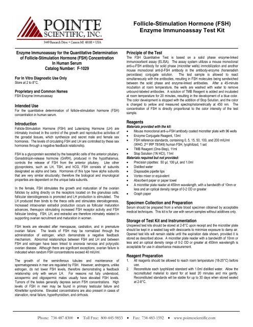
Follicle-Stimulation Hormone (FSH)<br />
Enzyme Immunoassay Test Kit<br />
Enzyme Immunoassay for the Quantitative Determination<br />
of Follicle-Stimulation Hormone (FSH) Concentration<br />
In Human Serum<br />
Catalog Number: F-1029<br />
For In Vitro Diagnostic Use Only<br />
Store at 2 to 8°C.<br />
Proprietary and Common Names<br />
FSH Enzyme Immunoassay<br />
Intended Use<br />
For the quantitative determination of follicle-stimulation hormone (FSH)<br />
concentration in human serum.<br />
Introduction<br />
Follicle-Stimulation Hormone (FSH) and Luteinizing Hormone (LH) are<br />
intimately involved in the control of the growth and reproductive activities of<br />
the gonadal tissues, which synthesize and secret male and female sex<br />
hormones. The levels of circulating FSH and LH are controlled by these sex<br />
hormones through a negative feedback relationship.<br />
FSH is a glycoprotein secreted by the basophilic cells of the anterior pituitary.<br />
Gonadotropin-release hormone (GnRH), produced in the hypothalamus,<br />
controls the release of FSH from the anterior pituitary. Like other<br />
glycoproteins, such as LH, TSH, and hCG, FSH consists of subunits<br />
designated as alpha and beta. Hormones of this type have alpha subunits<br />
that are very similar structurally; therefore the biological and imunological<br />
properties are dependent on the unique beta subunits.<br />
In the female, FSH stimulates the growth and maturation of the ovarian<br />
follicles by acting directly on the receptors located on the granulosa cells;<br />
follicular steroidogenesis is promoted and LH production is stimulated. The<br />
LH produced then binds to the theca cells and stimulates steroidogenesis.<br />
<strong>Inc</strong>reased intraovarian estradiol production occurs as follicular maturation<br />
advances, thereupon stimulating increased FSH receptor activity and FSH<br />
follicular binding. FSH, LH, and estradiol are therefore intimately related in<br />
supporting ovarian recruitment and maturation in women.<br />
FSH levels are elevated after menopause, castration, and in premature<br />
ovarian failure. The levels of FSH may be normalized through the<br />
administration of estrogen, which demonstrate a negative feedback<br />
mechanism. Abnormal relationships between FSH and LH and between<br />
FSH and estrogen have been linked to anorexia nervosa and polycystic<br />
ovarian disease. Although there are significant exceptions, ovarian failure is<br />
indicated when random FSH concentrations exceed 40 mIU/ml.<br />
The growth of the seminiferous tubules and maintenance of<br />
spermatogenesis in men are regulated by FSH. However, androgens, unlike<br />
estrogen, do not lower FSH levels, therefore demonstrating a feedback<br />
relationship only with serum LH. For reasons not fully understood,<br />
azospermic and oligospermic males usually have elevated FSH levels.<br />
Tumors of the testes generally depress serum FSH concentrations. High<br />
levels of FSH in men may be found in primary testicular failure and<br />
Klinefelter syndrome. Elevated concentrations are also present in cases of<br />
starvation, renal failure, hyperthyroidism, and cirrhosis.<br />
Principle of the Test<br />
The FSH Quantitative Test is based on a solid phase enzyme-linked<br />
immunosorbent assay (ELISA). The assay system utilizes a mouse monoclonal<br />
anti-α-FSH antibody for solid phase (microtiter wells) immobilization and another<br />
mouse monoclonal anti-β-FSH antibody in the antibody-enzyme (horseradish<br />
peroxidase) conjugate solution. The test sample is allowed to react<br />
simultaneously with the antibodies, resulting in FSH molecules being sandwiched<br />
between the solid phase and enzyme-linked antibodies. After a 45-minute<br />
incubation at room temperature, the wells are washed with water to remove<br />
unbound-labeled antibodies. A solution of TMB Reagent is added and incubated<br />
at room temperature for 20 minutes, resulting in the development of a blue color.<br />
The color development is stopped with the addition of Stop Solution, and the color<br />
is changed to yellow and measured spectrophotometrically at 450 nm. The<br />
concentration of FSH is directly proportional to the color intensity of the test<br />
sample.<br />
Reagents<br />
Materials provided with the kit:<br />
• Mouse monoclonal anti-α-FSH antibody coated microtiter plate with <strong>96</strong> wells<br />
• Enzyme Conjugate Reagent, 13ml<br />
• FSH reference standards, containing 0, 5, 15, 50, 100, and 200 mIU/ml<br />
(WHO, 2 nd IRP 78/549) human FSH, lyophilized, 1 set<br />
• TMB Reagent (One-Step), 11ml<br />
• Stop Solution (1N HCI), 11ml<br />
Materials required but not provided:<br />
• Precision pipettes: 50 µl, 100 µl, and 1.0ml<br />
• Distilled water<br />
• Disposable pipette tips<br />
• Vortex mixer or equivalent<br />
• Absorbent paper or paper towel<br />
• A microtiter plate reader at 450nm wavelength, with a bandwidth of 10nm or<br />
less and an optical density range of 0-2 OD or greater<br />
• Graph paper<br />
Specimen Collection and Preparation<br />
Serum should be prepared from a whole blood specimen obtained by acceptable<br />
medical techniques. This kit is for use with serum samples without additives only.<br />
Storage of Test Kit and Instrumentation<br />
Unopened test kits should be stored at 2-8°C upon receipt and the microtiter plate<br />
should be kept in a sealed bag with desiccants to minimize exposure to damp air.<br />
Opened test kits will remain stable until the expiration date shown, provided it is<br />
stored as described above. A microtiter plate reader with a bandwidth of 10nm or<br />
less and an optical density range of 0-2 OD or greater at 450nm wavelength is<br />
acceptable for use in absorbance measurement.<br />
Reagent Preparation<br />
1. All reagents should be allowed to reach room temperature (18-25°C) before<br />
use.<br />
2. Reconstitute each lyophilized standard with 1.0ml distilled water. Allow the<br />
reconstituted material to stand for at least 20 minutes and mix gently.<br />
Reconstituted standards will be stable for up to 30 days when stored sealed<br />
at 2-8°C.<br />
Phone: 734-487-8300 • Toll Free: 800-445-9853 • Fax: 734-483-1592 • www.pointescientific.com
Follicle-Stimulation Hormone (FSH)<br />
Enzyme Immunoassay Test Kit<br />
Assay Procedure<br />
1. Secure the desired number of coated wells in the holder.<br />
2. Dispense 50μl of standard, specimens, and controls into appropriate<br />
wells.<br />
3. Dispense 100μl of Enzyme Conjugate Reagent into each well.<br />
4. Thoroughly mix for 30 seconds. It is very important to have complete<br />
mixing in this setup.<br />
5. <strong>Inc</strong>ubate at room temperature (18-25°C) for 45 minutes.<br />
6. Remove the incubation mixture by flicking plate contents into a waste<br />
container.<br />
7. Rinse and flick the microtiter wells 5 times with distilled or deionized<br />
water. (Do not use tap water).<br />
8. Strike the wells sharply onto absorbent paper or paper towels to<br />
remove all residual water droplets.<br />
9. Dispense 100μl TMB Reagent into each well. Gently mix for 10<br />
seconds.<br />
10. <strong>Inc</strong>ubate at room temperature in the dark for 20 minutes.<br />
11. Stop the reaction by adding 100μl of Stop Solution to each well.<br />
12. Gently mix for 30 seconds. It is important to make sure that all the<br />
blue color changes to yellow color completely.<br />
13. Read the optical density at 450nm with a microtiter plate reader within<br />
15 minutes.<br />
Calculation of Results<br />
1. Calculate the mean absorbance value (A450) for each set of reference<br />
standards, specimens, controls and patient samples.<br />
2. Construct a standard curve by plotting the mean absorbance obtained<br />
from each reference standard against its concentration in mIU/ml on<br />
linear graph paper, with absorbance values on the vertical or Y-axis<br />
and concentrations on the horizontal or X-axis.<br />
3. Using the mean absorbance value for each sample, determine the<br />
corresponding concentration of FSH in mIU/ml from the standard curve.<br />
Example of Standard Curve<br />
Results of a typical standard run with optical density readings at 450nm<br />
shown in the Y axis against FSH concentration shown in the X-axis. This<br />
standard curve is for the purpose of illustration only, and should not be used<br />
to calculate unknowns. Each user should obtain his or her own data and<br />
standard curve in each experiment.<br />
FSH Standards (mIU/ml) Absorbance (450nm)<br />
0 0.058<br />
5 0.133<br />
15 0.265<br />
50 0.782<br />
100 1.483<br />
200 2.885<br />
Absorbance (450nm)<br />
3<br />
2<br />
1<br />
0<br />
0 50 100 150 200 250<br />
FSH Conc. (mIU/ml)<br />
Expected Values and Sensitivity<br />
Based on random selected outpatient clinical laboratory samples, the mean FSH<br />
values in males (N=100) and females (N=150) are 11 and 12 mIU/ml, respectively.<br />
The mean FSH values in post menopausal (N=60) and pregnant females (N=60)<br />
are 94 and 1.0 mIU/ml, respectively. The minimum detectable concentration of<br />
FSH by this assay is estimated to be 2.5 mIU/ml.<br />
Limitations of the Procedure<br />
1. Serum samples demonstrating gross lipemia, gross hemolysis, or turbidity<br />
should not be used with this test.<br />
2. The results obtained from the use of this kit should be used only as an<br />
adjunct to other diagnostic procedures and information available to the<br />
physician.<br />
References<br />
1. Marshall, J.C. Clinic in Endocrinol Metab 1975; 4:545.<br />
2. Cohen K.L. Metabolism 1977; 26:1165.<br />
3. Rebar, R.W., Erickson, G.F., and Yen, S.C.C. Fertil. Steril. 1982; 37:35.<br />
4. Abranham, G.E. Ed. Radioassay Systems in Clinic. Endocrinol. Marcel<br />
Dekker, <strong>Inc</strong>., New York (1981).<br />
5. Uotila M., Ruoslahti E. and Engvall E. J. Immunol. Methods 1981; 42:11.<br />
Manufactured for <strong>Pointe</strong> <strong>Scientific</strong>, <strong>Inc</strong>.<br />
5449 Research Drive, Canton, MI 48188<br />
European Authorized Representative:<br />
Obelis s.a.<br />
Boulevard Général Wahis 53<br />
1030 Brussels, BELGIUM<br />
Tel: (32)2.732.59.54 Fax:(32)2.732.60.03 email: mail@obelis.net<br />
Rev. 04/10 P803-<strong>F1029</strong>-01<br />
040203


