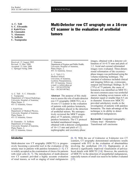
Multi-Detector row CT urography on a 16-row CT scanner in the ...
Multi-Detector row CT urography on a 16-row CT scanner in the ...
Multi-Detector row CT urography on a 16-row CT scanner in the ...

Create successful ePaper yourself
Turn your PDF publications into a flip-book with our unique Google optimized e-Paper software.
Eur Radiol<br />
DOI 10.1007/s00330-006-0383-2<br />
UROGENITAL<br />
A. C. Tsili<br />
S. C. Efremidis<br />
J. Kalef-Ezra<br />
D. Giannakis<br />
Y. Alamanos<br />
N. Sofikitis<br />
C. Tsampoulas<br />
<str<strong>on</strong>g>Multi</str<strong>on</strong>g>-<str<strong>on</strong>g>Detector</str<strong>on</strong>g> <str<strong>on</strong>g>row</str<strong>on</strong>g> <str<strong>on</strong>g>CT</str<strong>on</strong>g> <str<strong>on</strong>g>urography</str<strong>on</strong>g> <strong>on</strong> a <strong>16</strong>-<str<strong>on</strong>g>row</str<strong>on</strong>g><br />
<str<strong>on</strong>g>CT</str<strong>on</strong>g> <strong>scanner</strong> <strong>in</strong> <strong>the</strong> evaluati<strong>on</strong> of uro<strong>the</strong>lial<br />
tumors<br />
Received: 15 August 2005<br />
Revised: 31 May 2006<br />
Accepted: 23 June 2006<br />
# Spr<strong>in</strong>ger-Verlag 2006<br />
A. C. Tsili . S. C. Efremidis .<br />
C. Tsampoulas<br />
Department of Cl<strong>in</strong>ical Radiology,<br />
University Hospital of Ioann<strong>in</strong>a,<br />
Platia Pargis, 2,<br />
453 32 Ioann<strong>in</strong>a, Greece<br />
J. Kalef-Ezra<br />
Department of Medical Physics,<br />
University Hospital of Ioann<strong>in</strong>a,<br />
Platia Pargis, 2,<br />
453 32 Ioann<strong>in</strong>a, Greece<br />
D. Giannakis . N. Sofikitis<br />
Department of Urology,<br />
University Hospital of Ioann<strong>in</strong>a,<br />
Platia Pargis, 2,<br />
453 32 Ioann<strong>in</strong>a, Greece<br />
Y. Alamanos<br />
Department of Hygiene and Public Health,<br />
University Hospital of Ioann<strong>in</strong>a,<br />
Platia Pargis, 2,<br />
453 32 Ioann<strong>in</strong>a, Greece<br />
A. C. Tsili (*)<br />
Medical School,<br />
University of Ioann<strong>in</strong>a,<br />
Leoforos Panepistimiou,<br />
451 10 Ioann<strong>in</strong>a, Greece<br />
e-mail: a_tsili@yahoo.gr<br />
Tel.: +30-2651070708<br />
Fax: +30-2651097862<br />
Abstract The purpose of this study<br />
was to assess <strong>the</strong> role of multi-detector<br />
<str<strong>on</strong>g>row</str<strong>on</strong>g> <str<strong>on</strong>g>CT</str<strong>on</strong>g> <str<strong>on</strong>g>urography</str<strong>on</strong>g> (MD<str<strong>on</strong>g>CT</str<strong>on</strong>g>U), <strong>on</strong> a<br />
<strong>16</strong>-<str<strong>on</strong>g>row</str<strong>on</strong>g> <str<strong>on</strong>g>CT</str<strong>on</strong>g> <strong>scanner</strong> <strong>in</strong> <strong>the</strong> evaluati<strong>on</strong><br />
of patients with pa<strong>in</strong>less hematuria,<br />
with emphasis placed <strong>in</strong> <strong>the</strong> detecti<strong>on</strong><br />
of uro<strong>the</strong>lial tumors. We retrospectively<br />
reviewed <strong>the</strong> MD<str<strong>on</strong>g>CT</str<strong>on</strong>g> urographies<br />
of 75 patients, referred for<br />
pa<strong>in</strong>less hematuria. The <str<strong>on</strong>g>CT</str<strong>on</strong>g> protocol<br />
<strong>in</strong>cluded unenhanced images,<br />
obta<strong>in</strong>ed with a detector c<strong>on</strong>figurati<strong>on</strong><br />
of <strong>16</strong>×1.5 mm and pitch of 1.2,<br />
nephrographic and excretory-phase<br />
images, obta<strong>in</strong>ed with a detector collimati<strong>on</strong><br />
of <strong>16</strong>×0.75 mm and pitch of<br />
1.2. Axial and cor<strong>on</strong>al reformatted<br />
images were evaluated. Three-dimensi<strong>on</strong>al<br />
reformati<strong>on</strong> of <strong>the</strong> excretoryphase<br />
images was performed us<strong>in</strong>g <strong>the</strong><br />
volume-render<strong>in</strong>g technique. The<br />
standard of reference <strong>in</strong>cluded cl<strong>in</strong>ical<br />
and imag<strong>in</strong>g follow-up, cystoscopic,<br />
surgical and histologic f<strong>in</strong>d<strong>in</strong>gs. In 55<br />
(73%) of 75 patients, <strong>the</strong> cause of<br />
hematuria was identified <strong>on</strong> MD<str<strong>on</strong>g>CT</str<strong>on</strong>g>U;<br />
<strong>the</strong> most comm<strong>on</strong> cause was uro<strong>the</strong>lial<br />
cancer, <strong>in</strong>clud<strong>in</strong>g seven tumors with a<br />
diameter equal or smaller than 0.5 cm<br />
<strong>in</strong> diameter. Sixteen-<str<strong>on</strong>g>row</str<strong>on</strong>g> MD<str<strong>on</strong>g>CT</str<strong>on</strong>g>U<br />
provided satisfactory results <strong>in</strong> <strong>the</strong><br />
<strong>in</strong>vestigati<strong>on</strong> of patients with pa<strong>in</strong>less<br />
hematuria. The ma<strong>in</strong> advantage of <strong>the</strong><br />
technique is its ability to detect<br />
uroepi<strong>the</strong>lial malignancies.<br />
Keywords Computed tomography .<br />
Ur<strong>in</strong>ary tract . Hematuria .<br />
Transiti<strong>on</strong>al cell carc<strong>in</strong>oma<br />
Introducti<strong>on</strong><br />
<str<strong>on</strong>g>Multi</str<strong>on</strong>g>-detector <str<strong>on</strong>g>row</str<strong>on</strong>g> <str<strong>on</strong>g>CT</str<strong>on</strong>g> <str<strong>on</strong>g>urography</str<strong>on</strong>g> (MD<str<strong>on</strong>g>CT</str<strong>on</strong>g>U) is progressively<br />
becom<strong>in</strong>g a powerful tool <strong>in</strong> <strong>the</strong> evaluati<strong>on</strong> of <strong>the</strong><br />
ur<strong>in</strong>ary tract <strong>in</strong> patients with pa<strong>in</strong>less hematuria [1–5]. <str<strong>on</strong>g>CT</str<strong>on</strong>g><br />
is widely accepted as <strong>the</strong> exam<strong>in</strong>ati<strong>on</strong> of choice for patients<br />
with renal colic [6, 7]. The <strong>in</strong>troducti<strong>on</strong> of multi-detector<br />
<str<strong>on</strong>g>row</str<strong>on</strong>g> <str<strong>on</strong>g>CT</str<strong>on</strong>g> <strong>scanner</strong>s provided a highly accurate evaluati<strong>on</strong><br />
of renal masses, as well as stag<strong>in</strong>g of renal malignancies<br />
[8, 9]. With <strong>the</strong> use of 4-detector or 8-detector <str<strong>on</strong>g>row</str<strong>on</strong>g> <str<strong>on</strong>g>CT</str<strong>on</strong>g><br />
<strong>scanner</strong>s, <str<strong>on</strong>g>CT</str<strong>on</strong>g> <str<strong>on</strong>g>urography</str<strong>on</strong>g> dem<strong>on</strong>strated satisfactory results<br />
compared with IVU <strong>in</strong> <strong>the</strong> evaluati<strong>on</strong> of abnormalities<br />
<strong>in</strong>volv<strong>in</strong>g <strong>the</strong> uro<strong>the</strong>lium [10–13]. Raptopoulos et al.<br />
showed a significantly improved visualizati<strong>on</strong> of pelvicaliceal<br />
details with MD<str<strong>on</strong>g>CT</str<strong>on</strong>g>U [14]. However, <strong>the</strong> sensitivity<br />
of MD<str<strong>on</strong>g>CT</str<strong>on</strong>g>U <strong>in</strong> <strong>the</strong> detecti<strong>on</strong> of uroepi<strong>the</strong>lial malignancies,<br />
especially tumors of small size need to be fur<strong>the</strong>r evaluated<br />
[15].
With <strong>the</strong> <strong>in</strong>troducti<strong>on</strong> of <strong>16</strong>-<str<strong>on</strong>g>row</str<strong>on</strong>g> <str<strong>on</strong>g>CT</str<strong>on</strong>g> <strong>scanner</strong>s, <strong>the</strong><br />
radiologic evaluati<strong>on</strong> of <strong>the</strong> ur<strong>in</strong>ary tract is chang<strong>in</strong>g<br />
rapidly. The advantages of a <strong>16</strong>-<str<strong>on</strong>g>row</str<strong>on</strong>g> <str<strong>on</strong>g>CT</str<strong>on</strong>g> <strong>scanner</strong> are <strong>the</strong><br />
follow<strong>in</strong>g: fast scann<strong>in</strong>g (coverage of at least 48 mm l<strong>on</strong>g<br />
body secti<strong>on</strong>s per sec<strong>on</strong>d), acquisiti<strong>on</strong> of th<strong>in</strong> collimati<strong>on</strong><br />
and creati<strong>on</strong> of high-resoluti<strong>on</strong> multiplanar and threedimensi<strong>on</strong>al<br />
(3D) reformatted images, with excellent<br />
anatomic details [<strong>16</strong>, 17]. The purpose of our study was<br />
to assess <strong>the</strong> role of multi-detector <str<strong>on</strong>g>row</str<strong>on</strong>g> <str<strong>on</strong>g>CT</str<strong>on</strong>g> <str<strong>on</strong>g>urography</str<strong>on</strong>g>,<br />
us<strong>in</strong>g a <strong>16</strong>-<str<strong>on</strong>g>row</str<strong>on</strong>g> <str<strong>on</strong>g>CT</str<strong>on</strong>g> <strong>scanner</strong> <strong>in</strong> <strong>the</strong> evaluati<strong>on</strong> of patients<br />
present<strong>in</strong>g with pa<strong>in</strong>less hematuria, with emphasis placed<br />
<strong>in</strong> <strong>the</strong> detecti<strong>on</strong> of uro<strong>the</strong>lial tumors.<br />
Materials and methods<br />
This study was a retrospective review of <str<strong>on</strong>g>CT</str<strong>on</strong>g> urographies<br />
performed <strong>on</strong> a <strong>16</strong>-<str<strong>on</strong>g>row</str<strong>on</strong>g> <str<strong>on</strong>g>CT</str<strong>on</strong>g> <strong>scanner</strong> for <strong>the</strong> evaluati<strong>on</strong> of<br />
pa<strong>in</strong>less hematuria. The study <strong>in</strong>cluded 75 c<strong>on</strong>secutive<br />
patients (47 men and 28 women) with a mean age of 63<br />
years (age range: 21–87 years), who underwent multidetector<br />
<str<strong>on</strong>g>row</str<strong>on</strong>g> <str<strong>on</strong>g>CT</str<strong>on</strong>g> <str<strong>on</strong>g>urography</str<strong>on</strong>g> between March 2004 and<br />
January 2005, referred to <strong>the</strong> department of Radiology for<br />
pa<strong>in</strong>less hematuria. Five patients were younger than 40<br />
years old. Informed c<strong>on</strong>sent for this study was obta<strong>in</strong>ed<br />
from each patient.<br />
The cl<strong>in</strong>ical evaluati<strong>on</strong> of <strong>the</strong> patients <strong>in</strong>cluded a careful<br />
history and cl<strong>in</strong>ical exam<strong>in</strong>ati<strong>on</strong>. The laboratory analysis<br />
began with a comprehensive exam<strong>in</strong>ati<strong>on</strong> of <strong>the</strong> ur<strong>in</strong>e and<br />
ur<strong>in</strong>e sediment and cytologic analysis. All patients had an<br />
ultrasound exam<strong>in</strong>ati<strong>on</strong> of <strong>the</strong> abdomen, some performed<br />
at an outpatient cl<strong>in</strong>ic and <strong>the</strong> rest at our department.<br />
Patients with hematuria and a s<strong>on</strong>ographic exam<strong>in</strong>ati<strong>on</strong><br />
dem<strong>on</strong>strat<strong>in</strong>g a kidney mass were not <strong>in</strong>cluded <strong>in</strong> this<br />
study, s<strong>in</strong>ce <strong>the</strong>y were evaluated with a different protocol<br />
tailored for <strong>the</strong> <strong>in</strong>vestigati<strong>on</strong> of renal masses. Cystoscopic<br />
exam<strong>in</strong>ati<strong>on</strong> was carried out <strong>in</strong> all patients.<br />
In patients <strong>in</strong> whom cl<strong>in</strong>ical and imag<strong>in</strong>g evaluati<strong>on</strong><br />
revealed no cause for <strong>the</strong> hematuria, a follow-up was<br />
recommended. This <strong>in</strong>cluded <strong>the</strong> repeat of <strong>the</strong> ur<strong>in</strong>ary<br />
analysis and ur<strong>in</strong>e cytology every m<strong>on</strong>th for <strong>the</strong> first 4<br />
m<strong>on</strong>ths and <strong>the</strong>reafter at 6, 12, 24 and 36 m<strong>on</strong>ths.<br />
Immediate reevaluati<strong>on</strong> with repeat of <strong>the</strong> <str<strong>on</strong>g>CT</str<strong>on</strong>g> exam<strong>in</strong>ati<strong>on</strong><br />
and/or cystoscopy and ur<strong>in</strong>e cytology was recommended if<br />
any of <strong>the</strong> follow<strong>in</strong>g occurred: development of gross<br />
hematuria, abnormal ur<strong>in</strong>ary cytologic f<strong>in</strong>d<strong>in</strong>gs or irritat<strong>in</strong>g<br />
void<strong>in</strong>g symptoms <strong>in</strong> <strong>the</strong> absence of <strong>in</strong>fecti<strong>on</strong> [18].<br />
Technique<br />
Exam<strong>in</strong>ati<strong>on</strong>s were performed <strong>on</strong> a <strong>16</strong>-<str<strong>on</strong>g>row</str<strong>on</strong>g> <str<strong>on</strong>g>CT</str<strong>on</strong>g> <strong>scanner</strong><br />
with 24 mm scann<strong>in</strong>g span per rotati<strong>on</strong> (Mx8000 IDT,<br />
Philips) and a three-phase protocol was used. Unenhanced<br />
images were obta<strong>in</strong>ed from <strong>the</strong> kidneys through <strong>the</strong> ur<strong>in</strong>ary<br />
bladder us<strong>in</strong>g a slice collimati<strong>on</strong> of <strong>16</strong>×1.5 mm and a slice<br />
thickness of 3 mm. These images were scrut<strong>in</strong>ized to<br />
identify ur<strong>in</strong>ary tract calculi or calcificati<strong>on</strong>s, as well as to<br />
assist <strong>in</strong> <strong>the</strong> characterizati<strong>on</strong> of any detected renal masses.<br />
Intravenous n<strong>on</strong>-i<strong>on</strong>ic iod<strong>in</strong>ated c<strong>on</strong>trast material (120 ml,<br />
320 mg I/ml) was subsequently adm<strong>in</strong>istered at a rate of<br />
3 ml/sec, via mechanical <strong>in</strong>jector, followed by 250 ml of<br />
normal sal<strong>in</strong>e 0.9%, rapidly <strong>in</strong>fused by <strong>in</strong>travenous drip.<br />
The sec<strong>on</strong>d phase was <strong>the</strong> nephrographic phase obta<strong>in</strong>ed at<br />
100 sec delay, cover<strong>in</strong>g <strong>the</strong> area from <strong>the</strong> diaphragm to <strong>the</strong><br />
iliac crests and <strong>the</strong> third phase was <strong>the</strong> excretory phase<br />
start<strong>in</strong>g with 10-m<strong>in</strong>ute delay, cover<strong>in</strong>g <strong>the</strong> kidneys, ureters<br />
and <strong>the</strong> ur<strong>in</strong>ary bladder. The nephrographic phase was<br />
chosen for <strong>the</strong> detecti<strong>on</strong> and characterizati<strong>on</strong> of renal<br />
masses, as well as for stag<strong>in</strong>g, while <strong>the</strong> excretory phase<br />
was utilized for <strong>the</strong> evaluati<strong>on</strong> of abnormalities <strong>in</strong>volv<strong>in</strong>g<br />
<strong>the</strong> uro<strong>the</strong>lium. For <strong>the</strong>se two phases a secti<strong>on</strong> collimati<strong>on</strong><br />
of <strong>16</strong>×0.75 mm and a slice thickness of 1 mm were<br />
employed. The nephrographic and excretory phase images<br />
were rec<strong>on</strong>structed at 0.5 mm <strong>in</strong>tervals (50% overlap). All<br />
<strong>the</strong> scans were performed with a pitch of 1.2, at 120 kV,<br />
us<strong>in</strong>g a rotati<strong>on</strong> time of 0.5 sec. Both dose modulati<strong>on</strong><br />
(DOM) and automatic current sett<strong>in</strong>g (DoseRight) were<br />
used [19]. No oral c<strong>on</strong>trast material was given, <strong>in</strong> order to<br />
facilitate 3D reformatt<strong>in</strong>g. The entire <str<strong>on</strong>g>CT</str<strong>on</strong>g> <str<strong>on</strong>g>urography</str<strong>on</strong>g><br />
exam<strong>in</strong>ati<strong>on</strong> lasted 15 m<strong>in</strong>utes per patient <strong>on</strong> <strong>the</strong> average<br />
and every scan lasted 7–12 sec<strong>on</strong>ds <strong>on</strong> <strong>the</strong> average. Our <str<strong>on</strong>g>CT</str<strong>on</strong>g><br />
protocol is illustrated <strong>on</strong> Table 1.<br />
<str<strong>on</strong>g>CT</str<strong>on</strong>g> data <strong>in</strong>terpretati<strong>on</strong><br />
Image <strong>in</strong>terpretati<strong>on</strong> was d<strong>on</strong>e <strong>on</strong> a workstati<strong>on</strong> (MxView,<br />
Philips) and <strong>in</strong>cluded <strong>the</strong> study of <strong>the</strong> axial images of <strong>the</strong><br />
unenhanced scan. The evaluati<strong>on</strong> of <strong>the</strong> axial source<br />
images of <strong>the</strong> nephrographic and <strong>the</strong> excretory phase was<br />
Table 1 <str<strong>on</strong>g>CT</str<strong>on</strong>g> protocol for <strong>the</strong> <strong>in</strong>vestigati<strong>on</strong> of hematuria<br />
Unenhanced<br />
scan<br />
Nephrographic<br />
phase,<br />
100 sec<br />
Excretory<br />
phase,<br />
10 m<strong>in</strong><br />
kV, rotati<strong>on</strong> time (sec) 120/0.5 120/0.5 120/0.5<br />
mAsnom/mAssel 200/170 200/175 200/<strong>16</strong>0<br />
<str<strong>on</strong>g>Detector</str<strong>on</strong>g> collimati<strong>on</strong> <strong>16</strong>×1.5 <strong>16</strong>×0.75 <strong>16</strong>×0.75<br />
(mm)<br />
Pitch 1.2 1.2 1.2<br />
Slice thickness (mm) 3 1 1<br />
Rec<strong>on</strong>structi<strong>on</strong> 3 0.5 0.5<br />
<strong>in</strong>terval (mm)<br />
Area<br />
Kidneysur<strong>in</strong>ary<br />
bladder<br />
Diaphragmiliac<br />
crests<br />
Kidneysur<strong>in</strong>ary<br />
bladder<br />
C<strong>on</strong>trast material<br />
(iv)<br />
120 ml (320), 3 ml/sec and 250 ml<br />
sal<strong>in</strong>e 0.9%
difficult, due to <strong>the</strong> large number (approximately 400–800)<br />
of noisy images. For <strong>the</strong>se reas<strong>on</strong>s reformatted images, of<br />
4 mm slice thickness and at 3 mm <strong>in</strong>tervals, usually <strong>in</strong> <strong>the</strong><br />
transverse and cor<strong>on</strong>al planes (us<strong>in</strong>g <strong>the</strong> Extended<br />
Brilliance V.1.0.1.1. software) were used for <strong>in</strong>terpretati<strong>on</strong><br />
of <strong>the</strong> nephrographic and excretory phases. The time to<br />
generate <strong>the</strong>se images was less than <strong>on</strong>e m<strong>in</strong>ute. Wide<br />
w<strong>in</strong>dow sett<strong>in</strong>gs (b<strong>on</strong>e w<strong>in</strong>dow sett<strong>in</strong>gs, W: 1500, L: 500)<br />
were also used for <strong>the</strong> excretory phase, to better visualize<br />
caliceal details, as well as fill<strong>in</strong>g defects <strong>in</strong> <strong>the</strong> ur<strong>in</strong>ary<br />
collect<strong>in</strong>g system (Fig. 1). F<strong>in</strong>ally, three-dimensi<strong>on</strong>al (3D)<br />
rec<strong>on</strong>structi<strong>on</strong>s of <strong>the</strong> excretory phase data were performed,<br />
us<strong>in</strong>g <strong>the</strong> volume render<strong>in</strong>g technique and <strong>the</strong><br />
same software, obta<strong>in</strong>ed <strong>in</strong> <strong>the</strong> cor<strong>on</strong>al and/or oblique<br />
planes. Volume render<strong>in</strong>g technique was c<strong>on</strong>sidered ideal<br />
for <strong>the</strong> creati<strong>on</strong> of <strong>the</strong> 3D rec<strong>on</strong>structi<strong>on</strong>s [3], especially<br />
because it does not obscure superimposed structures, such<br />
as <strong>the</strong> pelvis and <strong>the</strong> ureters. The time required to create <strong>the</strong><br />
3D reformatted images was less than <strong>on</strong>e m<strong>in</strong>ute. The ma<strong>in</strong><br />
advantage of <strong>the</strong>se images was that <strong>the</strong>y were more familiar<br />
to our cl<strong>in</strong>ical colleagues, because <strong>the</strong>y closely resemble<br />
those of <strong>in</strong>travenous <str<strong>on</strong>g>urography</str<strong>on</strong>g>. Additi<strong>on</strong>ally, <strong>the</strong>se images<br />
could be more effectively used for preoperative plann<strong>in</strong>g,<br />
whenever it was necessary.<br />
An important issue <strong>on</strong> <str<strong>on</strong>g>CT</str<strong>on</strong>g> <str<strong>on</strong>g>urography</str<strong>on</strong>g> is <strong>the</strong> degree of<br />
ureteral opacificati<strong>on</strong>. In cases, where excreti<strong>on</strong> of <strong>the</strong><br />
c<strong>on</strong>trast material was detected <strong>on</strong> <str<strong>on</strong>g>CT</str<strong>on</strong>g> <str<strong>on</strong>g>urography</str<strong>on</strong>g>, <strong>the</strong> extent<br />
of ureteral opacificati<strong>on</strong> was evaluated. The ureter was<br />
divided <strong>in</strong>to proximal, middle and distal thirds. The<br />
proximal third was def<strong>in</strong>ed as <strong>the</strong> porti<strong>on</strong> of <strong>the</strong> ureter<br />
that extended from <strong>the</strong> renal pelvis to <strong>the</strong> superior iliac<br />
crests. The middle third was def<strong>in</strong>ed as <strong>the</strong> porti<strong>on</strong> of <strong>the</strong><br />
ureter between <strong>the</strong> superior iliac crests and <strong>the</strong> anterior<br />
<strong>in</strong>ferior iliac sp<strong>in</strong>es. The lower third was def<strong>in</strong>ed as <strong>the</strong><br />
segment of <strong>the</strong> ureter located between <strong>the</strong> anterior <strong>in</strong>ferior<br />
iliac sp<strong>in</strong>es and <strong>the</strong> ureterovesical juncti<strong>on</strong>. An opacificati<strong>on</strong><br />
score from 1 to 3 was assigned for each ureter based <strong>on</strong><br />
<strong>the</strong> length of <strong>the</strong> ureteral porti<strong>on</strong> opacified. A score of 1<br />
<strong>in</strong>dicated opacificati<strong>on</strong> of less than <strong>on</strong>e third of <strong>the</strong> ureter; a<br />
score of 2 <strong>in</strong>dicated opacificati<strong>on</strong> of less than two thirds of<br />
<strong>the</strong> ureter; a score of 3, opacificati<strong>on</strong> of more than two<br />
thirds of <strong>the</strong> ureteral segments up to <strong>the</strong> entire length of <strong>the</strong><br />
ureter.<br />
<str<strong>on</strong>g>CT</str<strong>on</strong>g> data correlati<strong>on</strong><br />
F<strong>in</strong>d<strong>in</strong>gs of MD<str<strong>on</strong>g>CT</str<strong>on</strong>g>U were retrospectively correlated with<br />
<strong>the</strong> results of cl<strong>in</strong>ical and imag<strong>in</strong>g follow-up, cystoscopy<br />
and/or ureteroscopy, as well as with surgical and histological<br />
f<strong>in</strong>d<strong>in</strong>gs, <strong>in</strong> surgical cases. More specifically, <strong>in</strong><br />
patients with ur<strong>in</strong>ary tract lithiasis c<strong>on</strong>firmati<strong>on</strong> of <strong>the</strong> <str<strong>on</strong>g>CT</str<strong>on</strong>g><br />
diagnosis was documented <strong>on</strong> <strong>the</strong> basis of o<strong>the</strong>r imag<strong>in</strong>g<br />
exam<strong>in</strong>ati<strong>on</strong>s (pla<strong>in</strong> films and ultrasound). C<strong>on</strong>venti<strong>on</strong>al<br />
cystoscopy and histologic results were used as standard of<br />
reference <strong>in</strong> patients with ur<strong>in</strong>ary bladder carc<strong>in</strong>omas.<br />
C<strong>on</strong>venti<strong>on</strong>al cystoscopy was also used to c<strong>on</strong>firm <strong>the</strong><br />
presence of benign prostatic hyperplasia (BPH) as <strong>the</strong><br />
cause of hematuria. In patients with transiti<strong>on</strong>al cell<br />
carc<strong>in</strong>oma of <strong>the</strong> pelvicaliceal system, as well as ureteral<br />
lesi<strong>on</strong>s, c<strong>on</strong>firmati<strong>on</strong> of <strong>the</strong> <str<strong>on</strong>g>CT</str<strong>on</strong>g> diagnosis was obta<strong>in</strong>ed<br />
based <strong>on</strong> o<strong>the</strong>r imag<strong>in</strong>g exam<strong>in</strong>ati<strong>on</strong>s (IVU, retrograde<br />
pyelography), ureteroscopic f<strong>in</strong>d<strong>in</strong>gs and f<strong>in</strong>ally <strong>the</strong> surgical<br />
and histologic f<strong>in</strong>d<strong>in</strong>gs. Surgery and histologic diagnosis<br />
c<strong>on</strong>firmed <strong>the</strong> presence of o<strong>the</strong>r malignancies (renal<br />
cancer, pancreatic and prostate cancer). For patients with<br />
ur<strong>in</strong>ary tract c<strong>on</strong>genital anomalies, <strong>the</strong> f<strong>in</strong>d<strong>in</strong>gs of IVU<br />
were used for c<strong>on</strong>firm<strong>in</strong>g <strong>the</strong> <str<strong>on</strong>g>CT</str<strong>on</strong>g> diagnosis.<br />
The <str<strong>on</strong>g>CT</str<strong>on</strong>g> images were <strong>in</strong>terpreted by <strong>on</strong>e radiologist<br />
(A. C. T), bl<strong>in</strong>ded to <strong>the</strong> results of <strong>the</strong> o<strong>the</strong>r exam<strong>in</strong>ati<strong>on</strong>s.<br />
Dosimetry<br />
For dosimetry <strong>the</strong> free-<strong>in</strong>-air Computed Tomography Dose<br />
Index was measured al<strong>on</strong>g <strong>the</strong> axis of rotati<strong>on</strong> us<strong>in</strong>g a<br />
pencil-shaped i<strong>on</strong> chamber, type 10×5 10.3 <str<strong>on</strong>g>CT</str<strong>on</strong>g> (Radcal<br />
Corp., California, USA). The effective dose was calculated<br />
us<strong>in</strong>g <strong>the</strong> ImPA<str<strong>on</strong>g>CT</str<strong>on</strong>g> <str<strong>on</strong>g>CT</str<strong>on</strong>g> Patient Dosimetry Calculator<br />
(versi<strong>on</strong> 0.99v) [20]. The software used is based <strong>on</strong> <strong>the</strong><br />
Fig. 1 A 70-year-old woman<br />
with fibroepi<strong>the</strong>lial polyp of <strong>the</strong><br />
right ureter. Cor<strong>on</strong>al (a) and<br />
transverse (b) reformatted<br />
images of <strong>the</strong> excretory phase<br />
dem<strong>on</strong>strate an el<strong>on</strong>gated fill<strong>in</strong>g<br />
defect (ar<str<strong>on</strong>g>row</str<strong>on</strong>g>), smoothly marg<strong>in</strong>ated,<br />
<strong>in</strong>volv<strong>in</strong>g <strong>the</strong> <strong>in</strong>travesical<br />
part of <strong>the</strong> right ureter and<br />
prolaps<strong>in</strong>g <strong>in</strong>to <strong>the</strong> ur<strong>in</strong>ary<br />
bladder. The lesi<strong>on</strong> is clearly<br />
seen with b<strong>on</strong>e w<strong>in</strong>dow sett<strong>in</strong>gs
Table 2 MD<str<strong>on</strong>g>CT</str<strong>on</strong>g>U diagnoses for <strong>the</strong> hematuria<br />
<str<strong>on</strong>g>CT</str<strong>on</strong>g> diagnoses<br />
Number of patients<br />
St<strong>on</strong>e disease<br />
Ureter 4<br />
Kidney 7<br />
Ur<strong>in</strong>ary bladder 2<br />
Passed st<strong>on</strong>e 1<br />
Cancer<br />
Ur<strong>in</strong>ary bladder <strong>16</strong><br />
Kidney 7<br />
Ureter 1<br />
Prostate 1<br />
Pancreas 1<br />
Benign tumors<br />
Kidney 1<br />
Ureter 1<br />
Ur<strong>in</strong>ary tract <strong>in</strong>flammati<strong>on</strong> 2<br />
Benign prostatic hyperplasia 9<br />
Pyeloureteric juncti<strong>on</strong> obstructi<strong>on</strong> 1<br />
C<strong>on</strong>genital anomalies 4<br />
M<strong>on</strong>te Carlo c<strong>on</strong>versi<strong>on</strong> coefficients obta<strong>in</strong>ed by J<strong>on</strong>es and<br />
Shrimpt<strong>on</strong> [21]. C<strong>on</strong>sider<strong>in</strong>g <strong>the</strong> fact that <strong>the</strong> tube current is<br />
modulated across <strong>the</strong> patient’s body, based <strong>on</strong> plann<strong>in</strong>g<br />
scan projecti<strong>on</strong> radiograph (PSPR) and body asymmetry<br />
(dose modulati<strong>on</strong> functi<strong>on</strong>), <strong>the</strong> mean mAs per rotati<strong>on</strong> at<br />
each phase for each <strong>in</strong>dividual patient was recorded and <strong>the</strong><br />
mean mAs per rotati<strong>on</strong> am<strong>on</strong>g all patients at each phase<br />
was calculated. To assess <strong>the</strong> probability for <strong>in</strong>ducti<strong>on</strong> of<br />
determ<strong>in</strong>istic effects, two groups of LiF:Mg,Ti <strong>the</strong>rmolum<strong>in</strong>escent<br />
dosimeters were fixed with hypo-allergic tape<br />
<strong>on</strong> <strong>the</strong> sk<strong>in</strong> at <strong>the</strong> abdomen of six c<strong>on</strong>secutive patients, over<br />
a two-week period (<strong>on</strong>e group at umbilicus and <strong>the</strong> o<strong>the</strong>r at<br />
midaxillary l<strong>in</strong>e <strong>in</strong> <strong>the</strong> same level).<br />
Results<br />
In 18 of 75 patients (24%) no ur<strong>in</strong>ary tract abnormality was<br />
seen <strong>on</strong> MD<str<strong>on</strong>g>CT</str<strong>on</strong>g>U. In <strong>16</strong> of <strong>the</strong>se patients <strong>the</strong> cl<strong>in</strong>ical<br />
evaluati<strong>on</strong>, <strong>in</strong>clud<strong>in</strong>g cystoscopy and ur<strong>in</strong>alysis revealed<br />
no cause for <strong>the</strong> hematuria. These cases have been<br />
followed-up for a period ranged from 12 to 20 m<strong>on</strong>ths<br />
and rema<strong>in</strong>ed negative. These cases were c<strong>on</strong>sidered as<br />
true-negative. We had two false-negative cases. One with a<br />
patient, <strong>in</strong> whom <strong>the</strong> hematuria was attributed to carc<strong>in</strong>oma<br />
of <strong>the</strong> prostate gland, not shown <strong>on</strong> MD<str<strong>on</strong>g>CT</str<strong>on</strong>g>U and a sec<strong>on</strong>d<br />
case, <strong>in</strong> which <strong>the</strong> ur<strong>in</strong>ary bladder was filled with blood<br />
clots. In this case it was impossible for <strong>the</strong> <str<strong>on</strong>g>CT</str<strong>on</strong>g> exam<strong>in</strong>ati<strong>on</strong><br />
to recognize a small tumor, with a diameter less than<br />
0.5 cm, as seen with c<strong>on</strong>venti<strong>on</strong>al cystoscopy. This tumor<br />
could probably be detected, if <strong>the</strong>re were no blood clots <strong>on</strong><br />
<strong>the</strong> ur<strong>in</strong>ary bladder dur<strong>in</strong>g <str<strong>on</strong>g>CT</str<strong>on</strong>g> scann<strong>in</strong>g, as it was detected<br />
<strong>on</strong> s<strong>on</strong>ography. This lesi<strong>on</strong> was characterized as transiti<strong>on</strong>al<br />
cell carc<strong>in</strong>oma (TCC) <strong>on</strong> histology.<br />
In 57 of 75 patients (76%) <strong>the</strong> cause of hematuria was<br />
identified. Table 2 dem<strong>on</strong>strates <strong>the</strong> different etiologies of<br />
hematuria, as shown <strong>on</strong> MD<str<strong>on</strong>g>CT</str<strong>on</strong>g>U.<br />
In 12 patients, <strong>the</strong> cause of hematuria was ur<strong>in</strong>ary tract<br />
lithiasis. Four patients had ureteral st<strong>on</strong>es, rang<strong>in</strong>g from 3.5<br />
to 7 mm <strong>in</strong> diameter, with no signs of obstructi<strong>on</strong> <strong>in</strong> three.<br />
These four patients were followed-up with pla<strong>in</strong> films and<br />
ultrasound. In <strong>on</strong>e patient renal calculi were also seen. In<br />
six patients <strong>the</strong> calculi were located <strong>in</strong> <strong>the</strong> pelvicaliceal<br />
system, c<strong>on</strong>firmed also <strong>on</strong> pla<strong>in</strong> films and s<strong>on</strong>ography and<br />
<strong>in</strong> two <strong>in</strong> <strong>the</strong> ur<strong>in</strong>ary bladder, c<strong>on</strong>firmed by cystoscopy. In<br />
six of <strong>the</strong>se cases ur<strong>in</strong>ary tract lithiasis was detected as <strong>the</strong><br />
<strong>on</strong>ly possible cause for <strong>the</strong> hematuria, s<strong>in</strong>ce both MD<str<strong>on</strong>g>CT</str<strong>on</strong>g>U<br />
and cl<strong>in</strong>ical evaluati<strong>on</strong> revealed no o<strong>the</strong>r cause. In <strong>the</strong> o<strong>the</strong>r<br />
six patients BPH was also detected <strong>on</strong> <str<strong>on</strong>g>CT</str<strong>on</strong>g>U, but<br />
c<strong>on</strong>venti<strong>on</strong>al cystoscopy did not c<strong>on</strong>firm this f<strong>in</strong>d<strong>in</strong>g as<br />
<strong>the</strong> cause for <strong>the</strong> hematuria. In <strong>on</strong>e additi<strong>on</strong>al patient <strong>on</strong>ly<br />
signs of obstructi<strong>on</strong> were detected, probably due to a<br />
Fig. 2 A 67-year old man<br />
with TCCs of <strong>the</strong> ur<strong>in</strong>ary<br />
bladder, <strong>the</strong> larger tumor<br />
extend<strong>in</strong>g <strong>in</strong>to <strong>the</strong> perivesical<br />
space. (a) Cor<strong>on</strong>al and<br />
sagittal (b) reformati<strong>on</strong>s of<br />
<strong>the</strong> excretory phase show a<br />
small polypoid lesi<strong>on</strong> (short<br />
ar<str<strong>on</strong>g>row</str<strong>on</strong>g>) of <strong>the</strong> ur<strong>in</strong>ary bladder<br />
wall and a larger tumor (l<strong>on</strong>g<br />
ar<str<strong>on</strong>g>row</str<strong>on</strong>g>) extend<strong>in</strong>g <strong>in</strong>to <strong>the</strong><br />
perivesical fat
Fig. 3 A 73-year old man with TCC of <strong>the</strong> ur<strong>in</strong>ary bladder with<br />
<strong>in</strong>volvement of <strong>the</strong> perivesical fat. (a, b) Cor<strong>on</strong>al reformatted images<br />
of <strong>the</strong> excretory phase show left hydr<strong>on</strong>ephrosis and ureteral<br />
dilatati<strong>on</strong>. A mass <strong>in</strong>volv<strong>in</strong>g <strong>the</strong> left lateral wall of <strong>the</strong> ur<strong>in</strong>ary<br />
passed st<strong>on</strong>e, as it was <strong>in</strong>dicated by his past history of renal<br />
colic.<br />
In <strong>16</strong> patients MD<str<strong>on</strong>g>CT</str<strong>on</strong>g>U dem<strong>on</strong>strated carc<strong>in</strong>oma of <strong>the</strong><br />
ur<strong>in</strong>ary bladder. There were two false-positive cases. In <strong>on</strong>e<br />
case a small tumor of <strong>the</strong> ur<strong>in</strong>ary bladder seen <strong>on</strong> MD<str<strong>on</strong>g>CT</str<strong>on</strong>g>U<br />
was not c<strong>on</strong>firmed by cystoscopy and was c<strong>on</strong>sidered to<br />
represent a blood clot. In <strong>the</strong> sec<strong>on</strong>d case, <str<strong>on</strong>g>CT</str<strong>on</strong>g> <str<strong>on</strong>g>urography</str<strong>on</strong>g><br />
showed thicken<strong>in</strong>g of <strong>the</strong> right anterolateral wall of <strong>the</strong><br />
ur<strong>in</strong>ary bladder, as well as enhancement after c<strong>on</strong>trast<br />
material adm<strong>in</strong>istrati<strong>on</strong>. The patient underwent c<strong>on</strong>venti<strong>on</strong>al<br />
cystoscopy and no abnormality of <strong>the</strong> ur<strong>in</strong>ary<br />
bladder wall was seen. In 14 patients MD<str<strong>on</strong>g>CT</str<strong>on</strong>g>U detected<br />
bladder with perivesical fat <strong>in</strong>filtrati<strong>on</strong> is detected. (c) Threedimensi<strong>on</strong>al<br />
VR image c<strong>on</strong>firms <strong>the</strong> presence of <strong>the</strong> ur<strong>in</strong>ary bladder<br />
mass, as well as <strong>the</strong> f<strong>in</strong>d<strong>in</strong>gs of ur<strong>in</strong>ary tract obstructi<strong>on</strong><br />
20 ur<strong>in</strong>ary bladder tumors, c<strong>on</strong>firmed at c<strong>on</strong>venti<strong>on</strong>al<br />
cystoscopy, of <strong>the</strong>se patients three had multiple lesi<strong>on</strong>s<br />
(<strong>on</strong>e had two lesi<strong>on</strong>s, <strong>on</strong>e three and <strong>on</strong>e four lesi<strong>on</strong>s),<br />
(Fig. 2). Histologic exam<strong>in</strong>ati<strong>on</strong> dem<strong>on</strong>strated TCCs <strong>in</strong> 12<br />
patients and undifferentiated carc<strong>in</strong>oma of <strong>the</strong> ur<strong>in</strong>ary<br />
bladder <strong>in</strong> <strong>on</strong>e case, with <strong>in</strong>volvement of <strong>the</strong> perivesical fat.<br />
In two patients with TCCs <strong>the</strong> tumor <strong>in</strong>filtrated <strong>the</strong><br />
perivesical fat (Fig. 3), while <strong>in</strong> <strong>on</strong>e patient <strong>the</strong>re was<br />
Fig. 4 A 69-year old man with TCC of <strong>the</strong> ur<strong>in</strong>ary bladder.<br />
Transverse reformatted image of <strong>the</strong> excretory phase shows a sessile<br />
mass (ar<str<strong>on</strong>g>row</str<strong>on</strong>g>) of <strong>the</strong> left anterolateral wall of <strong>the</strong> ur<strong>in</strong>ary bladder. In<br />
this patient <strong>the</strong> first c<strong>on</strong>venti<strong>on</strong>al cystoscopy was negative and <strong>the</strong><br />
lesi<strong>on</strong> was c<strong>on</strong>firmed <strong>on</strong> a repeat exam<strong>in</strong>ati<strong>on</strong><br />
Fig. 5 A 69-year old man with transiti<strong>on</strong>al cell carc<strong>in</strong>oma of <strong>the</strong><br />
ur<strong>in</strong>ary bladder. Sagittal reformatted image of <strong>the</strong> excretory phase<br />
shows a small polypoid lesi<strong>on</strong> (ar<str<strong>on</strong>g>row</str<strong>on</strong>g>) of posterior wall of <strong>the</strong><br />
ur<strong>in</strong>ary bladder. The lesi<strong>on</strong> was seen <strong>on</strong>ly <strong>on</strong> sagittal reformati<strong>on</strong>s
Fig. 6 A 62-year old man with TCC of <strong>the</strong> renal pelvis. (a)<br />
Transverse unenhanced image shows a soft-tissue mass (ar<str<strong>on</strong>g>row</str<strong>on</strong>g>) of<br />
<strong>the</strong> right renal pelvis. (b) Transverse reformatted image of <strong>the</strong><br />
nephrographic phase dem<strong>on</strong>strates enhancement of <strong>the</strong> mass<br />
Fig. 7 A 65-year old woman with TCC of <strong>the</strong> right renal collect<strong>in</strong>g<br />
system. Sagittal reformati<strong>on</strong> of <strong>the</strong> excretory phase depicts a mass<br />
(ar<str<strong>on</strong>g>row</str<strong>on</strong>g>) <strong>in</strong>volv<strong>in</strong>g <strong>the</strong> lower calyx<br />
(ar<str<strong>on</strong>g>row</str<strong>on</strong>g>). (c) Cor<strong>on</strong>al reformatted image of <strong>the</strong> excretory phase<br />
shows <strong>the</strong> lesi<strong>on</strong> as a fill<strong>in</strong>g defect with irregular marg<strong>in</strong>s, <strong>in</strong>volv<strong>in</strong>g<br />
<strong>the</strong> right renal pelvis (ar<str<strong>on</strong>g>row</str<strong>on</strong>g>)<br />
metastatic <strong>in</strong>volvement of <strong>the</strong> retroperit<strong>on</strong>eal lymph nodes,<br />
shown by MD<str<strong>on</strong>g>CT</str<strong>on</strong>g>U. In a third patient with TCC <strong>the</strong> tumor<br />
<strong>in</strong>filtrated <strong>the</strong> retrovesical fat and <strong>the</strong>re was also retroperit<strong>on</strong>eal<br />
lymhadenopathy. This patient had a previous<br />
hysterectomy, performed at an outpatient cl<strong>in</strong>ic, reported<br />
for benign causes. F<strong>in</strong>ally <strong>in</strong> <strong>on</strong>e patient c<strong>on</strong>venti<strong>on</strong>al<br />
cystoscopy was negative, while MD<str<strong>on</strong>g>CT</str<strong>on</strong>g>U showed a sessile<br />
mass of <strong>the</strong> ur<strong>in</strong>ary bladder wall (Fig. 4). The patient<br />
underwent a sec<strong>on</strong>d cystoscopy, which c<strong>on</strong>firmed <strong>the</strong> <str<strong>on</strong>g>CT</str<strong>on</strong>g><br />
f<strong>in</strong>d<strong>in</strong>gs and histology revealed a TCC. The size of ur<strong>in</strong>ary<br />
bladder carc<strong>in</strong>omas detected <strong>on</strong> MD<str<strong>on</strong>g>CT</str<strong>on</strong>g>U ranged from 0.4<br />
to 7 cm <strong>in</strong> diameter (mean size: 1.8 cm), <strong>in</strong>cluded were six<br />
lesi<strong>on</strong>s with a diameter equal or smaller than 0.5 cm,<br />
(Fig. 5).<br />
In six patients carc<strong>in</strong>oma was located <strong>in</strong> <strong>the</strong> pelvicaliceal<br />
system, characterized as TCCs histologically (Figs. 6, 7)<strong>in</strong><br />
five, and as TCC, with sarcomatous elements <strong>in</strong> <strong>on</strong>e patient<br />
(Fig. 8). These lesi<strong>on</strong>s measured 0.7–5 cm <strong>in</strong> maximal<br />
diameter (mean diameter: 2 cm). In two more patients<br />
MD<str<strong>on</strong>g>CT</str<strong>on</strong>g>U revealed a ureteral lesi<strong>on</strong>, measur<strong>in</strong>g 0.3 cm <strong>in</strong><br />
transverse diameter, proved histologically to be a TCC <strong>in</strong><br />
<strong>on</strong>e patient (Fig. 9) and a fibroepi<strong>the</strong>lial polyp, <strong>in</strong>volv<strong>in</strong>g<br />
Fig. 8 A 68-year old man with<br />
TCC of <strong>the</strong> left renal collect<strong>in</strong>g<br />
system with sarcomatous<br />
elements, which <strong>in</strong>vades renal<br />
parenchyma. (a) Transverse<br />
reformati<strong>on</strong> of <strong>the</strong> nephrographic<br />
phase depicts a left atrophic<br />
kidney with a mass (ar<str<strong>on</strong>g>row</str<strong>on</strong>g>)<br />
<strong>in</strong>volv<strong>in</strong>g <strong>the</strong> lower calyx and<br />
<strong>in</strong>vad<strong>in</strong>g renal parenchyma. The<br />
patient underwent left nephrectomy<br />
and ureterectomy. Six<br />
m<strong>on</strong>ths later, local recurrence<br />
and retroperit<strong>on</strong>eal lymphadenopathy<br />
was detected <strong>on</strong> a<br />
follow-up <str<strong>on</strong>g>CT</str<strong>on</strong>g> exam<strong>in</strong>ati<strong>on</strong> (b)
Fig. 9 A 70-year old man with TCC of <strong>the</strong> right ureter. Cor<strong>on</strong>al<br />
reformatted image of <strong>the</strong> excretory phase dem<strong>on</strong>strates a mass<br />
(ar<str<strong>on</strong>g>row</str<strong>on</strong>g>s) <strong>in</strong>volv<strong>in</strong>g part of <strong>the</strong> upper and middle third of <strong>the</strong> right<br />
ureter. The lesi<strong>on</strong> causes dilatati<strong>on</strong> of <strong>the</strong> right ureter, as well as right<br />
hydr<strong>on</strong>ephrosis and delay of <strong>the</strong> excreti<strong>on</strong> from <strong>the</strong> right kidney<br />
<strong>the</strong> <strong>in</strong>travesical part of <strong>the</strong> ureter (Fig. 1) <strong>in</strong> <strong>the</strong> o<strong>the</strong>r. In <strong>the</strong><br />
last case, <strong>the</strong> lesi<strong>on</strong> ma<strong>in</strong>ly detected with wide w<strong>in</strong>dow<br />
sett<strong>in</strong>gs, measured 0.2 cm <strong>in</strong> transverse diameter and did<br />
not cause ureteral dilatati<strong>on</strong>.<br />
In three patients renal cell carc<strong>in</strong>oma, pancreatic cancer<br />
and carc<strong>in</strong>oma of <strong>the</strong> prostate gland were <strong>the</strong> causes of<br />
hematuria, respectively. The first patient was presented<br />
with a kidney mass, metastatic to <strong>the</strong> retroperit<strong>on</strong>eal lymph<br />
nodes and liver and <strong>the</strong> sec<strong>on</strong>d patient with a carc<strong>in</strong>oma of<br />
<strong>the</strong> pancreatic tail <strong>in</strong>filtrat<strong>in</strong>g <strong>the</strong> left adrenal gland, <strong>the</strong><br />
per<strong>in</strong>ephric space and <strong>the</strong> left renal pelvis. In <strong>the</strong>se cases<br />
<strong>the</strong> diagnosis was c<strong>on</strong>firmed histologically follow<strong>in</strong>g<br />
diagnostic laparotomy <strong>in</strong> two and by biopsy of <strong>the</strong> prostate<br />
gland <strong>in</strong> <strong>the</strong> third patient.<br />
Benign prostatic hyperplasia was <strong>the</strong> cause of hematuria<br />
<strong>in</strong> n<strong>in</strong>e patients, c<strong>on</strong>firmed by c<strong>on</strong>venti<strong>on</strong>al cystoscopy,<br />
seven of <strong>the</strong>m had surgery and <strong>the</strong> o<strong>the</strong>r two were<br />
followed-up for at least <strong>on</strong>e year under medical treatment.<br />
Ur<strong>in</strong>ary tract <strong>in</strong>flecti<strong>on</strong> was detected as <strong>the</strong> cause of<br />
hematuria <strong>in</strong> two patients, a case of acute pyel<strong>on</strong>ephritis<br />
and a case of cystitis, proved by <strong>the</strong> cl<strong>in</strong>ical and laboratory<br />
data.<br />
C<strong>on</strong>genital anomalies identified <strong>on</strong> MD<str<strong>on</strong>g>CT</str<strong>on</strong>g>U such as<br />
pelvic kidney, two partial ureteral duplicati<strong>on</strong>s, <strong>on</strong>e<br />
complete ureteral duplicati<strong>on</strong> and <strong>on</strong>e case with c<strong>on</strong>genital<br />
obstructi<strong>on</strong> at <strong>the</strong> ureteropelvic juncti<strong>on</strong> were c<strong>on</strong>sidered<br />
<strong>the</strong> cause of hematuria <strong>in</strong> five patients. F<strong>in</strong>ally, <strong>in</strong> <strong>on</strong>e<br />
patient MD<str<strong>on</strong>g>CT</str<strong>on</strong>g>U showed a small renal angiomyolipoma.<br />
The <str<strong>on</strong>g>CT</str<strong>on</strong>g> diagnoses for patients younger than 40 years, <strong>in</strong><br />
our study, were <strong>the</strong> follow<strong>in</strong>g: ureteral calculus (n=2),<br />
c<strong>on</strong>genital anomalies (n=2) and a negative exam<strong>in</strong>ati<strong>on</strong><br />
(n=1).<br />
In 72 of 75 <str<strong>on</strong>g>CT</str<strong>on</strong>g> exam<strong>in</strong>ati<strong>on</strong>s <strong>the</strong>re was excreti<strong>on</strong> of <strong>the</strong><br />
c<strong>on</strong>trast material from <strong>the</strong> kidneys. In 50 (70%) of <strong>the</strong> 72<br />
<str<strong>on</strong>g>CT</str<strong>on</strong>g> urographies (100 ureteral segments out of 144)<br />
complete distenti<strong>on</strong> and opacificati<strong>on</strong> of <strong>the</strong> ureters was<br />
achieved (score 3). In 22 (30%) of <strong>the</strong> 72 exam<strong>in</strong>ati<strong>on</strong>s (44<br />
out of 144), <strong>the</strong>re were ureteral segments not completely<br />
distended or unopacified. These <strong>in</strong>cluded n<strong>on</strong>-opacificati<strong>on</strong><br />
of less than <strong>on</strong>e third of <strong>the</strong> ureter (score 1) for 20<br />
(14%) ureteral units, <strong>in</strong>volv<strong>in</strong>g <strong>the</strong> distal parts and n<strong>on</strong>complete<br />
opacificati<strong>on</strong> of less than two thirds of <strong>the</strong> ureters<br />
(score 2) for 24 (<strong>16</strong>%) ureteral segments, <strong>in</strong>volv<strong>in</strong>g <strong>the</strong><br />
middle and lower parts. In <strong>the</strong>se cases, cl<strong>in</strong>ical follow-up<br />
revealed no cause for <strong>the</strong> hematuria.<br />
The mean mAs and length of <strong>the</strong> irradiated regi<strong>on</strong> were<br />
<strong>16</strong>5 mAs and 40.8 cm dur<strong>in</strong>g <strong>the</strong> first phase, 175 mAs and<br />
19.2 cm dur<strong>in</strong>g <strong>the</strong> sec<strong>on</strong>d, <strong>16</strong>0 mAs and 39.6 cm dur<strong>in</strong>g<br />
<strong>the</strong> third phase, respectively. The effective dose of <strong>the</strong><br />
entire exam<strong>in</strong>ati<strong>on</strong> (scann<strong>in</strong>g <strong>in</strong> three phases and PSPR)<br />
was 29 mSv. The stomach and g<strong>on</strong>ads were <strong>the</strong> ma<strong>in</strong><br />
c<strong>on</strong>tributors to <strong>the</strong> effective dose, 28% and 17% respectively<br />
and <strong>the</strong> kidneys <strong>the</strong> organ with <strong>the</strong> highest dose of<br />
75 mGy, neglect<strong>in</strong>g current modulati<strong>on</strong>. Us<strong>in</strong>g <strong>the</strong> c<strong>on</strong>servative<br />
risk factor of 5.6% per Sv for <strong>the</strong> studied populati<strong>on</strong>,<br />
<strong>the</strong> nom<strong>in</strong>al probability for stochastic effects was estimated<br />
to be 1.6 10 −3 . The mean sk<strong>in</strong> dose assessed by <strong>in</strong> vivo<br />
dosimetry was 58±9 mGy.<br />
Discussi<strong>on</strong><br />
Pa<strong>in</strong>less hematuria is a comm<strong>on</strong> urological problem with a<br />
wide range of causes <strong>in</strong>clud<strong>in</strong>g <strong>in</strong>fecti<strong>on</strong>, st<strong>on</strong>e disease,<br />
tumors of <strong>the</strong> kidney and <strong>the</strong> ur<strong>in</strong>ary tract, drug toxicity and<br />
coagulopathy [22]. Traditi<strong>on</strong>al imag<strong>in</strong>g evaluati<strong>on</strong> of <strong>the</strong>se<br />
patients <strong>in</strong>cluded a cross-secti<strong>on</strong>al study of <strong>the</strong> upper<br />
ur<strong>in</strong>ary tract (s<strong>on</strong>ography, <str<strong>on</strong>g>CT</str<strong>on</strong>g>, or MR imag<strong>in</strong>g) and IVU<br />
[18, 23, 24]. The results of our study dem<strong>on</strong>strated that a<br />
wide variety of causes for <strong>the</strong> hematuria could be<br />
successfully detected with multi-detector <str<strong>on</strong>g>CT</str<strong>on</strong>g> <str<strong>on</strong>g>urography</str<strong>on</strong>g><br />
<strong>on</strong> a <strong>16</strong>-<str<strong>on</strong>g>row</str<strong>on</strong>g> <str<strong>on</strong>g>CT</str<strong>on</strong>g> <strong>scanner</strong>. Us<strong>in</strong>g a very th<strong>in</strong> collimati<strong>on</strong>, we<br />
were able to depict <strong>the</strong> entire renal collect<strong>in</strong>g system, <strong>the</strong><br />
ureters and <strong>the</strong> ur<strong>in</strong>ary bladder <strong>in</strong> <strong>on</strong>e acquisiti<strong>on</strong>, obta<strong>in</strong>ed<br />
<strong>in</strong> 7–12 sec<strong>on</strong>ds <strong>on</strong> <strong>the</strong> average, with a resoluti<strong>on</strong> we<br />
c<strong>on</strong>sider very close to that of IVU, although no direct<br />
comparis<strong>on</strong> of <strong>the</strong>se two studies was performed. <str<strong>on</strong>g>Multi</str<strong>on</strong>g>planar<br />
reformati<strong>on</strong>s and 3D rec<strong>on</strong>structi<strong>on</strong>s provided<br />
images with excellent anatomic details, as well as IVUlike<br />
images. MD<str<strong>on</strong>g>CT</str<strong>on</strong>g>U has been well accepted by our<br />
cl<strong>in</strong>ical colleagues and, <strong>in</strong> our <strong>in</strong>stituti<strong>on</strong> we have g<strong>on</strong>e<br />
from perform<strong>in</strong>g IVU several times a week to perhaps <strong>on</strong>ce<br />
a week.<br />
The most comm<strong>on</strong> cause of hematuria <strong>in</strong> our study<br />
populati<strong>on</strong> was uro<strong>the</strong>lial cancer, which was <strong>the</strong> cause of<br />
hematuria <strong>in</strong> 21 (28%) of 75 patients. Although our series
is small, we successfully detected seven (100%) of seven<br />
foci of upper tract malignancies and n<strong>in</strong>e (90%) of 10<br />
ur<strong>in</strong>ary bladder malignancies, <strong>in</strong>clud<strong>in</strong>g seven tumors with<br />
a diameter equal or smaller than 0.5 cm. MD<str<strong>on</strong>g>CT</str<strong>on</strong>g>U has<br />
dem<strong>on</strong>strated satisfactory results <strong>in</strong> <strong>the</strong> detecti<strong>on</strong> of<br />
uroepi<strong>the</strong>lial malignancies [5, 13, 15, 25]. Caoili et al. [5]<br />
detected 15 out of <strong>16</strong> foci of TCCs, <strong>in</strong>clud<strong>in</strong>g <strong>on</strong>e lesi<strong>on</strong> of<br />
5 mm <strong>in</strong> maximal diameter. The same group of<br />
<strong>in</strong>vestigators [15] retrospectively review<strong>in</strong>g 370 MD<str<strong>on</strong>g>CT</str<strong>on</strong>g><br />
<str<strong>on</strong>g>urography</str<strong>on</strong>g> exam<strong>in</strong>ati<strong>on</strong>s of patients suspected of ur<strong>in</strong>ary<br />
tract disease, identified 24 of 27 upper tract cancers,<br />
<strong>in</strong>clud<strong>in</strong>g five masses with a diameter equal or smaller than<br />
5 mm. In ano<strong>the</strong>r series, McCarthy and Cowan [25]<br />
perform<strong>in</strong>g MD<str<strong>on</strong>g>CT</str<strong>on</strong>g>U and retrograde pyelography <strong>in</strong> 106<br />
high-risk patients correctly identified all upper tract<br />
malignancies, with MD<str<strong>on</strong>g>CT</str<strong>on</strong>g>U prov<strong>in</strong>g more sensitive than<br />
retrograde pyelography (sensitivity: 98% versus 79%). In<br />
<strong>the</strong> same study <strong>on</strong>ly <strong>on</strong>e bladder cancer was not detected.<br />
In ano<strong>the</strong>r multi-<strong>in</strong>stituti<strong>on</strong>al study [13] of 350 patients<br />
with hematuria four patients had bladder TCCs, <strong>on</strong>e had an<br />
urachal adenocarc<strong>in</strong>oma and <strong>on</strong>ly <strong>on</strong>e patient had TCC of<br />
<strong>the</strong> upper tact, all detected <strong>on</strong> MD<str<strong>on</strong>g>CT</str<strong>on</strong>g>U. The above groups<br />
of <strong>in</strong>vestigators used ei<strong>the</strong>r a 4- or an 8-detector <str<strong>on</strong>g>row</str<strong>on</strong>g> <str<strong>on</strong>g>CT</str<strong>on</strong>g><br />
<strong>scanner</strong>. Our study dem<strong>on</strong>strated that <strong>16</strong>-<str<strong>on</strong>g>row</str<strong>on</strong>g> <str<strong>on</strong>g>CT</str<strong>on</strong>g> <str<strong>on</strong>g>urography</str<strong>on</strong>g><br />
could detect uroepi<strong>the</strong>lial malignancies, <strong>in</strong>clud<strong>in</strong>g<br />
lesi<strong>on</strong>s of small size, even though a larger number of<br />
patients are needed to support <strong>the</strong> above data. The<br />
satisfactory results of <strong>16</strong>-<str<strong>on</strong>g>row</str<strong>on</strong>g> <str<strong>on</strong>g>CT</str<strong>on</strong>g> <str<strong>on</strong>g>urography</str<strong>on</strong>g> <strong>in</strong> <strong>the</strong><br />
detecti<strong>on</strong> of ur<strong>in</strong>ary bladder neoplasms <strong>in</strong> this study,<br />
perhaps <strong>the</strong>y would obviate <strong>the</strong> need of perform<strong>in</strong>g<br />
c<strong>on</strong>venti<strong>on</strong>al cystoscopy <strong>in</strong> patients present<strong>in</strong>g with hematuria<br />
and positive results <strong>on</strong> MD<str<strong>on</strong>g>CT</str<strong>on</strong>g> <str<strong>on</strong>g>urography</str<strong>on</strong>g>.<br />
Of major c<strong>on</strong>cern with <str<strong>on</strong>g>CT</str<strong>on</strong>g> <str<strong>on</strong>g>urography</str<strong>on</strong>g>, as well as with<br />
IVU, is <strong>the</strong> necessity for complete distenti<strong>on</strong> and opacificati<strong>on</strong><br />
of <strong>the</strong> renal collect<strong>in</strong>g systems and ureters. There is<br />
always a <strong>the</strong>oretical possibility that small uro<strong>the</strong>lial lesi<strong>on</strong>s<br />
located <strong>in</strong> <strong>the</strong>se unopacified segments could be missed.<br />
O<strong>the</strong>r <strong>in</strong>vestigators, however, believe that <strong>the</strong> necessity for<br />
ureteral distenti<strong>on</strong> is simply a radiological myth and that<br />
uro<strong>the</strong>lial neoplasms are manifested at imag<strong>in</strong>g as fill<strong>in</strong>g<br />
defects or obstructi<strong>on</strong> and not as underfill<strong>in</strong>g of <strong>the</strong> uretes<br />
[4]. Even <strong>in</strong> cases of n<strong>on</strong>-complete distenti<strong>on</strong> and opacificati<strong>on</strong><br />
of <strong>the</strong> uretes <strong>the</strong> evaluati<strong>on</strong> of <strong>the</strong> unopacified<br />
segments <strong>on</strong> axial images may be satisfactory [3]. The high<br />
negative predictive value (100%) of <strong>the</strong> n<strong>on</strong>-opacified<br />
ureter for <strong>the</strong> presence of uroepi<strong>the</strong>lial malignancies, as<br />
showed <strong>in</strong> this study, perhaps justifies <strong>the</strong> above statement.<br />
As with o<strong>the</strong>r multiphasic abdom<strong>in</strong>al <str<strong>on</strong>g>CT</str<strong>on</strong>g> protocols, <str<strong>on</strong>g>CT</str<strong>on</strong>g><br />
<str<strong>on</strong>g>urography</str<strong>on</strong>g> exposes <strong>the</strong> patient to <strong>in</strong>creased radiati<strong>on</strong><br />
exposure, which has to be weighted aga<strong>in</strong>st <strong>the</strong> expected<br />
benefit from <strong>the</strong> exam<strong>in</strong>ati<strong>on</strong>. We estimated an effective<br />
dose of 29 mSv is required for our entire protocol<br />
(11.1 mSv for <strong>the</strong> unenhanced scan, 5.9 mSv for <strong>the</strong><br />
nephrographic phase, 11.3 mSv for <strong>the</strong> excretory phase and<br />
1 mSv for PSPR). This value is with<strong>in</strong> <strong>the</strong> range of 25–<br />
35 mSv for an average size male, given by Caoili et al. [5],<br />
for a four-phase protocol, <strong>on</strong> a 4-detector <str<strong>on</strong>g>row</str<strong>on</strong>g> <str<strong>on</strong>g>CT</str<strong>on</strong>g> <strong>scanner</strong>.<br />
The same group of <strong>in</strong>vestigators found that MD<str<strong>on</strong>g>CT</str<strong>on</strong>g>U<br />
exposure for <strong>the</strong> same protocol was similar to <strong>the</strong> radiati<strong>on</strong><br />
exposure result<strong>in</strong>g from <strong>the</strong> comb<strong>in</strong>ati<strong>on</strong> of IVU and<br />
c<strong>on</strong>venti<strong>on</strong>al s<strong>in</strong>gle-phase <str<strong>on</strong>g>CT</str<strong>on</strong>g> exam<strong>in</strong>ati<strong>on</strong>. McTavish et al.<br />
[26] reported a total effective dose of 22.6 mGy, us<strong>in</strong>g a<br />
three-phase protocol, <strong>on</strong> a 4-detector <str<strong>on</strong>g>row</str<strong>on</strong>g> <str<strong>on</strong>g>CT</str<strong>on</strong>g> <strong>scanner</strong>. Sk<strong>in</strong><br />
dose should be also taken <strong>in</strong>to account. In <strong>the</strong> present study<br />
<strong>the</strong> mean sk<strong>in</strong> dose of 58 mGy was similar to <strong>the</strong> 54.6 mGy<br />
mean dose reported by Nawfel et al. [27] and lower than<br />
that of 74.1 mGy reported by McTavish et al. [26].<br />
In c<strong>on</strong>clusi<strong>on</strong>, multidetector <str<strong>on</strong>g>row</str<strong>on</strong>g> <str<strong>on</strong>g>CT</str<strong>on</strong>g> <str<strong>on</strong>g>urography</str<strong>on</strong>g> <strong>on</strong> a <strong>16</strong>-<br />
<str<strong>on</strong>g>row</str<strong>on</strong>g> <str<strong>on</strong>g>CT</str<strong>on</strong>g> <strong>scanner</strong> dem<strong>on</strong>strated satisfactory results <strong>in</strong> <strong>the</strong><br />
<strong>in</strong>vestigati<strong>on</strong> of pa<strong>in</strong>less hematuria, be<strong>in</strong>g able to dem<strong>on</strong>strate<br />
a wide spectrum of diseases affect<strong>in</strong>g <strong>the</strong> ur<strong>in</strong>ary<br />
tract. An important advantage of <strong>the</strong> technique was its<br />
ability to detect uroepi<strong>the</strong>lial malignancies, although a<br />
larger series is needed to c<strong>on</strong>firm <strong>the</strong> effectiveness of <strong>16</strong>-<br />
<str<strong>on</strong>g>row</str<strong>on</strong>g> <str<strong>on</strong>g>CT</str<strong>on</strong>g>U <strong>in</strong> <strong>the</strong> detecti<strong>on</strong> of small-sized tumors.<br />
References<br />
1. Noroozian M, Cohan RH, Caoli EM,<br />
Cowan NC, Ellis JH (2004) <str<strong>on</strong>g>Multi</str<strong>on</strong>g>slice<br />
<str<strong>on</strong>g>CT</str<strong>on</strong>g> <str<strong>on</strong>g>urography</str<strong>on</strong>g>: state of <strong>the</strong> art. Br J Rad<br />
77:S74–S86<br />
2. Kawashima A, Vrtiska TJ, Hartman RP,<br />
McCollough CH, K<strong>in</strong>g BF (2004) <str<strong>on</strong>g>CT</str<strong>on</strong>g><br />
<str<strong>on</strong>g>urography</str<strong>on</strong>g>. Radiographics 24:S35–S54<br />
3. Joffe SA, Servaes S, Ok<strong>on</strong> S, Ho<str<strong>on</strong>g>row</str<strong>on</strong>g>itz<br />
M (2003) <str<strong>on</strong>g>Multi</str<strong>on</strong>g>-detector <str<strong>on</strong>g>row</str<strong>on</strong>g> <str<strong>on</strong>g>CT</str<strong>on</strong>g> <str<strong>on</strong>g>urography</str<strong>on</strong>g><br />
<strong>in</strong> <strong>the</strong> evaluati<strong>on</strong> of hematuria.<br />
Radiographics 23:1441–1455<br />
4. Coakley FV, Yeh BM (2003) Invited<br />
commentary. Radiographics 23:1455–<br />
1456<br />
5. Caoli EM, Cohan RH, Korobk<strong>in</strong> M,<br />
Platt J, Francis I, Faerber G, M<strong>on</strong>tie J,<br />
Ellis J (2002) Ur<strong>in</strong>ary tract abnormalities:<br />
<strong>in</strong>itial experience with multi-detector<br />
<str<strong>on</strong>g>row</str<strong>on</strong>g> <str<strong>on</strong>g>CT</str<strong>on</strong>g> <str<strong>on</strong>g>urography</str<strong>on</strong>g>. Radiology<br />
222:353–360<br />
6. Smith RC, Rosenfield AT, Choe KA,<br />
Essenmacher KR, Verga M, Clickman<br />
MG, Lange RC (1995) Acute flank<br />
pa<strong>in</strong>: comparis<strong>on</strong> of n<strong>on</strong>-c<strong>on</strong>trast enhanced<br />
<str<strong>on</strong>g>CT</str<strong>on</strong>g> and <strong>in</strong>travenous <str<strong>on</strong>g>urography</str<strong>on</strong>g>.<br />
Radiology 194:789–794<br />
7. Wang LJ, Ng CJ, Chen JC, Chiu TF,<br />
W<strong>on</strong>g YC (2004). Diagnosis of acute<br />
flank a<strong>in</strong> caused by ureteral st<strong>on</strong>es:<br />
value of comb<strong>in</strong>ed direct and <strong>in</strong>direct<br />
signs <strong>on</strong> IVU and unenhanced helical<br />
<str<strong>on</strong>g>CT</str<strong>on</strong>g>. Eur Rad 14:<strong>16</strong>34–<strong>16</strong>40<br />
8. Sheth S, Scatarige JC, Hort<strong>on</strong> KM,<br />
Corl FM, Fishman EK (2001) Current<br />
c<strong>on</strong>cepts <strong>in</strong> <strong>the</strong> diagnosis and management<br />
of renal cell carc<strong>in</strong>oma: role of<br />
multidetector <str<strong>on</strong>g>CT</str<strong>on</strong>g> and three-dimensi<strong>on</strong>al<br />
<str<strong>on</strong>g>CT</str<strong>on</strong>g>. Radiographics 21:S237–S254
9. Catalano C, Fraoli F, Laghi A, Napoli<br />
A, Pedic<strong>on</strong>i F, Danti M, Nardis P,<br />
Passariello R (2003) High-resoluti<strong>on</strong><br />
multidetector <str<strong>on</strong>g>CT</str<strong>on</strong>g> <strong>in</strong> <strong>the</strong> preoperative<br />
evaluati<strong>on</strong> of patients with renal cell<br />
carc<strong>in</strong>oma. AJR 180:1271–1277<br />
10. McNicholas MM, Raptopoulos VD,<br />
Schwartz RK, Sheiman RG, Zormpala<br />
A, Prassopoulos PK, Ernst RD,<br />
Pearlman JD (1998) Excretory phase<br />
<str<strong>on</strong>g>CT</str<strong>on</strong>g> <str<strong>on</strong>g>urography</str<strong>on</strong>g> for opacificati<strong>on</strong> of<br />
<strong>the</strong> ur<strong>in</strong>ary collect<strong>in</strong>g system.<br />
AJR 170:1261–1267<br />
11. Christ<strong>in</strong>e L, Sears G, Ward JF, Sears<br />
ST, Puckett MF, Kane CJ, Aml<strong>in</strong>g CL<br />
(2002) Prospective comparis<strong>on</strong> of<br />
computerized tomography and excretory<br />
<str<strong>on</strong>g>urography</str<strong>on</strong>g> <strong>in</strong> <strong>the</strong> <strong>in</strong>itial evaluati<strong>on</strong><br />
of asymptomatic microhematuria.<br />
J Urol <strong>16</strong>8:2457–2460<br />
12. O’ Malley ME, Hahn PF, Yoder IC,<br />
Gazelle GS, McGovern FJ, Mueller PR<br />
(2003) Comparis<strong>on</strong> of excretory phase,<br />
helical computed tomography with <strong>in</strong>travenous<br />
<str<strong>on</strong>g>urography</str<strong>on</strong>g> <strong>in</strong> patients with<br />
pa<strong>in</strong>less hematuria. Cl<strong>in</strong> Rad 58<br />
(4):294–300<br />
13. Lang EK, Macchia RJ, Thomas R,<br />
Wats<strong>on</strong> RA, Marberger M, Lechner G,<br />
Gayle B, Richter F (2002) Computerized<br />
tomography tailored for <strong>the</strong> assessment<br />
of microscopic hematuria.<br />
J Urol <strong>16</strong>7:547–554<br />
14. Raptopoulos V, McNamara A (2005)<br />
Improved pelvicaliceal visualizati<strong>on</strong><br />
with multidetector computed tomography<br />
<str<strong>on</strong>g>urography</str<strong>on</strong>g>; comparis<strong>on</strong> with helical<br />
computed tomography. Eur Rad 15<br />
(9):1834–1840<br />
15. Caoili EM, Cohan RH, Inampudi P,<br />
Ellis JH, Shah RB, Faerber GJ, M<strong>on</strong>tie<br />
JE (2005) MD<str<strong>on</strong>g>CT</str<strong>on</strong>g> Urography of upper<br />
tract uro<strong>the</strong>lial neoplasms. AJR<br />
184:1873–1881<br />
<strong>16</strong>. Prokop M (2003) General pr<strong>in</strong>ciples of<br />
MD<str<strong>on</strong>g>CT</str<strong>on</strong>g>. Eur J Rad 45:S4–S10<br />
17. Flohr TG, Schaller S, Stierstorfer K,<br />
Bruder H, Ohnesorge BM, Schoepf UJ<br />
(2005) <str<strong>on</strong>g>Multi</str<strong>on</strong>g>-detector <str<strong>on</strong>g>row</str<strong>on</strong>g> <str<strong>on</strong>g>CT</str<strong>on</strong>g> systems<br />
and image-rec<strong>on</strong>structi<strong>on</strong> techniques.<br />
Radiology 235:756–773<br />
18. Grossfeld GD, Litw<strong>in</strong> MS, Wolf S,<br />
Hricak H, Shuler C, Agerter D, Carroll<br />
P (2001) Evaluati<strong>on</strong> of asymptomatic<br />
microscopic hematuria <strong>in</strong> adults: The<br />
American Urological Associati<strong>on</strong> best<br />
practice policy- Part II: patient evaluati<strong>on</strong>,<br />
cytology, voided markers, imag<strong>in</strong>g,<br />
cystoscopy, nephrology evaluati<strong>on</strong><br />
and follow-up. Urology 57:604–610<br />
19. Karla MK, Maher MM, Toth TL,<br />
Schmidt B, Westerman BL, Morgan<br />
HT, Sa<strong>in</strong>i (2004) Techniques and applicati<strong>on</strong>s<br />
of automatic tube current<br />
modulati<strong>on</strong> for <str<strong>on</strong>g>CT</str<strong>on</strong>g>. Radiology<br />
233:649–657<br />
20. CRP (1991) 1990 Recommendati<strong>on</strong>s of<br />
<strong>the</strong> Internati<strong>on</strong>al Commissi<strong>on</strong> <strong>on</strong> Radiological<br />
Protecti<strong>on</strong>, ICRP rpublicati<strong>on</strong><br />
60 Annals of <strong>the</strong> ICRP 21<br />
21. J<strong>on</strong>es DG, Shrimpt<strong>on</strong> PC (1991) Survey<br />
of <str<strong>on</strong>g>CT</str<strong>on</strong>g> practice <strong>in</strong> UK Part 3:<br />
Normalized organ doses calculated<br />
us<strong>in</strong>g M<strong>on</strong>te Carlo techniques.<br />
NRPB-R2<br />
22. Grossfeld GD, Litw<strong>in</strong> MS, Wolf S,<br />
Hricak H, Shuler C, Agerter D, Carroll<br />
P (2001) Evaluati<strong>on</strong> of asymptomatic<br />
microscopic hematuria <strong>in</strong> adults: The<br />
American Urological Associati<strong>on</strong> best<br />
practice policy- Part I: def<strong>in</strong>iti<strong>on</strong>,<br />
detecti<strong>on</strong>, prevalence and etiology.<br />
Urology 57:599–603<br />
23. Webb JAW (1997) Editorial imag<strong>in</strong>g <strong>in</strong><br />
hematuria. Cl<strong>in</strong> Radiol 52:<strong>16</strong>7–171<br />
24. Amis ES (1999) Epitaph for <strong>the</strong> urogram.<br />
Radiology 213:639–640<br />
25. McCarthy CL, Cowan NC (2002)<br />
<str<strong>on</strong>g>Multi</str<strong>on</strong>g>detector <str<strong>on</strong>g>CT</str<strong>on</strong>g> <str<strong>on</strong>g>urography</str<strong>on</strong>g><br />
(MD-<str<strong>on</strong>g>CT</str<strong>on</strong>g>U) for uro<strong>the</strong>lial imag<strong>in</strong>g.<br />
Radiology 225(P):237<br />
26. McTavish JD, J<strong>in</strong>zaki M, Zou KH,<br />
Nawfel RD, Silverman SG (2002)<br />
<str<strong>on</strong>g>Multi</str<strong>on</strong>g>-detector <str<strong>on</strong>g>row</str<strong>on</strong>g> <str<strong>on</strong>g>CT</str<strong>on</strong>g> <str<strong>on</strong>g>urography</str<strong>on</strong>g>:<br />
comparis<strong>on</strong> of strategies for depict<strong>in</strong>g<br />
<strong>the</strong> normal ur<strong>in</strong>ary collect<strong>in</strong>g system.<br />
Radiology 225:783–790<br />
27. Nawfel RD, Judy PF, Schleipman AR,<br />
Silveramn SG (2004) Patient radiati<strong>on</strong><br />
dose at <str<strong>on</strong>g>CT</str<strong>on</strong>g> <str<strong>on</strong>g>urography</str<strong>on</strong>g> and c<strong>on</strong>venti<strong>on</strong>al<br />
<str<strong>on</strong>g>urography</str<strong>on</strong>g>. Radiology 232:126–132