Gunshot Wounds to the Spine: Literature Review ... - Ob.Gyn. News
Gunshot Wounds to the Spine: Literature Review ... - Ob.Gyn. News
Gunshot Wounds to the Spine: Literature Review ... - Ob.Gyn. News

Create successful ePaper yourself
Turn your PDF publications into a flip-book with our unique Google optimized e-Paper software.
A <strong>Review</strong> Paper<br />
<strong>Gunshot</strong> <strong>Wounds</strong> <strong>to</strong> <strong>the</strong> <strong>Spine</strong>:<br />
<strong>Literature</strong> <strong>Review</strong> and Report on<br />
a Migra<strong>to</strong>ry Intra<strong>the</strong>cal Bullet<br />
Edward Moon, MD, Dimitriy Kondrashov, MD, Mat<strong>the</strong>w Hannibal, MD, Ken Hsu, MD,<br />
and Jim Zucherman, MD<br />
Abstract<br />
Treatment of <strong>the</strong> complex injury <strong>to</strong> <strong>the</strong> spine produced<br />
by a gunshot wound remains controversial.<br />
Treatment depends on <strong>the</strong> physician’s ability <strong>to</strong> understand<br />
mechanism of injury, principles of medical management,<br />
diagnostic imaging, and surgical options.<br />
Antibiotics are an important component of treatment<br />
and should be continued for a minimum of 7 days in<br />
cases of wounds that both perforate <strong>the</strong> colon and<br />
injure <strong>the</strong> spine. Corticosteroids do not affect neurologic<br />
outcome and <strong>the</strong>refore should not be used.<br />
Decompression and removal of intracanal bullets<br />
at T12 and below may improve mo<strong>to</strong>r function. In<br />
select cases of cervical injuries, removal of intracanal<br />
bullet fragments may be justified, particularly with<br />
incomplete lesions. Regardless of injury level, newonset<br />
or progressive neurologic deterioration is an<br />
indication for urgent decompression. Optimal surgical<br />
timing remains a controversial issue, and more<br />
study is needed <strong>to</strong> develop treatment guidelines.<br />
Intra<strong>the</strong>cal migra<strong>to</strong>ry missiles represent a very rare subset<br />
of <strong>the</strong> gunshot wounds <strong>to</strong> <strong>the</strong> spine, and <strong>the</strong>ir treatment<br />
should be individualized. In this article, we review<br />
<strong>the</strong> literature and <strong>the</strong>n describe <strong>the</strong> case of a migra<strong>to</strong>ry<br />
intra<strong>the</strong>cal bullet in <strong>the</strong> lumbar spine of a patient who<br />
presented with cauda equina–type symp<strong>to</strong>ms.<br />
Initial Evaluation and Management<br />
<strong>Gunshot</strong> wounds <strong>to</strong> <strong>the</strong> spine are potentially devastating<br />
injuries that account for approximately 13% <strong>to</strong><br />
17% of all spinal cord injuries every year. 1 <strong>Gunshot</strong><br />
wounds are most common in <strong>the</strong> thoracic region, are<br />
Dr. Moon is Orthopaedic Surgery Resident, University of<br />
Washing<strong>to</strong>n, Seattle, Washing<strong>to</strong>n.<br />
Dr. Kondrashov is Orthopaedic <strong>Spine</strong> Surgeon, Dr. Hannibal<br />
is Orthopaedic <strong>Spine</strong> Surgeon, Dr. Hsu is Orthopaedic <strong>Spine</strong><br />
Surgeon, and Dr. Zucherman is Orthopaedic <strong>Spine</strong> Surgeon, St.<br />
Mary’s <strong>Spine</strong> Center, San Francisco, California.<br />
Address correspondence <strong>to</strong>: Dimitriy Kondrashov, MD, San<br />
Francisco Orthopaedics, One Shrader Street, Suite 650, San<br />
Francisco, CA 94117 (tel, 415-513-5865; fax, 415-221-0687; e-<br />
mail, dkondrash@yahoo.com).<br />
Am J Orthop. 2008;37(3):E47-E51. Copyright, Quadrant<br />
HealthCom Inc., 2008. All rights reserved.<br />
more likely than blunt trauma <strong>to</strong> cause complete spinal<br />
cord damage, and have <strong>the</strong>ir highest incidence in young<br />
minority males. 2-4 Initial management of a patient with<br />
a gunshot wound must include following standard<br />
trauma pro<strong>to</strong>cols, with maintenance of airway, breathing,<br />
and circulation taking precedence. Evaluation<br />
should be guided by area of injury: <strong>Gunshot</strong> wounds <strong>to</strong><br />
<strong>the</strong> neck may be complicated by injuries <strong>to</strong> <strong>the</strong> airway<br />
or esophagus; wounds <strong>to</strong> <strong>the</strong> thoracic region are at risk<br />
for damaging major organs, including <strong>the</strong> heart, lungs,<br />
and bowel; and sacral injuries are most often complicated<br />
by hemorrhage. 5,6<br />
“Periodic examination,<br />
preferably by <strong>the</strong> same<br />
physician, is needed <strong>to</strong> assess<br />
any deterioration in neurologic<br />
function...”<br />
After <strong>the</strong> patient is stabilized, <strong>the</strong> spinal injury should<br />
be thoroughly evaluated. Important his<strong>to</strong>ry may include<br />
information about type of weapon used, number of shots<br />
fired, and proximity of shot(s). This information provides<br />
important clues regarding extent of injury and will guide<br />
treatment decisions. The physical examination is equally<br />
important in assessing gunshot patients. A complete<br />
neurologic examination must be performed <strong>to</strong> document<br />
mo<strong>to</strong>r function, reflexes, and sensation at time of injury.<br />
Periodic examination, preferably by <strong>the</strong> same physician,<br />
is needed <strong>to</strong> assess any deterioration in neurologic function<br />
because it may affect treatment decisions. A rectal<br />
examination should also be performed. Entrance and<br />
exit wounds should be inspected and radiopaque markers<br />
placed over all wounds <strong>to</strong> help identify <strong>the</strong> gunshot path in<br />
radiographic studies.<br />
Initially, 2 orthogonal plain radiographic views of <strong>the</strong><br />
spine must be obtained <strong>to</strong> locate fragments of <strong>the</strong> bullet<br />
and detect fractures. This should be followed by computed<br />
<strong>to</strong>mography (CT), which is <strong>the</strong> study of choice, as it allows<br />
for more precise localization of <strong>the</strong> bullet fragments within<br />
<strong>the</strong> spinal canal or vertebral segments. 7 Although magnetic<br />
March 2008<br />
E47
<strong>Gunshot</strong> <strong>Wounds</strong> <strong>to</strong> <strong>the</strong> <strong>Spine</strong><br />
resonance imaging (MRI) is better for detecting soft-tissue<br />
damage and produces less artifact, its use remains controversial<br />
because of <strong>the</strong> potential for bullet fragments <strong>to</strong><br />
migrate and cause additional neural injury. 7,8 However, a<br />
detailed study of morbidity associated with MRI use after<br />
gunshot wounds <strong>to</strong> <strong>the</strong> spine has not been conducted, and<br />
numerous investiga<strong>to</strong>rs have demonstrated that MRI can be<br />
used safely in <strong>the</strong> appropriate clinical context. 7<br />
Role of Antibiotics<br />
and Steroids<br />
Aggressive medical management is indicated for all<br />
spinal gunshot wound patients. Tetanus prophylaxis is<br />
required, especially if immunization status is unknown.<br />
Moreover, broad-spectrum antibiotics should be started<br />
immediately, regardless of injury location and without<br />
delaying treatment for wound culture, which has limited<br />
utility in this setting. 7,9 Recommendations regarding optimal<br />
length of antibiotic treatment vary in <strong>the</strong> literature.<br />
The decision should be based on clinical assessment of<br />
<strong>the</strong> wound, injury location, and whe<strong>the</strong>r a viscus was<br />
perforated. Antibiotic prophylaxis of gunshot wounds<br />
that have not perforated <strong>the</strong> viscera should continue for a<br />
minimum of 48 <strong>to</strong> 72 hours. If <strong>the</strong> injury is complicated<br />
by viscus perforation before <strong>the</strong> bullet enters <strong>the</strong> spine,<br />
however, antibiotics should be continued for 7 <strong>to</strong> 14 days,<br />
particularly with colonic wounds. 10-12<br />
Roffi and colleagues 11 retrospectively studied 42 patients<br />
with gunshot wounds <strong>to</strong> <strong>the</strong> spine. In each case, <strong>the</strong> bullet<br />
perforated <strong>the</strong> viscus before entering <strong>the</strong> spine. Various<br />
combinations of antibiotics were used, including cefoxitin,<br />
gentamicin, clindamycin, and penicillin, for a minimum<br />
of 6 days. There were 3 spinal infections, 2 paraspinal<br />
abscesses, and 1 case of meningitis. More recently, Kumar<br />
and colleagues 10 found, in 13 patients with spinal gunshot<br />
wounds and colonic perforation, no spinal infection after<br />
treatment with antibiotics for 7 days. Optimal antibiotic<br />
treatment for spinal cord gunshot injuries after esophageal<br />
and upper airway perforation is unclear. There is a need for<br />
controlled studies <strong>to</strong> demonstrate <strong>the</strong> effects of antibiotics<br />
in <strong>the</strong>se situations. Regardless of viscus perforation, use of<br />
diverting colos<strong>to</strong>mies and surgical débridement does not<br />
appear <strong>to</strong> affect <strong>the</strong> rate of spinal infection. 7<br />
Recent Findings About Steroids. Until recently, <strong>the</strong><br />
role of corticosteroids in <strong>the</strong> treatment of spinal gunshot<br />
wounds was undetermined. Levy and colleagues 13 retrospectively<br />
reviewed 252 cases of spinal cord gunshot<br />
injuries and concluded that use of methylprednisolone did<br />
not affect neurologic outcome in ei<strong>the</strong>r incomplete or complete<br />
spinal injuries. Supporting this conclusion, Heary and<br />
colleagues 14 found that patients with spinal cord injuries<br />
from gunshots treated with methylprednisolone or dexamethasone<br />
showed no improvement in neurologic recovery<br />
compared with patients who did not receive steroid <strong>the</strong>rapy.<br />
Given <strong>the</strong> lack of efficacy, steroids should not be included<br />
in <strong>the</strong> treatment regimen for patients with spinal cord injuries<br />
from gunshots.<br />
Surgical Indications<br />
The decision <strong>to</strong> perform surgery depends on 4 main variables:<br />
neurologic status, spinal stability, bullet location,<br />
and injury level. Structured algorithms that may facilitate<br />
treatment decisions for blunt and penetrating chest and<br />
abdominal trauma were described by Bishop and colleagues.<br />
2 However, <strong>the</strong> mortality rate was not substantially<br />
higher for <strong>the</strong>se gunshot patient pro<strong>to</strong>cols than for<br />
treatments that deviated from <strong>the</strong> algorithms. 2 There is<br />
a continued need for well-studied pro<strong>to</strong>cols, specific <strong>to</strong><br />
spinal gunshot injuries, <strong>to</strong> simplify treatment decisions and<br />
improve <strong>the</strong> standard of care.<br />
Neurologic Status. Neurologic status is best evaluated<br />
in thorough serial examinations by a single experienced<br />
observer who can accurately document findings. Patients<br />
with a progressive or new-onset neurologic deficit with a<br />
radiologically identifiable cause—including patients with<br />
bone fragments, bullet fragments, or compressive epidural<br />
hema<strong>to</strong>ma—should be treated with urgent decompression<br />
regardless of o<strong>the</strong>r fac<strong>to</strong>rs. 2,7 In neurologically intact<br />
patients, <strong>the</strong>re are relatively few indications for surgery,<br />
as overly aggressive treatment may result in additional<br />
injury. 7 Recently, Medzon and colleagues 15 retrospectively<br />
reviewed 81 patients with cervical spine gunshot<br />
injuries and reported a low rate of fracture and instability<br />
in alert, neurologically intact patients. Decompression<br />
after complete and incomplete spinal cord injuries was<br />
studied by Stauffer and colleagues. 16 Of <strong>the</strong> 185 cases of<br />
gunshot paralysis, approximately half were treated with<br />
observation; <strong>the</strong> o<strong>the</strong>r half were treated with decompression.<br />
For complete injuries, <strong>the</strong>re was no statistical difference<br />
between those treated surgically and those treated<br />
nonsurgically. For incomplete injuries, 77% of nonsurgically<br />
treated patients and 71% of surgically treated patients<br />
showed neural improvement. Surgically treated patients<br />
also experienced a higher rate of complications, including<br />
wound infections, spinal fistulae, and late spinal instability.<br />
It is important <strong>to</strong> note that spinal injuries from gunshot<br />
wounds are not always accompanied by neurologic impairment.<br />
17 Recently, Klein and colleagues 17 reported a significant<br />
incidence of spinal injury in asymp<strong>to</strong>matic gunshot<br />
wound patients and recommended complete radiographic<br />
imaging <strong>to</strong> ensure that spinal injuries are not missed in this<br />
population.<br />
Spinal Stability. Criteria for spinal stability after gunshot<br />
injuries are not well established. Previously, <strong>the</strong> 3-<br />
column <strong>the</strong>ory for spinal stability popularized by Denis 18<br />
was applied <strong>to</strong> gunshot injuries of <strong>the</strong> spine. 7 Unlike blunt<br />
trauma, however, gunshot injuries provide a directional<br />
force on <strong>the</strong> static spine and may be less likely <strong>to</strong> cause<br />
instability, even in 2- or 3-column injury. 7 Flexion and<br />
extension x-rays of <strong>the</strong> cervical spine are indicated <strong>to</strong> visualize<br />
abnormal mobility of <strong>the</strong> spine. Ideally, <strong>the</strong>se studies<br />
are performed after immobilization for 2 weeks, by which<br />
point pain and muscle spasms have decreased. As already<br />
noted, MRI may also be indicated in <strong>the</strong> acute setting <strong>to</strong><br />
identify ligamen<strong>to</strong>us stability if <strong>the</strong>re is no significant risk<br />
E48 The American Journal of Orthopedics ®
E. Moon et al<br />
A B C A B<br />
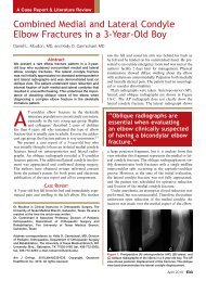
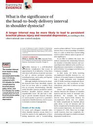
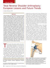
Figure 1. (A) Lateral x-ray of lumbosacral spine shows bullet<br />
lodged in spinal canal at L4. Note vertical orientation of missile<br />
and previous decompression and fusion at L4–S1 for isthmic<br />
spondylolys<strong>the</strong>sis. (B) Lateral x-ray myelogram shows even far<strong>the</strong>r<br />
cephalad migration of bullet <strong>to</strong> L2. (C) Anteroposterior x-ray<br />
of lumbosacral spine. Note 90° change in bullet orientation from<br />
lateral x-ray. Bullet is now oriented horizontally, which confirms<br />
its migration.<br />
<strong>to</strong> neurologic status. If <strong>the</strong>re is evidence of spinal instability,<br />
surgical intervention with instrumentation and fusion<br />
constructs may be indicated <strong>to</strong> prevent additional neurologic<br />
damage. 7<br />
Bullet Location and Level of Injury. Decisions<br />
regarding removal of bullets or bullet fragments depend<br />
on spinal canal proximity. Prospectively analyzing results<br />
of decompression on spinal injuries with intracanal bullets,<br />
Waters and Adkins 19 found statistically significantly<br />
improved mo<strong>to</strong>r function after surgical decompression<br />
of T12 <strong>to</strong> L4 lesions compared with nonsurgical treatment.<br />
However, <strong>the</strong>re were no significant neurologic<br />
improvements with surgical removal and decompression<br />
at o<strong>the</strong>r levels of <strong>the</strong> cervical and thoracic spine. There is<br />
a paucity of evidence as <strong>to</strong> <strong>the</strong> efficacy of bullet removal<br />
in <strong>the</strong> cervical and thoracic spine. Bono and Heary 7 advocated<br />
removing intracanal fragments from cervical-level<br />
injuries, particularly with incomplete lesions, because of<br />
<strong>the</strong> potential for 1 or 2 levels of recovery. However, <strong>the</strong><br />
authors did not believe that surgical removal is justified<br />
in thoracic-level injuries in which little functional return<br />
is sacrificed. 7<br />
Although gunshot wounds are most common in <strong>the</strong><br />
thoracic spine, injuries <strong>to</strong> <strong>the</strong> cervical spine are potentially<br />
more devastating <strong>to</strong> neurologic function. 2,5 Recently,<br />
Medzon and colleagues 15 analyzed <strong>the</strong> incidence of spinal<br />
cord injury and <strong>the</strong> stability of cervical spine fractures after<br />
gunshot wounds <strong>to</strong> <strong>the</strong> head and neck. Of <strong>the</strong> 81 patients<br />
identified over a 13-year period, 19 had sustained cervical<br />
spine fractures. Approximately 84% of patients with cervical<br />
spine fractures presented with ei<strong>the</strong>r neurologic deficits<br />
or altered mental status. Only 3 patients underwent operative<br />
stabilization and/or decompression for unstable cervical<br />
spine injuries; all 3 had associated neurologic deficits.<br />
The authors found a low incidence of unstable cervical<br />
spine fractures in patients who were alert and examinable<br />
and who showed no signs of neurologic deficit. They concluded<br />
that spinal precautions and/or a hard cervical collar<br />
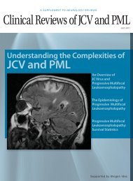
Figure 2. (A) Axial cut of computed <strong>to</strong>mography (CT) myelogram<br />
at L2 pedicle. Note intra<strong>the</strong>cal nature of missile. (B) Axial cut of<br />
CT myelogram shows layering of cerebrospinal fluid with intra<strong>the</strong>cal<br />
contrast, possible organized hema<strong>to</strong>ma, and clumped<br />
rootlets consistent with arachnoiditis.<br />
should not be maintained if <strong>the</strong>y are hindering emergent<br />
airway or hemodynamically stabilizing procedures, particularly<br />
in awake, neurologically intact patients. 15<br />
Special Indications for Surgery. A few special indications<br />
for surgery warrant fur<strong>the</strong>r discussion. If <strong>the</strong>re is evidence<br />
of cerebrospinal fluid leak, <strong>the</strong>n a lumbar subarachnoid<br />
drain should be inserted. For persistent cerebrospinal<br />
fluid leaks, open surgery with laminec<strong>to</strong>my and repair of<br />
<strong>the</strong> dural injury must be considered because of <strong>the</strong> risk for<br />
meningitis. 11,12 Lead in<strong>to</strong>xication from gunshot wounds <strong>to</strong><br />
<strong>the</strong> spine is ano<strong>the</strong>r rare complication. 20,21 If <strong>the</strong> bullet is<br />
located near facet joints or intervertebral discs, lead in<strong>to</strong>xication<br />
is more likely <strong>to</strong> occur, as synovial fluid can elute<br />
lead from <strong>the</strong> bullet. 7 Patients who have lead in<strong>to</strong>xication<br />
confirmed by peripheral blood lead levels or bone marrow<br />
biopsy should be treated with chelating agents and <strong>the</strong>n<br />
bullet removal if it can be safely accomplished. 7 <strong>Gunshot</strong><br />
wounds <strong>to</strong> <strong>the</strong> spine have also reportedly caused disc herniation<br />
and acute neurologic compromise. 22 Treatment of<br />
<strong>the</strong>se injuries is <strong>the</strong> same as for any o<strong>the</strong>r cause of disc herniation,<br />
with disc excision being <strong>the</strong> definitive procedure.<br />
Bullet removal is not absolutely required unless it can be<br />
done safely, without damaging surrounding structures.<br />
Timing of Surgery<br />
If surgery is <strong>to</strong> be pursued, <strong>the</strong> important issue of procedure<br />
timing should be addressed. Controversy continues<br />
over early versus delayed surgical management, and <strong>the</strong>re<br />
is no conclusive evidence for ei<strong>the</strong>r side of <strong>the</strong> debate.<br />
Interestingly, almost all <strong>the</strong>se studies fail <strong>to</strong> address <strong>the</strong> role<br />
of surgery in spinal injury after gunshot wounds. Cybulski<br />
and colleagues 23 retrospectively reviewed 88 patients with<br />
gunshot injuries at <strong>the</strong> conus or cauda equine level lesions<br />
and found no statistical difference in neurologic recovery<br />
for patients treated with decompressive laminec<strong>to</strong>my<br />
within 72 hours versus patients treated more than 72 hours<br />
after injury. Moreover, early versus delayed surgery or no<br />
surgical treatment at all may not significantly affect <strong>the</strong><br />
overall rate of complications or length of hospital stay. 24<br />
However, more randomized, controlled prospective studies<br />
March 2008 E49
<strong>Gunshot</strong> <strong>Wounds</strong> <strong>to</strong> <strong>the</strong> <strong>Spine</strong><br />
extremely challenging clinical problem. In <strong>the</strong> English-language<br />
literature, we found only one report of an intra<strong>the</strong>cal<br />
migra<strong>to</strong>ry missile (<strong>the</strong> patient presented with delayed<br />
radicular symp<strong>to</strong>ms). 28 In <strong>the</strong> next section, we describe <strong>the</strong><br />
case of a migra<strong>to</strong>ry intra<strong>the</strong>cal bullet in <strong>the</strong> lumbar spine of<br />
a patient who presented with cauda equina–type symp<strong>to</strong>ms.<br />
The patient was informed that his clinical findings would<br />
be submitted for publication.<br />
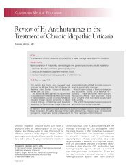
Figure 3. Anteroposterior x-ray myelogram shows missile at<br />
L3. Again note change in bullet orientation and in position from<br />
Figures 1 and 2.<br />
specifically addressing <strong>the</strong> timing of treatment after spinal<br />
gunshot injuries must be conducted <strong>to</strong> provide evidence for<br />
optimal management.<br />
A Caveat About High-Energy <strong>Wounds</strong>. One caveat<br />
is that <strong>the</strong> mentioned recommendations apply specifically<br />
<strong>to</strong> low-velocity, low-energy gunshot wounds. High-energy<br />
wounds caused by rifles or shotguns have different patterns<br />
of injury and wound characteristics that may increase<br />
<strong>the</strong> complexity of treatment decisions. Mirovsky and colleagues<br />
25 recently reported a case in which a high-velocity<br />
gunshot wound caused complete paraplegia, but without<br />
evidence that <strong>the</strong> spinal canal had been violated. Highenergy<br />
wounds may also cause more soft-tissue injuries,<br />
which are prone <strong>to</strong> infection. Studies performed on soldiers<br />
wounded in combat zones, where <strong>the</strong> majority of injuries<br />
are high-energy, have shown that surgical débridement is<br />
efficacious in preventing secondary complications. 26,27 It<br />
is likely that <strong>the</strong> same treatment principles apply <strong>to</strong> civilians<br />
who sustain high-energy wounds, which are becoming<br />
increasingly prevalent.<br />
Migra<strong>to</strong>ry Bullets<br />
Migration of retained missiles, which has been reported<br />
in <strong>the</strong> brain, blood vessels, and body cavities, presents an<br />
Case Illustration<br />
A man in his early 50s presented <strong>to</strong> us 6 months after<br />
being shot and treated. He had been shot 4 times from<br />
a short distance with a low-velocity 45-caliber handgun<br />
during a robbery. One bullet was lodged in <strong>the</strong> spine.<br />
The shoulder and abdomen had also sustained gunshot<br />
wounds. The patient underwent emergent explora<strong>to</strong>ry<br />
laparo<strong>to</strong>my at a nearby hospital. Initially, <strong>the</strong> spine wound<br />
was treated nonoperatively. The patient presented <strong>to</strong> us<br />
<strong>to</strong> seek a consultation regarding possible removal of <strong>the</strong><br />
bullet. He could ambulate only with cane or crutches and<br />
complained of lost sensation in <strong>the</strong> <strong>to</strong>es on <strong>the</strong> right and<br />
of being incontinent of bowel and bladder. His Oswestry<br />
score was 60 points. On a pain diagram, he indicated pain<br />
in <strong>the</strong> left hip, right anterior knee, right lateral calf, right<br />
dorsal medial foot, midline lower back and but<strong>to</strong>ck, bilateral<br />
posterior thigh, and plantar aspect of <strong>the</strong> right foot.<br />
On a 10-point scale, he rated his pain 3/10 at its best, 9/10<br />
at its worst, and 4/10 on average. On <strong>the</strong> McGill questionnaire,<br />
he described his pain as shooting, exhausting,<br />
unbearable, and numb. He could not sit for more than 1<br />
hour at a time. His pain was alleviated by bending forward<br />
and lying on his side.<br />
Prior surgical his<strong>to</strong>ry was remarkable for noninstrumented<br />
L4–S1 fusion for a high-grade isthmic spondylolys<strong>the</strong>sis<br />
(30 years earlier). Current medications included hydrocodone<br />
bitartrate and acetaminophen (Vicodin), morphine<br />
sulfate controlled-release (MS Contin), and gabapentin<br />
(Neurontin).<br />
The physical examination was remarkable for somewhat<br />
decreased lumbar lordosis. There was 50% loss of range of<br />
motion in forward flexion and extension, which was painful.<br />
Extension with rotation <strong>to</strong> ei<strong>the</strong>r side was painful. Flexion<br />
with rotation <strong>to</strong> ei<strong>the</strong>r side was painless. Lateral bending <strong>to</strong><br />
ei<strong>the</strong>r side was painful with 50% loss of motion. Sensation<br />
was abnormal with hypoes<strong>the</strong>sia on <strong>the</strong> right in <strong>the</strong> L4,<br />
L5, and S1 derma<strong>to</strong>mes <strong>to</strong> light <strong>to</strong>uch. Nei<strong>the</strong>r clonus nor<br />
Babinski sign could be elicited. Deep tendon reflexes were<br />
intact and symmetrical. The right extensor hallucis longus<br />
was 3/5 in strength, and <strong>the</strong> right gastrocnemius was 1/5 in<br />
strength. The rest of <strong>the</strong> mo<strong>to</strong>r examination was normal.<br />
The patient’s imaging studies have included plain x-<br />
rays, myelogram, and CT myelogram. The myelogram<br />
showed an intra<strong>the</strong>cal bullet, which migrated from L3<br />
<strong>to</strong> L2 during <strong>the</strong> myelogram procedure. It also showed a<br />
solid prior fusion and decompression at L4–S1. The bullet<br />
was seen as low as L4–L5 on plain x-rays and as high as<br />
L2 during myelography, confirming migration of <strong>the</strong> mis-<br />
E50 E48 The American Journal of Orthopedics ®
E. Moon et al<br />
sile. It also was observed spinning around its axis, changing<br />
its orientation in <strong>the</strong> spinal canal from horizontal <strong>to</strong><br />
vertical on different views (Figures 1–3). As mentioned,<br />
<strong>the</strong> patient could alleviate his pain by bending forward<br />
and lying on his side—results that could be explained by<br />
<strong>the</strong> change in bullet position with those postures. As <strong>the</strong><br />
bullet is a space-occupying lesion, his flexing forward<br />
(increasing <strong>the</strong> canal diameter) may also have alleviated<br />
<strong>the</strong> stenosis-type symp<strong>to</strong>ms.<br />
We consulted <strong>the</strong> army surgeon (see Acknowledgment).<br />
Bullet removal was recommended because of possible<br />
intra<strong>the</strong>cal lead <strong>to</strong>xicity and <strong>the</strong> potential for continued<br />
nerve rootlet microtrauma caused by bullet migration.<br />
However, <strong>the</strong> patient was cautioned that his traumatic cauda<br />
equina–type symp<strong>to</strong>ms might not change significantly<br />
after surgery.<br />
The surgery was performed with <strong>the</strong> patient in <strong>the</strong> prone<br />
position. Intraoperative fluoroscopy localized <strong>the</strong> bullet <strong>to</strong><br />
L2–L3, and L2–L3 lamino<strong>to</strong>my was performed <strong>to</strong> expose <strong>the</strong><br />
dural sac. Then a midline duro<strong>to</strong>my was performed <strong>to</strong> expose<br />
<strong>the</strong> intra<strong>the</strong>cal bullet, which was removed. The dural sac was<br />
closed and dural collagen patch with fibrin glue was applied.<br />
The patient had immediate pos<strong>to</strong>perative improvement<br />
in right leg symp<strong>to</strong>ms, and <strong>the</strong> improvement was still<br />
evident at 13-month follow-up. He was back <strong>to</strong> work in<br />
his physically demanding occupation. He had complete<br />
bowel control but no bladder control and was completely<br />
dependent on self-ca<strong>the</strong>terization. Residual pain was 60%<br />
in <strong>the</strong> legs and 40% in <strong>the</strong> lower back. However, <strong>the</strong> patient<br />
was still taking hydrocodone bitartrate and acetaminophen,<br />
morphine sulfate controlled-release, and gabapentin. His<br />
Oswestry score was improved (44 points).<br />
Summary<br />
A gunshot wound <strong>to</strong> <strong>the</strong> spine is a complex injury, and<br />
treatment remains controversial. Treatment depends on<br />
<strong>the</strong> physician’s ability <strong>to</strong> understand mechanism of injury,<br />
principles of medical management, diagnostic imaging, and<br />
surgical options. Antibiotics are an important component of<br />
treatment and should be continued for a minimum of 7 days<br />
in cases of wounds that both perforate <strong>the</strong> colon and injure<br />
<strong>the</strong> spine. Corticosteroids do not affect neurologic outcome<br />
and <strong>the</strong>refore should not be used.<br />
Decompression and removal of intracanal bullets at T12<br />
and below may improve mo<strong>to</strong>r function. In select cases of<br />
cervical injuries, removal of intracanal bullet fragments<br />
may be justified, particularly with incomplete lesions.<br />
Regardless of injury level, new-onset or progressive neurologic<br />
deterioration is an indication for urgent decompression.<br />
Optimal surgical timing remains a controversial issue,<br />
and more study is needed <strong>to</strong> develop treatment guidelines.<br />
Intra<strong>the</strong>cal migra<strong>to</strong>ry missiles represent a very rare subset<br />
of <strong>the</strong> gunshot wounds <strong>to</strong> <strong>the</strong> spine, and <strong>the</strong>ir treatment<br />
should be individualized.<br />
Authors’ Disclosure Statement<br />
and Acknowledgment<br />
The authors report no actual or potential conflict of interest<br />
in relation <strong>to</strong> this article.<br />
We thank Dr. Eugene Carragee, Chief of <strong>Spine</strong> Surgery at<br />
Stanford University, for his valuable counseling and advice.<br />
References<br />
1. Farmer JC, Vaccaro AR, Balders<strong>to</strong>n RA, Albert TJ, Cotler J. The changing<br />
nature of admissions <strong>to</strong> a spinal cord injury center: violence on <strong>the</strong> rise.<br />
J Spinal Disord. 1998;11(5):400-403.<br />
2. Bishop M, Shoemaker WC, Avakian S, et al. Evaluation of a comprehensive<br />
algorithm for blunt and penetrating thoracic and abdominal trauma. Am Surg.<br />
1991;57(12):737-746.<br />
3. Isiklar ZU, Lindsey RW. <strong>Gunshot</strong> wounds <strong>to</strong> <strong>the</strong> spine. Injury. 1998;29(suppl 1):<br />
SA7-SA12.<br />
4. Yoshida GM, Garland D, Waters RL. <strong>Gunshot</strong> wounds <strong>to</strong> <strong>the</strong> spine. Orthop Clin<br />
North Am. 1995;26(1):109-116.<br />
5. Kupcha PC, An HS, Cotler JM. <strong>Gunshot</strong> wounds <strong>to</strong> <strong>the</strong> cervical spine. <strong>Spine</strong>.<br />
1990;15(10):1058-1063.<br />
6. Naude GP, Bongard FS. <strong>Gunshot</strong> injuries of <strong>the</strong> sacrum. J Trauma. 1996;40(4):656-<br />
659.<br />
7. Bono CM, Heary RF. <strong>Gunshot</strong> wounds <strong>to</strong> <strong>the</strong> spine. <strong>Spine</strong> J. 2004;4(2):230-240.<br />
8. Bashir EF, Cybulski GR, Chaudhri K, Choudhury AR. Magnetic resonance imaging<br />
and computed <strong>to</strong>mography in <strong>the</strong> evaluation of penetrating gunshot injury of<br />
<strong>the</strong> spine. Case report. <strong>Spine</strong>. 1993;18(6):772-773.<br />
9. Gustilo RB. Current concepts in <strong>the</strong> management of open fractures. Instr Course<br />
Lect. 1987;36:359-366.<br />
10. Kumar A, Wood GW 2nd, Whittle AP. Low-velocity gunshot injuries of <strong>the</strong> spine<br />
with abdominal viscus trauma. J Orthop Trauma. 1998;12(7):514-517.<br />
11. Roffi RP, Waters RL, Adkins RH. <strong>Gunshot</strong> wounds <strong>to</strong> <strong>the</strong> spine associated with<br />
a perforated viscus. <strong>Spine</strong>. 1989;14(8):808-811.<br />
12. Romanick PC, Smith TK, Kopaniky DR, Oldfield D. Infection about <strong>the</strong> spine<br />
associated with low-velocity-missile injury <strong>to</strong> <strong>the</strong> abdomen. J Bone Joint Surg<br />
Am. 1985;67(8):1195-1201.<br />
13. Levy ML, Gans W, Wijesinghe HS, SooHoo WE, Adkins RH, Stillerman CB. Use<br />
of methylprednisolone as an adjunct in <strong>the</strong> management of patients with penetrating<br />
spinal cord injury: outcome analysis. Neurosurgery. 1996;39(6):1141-<br />
1148.<br />
14. Heary RF, Vaccaro AR, Mesa JJ, et al. Steroids and gunshot wounds <strong>to</strong> <strong>the</strong><br />
spine. Neurosurgery. 1997;41(3):576-583.<br />
15. Medzon R, Ro<strong>the</strong>nhaus T, Bono CM, Grindlinger G, Rathlev NK. Stability of<br />
cervical spine fractures after gunshot wounds <strong>to</strong> <strong>the</strong> head and neck. <strong>Spine</strong>.<br />
2005;30(20):2274-2279.<br />
16. Stauffer ES, Wood RW, Kelly EG. <strong>Gunshot</strong> wounds of <strong>the</strong> spine: <strong>the</strong> effects of<br />
laminec<strong>to</strong>my. J Bone Joint Surg Am. 1979;61(3):389-392.<br />
17. Klein Y, Cohn SM, Soffer D, Lynn M, Shaw CM, Hasharoni A. <strong>Spine</strong> injuries<br />
are common among asymp<strong>to</strong>matic patients after gunshot wounds.<br />
J Trauma. 2005;58(4):833-836.<br />
18. Denis F. The three column spine and its significance in <strong>the</strong> classification of acute<br />
thoracolumbar spinal injuries. <strong>Spine</strong>. 1983;8(8):817-831.<br />
19. Waters RL, Adkins RH. The effects of removal of bullet fragments retained in<br />
<strong>the</strong> spinal canal. A collaborative study by <strong>the</strong> National Spinal Cord Injury Model<br />
Systems. <strong>Spine</strong>. 1991;16(8):934-939.<br />
20. Grogan DP, Bucholz RW. Acute lead in<strong>to</strong>xication from a bullet in an intervertebral<br />
disc space. A case report. J Bone Joint Surg Am. 1981;63(7):1180-1182.<br />
21. Linden MA, Man<strong>to</strong>n WI, Stewart RM, Thal ER, Feit H. Lead poisoning<br />
from retained bullets. Pathogenesis, diagnosis, and management. Ann Surg.<br />
1982;195(3):305-313.<br />
22. Robertson DP, Simpson RK, Narayan RK. Lumbar disc herniation from a gunshot<br />
wound <strong>to</strong> <strong>the</strong> spine. A report of two cases. <strong>Spine</strong>. 1991;16(8):994-995.<br />
23. Cybulski GR, S<strong>to</strong>ne JL, Kant R. Outcome of laminec<strong>to</strong>my for civilian gunshot<br />
injuries of <strong>the</strong> terminal spinal cord and cauda equina: review of 88 cases.<br />
Neurosurgery. 1989;24(3):392-397.<br />
24. Fehlings MG, Perrin RG. The role and timing of early decompression for cervical<br />
spinal cord injury: update with a review of recent clinical evidence. Injury.<br />
2005;36(suppl 2):B13-B26.<br />
25. Mirovsky Y, Shalmon E, Blankstein A, Halperin N. Complete paraplegia following<br />
gunshot injury without direct trauma <strong>to</strong> <strong>the</strong> cord. <strong>Spine</strong>. 2005;30(21):2436-<br />
2438.<br />
26. Parsons TW 3rd, Lauerman WC, Ethier DB, et al. <strong>Spine</strong> injuries in combat<br />
troops—Panama, 1989. Mil Med. 1993;158(7):501-502.<br />
27. Splavski B, Vrankovic D, Saric G, Blagus G, Mursic B, Rukovanjski M. Early<br />
management of war missile spine and spinal cord injuries: experience with<br />
21 cases. Injury. 1996;27(10):699-702.<br />
28. Soges LJ, Kinnebrew GH, Limcaco OG. Mobile intra<strong>the</strong>cal bullet causing<br />
delayed radicular symp<strong>to</strong>ms [published correction appears in AJNR Am J<br />
Neuroradiol. 1988;9(5):890]. AJNR Am J Neuroradiol. 1988;9(3):610.<br />
This paper will be judged for <strong>the</strong> Resident Writer’s Award.<br />
March 2008 E49 E51