Imiquimod: A Comprehensive Therapy - Ob.Gyn. News
Imiquimod: A Comprehensive Therapy - Ob.Gyn. News
Imiquimod: A Comprehensive Therapy - Ob.Gyn. News

Create successful ePaper yourself
Turn your PDF publications into a flip-book with our unique Google optimized e-Paper software.
A SUPPLEMENT TO<br />
April 2009<br />
<strong>Imiquimod</strong>:<br />
A <strong>Comprehensive</strong> <strong>Therapy</strong><br />
Edward John Mayeaux, Jr, MD • Theodore Rosen, MD • Anita L. Nelson, MD<br />
Introduction to <strong>Imiquimod</strong>: A <strong>Comprehensive</strong><br />
<strong>Therapy</strong> for Health Care Professionals<br />
Edward John Mayeaux, Jr, MD<br />
This newsletter contains<br />
valuable information on<br />
diagnosis and treatment<br />
of several conditions commonly<br />
seen in practice: actinic keratosis<br />
(AK), superficial basal cell carcinoma<br />
(sBCC), and external genital<br />
warts (EGW). These neoplasms<br />
account for a large number of office<br />
visits each year, and their sequelae can be significant.<br />
Skin cancer is a growing concern for all patients,<br />
regardless of race, gender, or geography. Women have<br />
two unique concerns related to skin cancer: as adults,<br />
they may be facing personal consequences of years of<br />
inadequate sun-exposure protection, and as a health<br />
caregiver in their households, they need to remind their<br />
family members about the need for sun protection and<br />
disease screening. Risk factors for precancerous lesions<br />
and skin cancer are the same—frequent or intense<br />
sun exposure with greater risk in those with fair skin,<br />
Edward John Mayeaux, Jr, MD, is Professor, Department<br />
of Family Medicine; and Professor, Department of <strong>Ob</strong>stetrics<br />
and <strong>Gyn</strong>ecology, Louisiana State University Health Sciences<br />
Center, Shreveport.<br />
<strong>Imiquimod</strong> is indicated for<br />
actinic keratosis, superficial<br />
basel cell carcinoma, and<br />
external genital warts.<br />
light eyes and/or hair, exposure at<br />
younger ages, more frequent exposures,<br />
a history of skin disorders<br />
or damage, and altered immunity.<br />
Dr. Rosen reviews the clinical presentation,<br />
diagnosis, and treatment<br />
options for AK and sBCC.<br />
Human papillomavirus (HPV)<br />
infection is one of the most common<br />
sexually transmitted diseases in the United States.<br />
Although EGW will not develop in everyone with<br />
HPV infection, they are easily transmitted, and most<br />
sexual partners are infected by the time of a diagnosis.<br />
Dr. Nelson provides a review of HPV infection with<br />
emphasis on its role in the pathogenesis of EGW, and<br />
discusses multiple treatment options.<br />
The focus for treatment options is the role of<br />
imiquimod, a local immune modulator, which when<br />
topically applied triggers both innate and cell-mediated<br />
immune response. <strong>Imiquimod</strong>, with its unique mode<br />
of action and efficacy against a wide range of skin<br />
neoplasias, has shown itself to be a powerful tool in<br />
medical practice. Clinical trial data supporting the<br />
efficacy and safety of its use in the management of<br />
AK, sBCC, and EGW is presented herein.<br />
Educational support provided by Graceway Pharmaceuticals, LLC.
Developments in Identifying and<br />
Treating Non-Melanoma Skin Cancer<br />
A<br />
relatively easy and highly<br />
effective preventive counseling<br />
opportunity consists<br />
of warning patients to limit<br />
exposure to ultraviolet (UV) light<br />
both from natural (eg, the sun)<br />
and artificial (eg, tanning beds)<br />
sources. The benefits of judicious<br />
limiting of UV exposure are not<br />
only cosmetic, such as reduced<br />
wrinkling and lentigines (age<br />
spots), but also medical, such as lowered risk of skin cancer.<br />
Despite our best efforts to educate patients, many do<br />
not adhere to recommendations to avoid excessive UV<br />
exposure; thus, health care professionals need to be<br />
capable of early diagnosis and effective treatment of nonmelanoma<br />
skin cancer (NMSC) lesions. Fortunately,<br />
topical, field-directed treatment options are available that<br />
can be administered safely as an alternative to surgical<br />
intervention for select lesions.<br />
NMSC lesions include actinic keratosis (AK), superficial<br />
and nodular basal cell carcinoma (sBCC and nBCC),<br />
squamous cell carcinoma in-situ (SCCIS) (Bowen’s disease),<br />
and invasive squamous cell carcinoma (SCC). AK<br />
and SCC are part of a disease continuum that includes<br />
shared cellular genetic alterations which promote uncontrolled<br />
proliferation, morphology, and location. The<br />
presence of one or more AK lesions represents “the tip of<br />
the iceberg” and should be regarded as “a wake-up call.”<br />
To prevent AK lesions from developing into potentially<br />
lethal invasive SCC, early treatment is critical.<br />
BCC, the most common form of skin cancer, is characterized<br />
by a different pathology and a different set of<br />
UV-induced cellular genetic abnormalities. BCC is easily<br />
treatable when diagnosed early, but with time can<br />
become locally aggressive, damaging surrounding skin<br />
and other vital structures in close proximity. Neglected<br />
BCC can even invade bone or cartilage, although it<br />
rarely metastasizes. UV radiation is a modifiable risk<br />
factor for all NMSC lesions.<br />
Diagnosis<br />
A tentative differential diagnosis of NMSC lesions can<br />
be made based on appearance, although confirmation of<br />
Theodore Rosen, MD<br />
To prevent AK<br />
lesions from developing<br />
into potentially lethal<br />
invasive SCC, early<br />
treatment is critical.<br />
basal and SCCs almost always<br />
require biopsy. AK lesions are flat,<br />
scaly, and small, often with an underlying<br />
redness (Figure 1). Occasionally,<br />
AK lesions may be hypertrophic<br />
and therefore significantly raised<br />
above the skin surface. Nodular<br />
BCC lesions are often pearly or<br />
waxy with distinct borders and<br />
prominent blood vessels on the<br />
lesion’s surface. Invasive SCC lesions<br />
may resemble AK lesions but are thicker, more erythematous,<br />
have poorly defined borders, are generally<br />
rock hard and may be friable. Superficial BCC exhibits<br />
flat, red, scaly surfaces and sharp borders (Figure 2).<br />
Sclerotic or morpheoform BCCs are the rarest type<br />
and look like depigmented areas or scars. Both of<br />
these types of NMSC are much bigger than an AK<br />
lesion. Shave biopsy can be used to confirm most suspected<br />
NMSC, but punch biopsy is better utilized in<br />
order to penetrate the thickness of invasive SCC.<br />
Treatment Options<br />
The goal of treatment for all NMSC is complete<br />
removal of the tumor while preserving both function<br />
and cosmetic appearance as much as possible. Often<br />
overlooked, however, is the potential goal of preventing<br />
similar lesions from occurring on contiguous skin.<br />
Treatment modalities can be broadly divided into<br />
lesion-directed (surgical removal of the lesion only)<br />
Theodore Rosen, MD, is Professor, Department of Dermatology;<br />
and Chief, VA Dermatology Clinic, Baylor College of<br />
Medicine, Houston, TX.<br />
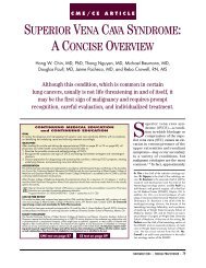
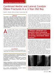
Figure 1. These lesions on a woman’s cheek are flat<br />
and scaly with underlying redness, typical of actinic<br />
keratosis.<br />
The Female Patient NEWSLETTER April 2009
Rosen<br />
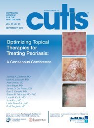
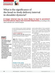
Figure 2. This lesion has the red, scaly surface and<br />
sharp borders typical of sBCC.<br />
and field-directed (broad-based<br />
topical therapy directed at the lesion<br />
and surrounding damaged skin).<br />
The National <strong>Comprehensive</strong><br />
Cancer Network states that in<br />
patients with low-risk sBCC, when<br />
surgery or radiation are contraindicated<br />
or impractical, topical therapy<br />
should be considered. 1 At this time,<br />
there are no consensus guidelines<br />
for management of AK, although<br />
field-directed therapy is becoming<br />
a first-line treatment in an attempt to achieve some measure<br />
of long-term suppression of carcinogenesis.<br />
Field-directed treatment<br />
is best suited for<br />
earliest manifestation<br />
of NMSC (AK) and<br />
for superficial forms of<br />
both BCC and SCC.<br />
leave residual depressed and/or hypopigmented scars.<br />
Excisional surgery with postoperative margin assessment<br />
and/or Mohs micrographic surgery with complete<br />
circumferential peripheral and deep margin<br />
assessment during removal are more extensive forms of<br />
surgery indicated for management of deeply invasive<br />
tumors, tumors ≥2 cm, incomplete excisions, recurrent<br />
neoplasms, and high-risk tumor locations.<br />
Radiation therapy is generally reserved for older<br />
patients (>60 years) and is indicated for use in the low-risk<br />
regions of the trunk and extremities. It is contraindicated<br />
for treatment of the genitalia, hands, and feet; in genetic<br />
conditions predisposed to skin cancers; and in patients<br />
with concomitant connective tissue diseases.<br />
Cryotherapy consists of rapid freezing of the lesion<br />
with liquid nitrogen and may be used for low-risk<br />
small, well-demarcated sBCC and<br />
AKs. In AK, response to cryotherapy<br />
is dependent on freeze<br />
times with higher rates of complete<br />
response seen in lesions frozen<br />
for longer than 5 to 20 seconds.<br />
Freeze times for sBCC are substantially<br />
longer. An expected<br />
outcome of cryotherapy, in addition<br />
to local blistering and crusting,<br />
is hypo-pigmentation due to<br />
destruction of melanocytes during<br />
the freezing process. This<br />
adverse effect may cause serious cosmetic defects, especially<br />
with repeated applications.<br />
Lesion-Directed Treatment: Surgery<br />
Surgical management of NMSC is considered the gold<br />
standard of treatment, and is indicated for all invasive<br />
tumors. Curettage and electrodessication (C&E) is<br />
used on low-risk lesions in non-hair bearing sites, is<br />
limited to removal of the epidermis and dermis only,<br />
and should be accompanied by pathologic confirmation<br />
of tumor type. 1 Though widely practiced, C&E can<br />
Field-Directed Treatment: Topical<br />
Field-directed treatment is best suited for the earliest<br />
manifestation of NMSC (AK) and for superficial forms<br />
of both BCC and SCC (Table). 5-Fluorouracil is an<br />
antineoplastic antimetabolite indicated in topical form<br />
for treatment of AK and sBCC. When used for AK,<br />
the various formulations are applied twice daily until<br />
the inflammatory response reaches the erosion stage<br />
Table. Therapies for Superficial Basal Cell Cancer and Actinic Keratosis 1<br />
Lesion-Directed Treatment<br />
Topical/Field-Directed Treatment<br />
Curettage and electrodesiccation <strong>Imiquimod</strong> (Aldara ® )<br />
Excision with POMA 5-Fluorouracil (Efudex ® , Carac ® , Fluroplex ® )<br />
Mohs resection (CCPDMA) Photodynamic therapy (Levulan ® , Metvix ® )<br />
Radiation therapy<br />
Diclofenac gel (Solaraze)<br />
Cryotherapy<br />
POMA = postoperative margin assessment<br />
CCPDMA = complete circumferential peripheral and deep margin assessment with frozen or permanent section<br />
The Female Patient NEWSLETTER April 2009
Non-Melanoma Skin Cancer<br />
(usually 2 to 4 weeks). The 5% formulation is applied<br />
twice daily for sBCC for at least 3 to 6 weeks and as<br />
long as 10 to 12 weeks until the lesion is obliterated. As<br />
with AK, complete healing may not be evident for 1 to<br />
2 months. While minimal post-therapy scarring is the<br />
rule, painful and unsightly local inflammatory reactions<br />
and ulceration may occur. The discomfort associated<br />
with treatment may interfere with adherence.<br />
Photodynamic therapy (PDT) involves the topical<br />
application of a photosensitizing drug to the affected<br />
area followed 14 to 18 hours later by illumination with<br />
a proprietary high-intensity blue light. 3 PDT is indicated<br />
for treatment of grade 1 or 2 AK on the face<br />
and/or scalp. Adverse events associated with PDT are<br />
limited to primarily mild to moderate local reactions,<br />
including scaling, crusting, itching, pain, erosion, and<br />
post-treatment hypopigmentation. A new PDT<br />
agent (methyl-amino-levulinic<br />
acid) with a different proprietary<br />
light source (red light) was recently<br />
FDA-approved; this agent may<br />
reduce both inconvenience of<br />
administration and pain attendant<br />
to this therapeutic modality.<br />
An NSAID gel formulation<br />
(diclofenac) is also indicated for the<br />
topical treatment of AK. 4 When<br />
applied twice daily for 60 to 90<br />
days to areas of AK, complete<br />
clearance rates at 30 days post<br />
treatment range from 31% to 47%.<br />
Local reactions including contact or irritant dermatitis,<br />
exfoliation, and dry skin may occur. This agent is not<br />
approved for any type of BCC or SCC.<br />
<strong>Imiquimod</strong> for Topical Treatment<br />
Thought to stimulate the immune system by induction,<br />
synthesis, and release of cytokines, 5% imiquimod<br />
cream is a novel topical agent. Upregulated immunity<br />
exerts a direct anticancer effect. 5 <strong>Imiquimod</strong> is indicated<br />
for the topical treatment of AK on the face or scalp in<br />
immunocompetent adults and for the treatment of<br />
biopsy-confirmed sBCC in immunocompetent adults<br />
with a maximum tumor diameter of 2.0 cm located on<br />
the trunk (excluding anogenital skin), neck, or extremities<br />
(excluding the hands and feet) only when surgical<br />
methods are less appropriate and patient follow-up can<br />
be assured. 6<br />
While surgery has<br />
been the gold standard<br />
for treatment of NMSC,<br />
field-directed therapies<br />
such as imiquimod offer<br />
many advantages.<br />
in AK in two phase III randomized studies of 436<br />
patients with 4 to 8 AK lesions located within a contiguous<br />
25 cm 2 treatment area on the face or balding<br />
scalp. 7 Patients were administered either imiquimod<br />
5% cream or matching vehicle two days per week for<br />
16 weeks followed by an 8-week follow-up period. In<br />
both studies, the complete and partial (≥75%) clearance<br />
rates were significantly greater with imiquimod.<br />
Notably, 48% of patients treated with imiquimod<br />
had an increase in AK lesion count above baseline during<br />
therapy with a complete clearance rate of new<br />
lesions that was similar to that observed for those<br />
present at baseline. The increase in lesion count was<br />
attributed to appearance of subclinical lesions that<br />
were not visible at baseline. At 8 weeks following<br />
completion of treatment, the median percent reduction<br />
in the number of AK lesions from baseline was<br />
83.3% in the imiquimod treatment<br />
group, indicating that half<br />
of patients treated had a least an<br />
83.3% reduction in the number of<br />
AK lesions counted in the treatment<br />
area at baseline. 7 Local skin<br />
reactions were more intense in<br />
the imiquimod group and increasing<br />
severity was associated with<br />
higher clearance rates. After 16<br />
months of follow up, imiquimod<br />
continued to provide a long-term<br />
clinical benefit in patients with<br />
complete AK lesion clearance. 8<br />
Two identical vehicle-controlled studies evaluated<br />
the use of imiquimod 5% cream applied 5 times per<br />
week for 6 weeks to 364 patients with sBCC. 9 Composite<br />
clearance rate (no clinical or histologic evidence<br />
or suspicious clinical evidence with no histologic evidence<br />
of sBCC at the 12-week posttreatment assessment)<br />
and histologic clearance rates were significantly<br />
higher in the patients who received imiquimod.<br />
Five-year long-term follow-up in 136 patients has<br />
shown continued sustained clearance with 90% of<br />
patients clear of sBCC lesions for 4 years, and 87% of<br />
patients clear for 5 years. 10 Evaluated in total, the most<br />
common adverse events reported in trials of imiquimod<br />
in either AK or sBCC consisted of application<br />
site reactions. The most commonly reported events<br />
were itching and burning, although in selected patients<br />
significant erythema and crusting may occur.<br />
<strong>Imiquimod</strong> Clinical Trials<br />
<strong>Imiquimod</strong> has been evaluated in multiple randomized-controlled<br />
clinical studies in AK and sBCC. 7-9<br />
The efficacy of imiquimod 5% cream was established<br />
Conclusion<br />
While surgery has been the gold standard for treatment<br />
of NMSC, field-directed therapies such as<br />
imiquimod offer many advantages. In AK, imiquimod<br />
The The Female Patient NEWSLETTER April 2009
Rosen<br />
use is associated with excellent response as a field-directed<br />
therapy that addresses not only visible AK lesions but<br />
subclinical foci. In sBCC, use of imiquimod 5 times per<br />
week produces long-term clearance of lesions.<br />
References<br />
1. National <strong>Comprehensive</strong> Cancer Network ® . NCCN Clinical<br />
Practice Guidelines in Oncology Basal Cell and Squamous Cell<br />
Skin Cancers. V.1.2009. Available at: www.nccn.org/professionals/<br />
physician_gls/PDF/nmsc.pdf. Accessed February 3, 2009.<br />
2. Efudex ® (fluorouracil) Topical Solutions and Creams<br />
Prescribing Information. Available at: www.valeant.com/<br />
fileRepository/products/PI/Efudex-40_Cream_5_Solution_<br />
2-5_PI_Apr04.pdf. Accessed February 3, 2009.<br />
3. Levulan ® Kerastick ® (aminolevulenic acid HCk) for Topical<br />
Solution, 20% Prescribing Information. Available at: www.<br />
dusapharma.com/duplicate-of-product-information.html.<br />
Accessed February 3, 2009.<br />
4. Solaraze (diclofenac sodium) Gel, 3% Prescribing Information.<br />
Available at: www.fda.gov/Cder/foi/label/2000/<br />
21005lbl.pdf. Accessed February 3, 2009.<br />
5. Krawtchenko N, Roewert-Huber J, Ulrich M, Mann I, Sterry W,<br />
Stockfleth E. A randomized study of topical 5% imiquimod vs.<br />
topical 5-fluorouracil vs cryosurgery in immunocompetent<br />
patients with actinic keratoses: A comparison of clinical and<br />
histological outcomes including 1-year follow-up. Br J Derm.<br />
2007;157(Suppl. 2):34-40.<br />
6. ALDARA ® (imiquimod) Cream, 5% Prescribing Information.<br />
Graceway Pharmaceuticals. Revised 11/07. Available at:<br />
www.aldara.com/pdfs/carcinoma_professional_pi.pdf.<br />
Accessed February 3, 2009.<br />
7. Lebwohl M, Dinehart S, Whiting D, et al. <strong>Imiquimod</strong> 5%<br />
cream for the treatment of actinic keratosis: Results from two<br />
phase III randomized, double-blind, parallel group, vehiclecontrolled<br />
trials. J Am Acad Dermatol. 2004;50(5):714-721.<br />
8. Lee PK, Harwell WB, Loven KH, et al. Long-term clinical<br />
outcomes following treatment of actinic keratosis with imiquimod<br />
5% cream. Dermatol Surg. 2005;31(6):659-664.<br />
9. Geisse J, Caro I, Lindholm J, Golitz L, Stampone P, Owens M.<br />
<strong>Imiquimod</strong> 5% cream for the treatment of superficial basal<br />
cell carcinoma: Results from two phase III, randomized,<br />
vehicle-controlled studies. J Am Acad Dermatol. 2004;50(5):<br />
722-733.<br />
10. Gollnick H, Barona CG, Frank RG. et al. Recurrence rate of<br />
superficial basal cell carcinoma following treatment with imiquimod<br />
5% cream: conclusion of a 5-year long-term follow-up study<br />
in Europe. Eur J Dermatol. 2008;18(6):677-682.<br />
The The Female The Female Patient Patient SUPPLEMENT NEWSLETTER November April 2009 2007
External Genital Warts:<br />
The Visible Consequences of Human Papillomavirus<br />
Human papillomavirus (HPV) is now recognized<br />
as one of the most common sexually<br />
transmitted infections (STIs) in the United<br />
States, accounting for more than 33% of new STI<br />
cases annually. 1 Incidence and prevalence are difficult<br />
to assess because HPV is not reportable, often asymptomatic,<br />
and produces antibodies<br />
in only 50% of cases. 2 Therefore,<br />
prevalence is likely to be significantly<br />
underestimated.<br />
Long-term health consequences<br />
of infection with high-risk HPV<br />
types can be devastating, ranging<br />
from precancerous cervical, vaginal,<br />
vulvar, and anal dysplasias to<br />
invasive squamous carcinoma and<br />
adenocarcinoma. One of the earliest<br />
clinical manifestations of both<br />
high- and low-risk HPV infections<br />
is exernal genital warts (EGW).<br />
Incidence<br />
It is estimated that 500,000 to 1 million new cases of<br />
EGW occur in the United States each year, and they<br />
account for 600,000 outpatient visits annually. 3 As of<br />
2004, 4% of men and 7.2% of women aged 19 to 59<br />
years reported having a diagnosis of EGW. Rates were<br />
highest among white women with higher education<br />
and greater income, and multiple lifetime partners<br />
also increased the risk. 4<br />
Clinical Presentations<br />
Genital warts in women can be found on the vulva,<br />
perineum, vagina, cervix, urethra, mouth, and larynx.<br />
Warts are generally located in areas exposed to friction<br />
during sexual activity, such as the posterior fourchette.<br />
Symptoms of discharge, itching, burning,<br />
soreness, or fissure are rare. 5 Women may note the<br />
presence of a mass, and if the warts become very large<br />
or superinfected, there may be tenderness, postcoital<br />
spotting, or odor.<br />
The classic appearance of condyloma acuminata is a<br />
raised, peaked, cauliflower-like lesion. However, they<br />
may also be rough or smooth papules or flat lesions<br />
Anita L. Nelson, MD<br />
Selection of treatment<br />
depends on wart<br />
size, number, anatomic<br />
site, morphology,<br />
adverse effects, provider<br />
experience, patient<br />
preference, and cost.<br />
(Figure 1). On thickly keratinized skin, they can appear<br />
white, grey, or flesh-toned. On mucosal surfaces, lowrisk<br />
HPV lesions tend to have finger-like projections<br />
that blend in color with the surrounding tissue. Flat<br />
genital warts can be hyperpigmented, white, or red,<br />
depending on local melanocytic involvement.<br />
Differential Diagnosis<br />
The differential diagnosis for<br />
EGW in women is extensive. It<br />
includes congenital vestibular<br />
papillomatosis, in which the vestibule<br />
is filled with symmetric,<br />
smooth, contoured projections.<br />
Close examination can distinguish<br />
this disorder because the<br />
projections are uniform in size<br />
and distribution and consist of<br />
many simple cylindrical projections<br />
from one surface instead of<br />
multiple projections arising from<br />
a common base. Other benign conditions to consider<br />
include sebaceous cysts, molluscum contagiosum (especially<br />
in women with HIV), skin tags, or benign nevi.<br />
The differential diagnoses for flat warts include condyloma<br />
lata, lichen sclerosis, vulvar hyperplasia, and psoriasis.<br />
For all genital warts, the possibility of dysplasia<br />
or invasive carcinoma must be considered.<br />
Anita L. Nelson, MD, is Professor, Department of <strong>Ob</strong>stetrics<br />
and <strong>Gyn</strong>ecology, Harbor-UCLA Medical Center,<br />
Torrance, CA.<br />
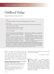
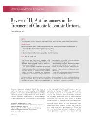
Figure 1. Condyloma acuminata may be raised, peaked,<br />
cauliflower-like lesions, or rough or smooth papules or<br />
flat lesions.<br />
The Female Patient NEWSLETTER April 2009
Nelson<br />
Table. Indications for Biopsy of EGW<br />
• Lesions surrounded by thickened skin, ulceration,<br />
or pigmentation<br />
• Raised, bleeding, red, or pigmented lesion<br />
• Indurated or fixed lesion<br />
• Lesions >2 cm<br />
• Lesions unresponsive to targeted therapy<br />
• Lesions in high-risk women (HIV, heavy smoking)<br />
Genital warts are routinely diagnosed clinically based<br />
on their visual appearance in bright light, occasionally<br />
aided by magnification. Biopsies are needed only for<br />
suspicious lesions, or when the diagnosis is uncertain<br />
(Table). Importantly, there is no place for HPV/DNA<br />
typing in routine clinical practice. 6<br />
Treatment<br />
As a virus, HPV is generally protected from serum factors,<br />
phagocytosis, and many other elements of the<br />
humoral immune system. The cellular immune system<br />
has the potential to identify virus-specific antigens on the<br />
surface of infected cells, but EGW<br />
are protected from these elements<br />
because they cannot penetrate into<br />
the epidermis. Even if infected cells<br />
can be tagged, it is difficult for the<br />
immune system to destroy them.<br />
Cytotoxic T cells also have only<br />
limited access to the epidermis,<br />
especially in the absence of inflammation.<br />
Therefore, without treatment,<br />
EGW can persist for months to years. Selection of<br />
treatment depends on wart size, number, anatomic site,<br />
morphology, adverse effects, provider experience, patient<br />
preference, and cost. 6<br />
Surgical Therapies<br />
Surgical excision under local anesthesia with iris scissors<br />
or tangential shave excision with a scalpel is appropriate<br />
for isolated, pedunculated condyloma that are attached<br />
by a slender stalk. However, surgical excision is generally<br />
reserved for very large exophytic lesions.<br />
Ablation can be done using cryotherapy, laser or<br />
loop electrical excision procedure (LEEP), depending<br />
on lesion site and treatment availability. Care must be<br />
taken to limit tissue destruction to the level of the<br />
papillary dermis. Cryotherapy of the cervix and vulva<br />
is cost-effective, and liquid nitrogen is preferred for<br />
<strong>Imiquimod</strong>’s mechanism<br />
of action as a local<br />
immune modulator is<br />
unique and indirect.<br />
urethral warts. The infected cells are destroyed, subsequently<br />
liquefying and shedding. Cryotherapy can<br />
be repeated every 1 to 2 weeks, provided there is progress<br />
in reducing the lesions. Cryotherapy is generally<br />
not used in the vagina because it is difficult to control<br />
depth of penetration through the vaginal mucosa, and<br />
adjacent bowel may be inadvertently injured.<br />
Laser ablation under anesthesia is particularly helpful<br />
for treating widespread lesions unresponsive to other<br />
therapies. Cervical and vaginal condyloma are also effectively<br />
treated with either carbon dioxide or vascular<br />
lasers because the depth of thermal damage can be controlled.<br />
Cases with more localized involvement can be<br />
laser-treated in the office setting. The LEEP is a treatment<br />
of choice in many settings for cervical warts and<br />
localized exophytic vulvar lesions.<br />
Medical Therapies<br />
Medical therapy can be used alone or in conjunction<br />
with surgery. The CDC’s Sexually Transmitted Diseases<br />
Treatment Guidelines, 2006 classified medical regimens<br />
into 2 categories: patient-applied and provider-administered.<br />
7 Considerations include patient preference,<br />
comfort, cost, and duration to clearance of warts.<br />
Patient preference is particularly important, as up to<br />
70% of patients have been previously treated. 8 Pregnancy<br />
status is also important,<br />
because some of these agents are<br />
contraindicated in pregnancy. 9<br />
Patient-Applied Treatments—<br />
Patient-applied treatments have<br />
the advantage of eliminating frequent<br />
visits to the health care<br />
professional. Direct patient costs<br />
of the drug should be balanced<br />
against copayments for office visits and the cost of<br />
time lost from work and transportation.<br />
Podofilox 0.5% gel is an antimitotic agent that prevents<br />
cell division and destroys warts by inducing local<br />
tissue necrosis. 10 It is applied twice daily for 3 consecutive<br />
days for 4 weeks. The treated surface must<br />
not exceed 10 cm 2 , and no more than 0.5 mL of the<br />
gel should be used per day. 9 This treatment provides<br />
clearance in 37% of patients. 11 Side effects are related<br />
to local inflammation. Recurrence has been reported in<br />
4% to 38% of patients, and long-term remission varies<br />
from 30% to 60%. 12,13<br />
<strong>Imiquimod</strong> 5% cream is a cell-mediated immune<br />
response modifier. It is applied to lesions 3 times a week<br />
(alternating nights) for up to 16 weeks. Patients should be<br />
advised to rub the cream well into the lesion, and to wash<br />
it off 6 to 10 hours after application. Most patients will<br />
The Female Patient NEWSLETTER April 2009
External Genital Warts<br />
develop localized erythema, but<br />
fewer than 10% will complain of<br />
pain. Those who experience pain<br />
can be advised to take a brief “holiday”<br />
from treatment. <strong>Imiquimod</strong> is<br />
a category C drug in pregnancy.<br />
<strong>Imiquimod</strong>’s mechanism of<br />
action as a local immune modulator<br />
is unique and indirect. It<br />
induces local interferon and<br />
cytokine release that triggers<br />
both innate and cell-mediated<br />
immune response, reducing the<br />
viral HPV load. 14 Complete<br />
clearance of warts is seen in 50% of patients, and<br />
more than 80% of women have at least a 50% reduction<br />
in wart area. 15,16 The effects of imiquimod are<br />
long term; wart recurrence is lowered in women<br />
treated with imiquimod. 9<br />
Provider-Applied Therapies—Podophyllin resin is an<br />
antimitotic agent that works in the same manner as<br />
podofilox. It is compounded in a 10% to 25% suspension<br />
in tincture of benzoin. Like podofilox, it should not be<br />
applied to areas greater than 10 mm 2 . Because of possibly<br />
serious systemic complications, some experts have recommended<br />
against its use in primary care settings. 12<br />
Recurrences following clinical trials have been reported<br />
in 23% to 65% of subjects. 12 Podophyllin should not be<br />
used in pregnancy.<br />
Bichloracetic acid (BCA)/trichloroacetic acid (TCA) is<br />
inexpensive and easy to apply. It denatures and precipitates<br />
proteins to kill wart cells. Concentrations vary<br />
from 50% to 95%. It must be applied carefully, and rapid<br />
drying helps reduce pain. Placing a protective “moat” of<br />
lidocaine ointment around the base of the wart can<br />
reduce run-off to adjacent tissue and consequent ulceration.<br />
12 Clearance rates of up to 80% can be expected,<br />
but multiple applications may be needed at week-long<br />
intervals. These agents are not absorbed systemically,<br />
and are safe in pregnancy.<br />
Newer Options<br />
The FDA has approved sinecatechins, 15%, for topical<br />
treatment of EGW. This is a botanical drug extract of<br />
green tea leaves. Catechins are a mixture of bioflavonoids,<br />
polyphenols, and antioxidants. A 0.5-cm strand of the<br />
ointment is applied 3 times a day in a thin layer for up to<br />
16 weeks. In clinical trials, complete clearance was<br />
achieved in 53.6% of subjects. 17<br />
The quadrivalent HPV vaccine is not a treatment for<br />
genital warts. However, it is particularly effective in<br />
reducing the risk of developing EGW by preventing<br />
Patient-applied<br />
methods offer many<br />
advantages, not only<br />
as stand-alone<br />
treatments, but also<br />
in conjunction with<br />
surgical therapies.<br />
infection with high-risk HPV<br />
types 6, 11, 16, and 18.<br />
Conclusion<br />
External genital warts represent<br />
a cosmetic and often symptomatic<br />
problem for patients. Given<br />
the wide variety of treatment<br />
options, it is more likely that clinicians<br />
will be able to design<br />
therapies that can meet the needs<br />
of individual patients. The<br />
patient-applied methods offer<br />
many advantages, not only as<br />
stand-alone treatments, but also in conjunction with<br />
surgical therapies.<br />
References<br />
1. The Henry J. Kaiser Family Foundation. Fact Sheet: Sexually<br />
Transmitted Diseases in the U.S. www.kff.org/womens<br />
health/1447-std_fs.cfm. Accessed February 2, 2009.<br />
2. Gerberding JL. Report to Congress: Prevention of Genital<br />
Human Papillomavirus Infection. Centers for Disease Control<br />
and Prevention, Department of Health and Human<br />
Services; 2004. www.cdc.gov/std/HPV/2004HPV%20Report.<br />
pdf. Accessed February 3, 2009.<br />
3. Fleischer AB Jr, Parrish CA, Glenn R, Feldman SR. Condylomata<br />
acuminata (genital warts): patient demographics and<br />
treating physicians. Sex Transm Dis. 2001;28(11):643-647.<br />
4. Dinh TH, Sternberg M, Dunne EF, Markowitz LE. Genital<br />
warts among 18- to 59-year-olds in the United States, national<br />
health and nutrition examination survey, 1999--2004. Sex<br />
Transm Dis. 2008;35(4):357-360.<br />
5. Mao C, Hughes JP, Kiviat N, et al. Clinical findings among<br />
young women with genital human papillomavirus infection.<br />
Am J <strong>Ob</strong>stet <strong>Gyn</strong>ecol. 2003;188(3):677-684.<br />
6. Workowski KA, Berman SM. HPV infection and genital<br />
warts. Sexually Transmitted Diseases Treatment Guidelines<br />
2006. MMWR Morb Mortal Wkly Rep. 2006;55<br />
(RR-11):62-67.<br />
7. Centers for Disease Control and Prevention. Workowski A,<br />
Berman SM. Sexually Transmitted diseases treatment guidelines,<br />
2006. MMWR Recomm Rep. 2006;55(RR-11):63-65.<br />
8. O’Mahony C, Law C, Gollnick HP, Marini M. New patientapplied<br />
therapy for anogenital warts is rated favourably by<br />
patients. Int J STD AIDS. 2001;12(9):565-570.<br />
9. Gunter J. Genital and perianal warts: new treatment opportunities<br />
for human papillomavirus infection. Am J <strong>Ob</strong>stet <strong>Gyn</strong>ecol.<br />
2003;189(3 Suppl.):S3-S11.<br />
10. Beutner KR, Wiley DJ, Douglas JM, et al. Genital warts and<br />
their treatment. Clin Infect Dis. 1999;28(Suppl. 1):S37-S56.<br />
11. Tyring S, Edwards L, Cherry LK, et al. Safety and efficacy of<br />
0.5% podofilox gel in the treatment of anogenital warts. Arch<br />
Dermatol. 1998;134:33-38.<br />
12. Wiley DJ, Douglas J, Beutner K, et al. External genital warts:<br />
diagnosis, treatment, and prevention. Clin Infect Dis. 2002;<br />
35(Suppl. 2):S210-S224.<br />
The Female Patient SUPPLEMENT NEWSLETTER April 2009
Nelson<br />
13. Maw RD. Treatment of anogenital warts. Dermatol Clin. 1998;<br />
16(4):829-834, xv.<br />
14. Sanclemente G, Herrera S, Tyring SK, et al. Human papillomavirus<br />
(HPV) viral load and HPV type in the clinical outcome<br />
of HIV-positive patients treated with imiquimod for<br />
anogenital warts and anal intraepithelial neoplasia. J Eur Acad<br />
Dermatol Venereol. 2007;21(8):1054-1060.<br />
15. Vilata JJ, Varela JA, Olmos L, et al. Validation and clinical use<br />
of the CECA, a disease-specific quality of life questionnaire<br />
for patients with anogenital condylomata acuminata. Acta<br />
Derm Venereol. 2008; 88(3):257-262.<br />
16. Edwards L, Ferenczy A, Eron L, et al. Self-administered<br />
topical 5% imiquimod cream for external anogenital warts.<br />
HPV Study Group. Human Papillomavirus. Arch Dermatol.<br />
1998;134(1):25-30.<br />
17. Veregen prescribing information. www.pharmaderm.com/<br />
pharmaderm/prescribing_info/veregen_pi.pdf. Accessed<br />
February 2, 2009.<br />
The Female Patient NEWSLETTER April 2009
Patient Counseling Guide for <strong>Imiquimod</strong><br />
Edward John Mayeaux, Jr, MD<br />
Patient counseling regarding<br />
proper use of imiquimod is<br />
essential to optimal and safe<br />
use. Counseling should be tailored<br />
to the patient and the indication,<br />
and should include discussion of<br />
the risks, benefits, success rates,<br />
and recurrence rates of treatment<br />
as well as strategies for prevention<br />
of further ultraviolet (UV) damage<br />
or human papillomavirus (HPV)<br />
exposure. Since actinic keratosis<br />
(AK) and superficial basal cell carcinoma (sBCC)<br />
develop after years of sun damage, these lesions will<br />
usually be found in patients older than 40 years of age.<br />
Patients with external genital warts (EGW) will generally<br />
be in their 20s or 30s, although they can be younger<br />
or older. Note that the safety for use in patients younger<br />
than age 12 has not yet been established.<br />
Patients often benefit from knowing that imiquimod<br />
does not act like any other anti-cancer therapies and is not<br />
an antiviral medication. <strong>Imiquimod</strong> has a unique mode of<br />
action and activates the body’s own immune system to<br />
fight AK, sBCC, and EGW. 2 All other therapies attempt<br />
to either cut out, damage, or poison the lesion. Manage<br />
patient treatment expectations by showing them images<br />
of before, during, and after treatment, and information on<br />
cure and recurrence rates. Consider having simple written<br />
materials available to reinforce the information given.<br />
Non-Melanoma Skin Cancer<br />
<strong>Imiquimod</strong> use should not be initiated until the skin in<br />
the treatment area is completely healed from any prior<br />
surgery or drug therapy. In patients undergoing treatment<br />
of non-melanoma skin cancers, it has been noted<br />
that the skin reaction to imiquimod follows a predictable<br />
pattern of erythema, edema, erosion and/or ulceration,<br />
weeping/exudates, and finally scabbing/crusting. The<br />
pattern of skin reactions reflects the inflammatory<br />
immune response in the skin. When treating visible AK<br />
lesions, imiquimod exposes and treats subclinical AK<br />
lesions that are not yet visible before they develop further.<br />
1 The clinical response of imiquimod on AK and<br />
sBCC lesions can be seen in Figures 1 and 2.<br />
Edward John Mayeaux, Jr, MD, is Professor, Department<br />
of Family Medicine; and Professor, Department of <strong>Ob</strong>stetrics<br />
and <strong>Gyn</strong>ecology, Louisiana State University Health Sciences<br />
Center, Shreveport.<br />
Manage patient treatment<br />
expectations by showing<br />
them images of before,<br />
during, and after treatment,<br />
and information on cure<br />
and recurrence rates.<br />
During and following treatment,<br />
all patients diagnosed with any<br />
type of AK or skin cancer should<br />
be reminded to practice simple<br />
skin cancer screening and prevention<br />
strategies. For screening in<br />
patients who have had a skin cancer,<br />
the National <strong>Comprehensive</strong><br />
Cancer Network (NCCN) recommends<br />
self examinations and<br />
skin evaluations every 6 to 12<br />
months for life (as in other situations,<br />
the US Preventive Services Task Force has more<br />
conservative recommendations). For prevention,<br />
patients should always wear a broad-spectrum sunscreen<br />
that blocks both UVA and UVB with a sun<br />
protection factor (SPF) of at least 15. Sunscreens that<br />
block both UVA and UVB include metal oxides, avobenzone<br />
(Parsol), and mexoryl (Anthelios, currently<br />
only available with SPF15 in the United States). They<br />
should be instructed to apply sunscreen to dry skin<br />
30 minutes before exposure, reapply 30 minutes after<br />
beginning exposure, and reapply after 2 to 3 hours of<br />
outdoor activity. Wearing protective clothing, especially<br />
hats, sunglasses, and coverings for the arms, legs,<br />
and abdomen, is important as well. Tanning beds<br />
should not be used.<br />
External Genital Warts<br />
Treatment of genital warts should begin with a discussion<br />
of the natural history of HPV. After diagnosis,<br />
patients often have little understanding of the disease<br />
process and high levels of anxiety related to the disease.<br />
Their initial expectations are usually centered on “curing”<br />
the disease with minimal pain, lifestyle changes, or<br />
health care visits. With time, these concerns often shift<br />
to treatment of lesions, impact on pregnancy, and longterm<br />
treatment efficacy. Effectively educating patients<br />
and addressing their concerns are vital for defining realistic<br />
treatment goals and achieving higher levels of<br />
patient satisfaction. 2<br />
Explain to patients that HPV genital infection is<br />
sexually transmitted and common among sexually<br />
active adults. 3 Also, clarify that treatment of genital<br />
warts may not reduce the risk of infecting current or<br />
future partners, nor does it protect against recurrence.<br />
Women with genital warts or those whose<br />
partners have been treated should be reminded of the<br />
importance of regular cervical cancer screening. 4<br />
10 The Female Patient NEWSLETTER April 2009
Mayeaux<br />
A<br />
B<br />
Figure 1. (A) During treatment of AK lesions with imiquimod, it is normal for the intense immune response to produce<br />
erythema, scabbing, and crusting. The greater intensity of the site reaction usually predicts a higher clearance rate.<br />
(B) The complete clearing of the AK lesions after severe site reaction during treatment can be a rough measure of<br />
treatment efficacy.<br />
A<br />
B<br />
Figure 2. (A) As with AK lesions, sBCC lesions during imiquimod treatment can be severe, indicating a high likelihood<br />
of clearance. (B) After treatment, complete clearing of the sBCC lesion.<br />
Explain to patients that when imiquimod is applied to<br />
the wart, activated immune cells travel to the area and<br />
work to eliminate the HPV-infected cells that are causing<br />
the warts. This helps the warts to clear. Note that<br />
imiquimod is pregnancy category C. It is indicated for<br />
use only on external HPV infections, and it is contraindicated<br />
for use on occluded mucus membranes or on the<br />
uterine cervix. 5 The response of EGW to imiquimod<br />
can be seen in Figure 3.<br />
Dosing of <strong>Imiquimod</strong><br />
The dosing parameters for imiquimod differ for AK,<br />
sBCC, and EGW. Patients undergoing treatment<br />
with imiquimod should be given directions for use<br />
and be advised of the importance of applying the<br />
cream in a safe manner. They should also be made<br />
aware of possibility of an intense local reaction. Dosing<br />
instructions are as follows 6 :<br />
Actinic Keratosis<br />
• Apply imiquimod 2 times per week to an area of<br />
skin approximately 25 cm 2 for 16 weeks. <strong>Imiquimod</strong><br />
may be applied to the face (eg, forehead or cheek) or<br />
scalp but not both simultaneously.<br />
• Rest periods of several days may be necessary in the<br />
event of an exaggerated local skin reaction. However,<br />
the total treatment period should not be extended<br />
beyond 16 weeks due to missed doses or rest periods.<br />
Superficial Basal Cell Carcinoma<br />
• Apply imiquimod 5 times per week (eg, Monday<br />
through Friday) for 6 weeks.<br />
The Female Patient NEWSLETTER April 2009 11
Patient Counseling Guide for <strong>Imiquimod</strong><br />
A<br />
B<br />
Figure 3. (A) Site reactions during treatment of EGW with imiquimod can be mild to moderate and include erythema,<br />
edema, and induration. (B) EGW after treatment with imiquimod.<br />
• Target tumors should have a maximum diameter of<br />
2 cm and be located on the trunk (excluding anogenital<br />
skin), neck, or extremities (excluding hands and feet).<br />
The treatment area should include a 1 cm margin<br />
around the tumor.<br />
• If there is clinical evidence of tumor following<br />
treatment, a biopsy or alternative intervention should<br />
be considered.<br />
External Genital Warts<br />
• Apply imiquimod 3 times per week until there is<br />
total clearance of external genital/perineal warts or<br />
for a maximum of 16 weeks. When applied to the<br />
treatment area, it should be gently rubbed in until it<br />
is no longer visible.<br />
• <strong>Imiquimod</strong> cream should be applied prior to bedtime<br />
and left on the skin for approximately 8 hours (6 to 10<br />
hours for EGW), then removed by washing the area<br />
with mild soap and water.<br />
• Describe to patients the exact area to be treated, and<br />
explain that the area should be washed with mild soap<br />
and water and be allowed to dry thoroughly before<br />
applying imiquimod. After applying imiquimod<br />
cream, patients should wash their hands.<br />
• Only one packet at a time of imiquimod should be<br />
used.<br />
Explain that local skin reactions (redness, burning,<br />
flaking, or crusting) in the treatment area are common.<br />
Patients should be instructed to contact you if reactions<br />
affect their daily activities or do not go away. A rest<br />
period of several days may be needed. Patients undergoing<br />
treatment for AK or sBCC should avoid sun exposure.<br />
Patients undergoing treatment for EGW may use<br />
non-occlusive cotton gauze dressings or cotton underwear.<br />
<strong>Imiquimod</strong> should not be used in the vagina.<br />
Because imiquimod is indicated for several conditions,<br />
be sure patients know which condition is being<br />
treated, possible side effects, and when to call you if<br />
needed. <strong>Imiquimod</strong>’s noninvasive regimen is a good<br />
alternative to surgery and most patients will appreciate<br />
the ease of treatment.<br />
Assistance with manuscript preparation was provided by<br />
Kate Martin, PharmD.<br />
References<br />
1. Berman B, Bienstock L, Kuritzky L, Mayeaux EJ Jr, Tyring SK.<br />
Actinic keratoses: Sequelae and treatments. J Fam Pract.<br />
2006;55(5):S1-S8.<br />
2. Mayeaux EJ Jr. External Genital Warts: An Update. The<br />
Female Patient. 2007;32(12):38-44.<br />
3. Centers for Disease Control. Human Papillomavirus: HPV<br />
Information for Clinicians. www.cdc.gov/std/hpv/commonclinicians/ClinicianBro-fp.pdf.<br />
Accessed February 6, 2009.<br />
4. Centers for Disease Control. Sexually Transmitted Disease<br />
Treatment Guidelines, 2006. www.cdc.gov/STD/treatment/<br />
2006/genital-warts.htm#warts3. Accessed February 7, 2009.<br />
5. Mayeaux EJ Jr, Dunton C. Modern management of external<br />
genital warts. J Low Genit Tract Dis. 2008;12:185-192.<br />
6. ALDARA ® Cream 5% (imiquimod) Prescribing Information.<br />
Graceway Pharmaceuticals. Revised 11/07. www.aldara.com/<br />
pdfs/carcinoma_professional_pi.pdf. Accessed February 6, 2009.<br />
12 The Female Patient NEWSLETTER April 2009 ALD020920