Subungual Extraosseous Chondroma in a Finger
Subungual Extraosseous Chondroma in a Finger
Subungual Extraosseous Chondroma in a Finger

Create successful ePaper yourself
Turn your PDF publications into a flip-book with our unique Google optimized e-Paper software.
A Case Report & Literature Review<br />
<strong>Subungual</strong> <strong>Extraosseous</strong> <strong>Chondroma</strong><br />
<strong>in</strong> a F<strong>in</strong>ger<br />
S. Alexander Rottgers, MD, Gutti Rao, MD, and Ronit Wollste<strong>in</strong>, MD<br />
<strong>Chondroma</strong>s are benign cartilage-produc<strong>in</strong>g tumors<br />
that are commonly found <strong>in</strong> tubular bones but<br />
seldom form <strong>in</strong> extraosseous soft tissues. These<br />
tumors must be dist<strong>in</strong>guished from their malignant<br />
counterparts by histology and biological behavior.<br />
The 3 types of extraosseous chondromas are <strong>in</strong>tra-articular/para-articular<br />
chondromas, juxtacortical chondromas,<br />
and chondromas of soft parts. Intra-articular/para-articular<br />
chondromas are histologically different <strong>in</strong> that they <strong>in</strong>clude<br />
benign-appear<strong>in</strong>g nuclei. 1 Juxtacortical chondromas and<br />
chondromas of soft parts tend to have mild nuclear atypia,<br />
despite a benign cl<strong>in</strong>ical course, and differ only <strong>in</strong> their<br />
association with periosteum and synovium, respectively. 2-9<br />
Juxtacortical chondromas are adjacent to bone and subperiosteum,<br />
whereas chondromas of soft parts are found <strong>in</strong> various<br />
tissue planes often associated with synovium.<br />
Here we report the case of a rare subungual extraosseous<br />
chondroma that presented atypically and that was therefore<br />
treated aggressively with disarticulation, despite an ultimately<br />
benign pathologic evaluation. The subungual location<br />
caused the tumor to obliterate the overly<strong>in</strong>g nail bed<br />
and nail plate, rais<strong>in</strong>g concern of a potentially malignant<br />
pathology dur<strong>in</strong>g <strong>in</strong>itial evaluation. In addition, the elderly<br />
male patient’s tumor was near the distal <strong>in</strong>terphalangeal<br />
(DIP) jo<strong>in</strong>t. Disarticulation was planned before surgery not<br />
only because of potential malignancy but also because of<br />
location. Resection followed by reconstruction of the nondom<strong>in</strong>ant,<br />
<strong>in</strong>dex f<strong>in</strong>ger distal phalanx would have required<br />
a more complex procedure, such as a sk<strong>in</strong> graft or a crossf<strong>in</strong>ger<br />
flap, without a significantly improved functional<br />
outcome. These options necessitate more surgery with the<br />
Dr. Rottgers is a Resident, Division of Plastic and Reconstructive<br />
Surgery, Department of Surgery, University of Pittsburgh Medical<br />
Center, University of Pittsburgh School of Medic<strong>in</strong>e, Pittsburgh,<br />
Pennsylvania.<br />
Dr. Rao is Associate Professor, Department of Pathology,<br />
Veterans Affairs Medical Center, University of Pittsburgh School<br />
of Medic<strong>in</strong>e, Pittsburgh, Pennsylvania.<br />
Dr. Wollste<strong>in</strong> is Assistant Professor, Division of Plastic and<br />
Reconstructive Surgery, Department of Surgery, University of<br />
Pittsburgh Medical Center, University of Pittsburgh School of<br />
Medic<strong>in</strong>e, Pittsburgh, Pennsylvania.<br />
Address correspondence to: Ronit Wollste<strong>in</strong>, MD, Scaife Hall,<br />
Suite 6B, 3550 Terrace Street, Pittsburgh, PA 15261 (tel, 412-<br />
648-2381; fax, 412-648-1987; e-mail, wollste<strong>in</strong>r@upmc.edu).<br />
Am J Orthop. 2008;37(11):E187-E190. Copyright Quadrant<br />
HealthCom Inc. 2008. All rights reserved.<br />
morbidity of a donor site and a return to the operat<strong>in</strong>g room<br />
for pedicle division <strong>in</strong> the f<strong>in</strong>ger-flap option. These options<br />
went aga<strong>in</strong>st the patient’s wish for m<strong>in</strong>imal surgery.<br />
In this patient, the paucity of subungual soft tissue caused<br />
the tumor to appear <strong>in</strong> a juxtacortical location, though it<br />
actually sat <strong>in</strong> a supraperiosteal tissue plane. As a result, the<br />
tumor was found <strong>in</strong> close opposition to the underly<strong>in</strong>g bone<br />
but lacked the classic radiologic f<strong>in</strong>d<strong>in</strong>gs of juxtacortical<br />
“These tumors must be dist<strong>in</strong>guished<br />
from their malignant<br />
counterparts by histology and<br />
biological behavior.”<br />
chondromas, such as bony saucerization and sclerosis. 8,9<br />
Hav<strong>in</strong>g a better understand<strong>in</strong>g of the nature of juxtacortical<br />
chondromas and chondromas of soft parts should aid<br />
surgeons <strong>in</strong> anticipat<strong>in</strong>g the diagnosis <strong>in</strong> the <strong>in</strong>stance of<br />
atypical presentation and should help make them more comfortable<br />
manag<strong>in</strong>g treatment decisions surround<strong>in</strong>g these<br />
histologically worrisome, yet benign lesions.<br />
Case Report<br />
A right-handed man <strong>in</strong> his mid-70s presented with a pa<strong>in</strong>less,<br />
slow-grow<strong>in</strong>g tumor with<strong>in</strong> the distal phalanx of the<br />
left <strong>in</strong>dex f<strong>in</strong>ger. Tumor growth was first noted after trauma<br />
to the f<strong>in</strong>ger 9 years before presentation; the patient had not<br />
sought medical treatment dur<strong>in</strong>g the <strong>in</strong>terven<strong>in</strong>g years.<br />
Past medical history <strong>in</strong>cluded a seizure disorder treated<br />
with lamotrig<strong>in</strong>e, hypertension treated with metoprolol, and<br />
A<br />
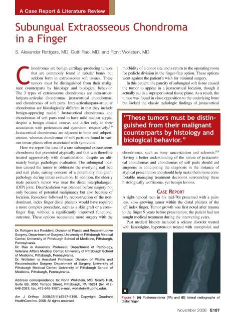
Figure 1. (A) Posteroanterior (PA) and (B) lateral radiographs of<br />
distal f<strong>in</strong>ger.<br />
November 2008<br />
E187
<strong>Subungual</strong> <strong>Extraosseous</strong> <strong>Chondroma</strong> <strong>in</strong> a F<strong>in</strong>ger<br />
Figure 2. Membrane (M) between bone (B) and tumor (T) (orig<strong>in</strong>al<br />
magnification ×100).<br />
hypercholesterolemia treated with simvastat<strong>in</strong>. The patient<br />
denied smok<strong>in</strong>g.<br />
Physical exam<strong>in</strong>ation revealed a 1×1-cm mass on the<br />
dorsum of the distal phalanx of the left <strong>in</strong>dex f<strong>in</strong>ger.<br />
No nail or residual nail bed was apparent. The tumor<br />
was round, white, hard, and smooth with no evidence<br />
of <strong>in</strong>flammation. The patient had full pa<strong>in</strong>less range of<br />
motion of the DIP jo<strong>in</strong>t. The hand exam<strong>in</strong>ation was otherwise<br />
unremarkable.<br />
“The cl<strong>in</strong>ical utility <strong>in</strong><br />
dist<strong>in</strong>guish<strong>in</strong>g between<br />
juxtacortical chondromas<br />
and chondromas of soft<br />
parts is unclear.”<br />
Radiographs showed a small <strong>in</strong>dentation <strong>in</strong> the dorsum<br />
of the distal phalanx with slight reactive sclerosis and no<br />
evidence of bony <strong>in</strong>vasion. There was no scallop<strong>in</strong>g of the<br />
bone. There was no stippl<strong>in</strong>g or other soft-tissue irregularity<br />
(Figure 1).<br />
The patient consented to excision and disarticulation<br />
of the DIP jo<strong>in</strong>t, as the diagnosis was not def<strong>in</strong>ite, and,<br />
though the prolonged cl<strong>in</strong>ical course suggested the lesion<br />
was benign, complete obliteration of the nail bed could<br />
represent malignant progression. The classic radiographic<br />
signs of juxtacortical chondromas, cortical erosion and<br />
overhang<strong>in</strong>g reactive sclerosis were absent, and there<br />
were no visible calcified masses to <strong>in</strong>dicate one of the<br />
other possible benign subungual hard-tissue masses.<br />
Furthermore, the patient was <strong>in</strong>terested <strong>in</strong> limited surgery<br />
without the need for complex reconstruction to preserve<br />
the tip of the f<strong>in</strong>ger. Dur<strong>in</strong>g surgery, disarticulation was<br />
performed, as the tumor could not be removed with safe<br />
marg<strong>in</strong>s. Frozen sections were not sent to pathology,<br />
and the distal phalanx/tumor was removed en bloc. The<br />
patient healed uneventfully.<br />
Figure 3. Sagittal cut of distal f<strong>in</strong>ger <strong>in</strong>cludes entire distal phalanx<br />
with tumor. Above the nail, membrane separates bone surface<br />
and tumor, and sk<strong>in</strong> covers tumor (top). Tumor size relative<br />
to bone can be appreciated (orig<strong>in</strong>al magnification ×10).<br />
On gross exam<strong>in</strong>ation, the pathologic specimen consisted<br />
of the distal phalanx with the neoplasm measur<strong>in</strong>g<br />
1.5×1.5 cm and about 1.0 cm <strong>in</strong> thickness <strong>in</strong>dent<strong>in</strong>g<br />
the phalanx. Surgical marg<strong>in</strong>s of 3 mm were obta<strong>in</strong>ed.<br />
The cut surface was translucent and firm. On microscopy,<br />
there was no erosion or <strong>in</strong>duction of sclerosis of<br />
contiguous cortex. The tumor was separated from the<br />
cortex by a periosteal fibrous membrane (Figure 2),<br />
and the other surface was covered with sk<strong>in</strong> (Figure<br />
3). The neoplasm was composed of mature adult hyal<strong>in</strong>e<br />
cartilage arranged <strong>in</strong> a lobular manner (Figure<br />
4). There were rare cartilage cells conta<strong>in</strong><strong>in</strong>g double<br />
nuclei (Figure 5). No calcifications were evident. The<br />
lesion was orig<strong>in</strong>ally diagnosed as juxtacortical chondroma,<br />
but with subsequent review of the literature we<br />
decided that the location of the tumor <strong>in</strong> a tissue plane<br />
superficial to the periosteum was more <strong>in</strong>dicative of a<br />
diagnosis of chondroma of soft parts.<br />
We have obta<strong>in</strong>ed the patient's <strong>in</strong>formed, written<br />
consent to publish his case report.<br />
Discussion<br />
Both juxtacortical chondromas and chondromas of soft<br />
parts present with local swell<strong>in</strong>g, a dist<strong>in</strong>ct mass, or pa<strong>in</strong>.<br />
Symptoms may be present for only a few weeks or for as long<br />
as 15 to 20 years, as was the case with our patient, suggest<strong>in</strong>g<br />
the benign nature of the lesions. 2,4,6,9,10 Our patient was a man<br />
<strong>in</strong> his mid-70s. Juxtacortical chondromas are most prevalent<br />
<strong>in</strong> young adults; mean age at diagnosis has ranged from 18.3<br />
to 26 years <strong>in</strong> different series, and the overall range is 6 to 70<br />
years. 2,10,11 Similarly, chondromas of soft parts are found <strong>in</strong><br />
all age groups; mean age <strong>in</strong> 1 case series was 34.5 years. 4,5<br />
The 3 largest case series of juxtacortical chondromas (12-23<br />
patients) had a small <strong>in</strong>creased prevalence of juxtacortical<br />
chondromas <strong>in</strong> male patients, but it is unknown if this is<br />
significant given the small numbers. 2,3,10 The 2 case series<br />
of chondromas of soft parts are larger (70 and 104 patients),<br />
but there is a male predom<strong>in</strong>ance <strong>in</strong> one and equality of sex<br />
prevalence <strong>in</strong> the other. 4,5<br />
E188 The American Journal of Orthopedics ®
S. A. Rottgers et al<br />
Figure 4. Hyal<strong>in</strong>e cartilage is mature and arranged <strong>in</strong> a lobular manner<br />
(orig<strong>in</strong>al magnification ×100).<br />
Figure 5. Cartilage cell conta<strong>in</strong><strong>in</strong>g double nuclei (arrow) (orig<strong>in</strong>al<br />
magnification ×400).<br />
Juxtacortical chondromas present adjacent to tubular<br />
bones, most commonly <strong>in</strong> the metaphyseal region. The<br />
tumor has a strong predilection for the hands and feet,<br />
as demonstrated by a review of all published cases that<br />
showed 51 of 183 tumors <strong>in</strong> the hand. 11 <strong>Chondroma</strong>s of<br />
soft parts have a similar aff<strong>in</strong>ity for the distal extremities.<br />
In one series, 89 of 104 cases were found <strong>in</strong> the hands or<br />
feet and 51 <strong>in</strong> the f<strong>in</strong>gers. 4 Another series showed 43 of 59<br />
tumors <strong>in</strong> the hands and 16 <strong>in</strong> the feet. 5<br />
Pathologically, juxtacortical chondromas are grossly<br />
described as firm, rubbery, white or bluish masses. They<br />
are lobulated and well circumscribed and can conta<strong>in</strong> various<br />
amounts of calcification impart<strong>in</strong>g a yellowish color<br />
or gritty texture to the lesion. 2,3,6,8 Most of the tumors are<br />
small—mean diameter is 2.6 cm—but lesions as large as<br />
parts is presence of mature hyal<strong>in</strong>e cartilage with a pronounced<br />
cellular element <strong>in</strong> regions of the tumor marked by<br />
mild cellular atypia. Cells may have plump, bilobed nuclei,<br />
<strong>in</strong>creased eos<strong>in</strong>ophilia, and occasional mitotic figures.<br />
Although these features would be <strong>in</strong>dicative of a low-grade<br />
malignancy if found <strong>in</strong> an <strong>in</strong>traosseous lesion, empiric<br />
experience with the 2 extraosseous counterparts shows<br />
these are benign tumors despite the pathologic characteristics.<br />
2-9 The tumor described <strong>in</strong> this report histologically fits<br />
<strong>in</strong>to either category; the only dist<strong>in</strong>guish<strong>in</strong>g feature seems<br />
to be whether it lies above or below the periosteum.<br />
The cl<strong>in</strong>ical utility <strong>in</strong> dist<strong>in</strong>guish<strong>in</strong>g between juxtacortical<br />
chondromas and chondromas of soft parts is unclear.<br />
These types of chondromas differ only <strong>in</strong> their anatomical<br />
location, and the dist<strong>in</strong>ction can confuse the diagnosis, as<br />
“Marg<strong>in</strong>al excision is the treatment of choice for extraosseous<br />
chondromas, but care must be taken to ensure that all tumor<br />
material is removed to avoid local recurrence.”<br />
8 cm have been described as be<strong>in</strong>g attached to larger bones,<br />
such as the femur. 2,3,8,10<br />
Similarly, descriptions of chondromas of soft parts<br />
state that most are well-demarcated rubbery or firm<br />
masses comparable <strong>in</strong> size to their juxtacortical counterparts.<br />
4,5 Other descriptions are of cystic, soft, friable, or<br />
calcified masses. This disparity may be easily expla<strong>in</strong>ed<br />
by a tendency to group multiple pathologies <strong>in</strong>to this category<br />
with ill-def<strong>in</strong>ed pathologic criteria. This was best<br />
illustrated by Lichtenste<strong>in</strong> and Goldman, 7 who categorized<br />
chondromas of soft parts <strong>in</strong>to 2 categories, a hyal<strong>in</strong>e<br />
cartilage tumor, which behaves just as other chondromas<br />
of soft parts do, and chondroid tumors, which were histologically<br />
immature and locally aggressive and may be<br />
more appropriately considered an <strong>in</strong>termediary between a<br />
chondroma and chondrosarcoma.<br />
On histologic exam<strong>in</strong>ation, the def<strong>in</strong><strong>in</strong>g characteristic<br />
of both juxtacortical chondromas and chondromas of soft<br />
<strong>in</strong> our patient’s case, <strong>in</strong> which the diagnosis was missed<br />
because the mass appeared <strong>in</strong> a juxtacortical location but<br />
was <strong>in</strong> the wrong tissue plane to produce the common<br />
radiologic f<strong>in</strong>d<strong>in</strong>gs. The dist<strong>in</strong>ction between extraosseous<br />
chondromas and chondroid tumors has much more cl<strong>in</strong>ical<br />
utility because the dist<strong>in</strong>ction <strong>in</strong>dicates a very real different<br />
predilection for local recurrence and spread.<br />
The typical radiographic f<strong>in</strong>d<strong>in</strong>gs of juxtacortical chondromas<br />
are well described. On pla<strong>in</strong> radiographs, 92% of<br />
juxtacortical chondromas cause visible erosion of the underly<strong>in</strong>g<br />
cortex, 67% cause sclerosis of the cortex, 67% show<br />
overhang<strong>in</strong>g osseous marg<strong>in</strong>s, particularly at the proximal<br />
side, and only 50% show soft-tissue masses and calcifications.<br />
These values <strong>in</strong>crease with magnetic resonance imag<strong>in</strong>g.<br />
10 Presence of a firm, slow-grow<strong>in</strong>g mass with these<br />
radiographic f<strong>in</strong>d<strong>in</strong>gs is highly suggestive of a juxtacortical<br />
lesion, but these f<strong>in</strong>d<strong>in</strong>gs are not seen <strong>in</strong> supraperiosteal<br />
chondromas of soft parts. These lesions produce radiograph-<br />
November 2008 E189
<strong>Subungual</strong> <strong>Extraosseous</strong> <strong>Chondroma</strong> <strong>in</strong> a F<strong>in</strong>ger<br />
ically visible soft-tissue masses or calcifications <strong>in</strong> only 60%<br />
of cases, and 8% have slight erosion of the bone. 5,12<br />
Only 4 cases of subungual extraosseous chondromas<br />
have been described <strong>in</strong> the English-language literature. 7,13,14<br />
None of these was diagnosed as a subungual juxtacortical<br />
chondroma. Three of the 4 reports did not <strong>in</strong>dicate whether<br />
the tumor was subperiosteal or supraperiosteal, and only the<br />
fourth <strong>in</strong>dicated that the tumor was <strong>in</strong> a supraperiosteal plane,<br />
as was observed <strong>in</strong> our patient’s case. 14<br />
There is always the potential confusion between extraosseous<br />
chondromas and malignant lesions. The dist<strong>in</strong>ction<br />
can often be based on degree of cellular atypia and number<br />
of mitoses. When this is not possible, cl<strong>in</strong>ical characteristics<br />
(eg, <strong>in</strong>vasion of medullary cavity of bone, periosteal reaction,<br />
lack of cortical sclerosis, larger size, pa<strong>in</strong>, rapid enlargement,<br />
ill-def<strong>in</strong>ed borders) are important clues to malignancy. 2,3 It<br />
should also be remembered that chondrosarcoma is much<br />
more prevalent <strong>in</strong> the deep musculature of the proximal<br />
extremities and is exceed<strong>in</strong>gly rare <strong>in</strong> the hand. 15<br />
The differential diagnosis of subungual lesions <strong>in</strong>cludes<br />
malignancies (eg, squamous cell carc<strong>in</strong>oma, basal cell carc<strong>in</strong>oma,<br />
malignant melanoma), but the hard-tissue masses<br />
found <strong>in</strong> this region (eg, enchondromas, subungual exostoses,<br />
subungual osteochondromas) are all benign, with the<br />
exception of malignant enchondromas, which are extremely<br />
rare. 14,16-18 Therefore, when one is presented with a subungual<br />
hard-tissue lesion lack<strong>in</strong>g the cl<strong>in</strong>ical signs of malignancy,<br />
as <strong>in</strong> our patient’s case, the lesion can usually be treated<br />
as a benign mass with local excision. Care must be taken to<br />
adequately assess the lesion pathologically to ensure that<br />
malignant foci are not harbored <strong>in</strong> the removed tumor.<br />
In the case presented, the decision to proceed with<br />
disarticulation was partially motivated by concern over<br />
the destructive and potentially malignant behavior of nailbed<br />
obliteration. In retrospect, loss of the nail bed was<br />
likely due to <strong>in</strong>creased pressure as the tumor distended<br />
the tightly adherent subungual soft tissue. This led to nail<br />
bed deformation and separation and possibly to vascular<br />
compromise to the germ<strong>in</strong>al and sterile matrices. Frozen<br />
section<strong>in</strong>g may have helped to alleviate these fears before<br />
disarticulation, but it must be remembered that, on <strong>in</strong>itial<br />
evaluation, chondromas found outside osseous structures<br />
exhibit concern<strong>in</strong>g atypical histology. 2-9 The review<strong>in</strong>g<br />
pathologist must be aware of the cl<strong>in</strong>ical scenario and<br />
must be knowledgeable about the benign behavior of<br />
extraosseous chondromas. If the same degree of cellular<br />
atypia were seen <strong>in</strong> <strong>in</strong>traosseous lesions, it would carry a<br />
much graver prognosis and necessitate more aggressive<br />
surgical management.<br />
Marg<strong>in</strong>al excision is the treatment of choice for extraosseous<br />
chondromas, but care must be taken to ensure that<br />
all tumor material is removed to avoid local recurrence.<br />
Studies have shown that curettage of the underly<strong>in</strong>g sclerotic<br />
bone is necessary <strong>in</strong> the juxtacortical subgroup of<br />
tumors to limit the rate of local recurrence. 11 In the case of<br />
a subungual location, this may require complex reconstruction<br />
with regimented follow-up.<br />
Authors’ Disclosure Statement<br />
The authors report no actual or potential conflict of <strong>in</strong>terest <strong>in</strong><br />
relation to this article.<br />
References<br />
1. Ste<strong>in</strong>er GC, Meushar N, Norman A, Present D. Intracapsular and paraarticular<br />
chondromas. Cl<strong>in</strong> Orthop. 1994;(303):231-236.<br />
2. Bauer TW, Dorfman HD, Latham JT Jr. Periosteal chondroma. A cl<strong>in</strong>icopathologic<br />
study of 23 cases. Am J Surg Pathol. 1982;6(7):631-637.<br />
3. Boriani S, Bacch<strong>in</strong>i P, Bertoni F, Campanacci M. Periosteal chondroma. A<br />
review of twenty cases. J Bone Jo<strong>in</strong>t Surg Am. 1983;65(2):205-212.<br />
4. Chung EB, Enz<strong>in</strong>ger FM. <strong>Chondroma</strong> of soft parts. Cancer. 1978;41(4):1414-<br />
1424.<br />
5. Dahl<strong>in</strong> DC, Salvador AH. Cartilag<strong>in</strong>ous tumors of the soft tissues of the<br />
hands and feet. Mayo Cl<strong>in</strong> Proc. 1974;49(10):721-726.<br />
6. Jaffe HL. Juxtacortical chondroma. Bull Hosp Jo<strong>in</strong>t Dis. 1956;17(1):20-29.<br />
7. Lichtenste<strong>in</strong> L, Goldman RL. Cartilage tumors <strong>in</strong> soft tissues, particularly <strong>in</strong><br />
the hand and foot. Cancer. 1964;17:1203-1208.<br />
8. Lichtenste<strong>in</strong> L, Hall JE. Periosteal chondroma; a dist<strong>in</strong>ctive benign cartilage<br />
tumor. J Bone Jo<strong>in</strong>t Surg Am. 1952;24(3):691-697.<br />
9. Marmor L. Periosteal chondroma (juxtacortical chondroma). Cl<strong>in</strong> Orthop.<br />
1964;(37):150-153.<br />
10. Woertler K, Blasius S, Br<strong>in</strong>kschmidt C, Hillmann A, L<strong>in</strong>k TM, He<strong>in</strong>del W.<br />
Periosteal chondroma: MR characteristics. J Comput Assist Tomogr.<br />
2001;25(3):425-430.<br />
11. Takada A, Nishida J, Akasaka T, et al. Juxtacortical chondroma of the hand:<br />
treatment by resection of the tumour and the adjacent bone cortex. J Hand<br />
Surg Br. 2005;30(4):401-405.<br />
12. Hondar Wu HT, Chen W, Lee O, Chang CY. Imag<strong>in</strong>g and pathological<br />
correlation of soft-tissue chondroma: a serial five-case study and literature<br />
review. Cl<strong>in</strong> Imag<strong>in</strong>g. 2006;30(1):32-36.<br />
13. Ayala F, Lembo G, Montesano M. A rare tumor: subungual chondroma.<br />
Report of a case. Dermatologica. 1983;167(6):339-340.<br />
14. Dumontier C, Abimelec P, Drape JL. Soft-tissue chondroma of the nail bed.<br />
J Hand Surg Br. 1997;22(4):474-475.<br />
15. Mahoney JL. Soft tissue chondromas <strong>in</strong> the hand. J Hand Surg Am.<br />
1987;12(2):317-320.<br />
16. Hodgk<strong>in</strong>son DJ. <strong>Subungual</strong> osteochondroma. Plast Reconstr Surg.<br />
1983;74(6):833-834.<br />
17. Hoehn JG, Coletta C. <strong>Subungual</strong> exostosis of the f<strong>in</strong>gers. J Hand Surg Am.<br />
1992;17(3):468-471.<br />
18. Lieb DA. <strong>Subungual</strong> osseous pathology. Cl<strong>in</strong> Podiatr Med Surg.<br />
1995;12(2):299-308.<br />
E190 E188 The American Journal of Orthopedics ®